Introduction
Spinal neoplastic lesions of metastatic origin severely affect patient quality of life. Metastasizing tumors show spinal manifestation in 30–70% of cases [2]. Most common primary tumors of vertebral metastases are localized in the breast, lung, prostate and kidney. Sur-gical intervention with corporectomy, body replacement and stabilization, although controversial, is still the best treatment option for spinal instability, neurological
complications and severe pain that cannot be managed conservatively or by local radiation [8,9]. However these extensive surgical procedures are often complicated by massive intraoperative blood loss. Compared with nor-mal vertebrae, tumorous vertebrae show extensive vas-cularization. The most highly vascularized vertebrae are those with metastases originating from renal cell and thyroid carcinoma [7]. Life-threatening blood losses have been described in cases without preoperative embolization [3, 6]. Several reports described technical Raphael Guzman Susan Dubach-Schwizer Paul Heini Karl-Olof Lovblad Daniel Kalbermatten Gerhard Schroth Luca Remonda
Preoperative transarterial embolization
of vertebral metastases
Received: 26 March 2003 Revised: 16 March 2004 Accepted: 7 April 2004
Published online: 16 September 2004 Ó Springer-Verlag 2004
Abstract The aim of this study was to evaluate the impact of preopera-tive devascularization of spinal metastases in relation to the pre-embolization tumor vascularization degree and in relation to the intra-operative blood loss. Twenty-four patients underwent preoperative transarterial embolization of hyper-vascular spinal metastases. Each tu-mor was assigned a vascularization grade (I–III) according to tumor blush after contrast agent injection in the main feeding artery. Emboli-zation was performed with polyvinyl alcohol particles in all patients. Surgical reports were reviewed in terms of estimated blood loss. A mild hypervascularization was found in three patients (group I), medium in six patients (group II) and exten-sive in 15 patients (group III). In 22 out of 24 patients embolization could be performed with a complete devascularization. In two patients, only partial embolization could be performed, due to the main feeding
artery arising from the artery of Adamkiewicz. In patients with complete devascularization the mean intraoperative blood loss was 1,900 ml, whereas in the two patients who were not embolized it was 5,500 ml. Intraoperative blood loss was not correlated to the vas-cularization grade. Angiography and embolization could be per-formed in all patients without caus-ing permanent neurologic deficit, skin or muscle necrosis. The sur-geons concluded that radical tumor resection after embolization was facilitated. Intraoperative blood loss is not correlated with the pre-inter-ventional vascularization degree, if complete devascularization can be achieved with embolization. Preop-erative embolization of vertebral hypervascular tumors is safe, effec-tive and facilitates tumor resection.
Keywords Spine Æ Metastases Æ Embolization Æ Interventional procedures Æ Blood loss R. Guzman
Department of Neurosurgery, University of Bern Inselspital, 3010 Bern, Switzerland
S. Dubach-Schwizer Æ K.-O. Lovblad G. Schroth Æ L. Remonda (&) Department of Neuroradiology, University of Bern Inselspital,
Freiburgstrasse 10, 3010 Bern, Switzerland E-mail: remonl@insel.ch
Tel.: +41-31-6322655 Fax: +41-31-6324872 P. Heini Æ D. Kalbermatten Department of Orthopedic Surgery, University of Bern Inselspital, 3010 Bern, Switzerland
aspects of vertebral tumor embolization [1, 7] and showed the efficacy of preoperative embolization on perioperative blood loss [3,4, 10].
In this paper we report our personal experience with the embolization of hypervascular vertebral tumors in a series of 24 patients. The safety of preoperative embo-lization and the tumor resectability in the surgeons’ appreciation is evaluated. We analyze the impact of the devascularization of the lesion in relation to the pre-interventional degree of tumor hypervascularization and in relation to the intraoperative blood loss. Further-more, we describe the use of the ‘‘Berner Spinalkatheter I-III,’’ a guiding catheter that was designed to obtain optimal position in the aorta abdominalis.
Materials and methods
Patient selection
We retrospectively reviewed the records of 24 patients (eight females and 16 males, mean age 64 years, range: 21– 80 years, Table 1) with spinal hypervascular metastases who had undergone preoperative angiography for transarterial embolization. In 14 patients, the primary neoplasm was a renal cell carcinoma, and in four patients it was thyroid cancer. In six patients, the primary tumors were of various origins (paraganglioma, pha¨ochromocy-toma, urothelial carcinoma, melanoma, esophageal
carcinoma and bronchial carcinoma). The presenting symptom was back pain in 16 patients (66.7%), neuro-logical deficit due to spinal cord compression and nerve root compression in eight patients (33.3%).
Imaging and embolization
All patients received a radiological work-up with plain films, CT and additional MRI (n=13) and/or myelogra-phy (n=4). Diagnostic and interventional angiographies were performed on a CAS 500 biplane digital subtraction angiography unit (Toshiba Medical Systems, Tokyo, Ja-pan). Patients underwent diagnostic angiography and embolization under local anesthesia using the femoral approach. Embolization was usually performed sub-sequent to diagnostic angiography or, occasionally, a few days later. Depending on the origin of the feeding arteries, various pre-shaped catheters were used for spinal angi-ography. To avoid unnecessary use of catheters for selective catheterization of the thoracic and lumbar seg-mental arteries, we decided to develop new catheters on the basis of a 5.2F catheter (Spinal Super Torque Johnson & Johnson, Division Cordis, Switzerland), with the same shape but different bend sizes. These different catheter sizes were developed with the support of Johnson & Johnson (Division Cordis, Switzerland). The inner diameter of the catheters (0.97 mm) allows their use as diagnostic catheters and as guiding catheters. Therefore,
Table 1 Patient data (TS thoracic spine, LS lumbar spine, SS sacral spine, CS cervical spine Patient Age Sex Primary
tumor Localization Embolization degree Time between embolization and surgery (days) Vascularization degree
1 76 m Renal cell carcinoma TS Partial embolization 1 III
2 76 f Renal cell carcinoma LS Embolization 1 III
3 77 m Renal cell carcinoma LS Embolization 1 III
4 27 m Pha¨ochromocytoma TS Embolization 0 III
5 44 f Glomus tumor SS Embolization 2 III
6 71 m Renal cell carcinoma TS Embolization 1 II
7 79 f Renal cell carcinoma CS Embolization 1 III
8 73 m Renal cell carcinoma SS Embolization 1 III
9 76 f Thyroid cancer TS Embolization 1 III
10 84 m Uorthelial carcinoma SS Embolization 1 I
11 73 m Renal cell carcinoma TS Embolization 3 III
12 74 m Melanoma CS Embolization 1 II
13 71 m Renal cell carcinoma LS Embolization 1 III
14 85 f Renal cell carcinoma TS Embolization 1 III
15 73 f Thyroid cancer LS Embolization 1 I
16 74 f Renal cell carcinoma TS Embolization 2 III
17 71 m Esophageal carcinoma LS Embolization 2 I
18 67 m Renal cell carcinoma TS Embolization 2 III
19 85 m Thyroid cancer CS Embolization 1 II
20 62 m Renal cell carcinoma TS Partial embolization 14 III
21 59 m Renal cell carcinoma TS Embolization 2 II
22 37 m Bronchial carcinoma LS Embolization 3 II
23 75 m Renal cell carcinoma CS Embolization 5 III
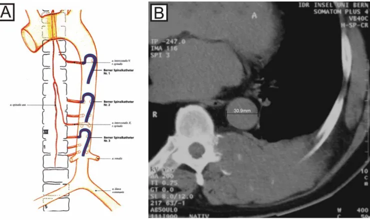
when stable position is achieved, direct coaxial introduc-tion of the microcatheter (usually a Tracker 10 or 18, Target Therapeutics, Fremont, CA, USA) is possible. Prior to the embolization procedure, the diameter of the aorta at the level of the tumor was measured, based on the CT image. Depending on the aortic diameter, one of three different catheter sizes (10 mm, 13 mm or 16 mm: Berner Spinalkatheter I-III) was chosen in order to get optimal stability during the intervention (Fig.1). The angio-graphic protocol included visualization of the vertebral arteries, thyrocervical trunk, costocervical trunk, and external carotid arteries for cervical lesions. This was also done in the segmental arteries within at least two levels above and below the tumor site, for thoracic and upper lumbar lesions; and in the lower lumbar arteries, medial sacral arteries and internal iliac arteries for lower lumbar lesions. In the thoracic and upper lumbar spine, addi-tional localization of the artery of Adamkiewicz was performed to avoid damage of the spinal cord during embolization or corporectomy. Selective angiography of the suspected feeding arteries was then performed. Depending on the tumor vascularization on the arterial angiogram, all lesions were assigned to three groups by
two independent radiologists (S.D.S., G.S.): group I for weak tumor blush after contrast injection, group II for medium tumor blush and group III for significant tumor blush. Polyvinyl alcohol (PVA) particles ranging in size from 45 lm to 350 lm were injected via a microcatheter (Tracker 10 or 18) coaxially inserted through the guiding catheter. In three cases coils were used to obliterate the segmental artery feeding the tumor. Intraoperative blood loss was determined by volume registration from the suction unit.
Surgical procedures
All the surgical procedures were performed at a mean of 2 days (range 0–14 days) after embolization. Anterior, posterior or combined anterior–posterior approaches were used depending on maximal tumor mass. Blood loss was estimated by the anesthesiologist.
Results
Angiographic results
Angiography and embolization could be performed in all patients without causing permanent neurologic deficit, skin or muscle necrosis. All tumors showed hy-pervascularization compared with the normal vertebral Fig. 1 The three sizes of the Berner Spinalkatheter. A Depending
on the aortic diameter, one of three catheter sizes is chosen in order to get an optimal stability during the intervention. B The inner diameter of this catheter allows its use as a diagnostic catheter and as a guiding catheter
blush on selective spinal angiograms. All tumors were extradural, with three localized in the sacrum, nine in the lumbar, eight in the thoracic and four in the cervical spine. The blood supply to the tumors originated from branches of the iliac artery in two patients, the lumbar segmental artery in seven patients, the intercostal artery in ten patients and from supraaortic vessels in five pa-tients. Tumor vascularization before embolization was weak in three patients (group I), medium in six patients (group II) and extensive in 15 patients (group III). No difference for the vascularization grade was found when comparing the lumbar, the thoracic and the cervical spine with a mean vascularization grade of 2.4, 2.8 and 2.5, respectively. In 22 of 24 patients, near complete embolization of the tumor vessels could be achieved. In two patients (cases 1 and 20), the tumor feeding artery also supplied the artery of Adamkiewicz and, conse-quently, only partial embolization could be performed. The use of different sizes of the Berner Spinalkatheter depending on the aortic diameter at the level of the tu-mor proved to be very useful—it was possible to achieve an optimal catheter stability during embolization. Fig.2
shows an illustrative case.
Surgical results
The tumors were approached from an anterior tran-scavitary route in six patients (25%), a posterior ap-proach in nine patients (37.5%) and a combined approach in nine patients (37.5%). Instrumentation was placed in all patients. Sixty percent of the patients were operated on within 1 day after embolization, 95% within 5 days. Mean duration of surgery was 4.7 h. The mean blood loss for all embolized patients was 1,900 ml. For the two partially embolized patients it was 5,500 ml. The intraoperative blood loss varied significantly, with a maximum of 6,000 ml (partially embolized) and a minimum of 1,000 ml. The mean blood loss only considering the cases of renal cell car-cinoma metastases was 2,400 ml. The mean blood loss estimation for all metastases except the cases of renal cell carcinoma metastases was 1,300 ml. The mean blood loss was 2,300 ml for lumbar, 2,200 ml for tho-racic and 1,350 ml for cervical spine tumors. There was no statistically significant correlation between the vas-cularization grade and the intraoperative blood loss after transarterial embolization (Fig.3). In two of the
Fig. 2 Illustrative case showing a metastasis of a renal cell carcinoma on the level of T7. A The CT section through vertebra T7 shows metastatic osseous destruction, mainly in the posterior part of the verte-bral body, as well as intraspinal epidural extension (arrows). B Selective injection of the segmental artery D7 through the Berner Spinalkatheter shows the tumor blush. C Through the coaxially intro-duced microcatheter (arrows), selective embolization of the tumor-feeding vessels and near complete devascularization can be achieved. D In this case, two coils were introduced in order to seal the segmental artery D7 (arrows)
24 patients (cases 1 and 20) the goal of the surgery could not be attained, due to excessive bleeding. In these patients, both of whom had hypervascularized metastases of a renal cell carcinoma, only a partial tumor reduction was achieved. The initial goal of the surgery for both patients was a vertebrectomy and instrumentation.
Discussion
Extensive surgical procedures in patients with hyper-vascular vertebral tumors are often complicated by excessive blood loss. Therefore, preoperative emboli-zation of spinal tumors is recommended to reduce intraoperative bleeding. Several investigators have re-ported results with preoperative endovascular emboli-zation of hypervascular spinal tumors and clearly showed its benefit in reduction of intraoperative blood loss [3, 4, 5, 6, 10]. In a paper by Berkefeld et al., the estimated intraoperative blood loss was 4,350 ml in non-embolized patients and 1,800 ml in patients with particle embolization [1]. More recently, in a series on spinal metastases from renal cell carcinoma, the median intraoperative blood loss was 1,500 ml (range 300–8,000 ml) [5]. In our series, the estimated mean blood loss for the 22 embolized cases was 1,900 ml. In cervical spine tumors, embolization is more difficult to perform than in lumbar or thoracic spine tumors, due to frequent anastomoses between the carotid, vertebral and subclavian arteries. The risk of cerebral or spinal
cord embolization is increased [12]. In our series no patient suffered neurological deterioration after the embolization procedure. Our results show that after the embolization the mean blood loss for cervical spine tumors was 1,350 ml. This stands in contrast with surgical reports of 9.5–15 l of blood loss when preoperative embolization was not performed [3]. In two patients (cases 1 and 20), only partial emboliza-tion could be performed, and the goal of surgery could not be achieved because of excessive bleeding, with blood losses of 5,000 ml and 6,000 ml respec-tively. These two cases illustrate the importance of preoperative transarterial embolization of hypervascu-lar vertebral metastases and demonstrate its potential influence on surgical success. Grading of tumor vas-cularization based on the tumor blush was thought to be prognostic for the intraoperative blood loss vol-ume. However, we found that there was no significant difference in blood loss among the three groups after radiological complete devascularization (Fig.3). This finding gives further evidence that preoperative embolization is effective.
From a qualitative, surgeons’ point of view, pre-operative tumor embolization facilitated surgical resection, primarily by reducing intraoperative bleeding and, hence, ensuring a good view of the surgical field. Furthermore, it increased the cleavage between tumor and dura or bone, which resulted from tumor shrink-age.
Choosing the size of our Berner Spinalkatheter based on the diameter of the aorta at the level of the involved segmental artery resulted in reduction of the time needed for stable positioning in thoracic and upper lumbar procedures. It led to a reduction of the number of catheters used for selective catheterization of the tho-racic and lumbar segmental arteries.
Conclusion
Preoperative transarterial embolization of hypervascular spinal tumors is a safe and effective procedure. Preop-erative embolization in hypervascular vertebral tumors reduces the perioperative blood loss and facilitates tumor resection. Appropriate techniques allow a considerable reduction of intervention time. Adapted guiding-catheter sizes increase the catheter stability and, therefore, reduce the risk of microcatheter displacement during embolization.
Acknowledgements Special thanks go to Mrs. Fu¨ gliskater from Johnson & Johnson, Division Cordis, Switzerland for supplying the illustration of the Berner Spinalkatheter I-III
Fig. 3 Bar plot showing the relation between the estimated blood loss in liters (y-axis) and the preembolization vascularization degree (x-axis). There is no statistically significant difference between the three groups
References
1. Berkfeld J, Scale D, Kirchner J, Heinrich T, Kollath J (1999) Hy-pervascular spinal tumors: Influence of the embolization technique on perioperative hemorrhage. AJNR AM J Neuroradiol 20:757–763
2. Boland PJ, Lene JM, Sundersen N (1982) Metastatic disease of the spine. Clin Orthop 1169:95–102
3. Gellad FE, Sadato N, Numaguchi Y, Levine AM (1990) Vascular metastatic lesions of the spine: preoperative embolization. Radiology 176:683–686 4. Go¨rich J, Solymosi L, Hasan I, Sittek
H, Majdali R, Reiser M (1995) Embolisation von Knochenmetastasen. Radiologe 35:55–59
5. Manke C, Bretschneider T, Lenhart M, Strotzer M, Neumann C, Gmeinwieser J, Feuerbach S (2001) Spinal metastases from renal cell carcinoma: effect of preoperative particle embolization on intraoperative blood loss. AJNR AM J Neuroradiol 22:997–1,003
6. Olerud C, Jonsson H Jr, Lofberg AM, Lorelius LE, Sjostrom L (1993) Embo-lization of spinal metastases reduces perioperative blood loss. 21 patients operated on for renal cell carcinoma. Acta Orthop Scand 64:9–12
7. Shi HB, Suh DC, Lee HK, Lim SM, Kim DH, Choi CG, Lee CS, Rhim SC (1999) Preoperative transarterial embo-lization of spinal tumors: emboembo-lization techniques and results. AJNR AM J Neuroradiol 20:2,009–2,015 8. Siegal T, Tiqva P, Siegal T (1985)
Vertebral body resection for epidural compression by malignant tumors, results of 47 consecutive operative procedures. J Bone Joint Surg 67:375– 382
9. Sundaresan N, Gailicich JH, Lane JM, Bains MS, McCormack P (1985) Treatment of epidural cord compression by vertebral body resection and stabilization. J Neurosurg 63:676–684 10. Sundaresan N, Choi IS, Hughes JEO,
Sachdev VP, Berenstein A (1990) Treatment of spinal metastases from kidney cancer by presurgical emboliza-tion and resecemboliza-tion. J Neurosurg 73:548– 554
11. Tadavarthy SM, Mo¨ller JH (1974) Polyvinyl alcohol (Ivalon): a new embolic material. AJR AM J Roentge-nol 125:609–616
12. Vetter SC, Strecker EP, Ackermann LW, Harms J (1997) Preoperative embolization of cervical spine tumors. Cardiovasc Intervent Radiol