Surg Radiol Anat (1998) 20:311-316
Surgical <
Radiologlc
Ana rny
Journal of Clinical Anatomy © Springer--Verlag 1998The reversed-flow medio-distal fasciocutaneous island thigh flap:
anatomic basis and clinical applications
F.T. Ballmer 1 and A.C. Masquelet 2
: Department of Orthopaedic Surgery, University of Berne, Inselspital, CH-3010 Berne, Switzerland
2 Service de Chirurgie Orthop6dique et R@aratrice, HSpital Avicenne, 125, route de Stalingrad, F-93000 Bobigny, France
S u m m a r y : A new f a s c i o c u t a n e o u s
reversed-flow island flap of the thigh is presented which is independent of the presence of perfused blood vessels below the level of the knee joint-line. The pedicle, which is supplied by the proxi- mal genicular a n a s t o m o t i c network, consists of the osteoarticular branch (OAB) and concomitant veins of the des- cending genicular artery. Based on cada- ver dissections the OAB arose in 23/30 s p e c i m e n s (77%) t o g e t h e r with the saphenous a~ery (SA). In 2/30 specimens (7%) the OAB originated directly from the superficial femoral artery and in 1/30 specimens (3%) the OAB was absent. The OAB gave off one to three cutaneous branches to the overlying skin in 26/30 specimens (87%). We were able to eleva- te a flap on the osteoarticular branch alone in 57%. Additional length could be added to the pedicle in 33% by including the most proximal part of the saphenous artery together with its first cutaneous branch. Thus, in 90% of the dissections a reversed-flow island flap could be raised which reached the proximal half of the
Correspondence to: F.T. Ballmer
leg, the knee and the most distal part of the thigh. We report our early clinical experience.
I 2 lambeau fascio-cutan6 m6dio-distal de la cuisse
flux invers6. Bases anatomiques et applications eliniques
R~sum6 : Un nouveau lambeau fascio-
cutan6 en ilot de la cuisse ~ flux invers6 est pr6sent6, dont la caract6ristique est d'etre ind6pendant de la pr6sence de vais- seaux sanguins perfus6s au-dessous du niveau de l ' i n t e r l i g n e articulaire du genou. Le p6dicule est constitu6 par le r6seau anastomotique proximal du genou centr6 sur le rameau ost6o-articulaire (ROA) de l'artbre descendante du genou (ADG) et ses veines satellites. Ce ROA a dt6 6tudi6 sur des dissections cadav6- riques, il naissait 23 fois sur 30 (77%) d'un tronc commun avec l'art~re saph~ne (AS), dans 2 cas sur 30 (7%) il naissait directement de l'art~re f6morale superfi- cielle, et dans 1 cas sur 30 (3%) il 6tait absent. Le R O A a b a n d o n n a i t 1 ~t 3 branches cutan6es aux t6guments adja- cents sur 26 des 30 sp6cimens (87%). I1 rut possible de lever un lambeau centr6 sur le ROA seul dans 57% des cas. I1 est p o s s i b l e d ' a l l o n g e r le p d d i c u l e en
incluant la partie la plus proximale de l'artare saphbne et sa premiere branche cutan6e dans 33 % des cas. En recourant ou non 5 cet artifice, sur 90% des dissec- tions, it aurait 6t~ possible de lever un lambeau en ilot h flux invers6 qui aurait atteint la partie proximale de la jambe, le genou et la partie le plus distale de la cuisse. Nos premiers cas cliniques sont pr6sent6s.
Key words: Fascio-cutaneous flap - -
Lower limb - - Descending genicular artery - - Osteoarticular branch
Soft tissue coverage of defects around the knee remains a reconsmactive challenge. Muscular or fasciocutaneous flaps from the calf are not always available, particu- larly if there has been previous trauma, utilization, o1" in through-kmee and espe- cially below-knee amputations. Various procedures for these difficult situations, including the distally based vastus latera- lis muscle flap [13], the vastus medialis muscle as a rotation flap [2] or as advan- cement flap [15], the popliteo-posterior thigh fasciocutaneous island flap [12], the lower posterolateral thigh flap [9], cross- leg flaps [3] and free flaps [7] have been described to accomplish this end. Other options are the reversed-flow saphenous
312 F.F. Ballmer and A.C. Masquelet: Medio-distal fasciocutaneous island thigh flap island flap [16], the saphenous postero-
medial cutaneous island thigh flap and the saphenous superomedial cutaneous island leg flap [5], the medial septocuta- neous island thigh flap [4], the sartorius myocutaneous island flap [14], the rever- sed flow saphenous island flap based on the medial inferior genicular a. [17], and the adipofascial flap based on the saphe- nous a. [10]. I n s p i r e d b y a p r e v i o u s report by Hegel and Masquelet [6], des- cribing a r e v e r s e d - f l o w m e d i a l knee osteoperiosteal flap, we present an anato- mic study and clinical experience of a new skin flap for closure of soft tissue defects around the knee and proximal half of the leg. This flap is a fasciocuta- neous island flap and is harvested from the medial aspect of the lower half of the thigh. It is supplied by reversed-flow in the osteoarticular branch of the descen- ding genicular a., thus being independent of the presence of perfused blood vessels below the level of the knee joint-line. Material and methods
The anatomic study was carried out on 15 fresh cadavers (30 dissections) of both sexes, and the ages ranged from the fifth to the ninth decade. Both legs were injec- ted with colored neoprene latex via the femoral a., and dissections were carried out after 2 days. Technical details of the dissection are described in the section. "Flap design and operative procedure". The vascular anatomy of the descending genicular a. ( D G A ) was studied with emphasis on the different combinations of its branches (the osteoarticular branch (OAB), the saphenous a. (SA) and the muscular branch (MB)). Special attention was given to the presence o f cutaneous branches arising from the O A B and to anastomoses of the latter with the arterial anastomotic circle of the knee. The geni- cular a n a s t o m o s e s are f o r m e d by the medial and lateral superior genicular, medial and lateral inferior genicular, middle genicular, anterior and posterior tibial recurrent, descending branch of the lateral circumflex femoral and circumflex fibular aa. [8, 18].
The following data were recorded: a) the distances between the medial joint-line of the knee and the origins of
the DGA, OAB, SA and MB,
b) the diameter at their origin of the DGA, OAB, SA and MB,
c) the length of the D G A before its division,
d) the number and diameter at their origins of the cutaneous branches of the OAB,
e) the distance between the medial joint-line of the knee and the origin of the
cutaneous branches of the OAB,
f) the pedicle lengths of two different vascular patterns (ie O A B alone and OAB + SA).
Clinical e x p e r i e n c e includes two patients with soft tissue defects around the knee.
Results
Anatomic study
Descending genicular a. (DGA)
The distal superficial femoral a. (SFA) or the proximal popliteal a. gave off three vascular axes: the OAB, the SA and the MB, each accompanied by two venae comitantes. W h e n two or three o f the above mentioned arteries arose from a common trunk, the latter was called the D G A [6]. In 27/30 specimens (90%) a D G A was found. The D G A arose from the SFA at an average of 14 cm (range 18.5 - 11.5 cm) above the medial joint line. Its average diameter at its origin measured 2.0 m m (range 1.5 - 2.6 ram). Within 1.6 cm of its origin (range 0.2 - 3.5 cm) it divided into its branches. The c o m b i n a t i o n s o f vessels f o r m i n g the D G A and their frequency are listed in Table 1.
Oswoarticular branch ( OAB )
T h e O A B arose in 2 3 / 3 0 s p e c i m e n s (77%) together with the S A ( O A B + SA + MB and OAB + SA). In 2/30 speci- mens (7%) the OAB originated directly f r o m the SFA. In 1/30 (3%) the O A B was absent. The OAB arose at an average of 12 cm (range 10 - 18 cm) above the medial joint-line. Its mean diameter at its point of origin was 1.3 m m (range 0.6 - 2 . 0 ram). The OAB ran in a distal direction on the posterior surface of the medial intermuscular septum alongside the adductor magnus tendon. In 16/30
Table 1. Combinations of vessels forming the descending genicular a. (DGA) and their frequen- cy (OAB = osteoarticular branch, SA = saphenous a., MB = muscular branch)
OAB + SA + MB 11/30 (37%)
OAB + SA 12/30 (40%)
OAB + MB 4/30 (13%)
No DGA 3/30 (10%)
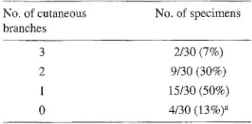
Table 2. Numbers of cutaneous branches arising from the OAB per dissected specimen (a including one specimen lacking an OAB)
No. of cutaneous No. of specimens
branches
3 2/30 (7%)
2 9/30 (30%)
1 15/30 (50%)
0 4/30 (13%) a
specimens (53%) the OAB divided into a medial and a lateral branch after having roached the medial femoral condyle, and in the remaining cases it divided more proximally. The terminal vessels of the lateral branch, spreading over the medial femoral condyle, constantly anastomosed with the genicular anastomoses, ie the medial superior genicular a. and, in many cases, through a well-developed anterior anastomosis with the lateral superior genicular a. The medial branch o f the OAB ran distally in front of the medial collateral ligament, crossing the joint line and anastomosing with the inferior geni- cular anastomoses, primarily with the medial inferior genicular a. The O A B gave off one to three cutaneous branches to the overlying skin in 26/30 specimens (87%). F o r n u m b e r s o f c u t a n e o u s branches per dissected s p e c i m e n see Table 2. The mean diameter of the cuta- n e o u s b r a n c h e s at their o r i g i n w a s 0.6 m m (range 0.2 - 0.9 ram). They arose between 3 and 14 cm above the medial joint-line. The cutaneous branches bifur- cated into a s c e n d i n g and d e s c e n d i n g branches, which formed a vascular arca- de with the n e x t a s c e n d i n g b r a n c h .
Saphenous a. (SA)
The origin of the SA was at an average of 12.5 cm (range 9.5 - 19 cm) above the medial joint-line. Its mean diameter at its source was 1.2 m m (range 0.7 - 1.8 mm).
F.F. Ballmer and A.C. Masquelet: Medio-distal fasciocutaneous island thigh flap 313
When originating in the adductor canal, it pierced the aponeurotic roof and came to lie in the loose fascial space bounded by the sartorius m. superficially, the adduc- tor magnus tendon posteriorly, and the vastus medialis m. anterolaterally. It ran distally in this space, finally passing bet- ween the sartorius and gracilis mm. to reach the medial side of the leg. The SA gave constantly branches to the overlying skin and to the sartorius m.
Muscular branch (MB)
The MB was not part of the pedicle of the flap but data are listed because of its ana- tomic proximity. It arose at an average of 12 cm (range 7 - 18.5 cm) above the medial joint-line and entered the vastus medialis m. after an average distance of 0.5 cm. Its mean diameter at the origin was 1.4 m m (range 0.6 - 2.5 ram). Addi- tional muscular branches to the vastus m e d i a l i s m. m a y o r i g i n a t e f r o m the DGA, OAB or SFA.
Venous drainage
In general, the venae comitantes of the three arterial axes joined to form the des- cending genicular v. which unified with the superficial f e m o r a l v. The v e n a e comitantes occasionally drained as two or three separate branches directly into the femoral v.
Patterns and lengths of the pedicles
Several factors determined the patterns and lengths of the pedicle: the localiza- tion of the origin of a reliable cutaneous branch from the OAB and the presence of a common trunk formed by the OAB and SA, as well as the very rare possibili- ty of absence of the OAB.A. The OAB was present and gave off a reliable cutaneous branch near its origin (Fig. 1). This pattern was encoun- tered in 17/30 specimens (57%). With this pattern the presence of a c o m m o n trunk (i.e. OAB + SA) is irrelevant, and the procedure is straightforward. After ligature of the OAB and its venae comi- tantes proximal to its cutaneous branch, a reversed-flow fasciocutaneous island flap could be raised tracing the vascular axis distally. This flap could be rotated on a
SFA SFA
Fig. 1
Medial view of right medio-distal thigh (on the right, proximal thigh; on the left, knee joint and proximal calf): The osteoar- ticular branch (OAB) gives off a good proximal (PCB)
and distal cutaneous branch
(DCB) . A reversedMflow
island flap can be raised after ligature of the OAB
proximal to the proximal c u t a n e o u s branch. The saphenous a. (SA) and mus- cular b r a n c h (MB) arise directly from the superficial femoral a. (SFA)
~
GAJ/\ y
J J b Fig. 2a, ba The OAB gives off no cutaneous branch but arises from a common trunk (ie DGA) with the SA which constantly gives off cutaneous branches. Note the anastomoses of the OAB with the superomedial (SMGA)
and superolateral genicular aa. (SLGA). b A reversed-flow island flap based on the OAB can be raised after ligature of the common trunk (i.e. DGA) and of the SA distal to its cutaneous branch. This pattern leng- thens the pedicle.
pivotal point in the area of the internal femoral condyle, where the vessels ana- stomosed constantly with the genicular anastomoses. Both the arterial supply and the venous drainage were retrograde in this situation. The mean pedicle length was 14 cm (range 10 - 15.5 cm).
B. The OAB was present and gave off a distal cutaneous branch, or the cuta- neous branch was absent (Fig. 2a), but the OAB arose from a common trunk (ie DGA) with the SA. The latter gave off constant cutaneous branches proximal to
the joint-line [4, 5]. This pattern was found in 10/30 specimens (33%). Note that the origin of a cutaneous branch f r o m the O A B was regarded as distal when originating in the distal half of the distance between the origin of the OAB and the joint-line. The SA and its venae comitantes were ligated proximal to the junction with the OAB (ie DGA) and dis- tal to the origin of the cutaneous branch (from the SA). A reversed-flow island flap could then be raised based on the OAB and SA with the constant cutaneous
314 F.F, Ballmer and A.C. MasqueIet: Medio-distal fasciocntaneous island thigh flap
Fig. 3
Arc of rotation: the proximaI half of the leg, the knee and the most distal part of the thigh can be reached
OAB with cutaneous branch near its origin
Yes No
i Reversed-flow flap OAB with or without cutaneus branch
on OAB Common trunk of OAB and SA
lISA retrogradely perfused i
...... .. .... ..
Reversed-flow flap i ,: Fitting flap in ; on SA ; : donor site defect : _ Yes /
Reversed-flow flap on OAB and SA
Fig. 5
Algorithm of intraoperative decision-making
C. The OAB was present and gave off a distal cutaneous branch, or the cuta- neous branch was absent but the OAB formed no c o m m o n trunk with the SA. The possible presence of a common trunk f o r m e d by the OAB and MB was not helpful for adding extra length to the pedicle due to the anatomic course and shortness of the MB. Finally, very rarely the OAB was absent. One of the afore- mentioned patterns was found in 3/30 specimens (10%), and a flap could not be raised as described in section A or B. If a retrograde circulation of the saphenous a., fed by perforating branches of the pos- terior tibial a. and the medial inferior genicular a., was present, it was possible to switch to a reversed-flow saphenous island flap as a salvage procedure (Fig. 4;1 [4, 5, 16, 17].
branch of the latter (Fig. 2b). The arterial flow and venous drainage were retrogra- de in the OAB, and orthograde in the SA and in its cutaneous branch. Choosing the pivotal point again in the region of the internal femoral condyle, the arterial sup- ply and venous drainage were the same as described above. The mean pedicle length was 17 cm (range 13.5 - 18 cm). Note that the mean pedicle length was 3 cm greater in this pattern, as the length of the SA from its origin to its cutaneous branch w a s added to the length o f the OAB. With patterns A and B the proxi- mal half of the leg could easily be rea- ched by adding a flap size of at least 15 x 5 cm. Furthermore, the arc of rotation of the flap a l l o w e d c o v e r a g e o f d e f e c t s around the knee and the most distal part of the thigh (Fig. 3).
Fig. 4
Reversed-flow saphenous island flap as alternative which is only possible if the saphenous a, is retro- gradely perfused
The algorithm of intraoperative deci- s i o n - m a k i n g is s u m m a r i z e d in Fig. 5.
Flap design and operative procedure
The patient is supine with a wedge under the opposite buttock to expose the medial aspect of the thigh. Before applying the tourniquet, the flap is outlined over the lower medial aspect o f the thigh. The pivotal point is located a p p r o x i m a t e l y over the palpable insertion of the adduc- tor magnus tendon. Elevation of the flap should include the aponeurosis of the vastus medialis m. and is carried out from the anterior incision, identifying any and all cutaneous branches and their origin. Careful elevation of the vastus medialis In. in a distal to p r o x i m a l d i r e c t i o n exposes the OAB, which lies just poste- rior to the medial intermuscutar septum. Following the
OAB
proximally to its ori- gin, the pattern of the D G A can be deter- mined. Depending on the anatomy and the desired length of the pedicle, one of the a b o v e - d e s c r i b e d p o s s i b l e p e d i c l e types chosen. It is very important to pro- tect the venae comitantes by leaving an adjacent strip of the medial intermuscular septum. After complete dissection of the pedicle, the skin layer is raised b y corn-F.F. Ballmer and A.C. Masquelet: Medio-distal fasciocutaneous island thigh flap 315
Fig. 6a-d
a Left below-knee amputation stump with a chronic pressure sore of the scar. b Outline of a 13 x 5 cm island flap on the medial thigh, c After elevation of the fas- ciocutaneous flap the anastomoses of the OAB with the vascular network on the medial femoral condyle are visible, d After generous excision of the scar the stump was covered and the donor side defect primarily closed
pleting the incision of the dorsal half. The donor defect is closed directly, if the skin layer does not exceed 6-7 cm in width. T h e l e n g t h a n d the f l e x i b i l i t y o f the p e d i c l e a l l o w r o t a t i o n o f the f l a p in various planes to perfectly fit in a defect.
Clinical applications
Case 1
A chronic pressure sore of the scar in a p a t i e n t w i t h a t r a u m a t i c b e l o w - k n e e amputation was present (Fig. 6a). A 13 x 5 c m r e v e r s e d - f l o w f a s c i o c u t a n e o u s island flap from the medio-distal thigh (Fig. 6b) was elevated, and the pedicte, consisting of the OAB and concomitant veins, was dissected distally to the ana- stomoses in the area of the medial femo-
ral condyle (Fig. 6c). Generous excision of the unstable scar and vulnerable skin o f the stump was p e r f o r m e d (Fig. 6d). The defect of the stump was covered by rotating the flap in the frontal and axial planes. The donor site defect was closed directly. The transferred flap was well perfused. The postoperative course was uneventful, with complete flap survival. Case 2
This patient had a patellar fracture which was approached through a longitudinal incision for open reduction and internal fixation. Postoperatively, skin necrosis developed with eventual exposure of the patella and the implants. After debride- ment the prepatellar defect was covered with the flap d e s c r i b e d in this article.
D i s c u s s i o n
Reversed-flow island flaps can be trans- ferred from a proximal to a distal part of the extremity. Harvested from the thigh, t h e y are thus u s e f u l to c o v e r d e f e c t s around the knee and the proximal half of the leg. To the best of our knowledge, the s a p h e n o u s i s l a n d f l a p b y T o r i i [16], b a s e d on the o r i g i n a l d e s c r i p t i o n b y Acland [1], the saphenous posteromedial cutaneous island thigh flap and the saphe- nous superomedial cutaneous island leg flap [5], the medial septocutaneous island thigh flap [4], the sartorius myocutaneous island flap [14], the reversed-flow saphe- nous island flap based on the medial infe- rior genicular a. [17], and the adipofascial flap of the lower leg based on the saphe- nous a. [10] are the only reversed-flow
316
fasciocutaneous island flaps of this anato- mic region. All these flaps are supplied by a retrograde circulation through ana- stomoses of the saphenous a. with the perforating branches of the posterior final and medial inferior genicular aa. After previous trauma these perforators may be damaged or even absent, as in through-knee amputation. T h e r e f o r e these flaps may all be unavailable.
In this article another reversed-flow fasciocutaneous island thigh flap is added to the armamentmium of the reconstructi- ve surgeon. In contrast, this flap is sup- plied by reversed-flow in the osteoarticu- lar branch of the descending genicular a. As the osteoaa:ticular branch anastomoses with the proximal genicular anastomotic network, ie mainly the medial superior genicular a., the presence of perfused blood vessels below the level of the knee joint-line is not a prerequisite. The slen- der and relatively long vascular pedicle allows rotation of the flap in different planes to perfectly fit in the defect.
Most of our anatomic data on the arteries forming the DGA are in good correspondence with the literature [4, 6]. Only the incidence of an independent ori- gin of these vessels from the femoral a. was found to be slightly higher by Bertel- li [4]. The f r e q u e n c y of cutaneous branches originating from the OAB in our dissections is consistent with the data published by Martin [11], who found in 31/36 specimens (86%) cutaneous branches from the OAB. Therefore it is theoretically possible to raise a flap on the OAB alone in more than 80% of cases. But to raise the longest possible pedicte we have chosen a vascular axis formed by the OAB and SA in 33% of our specimens. In 10% of our dissections it was not possible to raise a flap based on the OAB, or OAB and SA. Provided that the saphenous a. is retrogradely per- fused by the perforating branches of the posterior tibial and medial inferior geni- cular aa., a reversed-flow saphenous island flap may be elevated under these circumstances (Fig. 4) [4, 5, 16, t7]. However, it must be remembered that the flap presented in this study and based on the OAB or OAB and SA is not depen- dent on perfused blood vessels below the level of the joint-line of the knee.
F.F. Ballmer and A.C. Masquelet: Other methods for closure of defects around the knee, if flaps below the knee are unavailable, have been described. The distally based vastus lateratis muscle flap [13] i s bulky and delayed skin grafting due to superficial muscular necrosis has been observed by the authors. The vastus medialis m. as a rotation flap [2] covers only defects in the upper part of the knee and the vastus medialis myocutaneous/ myocutaneous-tendinous advancement flap [15] is useful only for small defects. If muscle flaps are used one should consider the degree of functional loss. The popliteo-posterior island [12] and posterolateral thigh flaps [9] are similar fasciocutaneous flaps, but require the lateral or prone position; which may be a disadvantage in anterior soft tissue defects. Free tissue transfer [7] is compli- cated, less economical and requires microsurgical expertise. The cross-leg flap [3] is a multi-stage procedure and the lower extremities are immobilized in an uncomfortable position over an extended time.
Conclusion
We believe that the reversed-flow fascio- cutaneous island flap raised on the OAB, or on the OAB and SA, has some advan- tages and will be a valuable addition to the armamentarium of procedures for coverage of soft-tissue defects around the knee and proximal half of the leg, when muscular or fasciocutaneous flaps from the calf are not otherwise available.
Acknowledgments This work was supported by a
grant from the Swiss Orthopaedic Society (SGO), by the Department of Orthopaedic Surgery, Insels- pital, University of Berne (Switzerland) and by the Laboratory of Anatomy, Ren6 Descartes Universi- ty, Paris (France).
References
1. Acland RD, Schusterman M, Godina M, Eder E, Taylor GI, Carlisle I (1981) The saphenous neurovascutar free flap. Plast Reconstr Surg 67:763-774
2, Arnold PG, Prunes-Carrillo F (1981) Vastus medialis muscle flap for functional closure of the exposed knee Joint. Plast Reconstr Surg 68:69-72
3. Barclay TL, Sharpe DT, C h i s h o l m EM (1983) Cross-leg fasciocutaneous flaps. Plast Reconstr Surg 72:843-846
Medio-distal fasciocutaneous island thigh flap 4. BertelIi JA (1992) The medial septumcuta- neous island thigh flap (25.10.91). Surg Radiol Anat 14:191-192
5. Bertelli JA (1992) The saphenous postero- medial cutaneous island thigh flap and the sapbenous supero-medial cutalaeous island leg flap (25.10.91). Surg Radiol Anat 14: 187-189
6. HerteI R, Masquelet AC (1989) The reversed- flow medial knee osteoperiosteal flap for ske- letal reconstruction of the leg. Description and anatomical basis. Surg RadioI Anat
11:257-262
7. Kasabian AK, Colen SR, Shaw WW, Pachter HL (1991) The role of microvascular free flaps in salvaging below-knee amputation stumps: A Review of 22 Cases. J Trauma 31: 495-501
8. Kirschner MH, Menck J, Hofmann GO (1996) Anatomic bases of a vascularized allo- genic knee joint transplantation: arterial Mood supply of the human knee joint. Surg RadioI Anat 18:263-269
9. Laitung JKG (1989) The lower posterolateral thigh flap. Br J Plast Surg 42:133-139 10. Lin SD, Lai CS, Chiu YT, Lin TM, Chou CK
(1996) Adipofascial flap of the lower leg based on the saphenous artery. Br J Plast Surg 49:390-395
11. Martin D, Bitonti-Grillo C, De Biscop J, Schott H, Mondie JM, Baudet J, Peri G (1991) Mandibular reconstruction using a free vascularised osteocutaneous flap from the internal condyle of the femur. Br J Plast Surg 42:397-402
12. Maruyama Y, Iwahira Y (1989) Popliteo-pos- terior thigh fasciocutaneous island flap for closure around the knee. Br J Plast Surg 42: 140-143
13. Swartz WM, Ramasastry SS, McGill JR, Noonan JD (1987) Distally Based Vastus Lateralis Muscle Flap for Coverage of Wounds About the Knee. Plast Reconstr Surg 80:255-263
14. Tang ML, Liu XY, Ren JW, Zhang DC, Li RS, Wen YM, Ge BF (1993) The sartorius myocutaneous island flap. Surg Radiol Anat 15:259-263
15. Tobin GR (1985) Vastus Medialis Myocuta- neous and myocutaneous-tendinous composi- te flaps. Plast Reconstr Surg 75:677-684 16. Torii S, Hayashi Y, Hasegawa M, Sugiura S
(1989) Reversed-flow saphenous island flap in the patient with below-knee amputation. Br J Plast Surg 42:517-520
17. Tsai CC, Lin SD, Lai CS, Chou CK, Lin TM (1995) Reconstruction of the upper Leg and knee with a reversed flow saphenous island flap based on the medial inferior genicular artery. Ann Plast Surg 35:480-484
18. Williams PL, Warwick R, Dyson M, Bannis- ter LH (1989) Gray's anatomy. Churchill Livingstone, Edinburgh London Melbourne New York
Received Februa~ 23, 1998 / Accepted in final form July 7, 1998