Relation between Disease Phenotype and HLA-DQ Genotype in Diabetic
Patients Diagnosed in Early Adulthood
ILSE WEETS1,VALERIE SIRAUX2, JEAN-CLAUDE DAUBRESSE3, IVO H. DE LEEUW5, FRANÇOISE
FÉRY2, BART KEYMEULEN1, GEORGES KRZENTOWSKI3, MICHEL LETIEXHE4, CHANTAL
MATHIEU6, FRANK NOBELS8, RAOUL ROTTIERS7, ANDRÉ SCHEEN4, LUC VAN GAAL5, FRANS C.
SCHUIT1, BART VAN DER AUWERA1, MAO RUI1, METER DE PAUW1, LEONARD KAUFMAN9,
FRANS K. GORUS1, THE BELGIAN DIABETES REGISTRY10
1 Diabetes Research Center (I.W., B.K., F.C.S., B.V.D.A., M.R., P.D.P., F.K.G.), Vrije Universiteit Brussel, B-l090 Brussels, Belgium; 2 Department of Endocrinology (V.S., F.F.), Hôpital Erasme, B-l 070 Brussels, Belgium;
3 Department of Endocrinology (J.-C.D., G.K.), Centre Hospitalier Universitaire, B-6000 Charleroi/B6040 Jumet, Belgium; 4Department of Diabetology (A.S., M.L.), Centre Hospitalier Universitaire Sart Tilman, B-4000 Liege, Belgium;
5 Department of Endocrinology (I.H.D.L., L.V.G.), Universitair Ziekenhuis Antwerpen, B-2650 Antwerp, Belgium; 6 Department of Endocrinology (C.M.), Universitaire Ziekenhuizen Gasthuisberg, B-3000 Leuven, Belgium; 7Department of Endocrinology (R.R.), Universitair Ziekenhuis Gent, B-9000 Ghent, Belgium;
8 Department of Endocrinology (F.N.), OLV Ziekenhuis, B-9300 Aalst, Belgium; 9 Department of Biostatistics (L.K.), Vrije Universiteit Brussel, B-l 090 Brussels, Belgium; 10 and Belgian Diabetes Registry, B-l 090 Brussels, Belgium
Abbreviations:
BDR Belgian Diabetes Registry BMI body mass index
CI confidence interval
GADA glutamate decarboxylase autoantibodies HbAlc glycosylated hemoglobin
HLA human leukocyte antigen genes IAA insulin autoantibodies
IA-2A insulinoma-associated antigen 2 antibodies ICA islet cell antibodies
INS insulin (gene)
JDF Juvenile Diabetes Foundation OR odds ratio
We investigated inaugural disease phenotype in relation to the presence or absence of diabetes-associated autoantibodies and human leukocyte antigen (HLA) DQ risk genotypes in adult-onset diabetic patients. Blood samples and questionnaires were obtained from 1584 recent-onset Belgian Caucasian patients (age 15-39 yr at diagnosis of primary diabetes) who were recruited by the Belgian Diabetes Registry over an 11-yr period. At clinical diagnosis, antibody-positive patients (n = 1198) were on average younger and had more symptoms, a more acute disease onset, lower body mass index, and random C-peptide levels, but higher insulin needs, glycemia, and prevalence of ketonuria, HLA-DQ, and 5' insulin gene susceptibility genotypes (P < 0.001 vs. antibody-negative patients; n = 386). In antibody-positive patients, these characteristics did not differ according to HLA-DQ genotype. However, in antibody-negative subjects, we found that patients were younger (P = 0.001); had a lower body mass index (P < 0.001), higher insulin needs (P = 0.014), and amylasemia (P = 0.001); and tended to have a higher glycemia and lower C-peptide in the presence of susceptible HLA-DQ genotypes. Differences according to HLA-DQ genotype subsisted after careful age-matching. In conclusion, we found no relation between initial disease phenotype and HLA-DQ genotype in antibody-positive diabetic young adults. In contrast, antibody-negative patients displayed more type 1-like features when carrying susceptible HLA-DQ genotypes known to promote the development of antibody-positive diabetes. The overrepresenta- tion of these susceptibility genotypes in antibody-negative patients suggests the existence of an immune-mediated disease process with as yet unidentified immune markers in a subgroup of seronegative patients. ( J Clin Endocrinol Metab 87: 2597-2605, 2002)
MOST CASES OF type 1 diabetes are believed to arise from the nearly complete destruction of insulin-producing β-cells by immune effector cells [1] . Although there are indications that the underlying pathological
process is often initiated in early childhood, the disease can become clinically manifest at any age, most cases arising after age 15 yr [1] [2] [3] [4] [5]. The disease presents along a clinical spectrum ranging from acute-onset
ketosis-prone diabetes to latent forms with slow progression to insulin deficiency after initial manifestation [6][7][8] [9]. Disease phenotype, underlying pathologic changes, and associated biological markers tend to change with age
diabetes from other forms of the disease with older age at clinical manifestation[3][4] [6] [9] . There is thus a need
for additional biological markers of adult-onset type 1 diabetes.
The American Diabetes Association and the World Health Organization recently proposed new classification criteria for diabetes in which the presence or absence of diabetes-associated autoantibodies plays a central role
[7][8]. Immunemediated type 1 diabetes (type 1A) is defined as hyperglycemia in the presence of autoantibodies
against β-cell antigens [7][8]. These patients preferentially carry specific HLA-DQA1-DQB1 risk haplotypes
(0301-0302 and/or 0501-0201) [7][8] . In initially noninsulin-requiring adult-onset patients, the presence of
autoantibodies has been associated with lower body mass index (BMI), less preserved residual β-cell mass, and more rapid evolution toward insulin dependency [6][16][17]. However, a number of diabetic patients, mostly of
African or Asian origin, presents with variable degrees of insulin deficiency in the absence of circulating diabetes autoantibodies [7][8]. A recent Japanese study reported a subgroup of diabetic patients with abrupt
clinical onset of diabetes in adulthood without detectable insulitis or diabetes-associated autoantibodies, but characterized by the presence of hyperamylasemia, hyperlipasemia, and lymphocytic infiltration of the exocrine pancreas [19]. Whether this subtype occurs in Caucasian populations has so far not been confirmed [20][21]. The
absence of autoantibodies at diagnosis does, however, not exclude the existence of an immunemediated β-cell destruction because we and others have occasionally observed the appearance of diabetes-associated
autoantibodies after clinical onset[22][23]. Likewise, it is conceivable that some patients may have lost
autoantibodies before diagnosis or present other immune markers than those currently searched for[23] . To
further improve the etiological classification of adult-onset diabetes, and hence etiology-based therapeutic schemes, it is important to try to relate clinical disease phenotype to biological markers of immune-mediated diabetes also in the absence of autoantibodies.
Therefore, the aims of the present report were: 1) to study the clinical phenotype of adult-onset diabetic patients after stratification for diabetes-associated autoantibodies, and 2) to investigate the possible interaction between the disease phenotype and HLA-DQ genotype in a representative population of adult-onset diabetic patients stratified according to the presence or absence of autoantibodies.
Figure 1. Number of included diabetic patients aged 15-39 yr, according to antibody status and HLA-DQ genotype.a Absence of gestational or secondary diabetes; Caucasian ethnicity; availability of demographic data
and blood samples for DNA, autoantibody, and chemical analysis within 18 months after diagnosis; and Belgian residency for at least 6 months before diagnosis.b From 1488 of 1584 included patients (94%), a standard
questionnaire was available with epidemiological, clinical, and locally determined biological data.c Positivity
for ICA, GADA, and/or IA-2A; negativity for ICA, GAD A, and IA-2A. d For the definition of HLA-DQ
SUBJECTS AND METHODS Subjects
Between January 1989 and December 1999, 1721 patients (age 15-39 yr) were consecutively recruited by the Belgian Diabetes Registry (BDR), a national data and sample bank for recent-onset diabetic patients and their first-degree relatives under age 40 [3]. Registration of newly diagnosed diabetic patients (all types of diabetes)
into the BDR is based on the voluntary reporting by pediatricians and endocrinologists participating in the BDR. Diabetes was diagnosed according to the criteria of the National Diabetes Data Group [24]. Comparison with a
subregion (Antwerp district) with nearly complete case ascertainment (94%) has demonstrated that the larger Belgian group of patients, with incomplete ascertainment (presently 55%), is representative for the Belgian diabetic patients with diagnosis before age 40 yr[25]. Of the 1721 patients, 1584 were included after verification
of the following criteria: 1) absence of gestational or secondary diabetes; 2) Caucasian ethnicity; 3) availability of demographic data and samples for DNA, autoantibody, and chemical analysis taken within the first 18 months after diagnosis; and 4) Belgian residency for at least 6 months before diagnosis ( Fig. 1 ). From 1488 included patients (94%), a standard questionnaire with epidemiological, clinical, demographic, and local biological data was available. Patients (n = 1584) were stratified according to the presence (immune-mediated type 1 diabetic patients) or absence (patients without evidence of immune-mediated type 1 diabetes) of autoantibodies at diagnosis or during follow-up [7][8]. A total of 1198 subjects (76%) showed positivity for at least one type of
autoantibody [islet cell cytoplasmic antibodies (ICA), glutamate decarboxylase antibodies (GADA), or
insulinoma-associated antigen 2 antibodies (IA-2A)] at first sampling and/or in any later available blood sample ( Fig. 1). Because patients could be included until 18 months after diagnosis, insulin antibodies were not considered in this analysis. It has, moreover, been shown that less than 1% of the diabetic patients diagnosed in young adulthood are positive for insulin autoantibodies (IAA) at clinical onset in the absence of ICA and GADA
[26] . Additional stratification was performed according to the presence or absence of HLA-DQ susceptibility
genotypes (Fig. 1). At diagnosis, patients were tentatively classified as type 1, type 2, or unclassifiable diabetes by their treating physicians who were unaware of the antibody or HLA-DQ status at that time.
The study was approved by the ethical committee of the BDR and the universities participating in its scientific projects. Informed consent was obtained from each subject and from the parents in case of minors in accordance with the Helsinki Declaration.
Antibody assays
ICA were measured with an indirect immunofluorescence assay using cryosections of fresh-frozen human blood group O pancreas for substrate[11]. The results were expressed in Juvenile Diabetes Foundation (JDF) units.
Autoantibodies against GADA and IA-2A were determined by liquid phase radio-binding assay using recombinant human 35 S-labeled intact glutamate decarboxylase (65 kDa isoform) and the 35 S-labeled
intracellular domain of insulinoma-associated antigen 2 for tracer, respectively [27]. Cut-off values for positivity
for ICA (>12 JDF units), GADA (>2.6% tracer bound), and IA-2A (>0.4% tracer bound) were established to secure 99% diagnostic specificity after omission of outlying values based on the analysis of 789 healthy control subjects [23]. The antibody assays performed repeatedly well in successive external quality control programs
(Immunology of Diabetes Workshops, Proficiency Testing of the University of Florida, Gainesville, FL, and of the Research Institute for Children, New Orleans, LA). In the latter program, our three assays achieved 100% diagnostic sensitivity, specificity, consistency, and validity. In the combinatorial islet autoantibody workshop assay, sensitivity adjusted for 99% specificity amounted to 73% for ICA and 85% for GADA (IA-2A was not yet available in our laboratory at the time of the workshop, i.e. 1995) [28] .
Genetic analysis
DNA was extracted from 5 ml potassium EDTA-blood by proteinase-K digestion of SDS-treated white blood cells and subsequent phenol extraction or salt precipitation. The second exons of the DQA1 and DQB1 alleles were simultaneously amplified by PCR using primer sets that were described previously [29]. Amplified DNA
was dot-blotted on nylon membranes, fixed by UV irradiation, and hybridized to a panel of 15 different digoxigenin-deoxyuridine triphosphate-labeled allele-specific oligonucleotide probes. After posthybridization washings, the membranes were incubated with antidigoxigenin antibody conjugated to alkaline phosphatase, and probe signals were visualized by chemiluminescent detection [30].
On the basis of the comparison of HLA-DQA1-DQB1 genotypes in 1866 antibody-positive recent-onset diabetic patients aged under 40 yr at diagnosis (recruited in Belgium between January 1989 and March 1999) and 750
nondiabetic Belgian residents, the following genotypes were found to confer significant susceptibility for type 1 diabetes in the Belgian population [31] : DQA1*0501-DQB1*0201/DQA1*0301-DQB 1*0302 [odds ratio (OR),
21.3; 95% confidence interval (CI), 12.2-38.0], homozygosity for DQA1*0301-DQB1*0302 (OR, 8.2; 95% CI, 3.2-22.8), or DQA1*0501DQB1*0201 (OR, 5.0; 95% CI, 2.8-8.8) and DQA1*0301-DQB1*0302 in combination with one of the neutral DQA1*-DQB1* haplotypes (0100-0501/0604/0605, 0102-0201, 0102-0502, 0301-0201, 0301-0301, 0301-0303, 0301-0401, 0401-0402 or 0501-0302) (OR, 4.3; 95% CI, 3.0-6.4). Overall, in subjects carrying one of the above susceptibility genotypes, the OR (95% CI) for type 1 diabetes amounted to 15.3 (11.6- 20.1)[31].
The variable number of tandem repeats polymorphism upstream from the insulin gene (INS) transcription initiation site (5INS) was detected by assessment of a diallelic adenine/thymidine polymorphism at position -23 of the INS gene which is in nearly complete linkage disequilibrium with the 5'INS variable number of tandem repeats. Genotypes were assessed by conventional PCR restriction fragment length polymorphism analysis with restriction enzyme HphI (Amersham Pharmacia Biotech, Uppsala, Sweden)[31] or by a 5'nuclease (TaqMan)
assay with fluorogenic probes [30] on the ABI PRISM 7700 Sequence Detection System (PE Applied Biosystems,
Foster City, CA). Other biological markers
Random C-peptide levels were determined on the serum samples collected for the analysis of diabetes-associated autoantibodies. The C-peptide assay was performed with a commercial kit (125I-human Cpeptide, guinea pig
antihuman C-peptide serum; Linco Research Inc., St. Charles, MO) that has its lower detection limit at 20 pmol/liter[33]. The same serum samples were analyzed for glucose and total activities of amylase and lipase using
a Vitros 950 IC analyser and dedicated Vitros slides (Ortho Clinical Diagnostics, Rochester, NY) according to the manufacturer's specifications [34].
Statistical analysis
Differences between groups were assessed by Mann-Whitney U test for continuous variables and by χ2 test with
Yates' correction or Fisher's exact test, whenever appropriate, for categorical variables. Multivariate analysis was performed by forward stepwise logistic regression with the genotype as the dependent variable and age, BMI, amylase activity, and random C-peptide levels as independent variables. C-peptide was log-transformed before multivariate analysis. All statistical tests were performed two-tailed by SPSS for Windows 10.0 (SPSS, Inc., Chicago, IL) for personal computers and were considered significant at P value less than 0.05 or, in case of k independent tests, at P value less than 0.05/ k (Bonferroni correction) [35].
TABLE 1 — Demographic and clinical characteristics of 1584 diabetic patients (all phenotypes) aged 15-39 yr at diagnosis
Characteristics Antibody-positive
patients (n = 1198)
Antibody-negative patients (n = 386)
P valuea
Age at diagnosis (yr)b 26 (20-31) 32 (26-36) <0.001
Male-to-female ratio [no. of patients (ratio)] 717/481 (1.49) 259/127 (2.04) 0.013 Presence of one or more symptomsc [no. of patients
(%)]
1099/1140 (96) 306/347 (88) <0.001
Duration of symptomsd (wk)b 4 (2-8) 6 (2-12) <0.001
BMIe(kg/m2)b 21 (19-23) 25 (22-30) <0.001
Daily insulin dose after stabilizationf(U/kg)b 0.47 (0.31-0.67) 0.34 (0.10-0.53) <0.001
Initial classification as type 1 by treating physician [no.
of patients (%)] 1139/1198 (95) 237/386 (61) <0.001
a Mann-Whitney U test was used for continuous variables and χ2 test for categorical variables; the threshold for statistical significance is P < 0.05/7 or P < 0.007 (Bonferroni adjustment).
b Median (interquartile range).
d Data were available for 963 antibody -positive and 302 antibody -negative patients. e Data were available for 1036 antibody -positive and 327 antibody -negative patients. f Data were available for 877 antibody -positive and 298 antibody -negative patients. RESULTS
Clinical and biological characteristics according to autoantibody status
At diagnosis, antibody -positive patients (n = 1198) differed significantly in terms of clinical presentation and biological characteristics from antibody-negative patients (n = 386) ( Fig. 1 ). Overall, antibody -positive subjects were younger (26 vs. 32 yr; P < 0.001), and they more frequently presented clinical symptoms of diabetes such as polyuria, polydipsia, fatigue, or weight loss (96 vs. 88%; P < 0.001) with an overall shorter duration of the prodromal phase (4 vs. 6 wk; P < 0.001); they were leaner at diagnosis (BMI, 21 vs. 25 kg/m2 ; P
< 0.001), had higher insulin requirements after stabilization (0.47 vs. 0.34 U/kg·d; P < 0.001), and were initially more often classified as type 1 patients by their treating physician (95 vs. 61%; P < 0.001) (Table 1). At diagnosis, 96% of the positive patients were treated with insulin, compared with 76% of the antibody-negative patients (P < 0.001). The reported male-to-female excess[14] tended to be more pronounced in the absence of antibodies (Table 1).
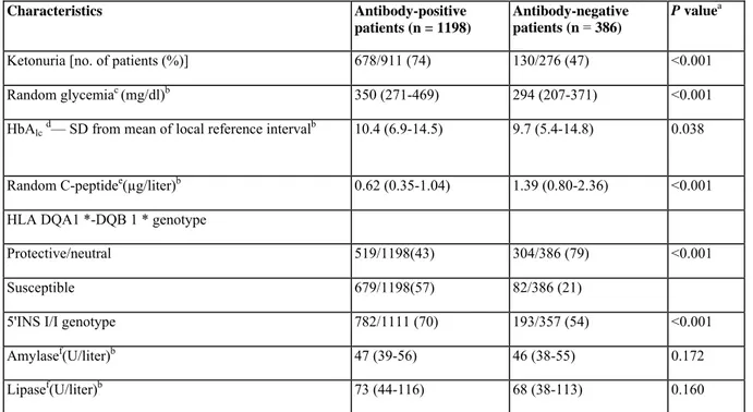
Compared with antibody-negative patients, antibody-positive subjects showed lower random C-peptide values (0.62 vs. 1.39 µg/liter; P < 0.001), despite higher random glycemia levels (350 vs. 294 mg/dl; P < 0.001), and had a higher prevalence of ketonuria (74 vs. 47%; P < 0.001), HLA-DQ (57 vs. 21%), and 5'INS (70 vs. 54%) risk markers (P < 0.001) (Table 2). The prevalence of susceptible HLA-DQ genotypes, but not of 5'INS I/I, was higher in antibody-negative patients than in control subjects (21 vs. 9%, P < 0.001; and 54 vs. 47%, P > 0.05, respectively) (Table 2). Both the high-risk heterozygous HLA-DQ genotype 0301-0302/0501-0201 and the presence of two susceptibility haplotypes (0301-0302 and/or 0501-0201 in homozygous or heterozygous combination) were significantly more prevalent in antibody -negative patients than in nondiabetic control subjects (7.5 vs. 1.9%, P < 0.001; and 14.8 vs. 4.5%, P < 0.001, respectively). Glycosylated hemoglobin (HbAlc)
levels tended to be higher in the presence of autoantibodies (P = 0.038) (Table 2). Serum amylase and lipase activities did not differ according to antibody status (Table 2). During follow-up [mean (range), 25 (2-60) months], antibody-positive subjects (n = 677) persistently showed lower BMI, higher insulin requirements, and lower random C-peptide levels than antibody-negative patients (n = 149; P < 0.001; data not shown). Also during follow-up, antibody-positive patients were more frequently treated with insulin (98 vs. 81%; P < 0.001). Regardless of HLA-DQ status, the presence of first-degree relatives with type 1 and/or type 2 diabetes was more frequently reported in antibody-negative patients than in antibody-positive subjects (34 vs. 20%; P < 0.001).
TABLE 2 — Biological characteristics of 1584 diabetic patients aged 15-39 yr at diagnosis
Characteristics Antibody-positive
patients (n = 1198)
Antibody-negative patients (n = 386)
P valuea
Ketonuria [no. of patients (%)] 678/911 (74) 130/276 (47) <0.001
Random glycemiac (mg/dl)b 350 (271-469) 294 (207-371) <0.001
HbAlcd— SD from mean of local reference intervalb 10.4 (6.9-14.5) 9.7 (5.4-14.8) 0.038
Random C-peptidee(µg/liter)b 0.62 (0.35-1.04) 1.39 (0.80-2.36) <0.001
HLA DQA1 *-DQB 1 * genotype
Protective/neutral 519/1198(43) 304/386 (79) <0.001
Susceptible 679/1198(57) 82/386 (21)
5'INS I/I genotype 782/1111 (70) 193/357 (54) <0.001
Amylasef(U/liter)b 47 (39-56) 46 (38-55) 0.172
Lipasef(U/liter)b 73 (44-116) 68 (38-113) 0.160
a Mann-Whitney U test was used for continuous variables and χ2 test for categorical variables; the threshold for statistical significance is P < 0.05/8 or P < 0.006 (Bonferroni adjustment).
c Data were available for 1109 antibody-positive and 346 antibody-negative patients. b Median (interquartile range).
d Data were available for 1019 antibody-positive and 326 antibody-negative patients. e Data were available for 1041 antibody-positive and 381 antibody-negative patients. f Data were available for 1028 antibody-positive and 377 antibody-negative patients.
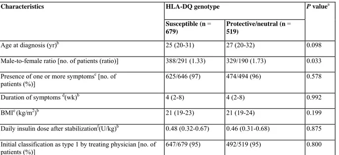
TABLE 3 — Demographic and clinical characteristics of 1198 antibody -positive diabetic patients aged 15-39 yr according to HLA-DQ genotype
Characteristics HLA-DQ genotype P valuea
Susceptible (n = 679)
Protective/neutral (n = 519)
Age at diagnosis (yr)b 25 (20-31) 27 (20-32) 0.098
Male-to-female ratio [no. of patients (ratio)] 388/291 (1.33) 329/190 (1.73) 0.033 Presence of one or more symptomsc [no. of
patients (%)] 625/646 (97) 474/494 (96) 0.578
Duration of symptoms d(wk)b 4 (2-8) 4 (2-8) 0.992
BMIe (kg/m2)b 21 (19-23) 21 (19-24) 0.199
Daily insulin dose after stabilizationf(U/kg)b 0.48 (0.32-0.67) 0.46 (0.31-0.68) 0.875
Initial classification as type 1 by treating physician [no. of patients (%)]
647/679 (95) 492/519 (95) 0.800
a Mann-Whitney U test was used for continuous variables and χ2 test for categorical variables; the threshold for statistical significance is P < 0.05/7 or P < 0.007 (Bonferroni adjustment).
b Median (interquartile range).
c Polyuria, polydipsia, weight loss, and/or fatigue.
d Data were available for 546 patients with susceptible and 417 patients with protective/neutral genotypes.
e Data were available for 592 patients with susceptible and 444 patients with protective/neutral genotypes.
f Data were available for 504 patients with susceptible and 373 patients with protective/neutral genotypes.
CLINICAL AND BIOLOGICAL CHARACTERISTICS ACCORDING TO HLADQ GENOTYPE
Antibody-positive patients.
There were no significant differences in clinical (Table 3) or biological (Table 4) characteristics according to HLA-DQ genotype in antibody-positive subjects, except for a trend toward older age at onset (P = 0.098) (Table 3) and higher male-to-female ratio (P = 0.033) (Table 3), random glycemia (P = 0.022), HbAlc levels (P =
0.046), and prevalence of the 5'INS I/I genotype (P = 0.038) (Table 4) in the absence of HLA-DQ susceptibility genotypes. Furthermore, during follow-up, we found no differences in functional residual β-cell mass, frequency of insulin treatment, or insulin needs for similar glycemic control after stratification for HLA-DQ risk
(susceptible, n = 380; protective/neutral, n = 297; data not shown). Antibody-negative patients.
In contrast to antibody -positive subjects, antibody -negative patients did markedly differ in terms of clinical presentation and biological markers at diagnosis according to the presence or absence of susceptible HLA-DQ genotypes. Subjects carrying susceptible genotypes were overall younger (28 vs. 33 yr; P = 0.001), had a lower BMI (23 vs. 26 kg/m2 ; P < 0.001), and tended to have higher insulin requirements after stabilization (0.41 vs.
0.32 U/kg·d; P = 0.014) (Table 5) to achieve the same degree of glycemic equilibration as reflected by similar HbAlc levels (Table 6). These patients were significantly more often classified as suffering from type 1 diabetes
by their treating physician compared with subjects carrying protective or neutral HLA-DQ genotypes (78% vs. 57%; P = 0.001) (Table 5). There was a tendency toward lower random C-peptide values (1.13 vs. 1.51 µg/liter; P = 0.034) in subjects with susceptible genotypes (Table 6). Amylase activity, but not lipase activity, was overall higher (P = 0.001) in subjects with the susceptible genotypes. However, this was not reflected in a higher prevalence of amylase activity above the upper limit of reference values (cut off, 113 U/liter).
TABLE 4 — Biological characteristics of 1198 antibody-positive diabetic patients aged 15-39 yr according to HLA-DQ genotype
Characteristics HLA-DQ genotype P valuea
Susceptible (n = 679)
Protective/neutral (n = 519)
Ketonuria [no. of patients (%)] 380/514 (74) 298/397 (75) 0.755
Random glycemiac (mg/dl)b 343 (270-450) 364 (277-499) 0.022
HbAlcd( SD from mean of local reference interval)b 10.0 (6.9-14.2) 10.9 (7.0-15.4) 0.046
Random C-peptidee(µg/liter)b 0.62 (0.37-1.02) 0.61 (0.34-1.06) 0.938
5'INS I/I genotype [no. of patients (%)] 428/631 (68) 354/480 (74) 0.038
Amylasef (U/liter)b 47 (39-56) 47 (39-56) 0.780
Lipasef (U/liter)b 73 (43-114) 76 (46-116) 0.192
α Mann-Whitney U test was used for continuous variables and χ2 test for categorical variables; the threshold for statistical significance is P < 0.05/7 or P < 0.007 (Bonferroni adjustment).
c Data were available for 635 patients with susceptible and 474 patients with protective/neutral genotypes. b Median (interquartile range).
d Data were available for 584 patients with susceptible and 435 patients with protective/neutral genotypes.
e Data were available for 586 patients with susceptible and 455 patients with protective/neutral genotypes.
f Data were available for 578 patients with susceptible and 450 patients with protective/neutral genotypes.
TABLE 5 — Demographic and clinical characteristics of 386 autoantibody -negative diabetic patients aged 15-39 yr according to HLA-DQ genotype
HLA-DQ genotype Characteristics
Susceptible (n = 82) Protective/neutral (n = 304)
P
Age at diagnosis (yr)b 28 (22-35) 33 (27-36) 0.001
Male-to-female ratio [no. of patients (ratio)] 57/25 (2.28) 202/102 (1.98) 0.695
Presence of one or more symptomsc [no. of patients (%)] 65/74 (88) 241/273 (88) 1.000
Duration of symptoms d (wk)b 4 (2-9) 6 (2-16) 0.147
BMIe(kg/m2)b 23 (20-27) 26 (22-30) <0.001
Daily insulin dose after stabilizationf(U/kg)b 0.41 (0.21-0.66) 0.32 (0.00-0.50) 0.014
Initial classification as type 1 by treating physician [no. of
patients (%)] 64/82 (78) 173/304 (57) 0.001
a Mann-Whitney U test was used for continuous variables and χ2 test for categorical variables; the threshold for statistical significance is P < 0.05/7 or P < 0.007 (Bonferroni adjustment).
b Median (interquartile range).
c Polyuria, polydipsia, weight loss, and/or fatigue.
d Data were available for 65 patients with susceptible and 237 patients with protective/neutral genotypes.
e Data were available for 67 patients with susceptible and 260 patients with protective/neutral genotypes.
f Data were available for 58 patients with susceptible and 240 patients with protective/neutralgenotypes.
Stepwise forward logistic regression analysis confirmed that in antibody-negative patients with onset in young adulthood, the presence of HLA-DQ susceptibility genes is associated with younger age at diagnosis (P = 0.014; OR, 1.052; 95% CI, 1.010-1.095) and independently of age also with BMI (P = 0.017; OR, 1.071; 95% CI, 1.013-1.133) and amylase activity (P = 0.051; OR, 0.983; 95% CI, 0.967-1.000). Likewise, when all 82 antibody -negative patients with HLA-DQ susceptibility genotypes were one by one matched to an antibody-negative patient of exactly the same age carrying a neutral or protective HLA-DQ genotype, thereby dissipating the age difference between both groups (median age, 28 yr; interquartile range, 22-35 yr, in both groups), the trend toward lower BMI (23 vs. 25 kg/m2 ; P = 0.026), more frequent classification as type 1 diabetes (78 vs. 60%; P
= 0.018), lower random C-peptide values (1.12 vs. 1.65 µg/liter; P = 0.009), and higher amylase activity (50 vs. 47 U/liter; P = 0.05) in the presence of HLA-DQ susceptibility genotypes persisted (Table 7). Furthermore, after a median follow-up of 25 months (range, 20-60 months), antibody -negative patients still presented a lower BMI (P = 0.001) and tended to have higher insulin needs (P = 0.073) to achieve similar glycemic control in the
presence of HLA-DQ risk genotypes (n = 29) than in their absence (n = 120; data not shown). Neither at diagnosis nor after follow-up was there a significant difference in the frequency of insulin treatment between patients with or without HLA-DQ risk genotypes.
CLINICAL AND BIOLOGICAL CHARACTERISTICS ACCORDING TO OTHER VARIABLES
Antibody-negative patients with HbAlc values above percentile 50 (i.e. HbAlc ≥ 10.3 SD above mean of local
reference interval) presented symptoms more frequently (P = 0.004) and had a higher prevalence of ketonuria (P < 0.001), higher insulin requirements (P< 0.001), higher glycemia at diagnosis (P < 0.001), and lower random C-peptide levels (P < 0.001) compared with antibody-negative patients with HbAlc values below percentile 50
(data not shown). Similar findings were observed in antibody-positive patients (data not shown) or when comparing patients with HbAlc below percentile 33 and above percentile 66 (data not shown). Regardless of
antibody and HLA-DQ status, there was no difference in the prevalence of the 5'INS I/I risk genotype according to BMI (patients with BMI > percentile 50 vs. BMI < percentile 50, or patients with BMI > percentile 66 vs. BMI < percentile 33) or in BMI according to 5'INS status (data not shown).
TABLE 6 — Biological characteristics of 386 autoantibody-negative diabetic patients aged 15-39 yr according to HLA-DQ genotype
HLA-DQ genotype Characteristics
Susceptible (n = 82) Protective/neutral (n = 304)
P valuea
Ketonuria [no. of patients (%)] 33/64 (52) 97/212 (46) 0.501
Random glycemiac(mg/dl)b 309 (207-415) 291 (207-365) 0.130
HbAlcd ( SD from mean of local reference
interval)b 8.6 (4.4-14.7) 9.7 (5.4-14.8) 0.651
Random C-peptidee(µg/liter) 1.13 (0.59-2.11) 1.51 (0.88-2.38) 0.034
5'INS I/I genotype [no. of patients (%)] 46/76 (61) 147/281 (52) 0.252
Amylasef(U/liter)b 50 (44-57) 44 (37-54) 0.001
Lipasef(U/liter)b 70 (38-113) 67 (37-114) 0.627
a Mann-Whitney U test was used for continuous variables and χ2 test for categorical variables; the threshold for statistical significance is P < 0.05/7 or P < 0.007 (Bonferroni adjustment).
c Data were available for 71 patients with susceptible and 275 patients with protective/neutral genotypes. b Median (interquartile range).
d Data were available for 67 patients with susceptible and 259 patients with protective/neutral genotypes.
e Data were available for 81 patients with susceptible and 300 patients with protective/neutral genotypes.
f Data were available for 80 patients with susceptible and 297 patients with protective/neutral genotypes.
TABLE 7 — Clinical and biological characteristics of 164 age-matched autoantibody-negative diabetic patients aged 15-39 yr according to HLA-DQ genotype
HLA-DQ genotype Susceptible Protective/neutral Characteristics (n = 82) (n = 82) P BMIc (kg/m2 )b 23 (20-27) 25 (22-29) 0.026
Daily insulin dose after stabilizationd (U/kg)b 0.41 (0.21-0.66) 0.36 (0.15-0.49) 0.195
Initial classification as type 1 by treating physician [no. of patients (percent)]
64/82 (78) 49/82 (60) 0.018
Random C-peptidee(µg/liter) 1.12 (0.59-2.11) 1.65 (0.99-2.50) 0.009
Amylasef(U/liter)b 50 (44-57) 47 (37-53) 0.050
a Mann-Whitney U test was used for continuous variables and χ2 test for categorical variables; the threshold for statistical significance is P < 0.05/5 or P < 0.010 (Bonferroni adjustment).
c Data were available for 67 patients with susceptible and 70 patients with protective/neutral genotypes. b Median (interquartile range).
d Data were available for 58 patients with susceptible and 67 patients with protective/neutral genotypes.
e Data were available for 81 patients with susceptible and 81 patients with protective/neutral genotypes.
DISCUSSION
Adult-onset diabetes presents along a clinical spectrum ranging from acute ketosis-prone onset to latent forms diagnosed by the presence of chronic complications[1][2][3][4][5]M[7][8] l9] . It is often difficult to classify patients
at diagnosis solely on clinical grounds m[8] . Therefore, both disease classification and etiology-driven therapy
would benefit from the optimal use of biological markers related to the underlying pathological process at diagnosis.
In the present study, antibody-positive patients with primary diabetes diagnosed in adulthood were on average leaner and showed an earlier, more acute, and more severe clinical presentation, a less preserved β-cell mass, higher insulin requirements after stabilization, and a higher frequency of HLA-DQ and 5'INS risk genotypes than antibody-negative patients, in agreement with previous observations [4][16][17]mm[38]m . Both HLA-DQ risk
genotypes and a more severe metabolic decompensation occur more frequently in childhood than in adult-onset immune-mediated diabetes [3][10][12][25]mm . We therefore wondered whether the overall less severe clinical
presentation in adult-onset type 1 diabetes was primarily associated with the lower prevalence of HLA-DQ risk markers or rather with older age at diagnosis per se. Hence, we investigated phenotype-genotype relations in antibody-positive and antibody-negative diabetic patients diagnosed in (early) adulthood.
The presence of high-risk HLA-DQ genotypes is not a necessary condition for the development of immune-mediated diabetes because 43% of the antibody-positive diabetic patients lacked such risk markers. The prevalence of the high-risk HLADR3/DR4 genotype in our population of childhood and adult-onset diabetic patients is similar to the prevalence reported in other studies [10][12][25][39] . Especially in the absence of
susceptible HLA-DQ alleles, other genetic risk factors might be needed as suggested by the fact that the prevalence of the 5'INS I/I susceptibility genotype tended to be higher in antibody-positive subjects carrying neutral or protective HLA-DQ genotypes, as shown previously [30] . In the presence of autoantibodies, we
observed no differences in clinical or biological characteristics according to HLA-DQ risk; this is at variance with earlier Scandinavian reports of a milder disease onset in HLA-DR3-positive type 1 patients [41] and of a
more severe presentation in association with an increased HLA-DQB1 linked genetic risk load in diabetic children[42] . However, in these studies all patients were under age 20, and the different genotype groups had not
been matched for age or antibody status, two important confounders. In line with our findings, a British study [39]
concluded that the HLA-DQ genotype background may not have a major role in disease severity in terms of insulin requirements once autoimmunity has been established in adult-onset diabetes.
In antibody-negative patients, HLA-DQ markers of immunemediated type 1 diabetes were overrepresented compared with the background population as reported previously [39] . Antibody-negative subjects displayed
more type 1-like features in the presence of HLA-DQ risk than in their absence; they developed hyperglycemia at a younger age in the presence of genetic markers associated with earlier disease onset in immune-mediated type 1 diabetes [10][11][12], were leaner overall, and tended to have less preserved C-peptide levels and higher
insulin needs. These trends persisted even after adjustment for age at diagnosis and during follow-up. Not surprisingly, antibody-negative patients were more frequently classified as having type 1 diabetes in the presence of HLA-DQ susceptibility genotypes than in their absence by physicians unaware of HLA and antibody status at that time. In carriers of HLA-DQ risk genotypes, the clinical and biological characteristics are suggesting the existence of an immune-mediated β-cell destructive process despite the absence of diabetes-associated autoantibodies. In insulin-dependent diabetes, antibodies indeed sometimes appear only months to years after diagnosis [22][23]. It is conceivable that certain patients have seroconverted to antibody negativity in the
preclinical phase or present other immune markers than those currently searched for[23] [43] [44] . On the basis of
previous observations, it can be safely concluded that in the age categories studied there are very few, if any, recent-onset diabetic patients negative for the diabetes-associated autoantibodies measured in the present study (ICA, GADA, and IA-2A) but positive for IAA (<1%) [26] or for antibodies against a 38-kDa glycated islet cell
membrane-associated protein (0%) [43] . At variance with a smaller study in Italian patients diagnosed in the age
range 30-54 yr[38], but in line with the study of Pietropaolo et al. [45] , our results suggest that genetic risk markers
known to promote the clinical onset of antibody -positive diabetes [1][31] may indicate the presence of an
immune-mediated disease process with yet unknown immune markers in patients with a type 1-like phenotype, but negative for the presently used antibody markers.
A subgroup of Japanese insulinopenic antibody -negative patients with acute diabetes onset in adulthood, low HbAlc levels, and elevated serum activities of pancreatic enzymes at diagnosis has recently been described[19].
Neither the present large study nor preliminary reports [20][21][46] could document the frequent occurrence of this
type of idiopathic type 1 diabetes in Caucasians. In our Belgian antibody-negative Caucasian patients, a more severe metabolic decompensation, higher insulin needs, and less preserved β-cell mass were associated with high
rather than low HbAlc levels, which is clearly discordant with the Japanese findings [19] but in line with a
preliminary European report[20]. Furthermore, we did not identify subjects with elevated amylasemia or lipasemia
among the subjects lacking genetic and antibody markers of immunemediated type 1 diabetes. Rather, amylase activity was lowest in antibody-negative subjects without HLA-DQ risk genotypes. It is unknown whether these lower amylase levels reflect increased enzyme clearance, decreased amylase synthesis due to relative acinar insulinopenia, or less leakage of enzymes from acinar cells [34].
We have observed a higher reported prevalence of familial diabetes in antibody-negative subjects compared with antibody-positive patients regardless of HLA-DQ genotype. It may be warranted to search for mutations
associated with maturity onset diabetes of the young and mitochondrial diabetes because these inherited forms of diabetes are likely to be more prevalent in antibody-negative subjects with a familial history of diabetes and lacking HLA-DQ risk genotypes [44][47][48].
The present report confirms the marked male-over-female excess in adult-onset diabetes [14]. This excess tended
to be more pronounced in antibody-negative patients compared with antibody -positive subjects. It is conceivable that in male subjects with already decreased β-cell function and/or mass increased BMI, a prominent risk factor for type 2 diabetes [7] , and the more visceral distribution of body fat may preferentially accelerate the clinical
onset of diabetes as a consequence of higher metabolic demands due to increased insulin resistance [14].
Alternatively, one may wonder whether a higher BMI may (in part) reflect a better ability to (partially) restore β-cell mass (and hence C-peptide and insulin levels) when confronted with ongoing β-β-cell destruction and/or peripheral insulin resistance [49]. In the light of a recent report on the association between the 5'INS I/I and
juvenile obesity, we failed to detect a possible relation between BMI and 5'INS genotype[50].
In conclusion, antibody-positive adult-onset patients develop diabetes overall at a younger age and, regardless of HLA-DQ genotype, have a more severe clinical presentation at diagnosis than antibody-negative subjects. In the absence of autoantibodies, patients carrying HLA-DQ-linked risk have on average an earlier disease onset and, regardless of age, show more type 1-like features than subjects lacking HLA-DQ risk genotypes. Genetic risk markers of seropositive type 1 diabetes may thus indicate the existence of an immune-mediated disease process in patients lacking conventional diabetes autoantibodies. Idiopathic type 1 diabetes with acute onset and increased amylase activity is not a frequent phenotype in Caucasians.
Acknowledgments
We thank the Vlaamse Diabetes Vereniging, the Association belge du Diabète, and the following members of the Belgian Diabetes Registry for their invaluable help in recruiting diabetic patients and first-degree relatives and/or in the handling of blood samples: E. Anckaert, P. Arnouts, J.-P. Baeyens, E. Balasse, H. Becq, J. Beirinckx, L. Berghmans, W. Bettens, A. Bocquet, A. Bodson, T. Boereboom, R. Bouillon, M. Buysschaert, A. Carlier, M. Cardon, A. Chachati, N. Christophe, S. Claes, I. Claeys†, L. Claeys, P. Cochez, M. Coeckelberghs, I. M. Colin, J.-L. Coolens, P. Coremans, F. Coucke, W. Coucke, E. Couturier, R. Craen, S. Daens, C. Daubresse, J. De Cock, P. Decraene, I. De Feyter, R. De Hauwere, J. De Lepeleire, B. Delgrange, C. Delvigne, R. Demaeseneer, P. De Monie, G. De Proft, L. Derdelinckx, P. De Roeck, J. De Schepper, S. De Weer, P. De Winter, L. Dooms, E. Duvivier, L. Emsens, C. Ernould, A. Eykens, A. Fassotte, N. Gaham, K. Garmijn, J. Gérard, C. Gillet, I. Gios, P. Goetstouwers, C. Guiot, A. Haemers, S. Haemers, F. Hay, J. Helmer, C. Herbaut, G. Heremans, V. Immegeers, R. Janssen, P. Jopart, J. Koumans, G. Lamberigts, C. Lauwers, M.-C. Lebrethon, P. Lefèbvre, P. Lemmens, I. Leunckens, T.-T. Lim, C. Litvine, K. Logghe, M. Maes, W. Maes, Y. Maus, R. Meulepas, J. Michiels, J. Mockel, J. Monballyu, G. Moorkens, W. Musch, D. Nicolaij, G. Op de Beeck, F. Peiffer, M.-C. Pelckmans, H. Penninckx, O. Peters, M. Pieron, L. Pleysier, K. Poppe, P. Prims, A. Purnode, G. Raes, C. Righes, D. Rocour-Brumioul, M.-P. Roggemans, R. Rooman, C. Roose, B. Roth, J. Schets, L. Schillemans, I. Schoemaker, S. Schrans, J. Schutyser, O. Segers, J.-C. Sodoyez†, G. Somerst, D. Staessen, M.-P. Stassen, A. Stroobant, P. Taelman, L. Terriere, M. Tersago, J. Teuwen, G. Thenaers, P. Thielemans, G. Thiry-Counson, U. Timmermans, J. Tits, A. Torfs, K. Van Acker, A. Van Baarle, J. Van Boxem, P. Van Crombrugge, L. Van Damme, L. Van de Mierop, P. Van den Bogaert, E. Vandenbussche, B. Van der Henst, M. Vanderijst, H. Vanderstappen, W. Van Dessel, C. Van Deun, P. Van de Wouwer, D. Van Doom, E. Van Fleteren, J. Van Haegenborgh, M. Van Hecke, E. Van Hollebeke, W. Van Horen, S. Van Imschoot, R. Van Landeghem, F. Van Loon, S. Vanneste, D. Van Nimmen, C. Van Parijs, H. Van Ravensteyn, P. Van Rooy, P. Van Vaerenbergh, J. Verbeeck, R. Verbiest, C. Vercammen, E. Vercruyssen, K. ver Elst, A. Vertruyen, A. Verhaegen, H. Verhaegen, J. Vertommen, E. Weber, I. Weemaes, L. Wouters, and G. Wyffels. We are indebted to N. Alaerts, M. Bodson, V. Claessens, A. Demarré, T. Demesmaeker, L. De Pree, S. Exterbille, T. Ghysels, P. Goubert, C. Groven, A. Ivens, D. Kesler, F. Lebleu, G. Schoonjans, and H. Thomas for excellent technical assistance, (†, Deceased.)
JDF standard for ICA determination and cDNA for preparation of 35 S-recombinant human GAD65 were
donated by Dr. Å. Lernmark (University of Washington, Seattle, WA) and by Dr. A. Falorni (when at the Karolinska Institute, Stockholm, Sweden). Human IA-2ic cDNA was a gift from Dr. M. Christie (King's College School of Medicine and Dentistry, London, UK).
This work was supported by the research council of the Vrije Universiteit Brussels (research fellowship to I.W.), the Belgian Diabetes Association (research fellowship to V.S.), the Belgian Fonds voor Wetenschappelijk Onderzoek (Grants 3-0113-97 and 6-0319-01, and LevenslijnDiabetes project 7.0021.96; postdoctoral research fellowship to C.M.), and by Bayer Corp., Life Scan, Novo Nordisk, Ortho Clinical Diagnostics, and Roche. The Belgian Diabetes Registry is supported financially by grants from the Ministries of Public Health of the Flemish and French Community.
References
1. Krolewski AS, Warram JH, Rand LI, Kahn CR 1987 Epidemiologic approach to the etiology of type 1 diabetes mellitus and impaired glucose tolerance in adults. N Engl J Med 317:1390-1398 Citation
2. Leslie DRG, Elliott RB 1994 Early environmental events as a cause of IDDM. Evidence and implications. Diabetes 43:843-850 Abstract
3. Gorus FK, Belgian Diabetes Registry 1997 Diabetes registries and early biological markers of insulin-dependent diabetes mellitus. Diabetes Metab Rev 4:247-274 Citation
4. Östman J, Lernmark Å, Landin-Olsson M, Sundkvist G, Palmer J, Arnqvist H, Blohme G, Lithner F, Littorin B, Nyström L, Schersten B, Wibell L 1996 Autoimmune (type 1) diabetes in young adults in Sweden. Horm Metab Res 28:348-350 Citation
5. Mølbak AG, Christau B, Marner B, Borch-Johnsen K, Nerup J 1994 Incidence of insulin-dependent diabetes mellitus in age groups over 30 years in Denmark. Diabet Med 11:299-303
6. Zimmet PZ 1995 The pathogenesis and prevention of diabetes in adults. Diabetes Care 18:1050-1064 Citation
7.1997 Report of the Expert Committee on the Diagnosis and Classification of Diabetes Mellitus. Diabetes Care 20:1183-1197
8. Alberti KGMM, Zimmet PZ, for the WHO consultation 1998 Definition, diagnosis and classification of diabetes mellitus and its complications. Part 1, Diagnosis and classification of diabetes mellitus: provisional report of a WHO consultation. Diabet Med 15:539-553 Abstract
9. Gale EAM, Gillespie KM 2001 Diabetes and gender. Diabetologia 44:3-15 Abstract
10. Karjalainen J, Salmela P, Ilonen J, Surcel HM, Knip M 1989 A comparison of childhood and adult type 1 diabetes mellitus. N Engl J Med 320:881-886 Abstract
11. Vandewalle CL, Decraene T, Schuit FC, De Leeuw IH, Pipeleers DG, Gorus FK, Belgian Diabetes Registry 1993 Insulin autoantibodies and high titre islet cell antibodies are preferentially associated with the HLA DQA1 *0301DQB*0302 haplotype at clinical onset of type 1 (insulin-dependent) diabetes mellitus before age 10 years, but not at onset between age 10 and 40 years. Diabetologia 36:1155-1162 Abstract
12. Caillat-Zucman S, Garchon HJ, Timsit J, Assan R, Boitard C, Djilali-Saiah I, Bougnères P, Bach JF 1992 Age-dependent HLA genetic heterogeneity of type 1 insulin-dependent diabetes mellitus. J Clin Invest 90:2242-2250 Abstract
13. Pipeleers D, Ling Z 1992 Pancreatic β-cells in insulin-dependent diabetes. Diabetes Metab Rev 8:209-227 Citation
14. Weets I, Van der Auwera BJ, Schuit FC, Du Caju MV, Décochez K, Van Autreve J, De Leeuw IH, Keymeulen B, Mathieu C, Rottiers R, Dorchy H, Quartier E, Gorus FK, Belgian Diabetes Registry 2001 Male-to-female excess in diabetes diagnosed in early adulthood is not specific for the immunemediated form nor is it
HLA-DQ restricted: possible relation to increased body mass index. Diabetologia 44:40-47 Abstract
15 Pozzilli P, Visalli N, Buzzetti R, Cavallo MG, Marietti G, Hawa M, Leslie RD, IMDIAB Study Group 1998 Metabolic and immune parameters at clinical onset of insulin-dependent diabetes: a population-based study. Metabolism 47:1205-1210 Abstract
16. Turner R, Stratton I, Horton V, Manley S, Zimmet P, Mackay IR, Shattock M, Bottazzo GF, Holman R, for UK Prospective Diabetes Study (UKPDS) Group 1997 UKPDS 25: autoantibodies to islet cytoplasm and glutamic acid decarboxylase for prediction of insulin requirements in type 2 diabetes. Lancet 350:1288-1293 Abstract
17. Groop L, Bottazzo GF, Doniach D 1986 Islet cell antibodies identify latent type 1 diabetes in patients aged 35-75 years at diagnosis. Diabetes 35:237-241 Abstract
18. Deleted in proof.
19. Imagawa A, Hanafusa T, Miyagawa J-I, Matsuzawa Y, for the Osaka IDDM Study Group 2000 A novel subtype of type 1 diabetes mellitus characterized by a rapid onset and an absence of diabetes-related antibodies. N Engl J Med 342:301-307 Abstract
20. Carreras G, Mauricio D, Pérez A, De Leiva A 2000 Can all newly diagnosed subjects without type 1
diabetes-associated autoimmune markers be classified as type lb diabetic patients? Diabetes Care 23:1715-1716 Citation
21. Tiberti C, Buzzetti R, Anastasi E, Dotta F, Vasta M, Petrone A, Cervoni M, Torresi P, Vecci E, Multari G, Di Mario U 2000 Autoantibody negative new onset type 1 diabetic patients lacking high risk HLA genes in a Caucasian population: are these type lb diabetes cases? Diabetes Metab Res Rev 16:8-14 Abstract
22. Landin-Olsson M, Arnquist HJ, Blohmé G, Littorin B, Lithner F, Nyström L, Schersten B, Sundkvist G, Wibell L, Ostman J, Lernmark A 1999 Appearance of islet cell autoantibodies after clinical diagnosis of diabetes mellitus. Autoimmunity 29:57-63 Abstract
23. Decochez K, Tits J, Coolens J-L, Van Gaal L, Krzentowski G, Winnock F, Anckaert E, Weets I, Pipeleers DG, Gorus FK, Belgian Diabetes Registry 2000 High frequency of persisting or increasing islet-specific autoantibody levels after diagnosis of insulin-requiring type 1 diabetes presenting before 40 years of age. Diabetes Care 23:838-844 Abstract
24. National Diabetes Data Group 1979 Classification and diagnosis of diabetes mellitus and other categories of glucose intolerance. Diabetes 28:1039-1057
25. Vandewalle CL, Coeckelberghs MI, De Leeuw IH, Du Caju MV, Schuit FC, Pipeleers DG, Gorus FK, Belgian Diabetes Registry 1997 Epidemiology, clinical aspects, and biology of IDDM patients under age 40 years. Diabetes Care 20:1556-1561 Abstract
26. Vandewalle C, Falorni A, Svanholm S, Lernmark A, Pipeleers DG, Gorus FK, Belgian Diabetes Registry 1995 High diagnostic sensitivity of glutamate decarboxylase autoantibodies in insulin-dependent diabetes mellitus with clinical onset between age 20 and 40 years. J Clin Endocrinol Metab 80:846-851 Full Text 27. Vandewalle C, Falorni A, Lernmark Å, Goubert P, Dorchy H, Coucke W, Semakula C, Van der Auwera B, Kaufman L, Schuit FC, Pipeleers DG, Gorus FK, Belgian Diabetes Registry 1997 Associations of GAD65- and IA-2-autoantibodies with genetic risk markers in new-onset IDDM patients and their siblings. Diabetes Care 20:1547-1552 Abstract
28. Verge CF, Stenger D, Bonifacio E, Colman PG, Pilcher C, Bingley PJ, Eisenbarth GS, Participating Laboratories 1998 Combined use of autoantibodies (IA2ab, GADab, IAA, ICA): combinatorial islet autoantibody workshop. Diabetes 47:1857-1866 Abstract
29. Heimberg H, Nagy ZP, Somers G, De Leeuw I, Schuit FC 1992 Complementation of HLA-DQA and -DQB genes confers susceptibility and protection to insulin-dependent diabetes mellitus. Hum Immunol 320:10-17 Abstract
30. Van der Auwera BJ, Schuit FC, Lyaruu I, Falorni A, Svanholm S, Vandewalle CL, Gorus FK, Belgian Diabetes Registry 1995 Genetic susceptibility for insulin-dependent diabetes mellitus in Caucasians revisited: the importance of diabetes registries in disclosing interactions between HLA-DQ- and insulin gene-linked risk. J Clin Endocrinol Metab 80:2567-2573 Full Text
31. Van der Auwera B, Schuit FC, Weets I, Ivens A, Van Autreve J, Gorus FK, Belgian Diabetes Registry 2002 Relative and absolute HLA-DQA1-DQB1 linked risk for developing type 1 diabetes before age 40 years in the Belgian population: implication for future prevention studies. Hum Immunol 63:40-50 Abstract
32. Livak KJ, Marmaro J, Todd JA 1995 Towards fully automated genome-wide polymorphism screening. Nat Genet 9:341-342 Citation
33. Keymeulen B, Ling Z, Gorus FK, Delvaux G, Bouwens L, Grupping A, Hendrieckx C, Pipeleers-Marichal M, Van Schravendijk C, Salmela K, Pipeleers DG 1998 Implantation of standardized β-cell grafts in a liver segment of IDDM patients: graft and recipient characteristics in two cases of insulin-independence under maintenance immunosuppression for prior kidney graft. Diabetologia 41:452-460 Abstract
34. Semakula C, Vandewalle C, Van Schravendijk C, Sodoyez JC, Schuit FC, Foriers A, Falorni A, Craen M, Decraene P, Pipeleers DG, Gorus FK, Belgian Diabetes Registry 1996 Abnormal circulating pancreatic enzyme activities in more than twenty-five percent of recent-onset insulin-dependent diabetic patients: association of hyperlipasemia with high-titer islet cell antibodies. Pancreas 12:321-333 Abstract
35. Bland JM, Altman DG 1995 Multiple significance tests: the Bonferroni method. BMJ 310:170 Citation 36. Tuomi T, Groop L, Zimmet PZ Rowley MJ, Knowles W, Mackay IR 1993 Antibodies to glutamic acid decarboxylase reveal latent autoimmune diabetes mellitus in adults with a non-insulin-dependent onset of disease. Diabetes 42:359-363 Abstract
37. Arnqvist HJ, Littorin B, Nyström L, Schersten B, Ostman J, Blohme G, Lithner F, Wibell L 1993 Difficulties in classifying diabetes at presentation in the young adult. Diabet Med 10:606-613 Abstract
38. Bruno G, De Salvia A, Arcari R, Borra M, Grosso N, Carta Q, Trovati M, Veglio M, Pagano G, Piedmont Study Group for Diabetes Epidemiology 1999 Clinical, immunological, and genetic heterogeneity of diabetes in an Italian population-based cohort of lean newly diagnosed patients aged 30-54 years. Diabetes Care 22:50-55 Abstract
39. Horton V, Stratton I, Bottazzo GF, Shattock M, Mackay I, Zimmet P, Manley S, Holman R, Turner R, for the UK Prospective Diabetes Study (UKPDS) Group 1999 Genetic heterogeneity of autoimmune diabetes: age of presentation in adults is influenced by HLA DRB1 andDQBl genotypes (UKPDS 43). Diabetologia 42:608-616 Abstract
40. Sabbah E, Savola K, Ebeling T, Kulmala P, Vahasalo P, Ilonen J, Salmela PI, Knip M 2000 Genetic, autoimmune, and clinical characteristics of childhood and adult-onset type 1 diabetes. Diabetes Care 23:1326-1332 Abstract
41. Ludvigsson J, Samuelsson U, Beauforts C, Deschamps I, Dorchy H, Drash A, Francois R, Herz G, New M, Schober E 1986 HLA-DR3 is associated with a more slowly progressive form of type 1 (insulin-dependent) diabetes. Diabetologia 29:207-210 Abstract
42. Veijola R, Knip M, Reijonen H, Vahasalo P, Puukka R, Ilonen J 1995 Effect of genetic risk load defined by HLA DQB1 polymorphism on clinical characteristics of IDDM in children. Eur J Clin Invest 25:106-112 Abstract
43. Winnock F, Christie MR, Batstra MR, Aanstoot HJ, Weets I, Decochez K, Jopart P, Nicolaij D, Gorus FK; The Belgian Diabetes Registry 2001 Autoantibodies to a 38 kDa glycosylated islet cell membrane antigen are specific markers of (pre)type 1 diabetes: association with autoantibodies against IA-2 and islet cell antigens. Diabetes Care 24:1181-1186 Abstract
44. Lernmark Å 2000 Rapid-onset type 1 diabetes with pancreatic endocrine dysfunction. N Engl J Med 342:344-345 Citation
45. Pietropaolo M, Becker DJ, LaPorte RE, Dorman JS, Riboni S, Rudert WA, Mazumdar S, Trucco M 2002 Progression to insulin-requiring diabetes inseronegative prediabetic subjects: the role of two distinct HLA-DQ high-risk haplotypes. Diabetologia 45:66-76 Abstract
46. Piñero-Liloña A, Litonjua P, Aviles-Santa L, Raskin P 2001 Idiopathic type 1 diabetes in Dallas, Texas. A 5-year experience. Diabetes Care 24:1014-1018 Abstract
47. Lehto M, Wipemo C, Ivarsson SA, Lindgren C, Lipsanen-Nyman M, Weng J, Wibell L, Widen E, Tuomi T, Groop L 1999 High frequency of mutation in MODY and mitochondrial genes in Scandinavian patients with familial earlyonset diabetes. Diabetologia 42:1131-1137 Abstract
48. Dussoix P, Vaxillaire M, Iynedjian PB, Tiercy JM, Ruiz J, Spinas GA, Berger W, Zahnd G, Froguel P, Philippe J 1997 Diagnostic heterogeneity of diabetes in lean young adults. Classification based on immunologic and genetic parameters. Diabetes 46:622-631 Abstract
49 Withers DJ, Burks DJ, Towery HH, Altamuro SL, Flint CL, White MF 1999 Irs-2 coordinates lgf-1 receptor-mediated β-cell development and peripheral insulin signaling. Nat Genet 23:32-40 Abstract
50. Le Stunff C, Fallin D, Schork NJ, Bougnères P 2000 The insulin gene VNTR is associated with fasting insulin levels and development of juvenile obesity. Nat Genet 26:444-446 Abstract