HAL Id: hal-00425366
https://hal.archives-ouvertes.fr/hal-00425366
Submitted on 21 Oct 2009HAL is a multi-disciplinary open access archive for the deposit and dissemination of sci-entific research documents, whether they are pub-lished or not. The documents may come from teaching and research institutions in France or abroad, or from public or private research centers.
L’archive ouverte pluridisciplinaire HAL, est destinée au dépôt et à la diffusion de documents scientifiques de niveau recherche, publiés ou non, émanant des établissements d’enseignement et de recherche français ou étrangers, des laboratoires publics ou privés.
X-ray spectroscopy characterization of Ar17+ produced
by an ECRIS in the afterglow mode
Christophe Prigent, Emily Lamour, Laurent Maunoury, Fabien Noury, J.Y.
Pacquet, Jean Marc Ramillon, J. -P. Rozet, Dominique Vernhet, Martino
Trassinelli
To cite this version:
Christophe Prigent, Emily Lamour, Laurent Maunoury, Fabien Noury, J.Y. Pacquet, et al.. X-ray spectroscopy characterization of Ar17+ produced by an ECRIS in the afterglow mode. Journal of Physics: Conference Series, IOP Publishing, 2009, 163, pp.012111. �10.1088/1742-6596/163/1/012111�. �hal-00425366�
X-ray spectroscopy characterization of Ar
17+produced by an
ECRIS in the afterglow mode
C. Prigent1) , E. Lamour1), J. Mérot1), B. Pascal1), J.-P. Rozet1), M. Trassinelli1),
D. Vernhet1), J.-Y. Pacquet2), L. Maunoury3), F. Noury3), J.-M. Ramillon3)
1) Institut des NanoSciences de Paris, CNRS UMR 75-88, Université Pierre et Marie
Curie, Paris 6, 75015 Paris, France
2) GANIL, bd Henri Becquerel, BP 55027, F-14076 Caen cedex 05, France, EU 3) CIMAP, 6 bd du Maréchal Juin, F-14050 Caen cedex 04, France, EU
Corresponding author’s e-mail address: christophe.prigent@insp.jussieu.fr
Abstract. An electron–cyclotron–resonance ion source (ECRIS) operating in the afterglow
mode allows to produce pulsed highly charged ion beams. We present a characterization of the
first production of pulsed Ar17+ ions from an ECRIS using X-ray spectroscopy techniques. An
ion current increase of a factor 3-4 compared to the continuous operating mode with a short rise time (< 100 µs) and a full width at half maximum of 500 µs has been obtained.
1. Introduction
Electron – cyclotron – resonance ion sources (ECRIS) are devices designed to produce intense beams of highly charged ions (HCI). They are mirror machines where the plasma is heated by a radio frequency (RF) in the GHz range [1]. A resonant coupling between wave and electron occurs when
electrons cross a particular magnetic surface – the resonant surface – where the Larmor frequency matches the wave frequency. It leads to an increase of the perpendicular velocity (relative to the magnetic flux lines) and the heated electrons are trapped in a magnetic bottle (minimum – B structure). When the RF power is turned off, the electrons cool down in a short time, the confinement gets worse and consequently the electron density is reduced. The potential barrier is therefore lost, and all ions can escape from the trap. The extracted ion current of heavy ions in high charge states can increase dramatically in a short time (around few ms), determined principally by the electron confinement lifetime. This scenario is well known as the afterglow pulse [2]. Effects occurring in the
afterglow mode may be optimized to a few microamperes of pulsed particle current of high charge state and a few hundreds of μs long at a frequency of a few Hz. Several ways to increase the amplitude and/or to drop the pulse duration have been proposed and supported by experimental and theoretical studies, as adding a pulsed magnetic field to change the magnetic mirror gradient [3], or injecting a
pulsed ion beam instead of neutral gas [4]. The afterglow mode allows good confinement to obtain the
desired charge state, while continuous operating mode (cw) leads to a compromise between confinement and steady state extraction of ions.
At present, the afterglow operation mode in the ECRIS has been mostly developed in a few applications as beam-injector of high-energy (> MeV/A) accelerators. Acceleration of heavy ions demands very short pulses (<1 ms) in high charge states. In particular in the frame of the CERN
program, lead ions in charge states up to 28+ have been obtained with a peak current around 100 eμA, a bunch duration of 400 μs and at a repetition frequency of 1 to 5 Hz [5, 6]. It is also of big interest for
atomic physics experiments making direct use of H-like or even bare ions in the low energy regime (i.e. a few keV/q). However, control of pulse widths, optimization of the operation conditions and measurements of bare or H-like ions currents are not trivial. For most HCI of medium Z (like argon), a complete characterization of the ion beams delivered in the afterglow mode is needed, since the current is around a few enA or lower, and possible contamination by ions having close m/q ratio may occur. Classical measurement techniques, based on ion current measurements in a Faraday cup, lead to ambiguous results.
To overcome this problem, we make use of more sophisticated X-ray spectroscopy techniques. Having Ar ions of a few hundreds of keV impinging on a thin carbon foil, we have been able to characterize, for the first time, the production of Ar17+ delivered in the afterglow mode. The ion-solid
interaction leads to K-line emission coming from the deexcitation of projectile atomic states populated by charge transfer from the target (single or multiple capture processes occur). As the lifetime of these excited states is well below a ps, X–ray spectroscopy allows to perform measurement on ultra short time scale. Consequently, measurements of the X–ray yield over a few ms show a direct image of the ion bunch in terms of intensity and time distribution. Moreover, this emission provides a clear signature of both the atomic number and the charge state of the emitting projectile. Characteristic K lines in the 3-4 keV range have been observed, allowing for a precise identification and quantification of the Ar17+ ion beam extracted from the plasma explosion after the RF power has been turned off.
In the next section, we will present the experimental set-up together with the main results. A conclusion with outlook will be given in the last part.
2. Experiment and results
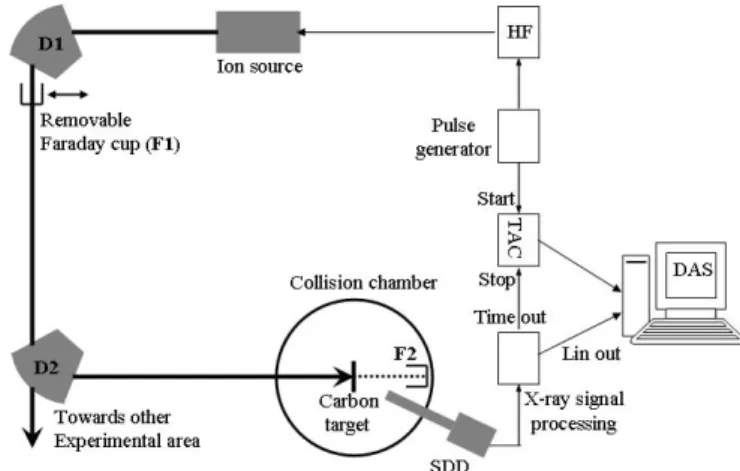
The experimental set-up is schematically depicted in figure 1. The ECRIS used in these studies was the SUPERSHyPIE ion source [7] of GANIL (Grand Accélérateur National d’Ions Lourds) at the
ARIBE facility (French acronym for “accelerator dedicated to research with low energy ions”). The RF power supply provides a microwave at 14.5 GHz frequency with a maximum power of 1 kW during the pulse. The repetition rate is fixed at 10 Hz for all the measurements reported here, with rise and fall time of the pulse generator (Tektronix AFG3021) of 18 ns. The time required to obtain the full RF power is 5 μs. The current set for the magnetic coils of the ECRIS is optimized in order to get the best possible plasma confinement (1.2 T at the injection side and 1 T at the extraction side). The extraction voltage is 15 kV and in order to keep it stable, a high voltage capacity of 1 μF has been added at the output of the High Voltage (HV) power supply. This capacity is mounted in parallel to the 30 MΩ resistance to the ground. For all experiments O2 was used as support gas. The ion bunch
current extracted from the source can be measured with a Faraday cup (F1 in fig. 1) located at the focal plane of the q/m analyzing dipole (D1 in fig. 1). The Faraday cup is connected to ground via a 100 kΩ resistance across which the voltage drop is read out with an oscilloscope (Tektronix TDS1012). From the first dipole to the collision chamber, the ion beam is transported along a beam line of 14 m equipped with several distributing dipoles (one used, D2 shown in fig. 1), 12 quadrupoles and 6 magnetic steerers. The vacuum after the first dipole is around 10-7 mbar while it reaches 10-9 in
the last section of the beam line up to the collision chamber. The ion beam is directed in the collision chamber onto a thin (15 μg/cm2) carbon foil. For beam current measurements, the target is removed
and the beam enters a biased Faraday cup (F2). An estimation of the ion beam transmission may be extracted comparing the measured ion current in the first Faraday cup (F1) with the value recorded in the collision chamber (F2). We found 10 – 15 % in the cw mode as well as in the afterglow mode.
When the target is “in beam”, the X rays characterizing the ion-solid interaction are recorded by a Silicon-Drift-Detector (SDD) (XFlash model detector from RONTEC) of well known efficiency [8].
The X-ray yield is measured also during operation in the continuous mode, thus allowing a well controlled relationship between instantaneous current and X-ray yield in the afterglow mode. A typical
spectrum, showing np → 1s transitions in Arq+ ions resulting from multiple captures of target electrons
in highly excited states of Ar17+, is given in figure 2. No additional lines are recorded, proving without
ambiguity the sole presence of Ar17+ ions in the incident beam. Nevertheless, to avoid any spurious
signal recording, an energy window on these transitions was set in the data acquisition system (DAS). Thus, only X-ray emission from 2.9 to 4 keV contributes to the recorded time spectra.
Figure 1. Scheme of the experimental set-up together with the
associated electronics and data acquisition system (DAS).
Figure 2. Typical spectrum showing the
2p1s and np1s with n > 2 transitions in
Arq+ resulting from multiple capture of
carbon target electrons in highly excited
states of Ar17+ ions at 255 keV.
In afterglow mode, X rays are recorded as a function of time delay after the end of RF pulse. A time to amplitude converter (TAC) of 2 ms dynamic range is started 300 μs before the end of the RF pulse and stopped by a time signal from the SDD with negligible delay. Time resolution can be as good as a few tens of ns, but was limited here to ~ 6 μs / channel. Great care was taken to insure that single stop condition over the dynamic range was achieved: a typical 0.1 probability of X-ray recording per shot was obtained by limiting the solid angle of the SDD with well defined slits.
20 ms 30 ms time (µs) 0 500 1000 1500 Intens ity (a .u .) 0 10 20 30 40 50 50 ms time (µs) 0 500 1000 1500 2000 15 ms In te n s it y ( a .u .) 0 10 20 30 40 50 60
Figure 3. Time spectra for four HF pulse lengths (from 15 to
50 ms with a repetition rate of 10 Hz) exhibiting the X-ray time distribution after the turnoff of the RF power (indicated by vertical dashed lines).
HF activation time (ms) FWHM (µs) Rising time (µs) Decay time (µs) 15 800 240 630 20 500 100 500 30 500 240 1000 50 ~ CW ~ CW ~ CW
Table 1. Full Width Half Maximum (in ms),
rising and decay time (in μs) for different HF activation time.
Figure 3 shows spectra of the X-ray emission recorded for different RF pulse widths namely of 15, 20, 30 and 50 ms. These four spectra have been recorded for the same acquisition time (2000 s) and for constant values of the tuning parameters of the ECRIS. Measurements have been found highly reproducible and used as a control during two weeks for the investigation of the ion-cluster dynamics.
For RF pulse durations ≤ 30 ms, the afterglow current is found to be around 3-4 times higher than in cw mode, analogously to previous measurements performed with lower argon ion charge states [9].
Starting from 50 ms, the gain factor is not significant and the X-ray distribution tends towards a cw mode. The full width at half maximum of the afterglow bunch is shorter for 20 and 30 ms RF pulse duration (around 500 μs) than for 15 ms (800 μs). These results show evidence for an optimum pulse duration to efficiently and rapidly empty the ion reservoir in the confinement volume. Moreover, the rise time is found to be a bit faster when using a 20 ms RF pulse width than 15 or 30 ms (see Table 1). A short rise time may be interesting for measurements based on coincidence techniques as in ion – matter interaction studies.
We remark that the decrease of the ion current with the increase of the RF pulse length (heating time) is opposite to what has been previously measured [6]. The authors show a drop of a factor 5 in the ion current when decreasing the RF length from 50 to 20 ms with the same repetition rate. Contrary to these studies where medium ion charge states are considered (i.e. maximum in the ion charge state distribution as Pb27+, Xe20-22+ [10]), here, we explore, with Ar17+ ions, the highly charged
states. This opposite behaviour suggests different charge breeding mechanisms for these two kinds of charge state.
3. Conclusion
We have presented the first characterization of pulsed Ar17+ ion beams from an ECRIS by x–ray
spectroscopy techniques in the keV range. In our experimental conditions, an optimal RF width of 20 ms has been obtained leading to an ion current increase of a factor 3-4 with respect to the cw mode, a short rise time of less than 100 μs and a full width at half maximum of 500 μs.
Combined with theoretical developments, the use of x–ray spectroscopy in the afterglow mode could give additional information to climb a new step in understanding of plasma and highly charged ion production in the ECR ion sources. In particular, x–ray spectroscopy acts as a probe on short time scales of the plasma itself and sheds some light on the competition between the heating, the breeding and the de-confinement time.
Acknowledgement
This work has been supported by the French national agency of research (ANR: Agence Nationale de Recherche) contract ANR 06 BLAN 0233.
1[] Geller R. et al., « ECRIS : the electron cyclotron ion source « , Annu. Rev. Part. Sci. 40, 15 (1990).
2[] Briand P. et al., NIMA, 294, 673 (1990).
3[] Hill. C.E. and Langbein K., Rev. Sci. Instrum., 67 (3), 1328 (1996). Mühle C. et al., Rev. Sci. Instrum, 67 (3), 1331
(1996).
4[] Shirkov G. et al. NIM B, 95, 527 (1995). Shirkov G et al., Rev. Sci. Instrum., 67 (3), 1158 (1996).
5[] Sortais P., Rev. Sci. Instrum 63 (4), 2801 (1992).
6[] Hill C.E. et al., Rev. Sci. Instrum., 73 (2), 564 (2002).
7[] Maunoury L. et al., Rev. Sci. Instrum., 73 (2), 561 (2002).
8[] Lamour E. et al., submitted to Rev. Sci. Instrum.
[9] Müller L., et al, Rev. Sci. Instrum, 73 (3) 1140 (2002).