HAL Id: hal-01944067
https://hal.umontpellier.fr/hal-01944067
Submitted on 25 May 2021
HAL is a multi-disciplinary open access
archive for the deposit and dissemination of
sci-entific research documents, whether they are
pub-lished or not. The documents may come from
teaching and research institutions in France or
abroad, or from public or private research centers.
L’archive ouverte pluridisciplinaire HAL, est
destinée au dépôt et à la diffusion de documents
scientifiques de niveau recherche, publiés ou non,
émanant des établissements d’enseignement et de
recherche français ou étrangers, des laboratoires
publics ou privés.
Distributed under a Creative Commons Attribution| 4.0 International License
Jean-François Rossi
To cite this version:
Jean-François Rossi. Targeted Therapies in Adult B-Cell Malignancies. BioMed Research
Interna-tional , Hindawi Publishing Corporation, 2015, 2015, pp.1-16. �10.1155/2015/217593�. �hal-01944067�
Review Article
Targeted Therapies in Adult B-Cell Malignancies
Jean-François Rossi
1,21Department of Hematology, University Hospital, CHU Saint Eloi, 80 avenue Augustin Fliche, 34295 Montpellier Cedex 05, France
2Universit´e Montpellier I, UFR M´edecine, 34396 Montpellier, France
Correspondence should be addressed to Jean-Franc¸ois Rossi; jeanfrancoisrossi@me.com Received 13 March 2015; Revised 3 May 2015; Accepted 5 May 2015
Academic Editor: Haiqing Ma
Copyright © 2015 Jean-Franc¸ois Rossi. This is an open access article distributed under the Creative Commons Attribution License, which permits unrestricted use, distribution, and reproduction in any medium, provided the original work is properly cited. B-lymphocytes are programmed for the production of immunoglobulin (Ig) after antigen presentation, in the context of T-lymphocyte control within lymphoid organs. During this differentiation/activation process, B-T-lymphocytes exhibit different restricted or common surface markers, activation of cellular pathways that regulate cell cycle, metabolism, proteasome activity, and protein synthesis. All molecules involved in these different cellular mechanisms are potent therapeutic targets. Nowadays, due to the progress of the biology, more and more targeted drugs are identified, a situation that is correlated with an extended field of the targeted therapy. The full knowledge of the cellular machinery and cell-cell communication allows making the best choice to treat patients, in the context of personalized medicine. Also, focus should not be restricted to the immediate effects observed as clinical endpoints, that is, response rate, survival markers with conventional statistical methods, but it should consider the prediction of different clinical consequences due to other collateral drug targets, based on new methodologies. This means that new reflection and new bioclinical follow-up have to be monitored, particularly with the new drugs used with success in B-cell malignancies. This review discussed the principal aspects of such evident bioclinical progress.
1. Introduction
B-lymphocytes are programmed for immunoglobulin (Ig) production directed against pathogens via the B-cell recep-tor (BCR) activation. During this maturation process, B-lymphocytes exhibit different surface markers, activation of intracellular pathways, metabolism modulation, protein synthesis, and interaction with their microenvironment. B-lymphocyte ontogeny takes place in lymphoid organs leading to plasma cells that migrate into the bone marrow or mucosa-associated tissues. Recently, progress in biology knowledge has resulted in a large number of targeted therapies, designed against surface markers, cell signaling pathways, cell cycle and apoptosis machinery, key molecules involved in cellular metabolism, in proteasome, and in immune modulation, and niche disruption. Rituximab, an anti-CD20 monoclonal antibody (mAb), was the first targeted therapy successfully developed in lymphoma and chronic lymphocytic leukemia of B-cell type (B-CLL) [1–3]. A synergy with chemotherapy was demonstrated in all B-cell malignancies (B-CM) express-ing CD20 molecules, with significant prolongation of the
progression-free survival (PFS) and overall survival (OS) [4]. Beyond rituximab, there are new molecules directed against several factors. This includes (1) other surface markers, including not only other B-cell markers but also receptors (R) of survival/growth factors, such as B-cell activating fac-tor/A proliferation-inducing ligand (BAFF/APRIL)R, inter-leukin (IL) 6R, IL7R, vascular endothelial growth factor (VEGF)R, epithelial growth factor (EGF)R, stromal cell-derived factor- (SDF-)1R or chemokine receptor type 4 (CXCR4), and insulin-like growth factor (IGF)1R; (2) key points for signaling pathways such as inhibitors of Bruton’s tyrosine kinase (BTK), phosphoinositide 3-kinase (PI3K), and spleen tyrosine kinase (Syk); (3) inhibitors of cell cycle regulators; (4) proteasome inhibitors and nuclear factor kappa-B (NF𝜅-B) inhibitors; (5) metabolism inhibitors, such as antilacticodehydrogenase (LDH); (6) immune modulators; and (7) inhibitors of the interaction between tumor cells and its microenvironment. The complexity of these ther-apeutic options requires new reflection and approach and new drug combinations based on biological data in order to create optimal conditions for such new age in personalized
Volume 2015, Article ID 217593, 16 pages http://dx.doi.org/10.1155/2015/217593
medicine, including methodologies, follow-up to evaluate quality of life, and safety and tolerability not only just after the administration, but also after a long treatment period due to improved survival [5] (Tables1,2, and3).
2. Cell Surface Markers
2.1. B-Cell Markers. B-lymphocyte differentiation is
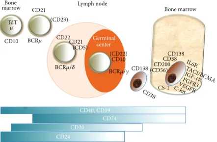
associ-ated with the expression of a variety of cell surface markers, including CD19, CD20, C22, CD40, surface Ig, and BAFF-R present at the different maturation steps, excluding the end-stage of this B-cell lineage, plasma cells [6–9]. Conversely, plasma cells also share CD19 and other surface markers such as CD38, CD138, CS-1, CD200, CD56, CD45, and other different markers [10] (Figure 1). In addition, all markers are currently used to define normal and malignant plasma cells, thus allowing evaluation of minimal residual disease, and to establish the true complete response (CR) expressed by the return of normal plasma cells inside the bone marrow niche [11]. Targeting surface B-cell markers also leads to cell signaling, as observed with CD19, CD20, CD5, and CD22 that are B-cell receptor (BCR) coreceptors with either stimulatory or inhibitory activities.
Rituximab, the first anti-CD20 mAb used in humans, has been shown clinically beneficial in B-CM, including non-Hodgkin’s lymphoma (NHL) and B-CLL [1–3,12]. This agent was also developed as a maintenance therapy with a significant prolongation of the therapeutic response. How-ever, empirism was associated with the early development of rituximab, and the usual dose of 375 mg/m2 was not chosen on a biological basis. The choice of the efficient dose based on biological criteria was only made in the context of B-CLL, with the demonstration of biological efficacy through the quantification of the CD20 molecules at the cell surface and the engagement of the Antibody-Dependent Cell Cytotoxicity (ADCC) [13]. Maintenance therapy with rituximab was rather based on commercial reasons even though treatment prolongation for two years was associated with an improved PFS. In fact, treatment prolongation should have been based on the control of residual disease associated with improved survival. The dose was similar to the one used in chemoimmunotherapy, but with different options for the administration schedule due to a lack of biological rationale, except that ADCC with Natural Killer (NK) cell activation may delay the time of the optimal response.
Delayed complications have been neglected in this con-text. The first observations were made in patients treated for Rheumatoid Arthritis (RA) who experienced reduction of immune response to vaccination [14] and reactivation of viral infections, not only hepatitis also observed in the context of B-CM, but also progressive multifocal leukoencephalopathy (PML), a lethal encephalitis caused by the polyomavirus JC [15]. We recently observed one case of PML with a severe immune defect due to chemoimmunotherapy and autologous transplantation for NHL, 15 years after the initiation of the therapy (personal data). The fact that similar observations were not so frequent was due to three factors: (1) lack of substantial long-term survivors; (2) the patients having RA had a more pronounced immunodepressed status due
to the exposition to several immune modulators, such as corticotherapy, methotrexate, or anti-Tumor Necrosis Factor (TNF) for example; and (3) relative limited efficacy of rituximab in depleting memory B-cells and plasma cell com-partment within lymphoid organs [16]. This is not probably the end of the story and longer observation period is needed, particularly with improved efficacy and prolonged patient survival due to new efficient molecules, including the more efficient anti-CD20 mAbs and new targeted therapies. The subcutaneous (SC) form of rituximab was developed as equivalent to the IV formulation. However, the lymph node compartment being the target organ after SC administration was not considered. Had this been taken into account, one could predict a better activity and a better clinical use of the drug. The therapeutic strategy should change, and the current long-term maintenance therapy with rituximab should be avoided. In addition, drug agencies have to prolong patient observation beyond therapeutic response and to analyze the immune response with functional markers, for example, after vaccination [17]. Considerable progress was made in understanding the structure and the functions of CD20 molecules and anti-CD20 mAbs. Binding of the mAbs to their target supports three types of action: intracellular signals leading to programmed cell death, binding to C1q molecules inducing complement-mediated cell lysis, and Fc/FcR interaction or antibody-dependent cell cytotoxicity, particularly with NK lymphocytes [7]. Rituximab, Yttrium-90 ibritumomab, iodine-131 tositumomab, and ofatumumab are all anti-CD20 mAbs approved for different indications and countries, while others are used in clinical trials [7]. Yttrium-90 ibritumomab is an effective therapeutic agent for lymphoma, particularly in the treatment consolidation after immunochemotherapy induction as a first-line treatment for large B-cell lymphoma [18].
As CD19 is expressed by the B-cell lineage, from pro/pre-B-cells to plasma cells, anti-CD19 mAbs may represent good candidates for the treatment of B-CM [8]. Blinatumomab is a bispecific T-cell engager that specifically targets CD19 and CD3 antigens. This bispecific mAb was approved in December 2014 for Acute Lymphoblastic Leukemia (ALL) in USA [19]. In addition, CD19 was used as engineered receptors grafted onto immune effector cells, particularly on T-cells, to generate chimeric antigen receptor T-cells (CAR-T) that express a fusion protein comprised of an anti-CD19 mAb with CD28 costimulatory and CD3-𝜁 chain signaling domain. This novel technology was developed as adoptive transfer of CAR-T for ALL of B-cell type [20].
The success of rituximab has encouraged developers to propose other mAbs targeting different surface B-cell markers, such as anti-CD22 inotuzumab ozogamicin (CMC-544) or epratuzumab, combined with rituximab [21–23], anti-CD37 particularly for B-CLL [9], and anti-CD74 directed against a component of the HLA DR (milatuzumab) [12,24]. Epratuzumab induces a marked decrease of CD22, CD19, CD21, and CD79b molecules on the B-cell surface and immune modulation on Fc𝛾R-expressing monocytes, NK cells, and granulocytes via trogocytosis [25]. Downstream the receptor, immune signaling involves specific tyrosine residues that are phosphorylated upon receptor activation.
T a ble 1: C linical tr ials fo r B -cell chr o nic ly m p ho cy ti c leuk emia (B-CLL), lym p h o m a (NHL = n o n -H o d gk in ’s ly m p ho ma), an d m ul ti p le m ye lo ma, b as ed o n res ea rc h m ade by u sin g ke y w o rd s fo r th e diff er en t d is ea se s, th ro ug h h tt p s://c linical tr ials.g o v/ ,a s o f M ar ch 13, 2015. VEGF :v as cu la r endo thelial gr o w th fac to r; E GFR: ep it helial gr o w th fac to r; IGF -1 R: in su lin gr o wt h fac to r re cep to r typ e 1; B TK: B ru to n’ s tyr osine k inas e; PI3k: p hosp ho inosi tide 3-kinas e; H D A C: hist o n e d eacety las e; CAR -T :c himer ic an ti ge n recep to r-T lym p h o cyt es; C dk: cy clin-dep enden t kinas e; D KK: Dic k ko p f-r ela ted p ro tein; AD C C = an tib o d y-dep enden t cel l cyt o to xici ty ;CD C = co m p lemen t-dep enden t cy to to xici ty ;A = dir ec t ap o p tos is; M = m o u se ;H = h uma nized; C h = chimer ic; C = cyt o to xici ty ;Pha g. = p h ag o cyt osis; D o xo .= d o xo rub icin; C yt o to x. = cyt o to xici ty . T yp e o f mA b b io logical ac ti vi ty N u m b er o f st udies Ly m p h o m a 22 4 6 st udies B-CLL 19 65 st udies M u lt ipl e m ye lom a 19 08 st udies M o no cl o n al an ti b o dies An ti -CD1 9 65 34 3 B lina tumo m ab CD1 9/CD3 An ti -CD20 101 7 329 20 R it u xi m ab (CLL, NHL) C h Ig G 1 A D C C ,C D C ,A O fa tumum ab (C L L ) HI gG1 CD C+ 58 57 — Ob in u tuzu m ab AD C C + 19 26 — Oc re li zu m ab AD C C + 3 1 — Ve lt u zu m ab AD C C ,C D C 7 1 — Ibr itu moma b ti u xe ta n (N H L ) MI gG1 T osi tu moma b (naked or 13 1 I) HI gG1 4 0 5 NHL An ti -CD2 2 16 21 — Epra tu zu m ab (n ak ed or 90 Y) HI gG1 tr og o cyt osis Ep ra tu zu m ab immun ot ox in An ti -CD2 5, LMB-2 An ti -T A C (Fv)PE3 8, C 2 — — An ti-CD3 8 d ar at um uma b HI gG1, A D C C, CD C, A — — 6 An ti -CD4 0 11 19 20 Da ce tu zu m ab HI gG1, A D C C, p h ag . An ti -T R A IL 2cona tu m u ma b A go n ist H IgG1, A An ti -CD45 7 9 5 BC 8 13 1 I/B C 8 90 Y MI gG1 An ti -CD7 4 hLL1 mi la tu zu m ab 14 2 (+do xo) H igG1, A /C yt o to x. An ti -CD80 galixima b HI gG1, A D C C 4 1 — An ti-CTL A4 ip ilim u ma b H IgG1, A D C C 7 14 3 An ti -PD-1 n iv o luma b HI gG4 2 9 1 P idilizuma b H IgG1 — 1 1 An ti -V EGF (s o ra fe nib , 21 6 13 b evacizuma b) CI gG1 An ti-I GF -1R HI gG1 1 1 3 An ti-IL6, si lt uxi m ab ,a n d C IgG1 7 to ci li zu ma b HI gG1 — — 2
Ta b le 1: C o n ti n u ed . T yp e o f mA b b io logical ac ti vi ty N u m b er o f st udies Ly m p h o m a 22 4 6 st udies B-CLL 19 65 st udies M u lt ipl e m ye lom a 19 08 st udies BT K in h ib itor Ibr u tin ib 94 7 3 spe br u tin ib ,an d ON O -4 05 9 P I3 k inas e/Ak t/m T O R /P IM/MEK in hib it o r Id el al isib ,d uvelisib ,a nd TG R -1 20 2 23 14 8 RP6 53 0 (d ual P I3K 𝛿𝛾 ) MK 2206, A M G 31 9 LG H 4 47 ,B Y L 71 9 P ic tilisib (GD C -0 941) GS K1120 212, G SK 11 01 83 N elfina vir ,CUCD-9 07 P ro te as o me in hib it o r Ix azo mib ,s alinosp o ra mide , 20 6 77 Fila nesib ,o p ro zo mib ,a n d la pa tinib HD A C V o ri nost at ,r ico linost at, 13 5 26 56 p anob inost at ,g iv inost at, 4SC20 2, en ti nost at , q u isinost at, ro ci linost at , tacedinaline ,a b exinost at, an d CD X1 01 CAR -T CD1 9, C D3 0, CD20, C D22, an d CD13 8 13 24 3 Oth er d ru gs So ma to st at in an alog 1 (pasir eo tide) 1 An ti-D KK1 13 6 5 CD K inhib it o rs An ti -EGFR (erlo tinib , 2 — — cr izo tinib)
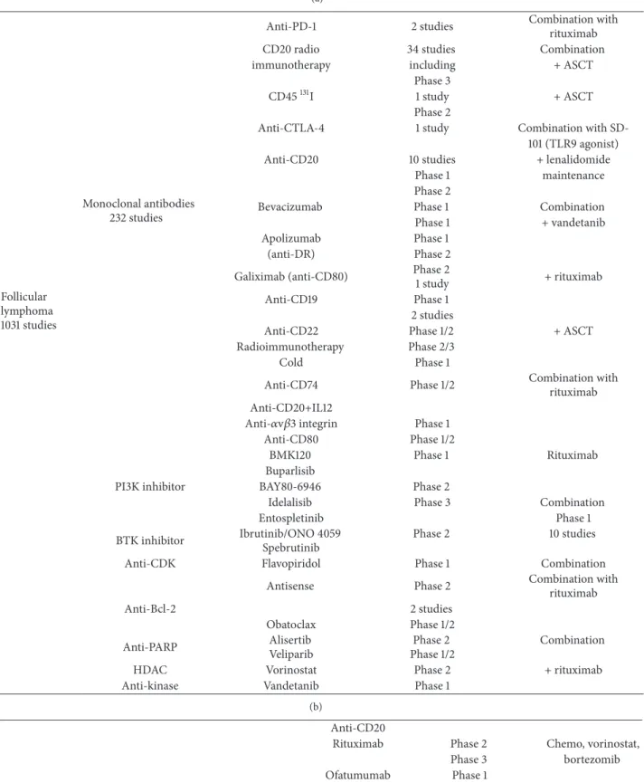
Table 2: Clinical trials for lymphoma: (a) for follicular lymphoma; (b) for Mantle cell lymphoma, based onhttps://clinicaltrials.gov/, as of March 13, 2015. PD-L-1: programmed death-1 ligand 1; CTLA-4: cytotoxic T-lymphocyte-associated protein 4; HDAC: histone deacetylase; PI3k: phosphoinositide 3-kinase; BTK: Bruton’s tyrosine kinase; Cdk: cyclin-dependent kinase; bcl2: B-cell lymphoma 2; PARP: poly(ADP-ribose) polymerase; ASCT: autologous stem cell transplantation.
(a) Follicular lymphoma 1031 studies Monoclonal antibodies 232 studies
Anti-PD-1 2 studies Combination withrituximab
CD20 radio 34 studies Combination
immunotherapy including + ASCT
Phase 3
CD45131I 1 study + ASCT
Phase 2
Anti-CTLA-4 1 study Combination with
SD-101 (TLR9 agonist)
Anti-CD20 10 studies + lenalidomide
Phase 1 maintenance
Phase 2
Bevacizumab Phase 1 Combination
Phase 1 + vandetanib
Apolizumab Phase 1
(anti-DR) Phase 2
Galiximab (anti-CD80) Phase 2
1 study + rituximab
Anti-CD19 Phase 1
2 studies
Anti-CD22 Phase 1/2 + ASCT
Radioimmunotherapy Phase 2/3
Cold Phase 1
Anti-CD74 Phase 1/2 Combination with
rituximab Anti-CD20+IL12
Anti-𝛼v𝛽3 integrin Phase 1
Anti-CD80 Phase 1/2
BMK120 Phase 1 Rituximab
Buparlisib
PI3K inhibitor BAY80-6946 Phase 2
Idelalisib Phase 3 Combination
Entospletinib Phase 1
BTK inhibitor Ibrutinib/ONO 4059 Spebrutinib
Phase 2 10 studies
Anti-CDK Flavopiridol Phase 1 Combination
Antisense Phase 2 Combination with
rituximab
Anti-Bcl-2 2 studies
Obatoclax Phase 1/2
Anti-PARP AlisertibVeliparib Phase 1/2Phase 2 Combination
HDAC Vorinostat Phase 2 + rituximab
Anti-kinase Vandetanib Phase 1
(b)
Anti-CD20
Rituximab Phase 2 Chemo, vorinostat,
Phase 3 bortezomib
(b) Continued. Ublituximab90Y/131I + lenalidomide maintenance + ASCT 51 studies Anti-CD56131I
3 studies Phase 1 + ASCT
Anti-VEGF Monoclonal antibodies
158 studies bevacizumab Phase 2 3 studies
Anti-VEGF kinase
(cediranib) Phase 1 + bevacizumab
Anti-transferrin R Phase 1
Anti-CTLA4 Phase 1/2 4 studies
Anti-HLA DR 2 studies Phase 1
Anti-CD22 Phase 1 1 study
Anti-CD2290Y Phase 1/2 + anti-CEA In111 1 study Mantle cell
lymphoma 860 studies
Anti-CD22 In111 Phase 1/2 Anti-𝛼-v 𝛽3 integrin Phase 1
Anti-CD19 Phase 1/2
Anti-CD74 Phase 1/2 + veltuzumab (humanized MoAb)
Anti-IGF-1R Phase 1/2
ganitumab
Anti-TRAIL R2 Phase 1 + bortezomib/vorinostat
conatumumab
Anti-PI3K Idelalisib Phase 1 Chemo/rituximab
BKM120 Phase 1 + rituximab
Anti-BTK Ibrutinib Phase 1 Chemo/rituximab
Anti-cdk Flavopiridol Phase 1 + chemo/rituximab
mTOR inhibitor Temsirolimus Phase 2 + rituximab
Phase 1/2 + cladribine/rituximab
Anti-endosialin/TEM1 Phase 1
HDAC Romidepsin Phase 1/2 + rituximab/lenalidomide
Anti-bcl2 Oblimersen Phase 2 + rituximab
Obatoclax + chemo/rituximab
Aurora-kinase inhibitor Alisertib Phase 2 +/− rituximab
Dehydrogenase inhibitor CPI-613 Phase 1 + bendamustine/rituximab
HDAC Vorinostat Phase 1/2 + chemo
Toll-R agonists CPG 7909 Phase 2 + chemo
These phosphorylation sites are frequently found in one of the three types of Immunoreceptor tyrosine-based regula-tory motifs (ITRM), including IT activation M (ITAM), IT inhibition M (ITIM), and IT switch M (ITSM) for SLAM/CD150 and related receptors of the CD2 subfamily [26]. Generally, ITIMs recruit the SH2 domain-containing tyrosine phosphatase SHP-1 or SHP-2, and phosphorylated ITAMs are recognized by SYK in B-cells [27,28].
Epratuzumab combined with rituximab was associated with a high response rate including 42.4% of CR rate with 60% of the patients having 3-year remission, for untreated patients with follicular lymphoma (FL) [21]. This relatively high response rate is not superior to that observed with
other treatments, but it opens the pathway for targeted therapy without chemotherapy. However, the combination of two mAbs is less cost-effective compared to new targeted drugs used orally; a decision was made to discontinue its development. A possible way for such development would be radioimmunotherapy and utilizing Yttrium-90 epratuzumab or other combination of CD22 with calicheamicin, or with PE38, a fragment of Pseudomonas exotoxin or novel anti-CD22 mAb that blocks anti-CD22 ligand binding, or second generation ADCC with linkers and more potent toxins, particularly tried in ALL [22,23].
CD19, CD200, CD38, CD138, CD56, and CS-1 are major targets expressed on Multiple Myeloma (MM) cells. MAbs
T a ble 3: C linical tr ials fo r m ul ti p le m ye lo ma, b as ed o n h tt p s://c linical tr ials.g o v/ , as o f M ar ch 13, 2015. ASCT : au to log o u s st em cell tra n sp la n ta ti o n ; P D-L -1: p rogra mmed de at h-1 liga n d 1; CTL A -4 : cyt o to xic T -l ym p ho cy te -ass o cia te d p ro te in 4; IG F -1R: in sulin gr o wt h fac to r-1 recep to r; KIR: killer cell Ig -lik e recep to r; D KK: Dic k ko pf -r el at ed prot ei n ; BT K : Br ut on ’s ty ro si n e k inas e; P I3 k :pho sphoi no si ti d e 3-k inas e; PA R P: p oly (A DP -r ib o se ) p oly me ra se . M u lt ipl e m ye lom a 19 08 st udies M o no cl o n al an ti b o dies 82 st udies An ti -CD3 8 4 st udies P has e 2 An ti -IL6 sil tu xima b 5 st udies C o m b ina ti o n An ti -CD4 0 4 st udies P has e 1/2 An ti -t ra nsf er ri n R 1 st u dy Phas e 1/2 An ti-GM2 1 st u d y P h as e 1/2 An ti -CD6 6 90 Y 1s tu d y +A SC T An ti -CD45 90 Y 1 st u d y + al log eneic tra n sp la n ta ti o n An ti -A dhesio n M o l1 1 st ud y P has e 1 A n ti -C D 38 1 st u d y C ombi n at ion An ti -PD L 1 3 st udies P has e 2 w it h vaccina ti o n C o m b ina tio n w it h lenalido mide An ti-I GF1R 1 st u d y P h as e 1 B evacizuma b 3 st udies P has e 2 co m b ina ti o n An ti -KIR 4 st udies P has e 1 and Phas e 2 An ti -CTL A -4 2 st udies + allog eneic T . An ti -CD5 2 3 st udies + allog eneic T . An ti -D KK1 1 st u dy R ando mize d Phas e 2 An ti -CD2 0 90Y/ 13 1I 2 st udies +A SC T C o ld: 3 st udies C o m b ina ti o n An ti -CD5 6 1 st ud y P has e I E lo tuzuma b 5 st udies R an do mized Phas e 1/2 An ti-GRP7 8 (P A T -S M6) 1 st ud y P has e 1 An ti-CX C R4 1 st u d y P h as e 1b BT K in h ib itors Ib ru tinib AC P -1 9 6 /AC P -3 19 2 st udies (+ ca rfilzo mib) 3 st udies Phas e 2 Phas e 1b PI3 k inas e in h ib it o rs Id el al is ib ,B Y L 71 9, C U D C -9 0 7,n el fi n av ir ,S O M 23 0L A R ,a n d so ra fe n ib Phas e 1/ 2 An ti -CD K Dinacic lib C o m b ina tio n P has e 1/2 An ti p ro te as o me Ca rfil zo m ib Ix azo mib Op ro zo mib M ar izo mib 10 st udies P has e 3 Phas e 1 HD A C Rico lin o st at , vor ino st at C o m b ina tio n P has e 1b PA RP in hib it o r A B T -888 C o m b ina tio n P has e 1
CD20
CD74
TdT
Bone marrow Bone
marrow Lymph node
Germinal center CD24 CD138 CD38 CD200 (CD56) CS-1 CD138 CD22 CD21 (CD5) (CD22) CD10 CD21 CD10 (CD23) CD40, CD19 𝜇 BCR𝜇 BCR𝜇/𝛿 BCR𝜇/𝛾 CD38 IL6R TACI/B CMA IGF-1R FGFR3 VEGFR C-ki t
Figure 1: Surface markers of B-cell lineage present at the principal stages of differentiation, as targets for therapy. TdT: terminal deoxynucleotide transferase. TACI/BCMA: transmembrane activator and CAML interactor/B-cell maturation antigen. IGF-1R: insulin growth factor-1 receptor. EGFR: epithelial growth factor receptor. VEGFR: vascular endothelial growth factor receptor. IL6R: interleukin 6 receptor. FGFR3: fibroblast growth factor receptor type 3. c-Kit: CD117. BCR: B-cell receptor.
against such molecules have been clinically developed [29]. Elotuzumab, a humanized mAb IgG1 antibody that targets CS-1 (SLAMF7), a cell surface glycoprotein with major expression in MM cells, has been shown to support very active ADCC [30]. It has been combined with lenalidomide and dexamethasone in patients having relapsed MM with promising results, 90% of the patients achieving a partial response (PR) with PFS exceeding 2 years [30]. A Phase III clinical study is ongoing and is due to be completed by 2017. Daratumumab is a humanized antibody against CD38 [31], a cell surface protein strongly expressed in MM [32]. CD38 is also expressed on malignant cells from B-CLL, mantle cell lymphoma (MCL), transformed FL, and clinical trials are ongoing with daratumumab in these diseases [31]. Some MM cells expressed CD56 and lorvotuzumab, an mAb against CD56, conjugated to mertansine has been developed in early clinical studies for MM [33]. CD200 is an immunosuppres-sive molecule overexpressed in several hematological malign-ancies including B-CLL, MM, and acute myeloid leukemia (AML) [34,35]. Early clinical trials are ongoing in these dis-eases or in different models of immunotherapeutic strategies in AML [35]. Syndecan-1 (CD138) belongs to heparan sul-fate proteoglycans that is highly expressed at the cell sur-face of MM cells [32,36]. In addition, cell surface CD138 acts as a functional coreceptor for chemokines and growth factors in the plasma cell niche. Soluble form of syndecan-1 can accu-mulate survival factors within the microenvironment, repre-senting a sort of sponge for these factors around the tumor cells [36]. Therefore, targeting this molecule is of potential clinical interest, due to a mixed activity on both the tumor cells and its cell niche, making the molecule attractive for radioimmunotherapy [37]. Different mAbs have been devel-oped in early clinical phases including anti-CD40 mAbs such as lucatumumab, dacetuzumab, or mAb directed against HM1.24, the XmAb 5592 [38]. A total of 91 studies with
mAbs are registered (https://clinicaltrials.gov/ct2/results? term=monoclonal+antibodies+in+multiple+myeloma&Search= Search) in MM patients, as of March 13, 2015.
For all of these mAbs and similarly to rituximab, clinical efficacy was only observed with combination strategies, par-ticularly with other active drugs, depending on the response or the refractoriness to prior therapies, including borte-zomib, IMiDs such as lenalidomide plus dexamethasone, and other new active drugs including approved molecules such as pomalidomide and carfilzomib, or other new targeted molecules.
2.2. Survival/Proliferation Factor Receptors. Upon
recogni-tion of foreign antigens, mature naive B lymphocytes are activated, leading to the production of short-lived plasma cells, followed by their proliferation and differentiation into memory B-lymphocytes and long-lived plasma cells for durable Ig production [39, 40]. Along these different steps, B-lymphocytes respond to diverse signals or sur-vival/proliferation factors, including BAFF/APRIL, BCR, IL6, VEGF, EGF, and IGF-1 [39]. By blocking the specific receptor or neutralizing the ligand, the activation of signaling pathway is not delivered into the cell, leading to tumor cell growth and/or survival arrest. BAFF and APRIL belong to the TNF family that binds to the TNFR-like receptors transmembrane activator, particularly interacting with three receptors, cal-cium modulator and cyclophilin ligand interactor (TACI), B-cell maturation antigen (BCMA), and BAFFR for only BAFF [41, 42]. APRIL is produced by hematopoietic cells, particularly by osteoclasts [43]. The inhibition of BAFF and APRIL using a soluble receptor, TACI-Ig or atacicept (SeronoMerck Inc.), in a culture of myeloma cell lines causes rapid cell death [44] and inhibits myeloma growth in a coculture system with osteoclasts [45]. We used this drug in a Phase I study, in patients with MM and macroglobulinemia,
with promising results, but this drug was mainly developed in dysimmune diseases [46–48].
Different mAbs against IL6 or soluble IL6R have been developed, particularly siltuximab, an anti-IL6 mAb, and tocilizumab, an antisoluble IL6R. Siltuximab has been recently registered for Castleman’s disease in Europe and USA. Tocilizumab is registered for some dysimmune diseases refractory to anti-TNF worldwide and Castleman’s diseases only in Japan [49].
IGF-1 represents the main cell communication factor produced by plasma cells and bone marrow stromal cells [50]. Inhibitors of IGF-1, including dalotuzumab and picrop-odophyllin, have been tested in cancers including early clini-cal phases of MM [51,52]. However, as observed for IL6, the use of such specific inhibitors in very advanced diseases did not show any clinical benefit due to intraclonal heterogeneity, with the emergence of tumor cell independence from their microenvironment in addition to other growth factors [53].
3. Intracellular Targets
3.1. Cell Signaling Markers. The activation of the BCR is
a major signaling pathway for B-lymphocyte function. The BCR is a multiprotein structure with a surface transmem-brane Ig noncovalently associated with the Ig𝛼 (CD79A) and Ig𝛽 (CD79B) chains [54, 55]. Antigen binding to the BCR causes receptor aggregation and engagement of the signal transduction via the phosphorylation of the receptor’s cytoplasmic tyrosine-based activation motifs (ITAMs) by recruited SRC-family kinases, including LYN, FYN, BLK, and LCK [54]. Then, the activation of phosphoinositide 3-kinase (PI3K3𝛿) mediates the conversion of phosphatidylinositol-4,5-bisphosphate to phosphatidylinositol-3,4,5-trisphosphate and ultimately recruits BTK [56]. BTK phosphorylation targets phospholipase C𝛾2 (PLC𝛾2), with activation of NF𝜅B and mitogen-activated protein kinase pathways. Antigen-independent signaling has been involved in B-CM which results in constitutive or aberrant BCR signaling, making BTK a major target for such diseases [57]. Ibrutinib (PCI-32765, Imbruvica, J&J Inc.) has been developed in B-CLL and B-cell lymphoma and is now approved for MCL and B-CLL. In a Phase II clinical study, a dramatic response rate was observed in both diseases, particularly in MCL with refractory disease (objective response rate (ORR) 68% including 21% CR) with a median PFS of 13.9 months [55]. Ibrutinib inhibits the adhesion mediated by chemokine and integrin to their microenvironment. This biological effect is associated with lymphocytosis and nodal shrinkage. This lymphocytosis decreased generally at the end of cycle 2. Tolerability was acceptable and adverse events included diar-rhea (50%), fatigue (44%), nausea (38%), cough (31%), and myalgia (25%). As ibrutinib is metabolized by cytochrome P450 enzyme 3A (CYP3A), coadministration with CYP3A inhibitors or inducers can interfere with other drugs and may be responsible for some toxicity. Ibrutinib was associated with chemotherapy, bendamustine, or the CHOP-R regimen that associates cyclophosphamide, doxorubicin, vincristine, prednisone, and anti-CD20 mAbs, particularly in B-cell lymphoma [55]. Currently, ibrutinib is used in naive patients,
especially for B-CLL and small lymphocytic lymphoma. It showed a high response rate of 71% including 13% CR, with estimated PFS and OS at 2 years of 96.3% and 96.6%, respec-tively, at the daily dose of 420 mg [58]. In MM, the overexpres-sion of BTK varied, being more present in MM than in mon-oclonal gammopathy of undetermined significance (MGUS), with also some interindividual variability of the expression level. Despite this variability, ibrutinib was associated with a high response rate in patients with refractory MM [59].
Ibrutinib has promising activity in other B-CM, including atypical B-cell lymphoproliferative disorders (personal data) and dysimmune diseases. The active dose could be correlated with the expression level of the targeted molecule, and its measurement could be a guide to optimize the clinical effi-cacy. In addition, since this drug is also active in patients with poor prognostic factors such as p53 mutation or other acquired genetic modifications, there is a need to define new markers of interest and new therapeutic combina-tions including immune therapy to prolong the therapeutic response. Knowing the mechanisms of resistance, the effect on the normal B-cell compartment and other immune cells, the status of the immune response and following the residual disease may contribute to addressing the question of the optimal treatment duration, to avoid the mistakes made with IMiDs in MM [60]. Some resistance mechanisms have been studied, including NF𝜅B pathway and KRAS mutations. The effects of ibrutinib on normal immune cells begin to be studied, including IL2-inducible kinase that promotes a T helper 1 response, a depletion of the B-cell memory and long-lived plasma cell compartment, thus reducing a recall response or a new antigen-dependent response.
Several other BTK inhibitors are in clinical development, including ONO-4059 (Gilead Inc., USA) and AVL-292 (Cel-gene Inc., USA) which are reversible inhibitors of BTK. In addition, there are multikinase inhibitors, such as LFM-A13, which inhibits BTK and polo-like kinase (PLK), fostama-tinib, which inhibits the 𝛿 isoform of PI3K and Syk [61], and dasatinib, initially developed as an inhibitor of tyrosine kinase for CML patients, which is also a BTK inhibitor.
The PI3 kinase (PIK)/AKT/mTOR pathway is an impor-tant signalling pathway for cellular functions, particularly growth and metabolism control. Different classes and iso-forms of PI3Ks exist that are associated with large possibilities of inhibition leading to a great number of molecules inhibit-ing this pathway. IPI-145 inhibits PI3K𝛿 and 𝛾, and it was developed in hematological malignancies. BAY 80-6946 pre-dominantly inhibits PI2K𝛼, 𝛿 isoforms, as well as INK1117, a PI3K𝛼 inhibitor, and more than 30 other compounds. Among them, idelalisib (GS-1101, Zydelig, Gilead Inc.), a speci-fic inhibitor of class I isoform p110𝛿 was approved on July 23, 2014, in USA for the treatment of FL and CLL and B-cell small lymphocytic lymphoma [62]. This molecule is also active in other B-CM.
The combination of these kinases inhibitors with mAbs requires the evaluation of the impact of such molecules on effector cells, particularly NK lymphocytes. Ibrutinib did not inhibit complement activation or complement-mediated lysis. In contrast, ibrutinib and idelalisib strongly inhibited cell-mediated mechanisms induced by anti-CD20 mAbs,
particularly the activation of NK lymphocytes [63]. In addi-tion, idelalisib reduces T-regulator lymphocytes (T-regs) and could have a positive impact on tumor cells [64].
3.2. Cell Cycle, Proteasome, and Apoptosis Machinery. In
cancer, proliferation depends on different proteins involved in cell-cycle regulation, particularly alterations of the cyclin-dependent kinase (CDK) CDK4/6-INK4-Rb-E2F cascade [65]. Resistance to chemotherapy is mediated by dysregu-lation of the cell-cycle machinery [66]. Overexpression of cyclins (e.g., cyclins D1 and E1), amplification of CDKs (e.g., CDK4/6), inactivation of critical CDK by CDK inhibitors (I) (e.g., p16Ink4a, p15Ink4b, p21waf1, and p27kip1), loss of retinoblastoma (Rb) expression, and loss of binding of CDKIs to CDKs (e.g., INK4 binding to CDKs) occur frequently in human malignancies [65]. Defects of apoptotic pathways are often observed in hematologic malignancies, involving the global repression of transcription by drugs that inhibit CDK7/9 [67]. Transcriptional CDKIs downregulate a great number of short-lived antiapoptotic proteins. This includes the antiapoptotic proteins myeloid cell leukemia-1 (Mcl-1) particularly critical in hematologic malignancies, the B-cell lymphoma extra long (Bcl-xL) and the XIAP (X linked IAP), D-cyclins, c-myc, Mdm-2 (leading to p53 stabilization), p21waf1, proteins whose transcription is mediated by nuclear factor-kappa B (NF-𝜅B), and hypoxia-induced VEGF [68].
Molecules that interfere with CDKs have been developed, either targeting a broad spectrum of CDKs or a specific type of CDK or targeting CDKs as well as additional kinases such as VEGFR or platelet-derived growth factor-R (PDGFR). More than 10 molecules have gone through clinical trials, including multi-CDK inhibitors such as flavopiridol (Sanofi-Aventis Inc.), a semisynthetic flavonoid, known as a pan-CDK inhibitor, developed in a large panel of hematological malignancies, SNS-032 (BMS-387032, Sunesis, BMS Inc.) developed in B-CLL, MM, and NHL, dinaciclib (SCH 727965, Merck Inc.) and PD0332991 (Pfizer Inc.) developed in various hematological malignancies, and R-roscovitine (seliciclib, CYC202, Cyclacel Inc.) [69]. The combinations of such inhibitors with cytotoxic agents but also with novel and targeted agents, including histone deacetylase inhibitors and PKC activators, NF𝜅B pathway modulators, and probably BTK and PI3K inhibitors, are programmed for clinical trials. The ubiquitin proteasome pathway plays a critical role in regulating many processes in the cell, which are important for tumor cell growth and survival. Bortezomib was the first clinical success in some cancers and has prompted the development of the second generation of proteasome inhibitors. The ubiquitin proteasome system represents the major pathway for intracellular protein degradation, with a complex mechanism involving at least six components: ubiquitin (Ub), the activating (E1), a group of Ub-conjugating enzymes (E2), a group of Ub ligases (E3), the proteasome, and the deubiquitinases, a process that is highly controlled in normal cells, but frequently dysregulated in cancers [70].
Chemotherapy designed cytotoxic drugs which are active through impairing mitosis or fast-dividing cells by various mechanisms including damaging DNA and inhibition of the
cellular machinery involved in cell division. The number of dividing cells is estimated by the mitotic index, the presence of Ki-67 positive cells on tumor samples, or the percentage of cell cycling in S phase. Such analysis may guide the prescription of cytotoxic drugs, particularly for cancers with variable percentage of cycling cells like in MM with high proliferative index superior to 4% of cells in S phase [71]. The inhibition of NF𝜅B activity modified the degradation of cell cycle-related molecules and perturbed proapoptotic and antiapoptotic protein balance, endoplasmic reticulum stress and inhibited angiogenesis and DNA repair, all the mechanisms that contribute to apoptosis of tumor cells. NF𝜅B that is constitutively active in a large proportion of cancers and is bound to its inhibitor I𝜅B within the cytoplasm, and inhibition of proteasome activity prevents degradation of I𝜅B and subsequent activation and translocation of NF𝜅B to the nucleus. Proteasome inhibitors may induce cell cycle arrest by interfering with the degradation of cell cycle regulators including cyclins. There are several inhibitors of proteasome that are used in clinic for hematological malignancies, parti-cularly for MM and MCL, and used in combination with different other drugs such as IMiDs, other cytotoxic mole-cules, and dexamethasone. Major proteasome inhibitors include bortezomib, carfilzomib, but also NPI-0052, a 𝛽-lactone derived from the marine bacterium Salinispora
trop-ica, MLN9708, CEP-18770, ONX0912, or inhibitor of the
immunoproteasome such as ISPI-101 or PR-924 [70]. Apoptosis is a common process of cell death for all multicellular eukaryotic organisms leading to the eradication of damaged or infected cells. Apoptosis is initiated by two sig-naling pathways, the intrinsic or mitochondrial pathway and the extrinsic or death receptor pathway, that is, Fas/CD95 that binds to specific cell surface receptors. The intrinsic pathway, with members of BCL2 family, is more commonly perturbed in lymphoid malignancies, including mutation of the tumor suppressor gene TP53, which normally acts to activate certain BH3-only proteins, and the overexpression of BCL2 [72].
Obatoclax (GX15-070) is a pan-BCL2 family inhibitor, binding to BCL2, BCLxL, BCLw, and MCL1. Therapeutic response with obatoclax in clinical trials has been reported to be low and its development has been halted [68]. The natural product gossypol and its synthetic derivative AT-101 bind to BCL2, BCLxL, and MCL1 with clinical activity only when combined to rituximab for FL [73]. Antiapoptotic BCL2 proteins antagonize death signaling by heterodimer forma-tion through binding at the BH3 domain of the protein. New molecules, BH3 mimetics were designed to functionally anta-gonize BCL2 protein family [74]. ABT-737 and its orally available analogue ABT-263 (navitoclax) bind and inhibit BCL2, BCLxL, and BCLw with high affinity, and it is developed in clinical phases, as well as ABT-199 which may be considered as the most active drug in the BCL2 family inhibitors. ABT-199 has shown high response rate (87%) in relapsed/refractory B-CLL, including bulky disease, fludarabine-refractory disease, and del17p patients [75], as well as for FL, Waldenstr¨om’s disease, and MCL [73]. ABT-199 induces apoptosis within 8 hours and the most significant dose-limiting toxicity is tumor lysis syndrome. In addition, ABT-199 may be combined to chemotherapy, demethylating
agents, histone deacetylase inhibitors, and novel targeted drugs such as ibrutinib and idelalisib [76].
3.3. Metabolic Process. In the early twentieth century,
War-burg first discovered that cancer cells preferentially consume glucose and metabolize it to lactate in the presence of oxygen, named aerobic glycolysis [77]. Accumulated evidence was made to support that this metabolic way was predominant for hematological malignancies in leukemias and T-cell lymphoma, with both inducers of Warburg effect, PKM2, and HIF-1𝛼, reported to be involved in AML and connected to epigenetic control of gene expression [78,79]. This metabolic process facilitates cancer progression by resisting induction of apoptosis and promoting tumor metastasis or indepen-dence of the cancer cell microenvironment. Hypoxia is a major factor that contributes to the Warburg effect, for rapid energetic production for the cancer cell, a process favored by changes within the microenvironment. Blocking glycolysis causes a rapid dephosphorylation of BAD protein at Ser112, leading to BAX localization to mitochondria and impor-tant cell death, also observed in multidrug resisimpor-tant cells [80]. The uptake of fluorodeoxyglucose positron emission tomography in cancers demonstrates the key role of glucose in the proliferation of cancer cells [81]. The generic drug dichloroacetate is a small orally available molecule known to block the pyruvate dehydrogenase kinase. It has thus been proposed in various cancers including rare patients with hematological malignancies and its use was associated with some success [82]. Through the reduction of SIRT1, the inhibition of LDH-A provides a way of altering p53 acetylation status and the downstream induction of p53 target genes selectively in cancer cells [83]. Other target is represented by peroxisome proliferator-activated receptor (PPAR), a group of nuclear receptor proteins that function as transcription factors regulating gene expression. PPAR-𝛼 is particularly implicated in lipid and lipoprotein metabolism and inflammation. Fenofibrate, a PPAR-𝛼 agonist, has been shown to induce apoptosis on certain cancer cells via acti-vation of NF-𝜅B pathway [84]. Inhibitors for PPAR-𝛾 may
enhance the activity of radiation therapy in cancer [85]. There are several compounds that modulate glycolytic metabolism. This includes 2-deoxyglucose that inhibits phos-phorylation of glucose hexokinase (HK), lonidamine, that inhibits glycolysis and mitochondrial respiration, HK, 3-bromopyruvate that inhibits HK and acts as an alkylating agent, imatinib that inhibits bcr-abl tyrosine kinase but also decreases HK and G6PD, and oxythiamine that inhibits pyruvate dehydrogenase [86, 87]. Specific LDH inhibitors have been developed, including AT-101, FX-11, galloflavin, N-hydroxyindole-based molecules [88], or new molecules in development by different companies. Such new molecules represent a new potent way to modulate or prevent chemore-sistance. In addition, they may have some impact on immune cells [89].
4. Targeting Microenvironment
4.1. Immune Therapy. The tumoral microenvironment, and
particularly immune cells, is involved in the tumor cell
control or expansion. Since many years, it has been rec-ognized that T-infiltrating lymphocytes (TIL), a mixture of different cells (Treg, T helper, T cytotoxic cells, etc.) when expanded ex vivo, may support some clinical efficacy [90,91]. Nowadays, the different cell subpopulations associated with a particular function (i.e., facilitating or repressing tumor cells) may orientate the clinical prognosis and the response to therapeutic agents [92–98]. Targeting cancer cells via the immune system depends on the presence of effector cells that recognize and kill cancer cells. Recognition may be specific for adaptative response, that is, cytotoxic T-cells via antigen presentation. In the context of innate response, there are other mechanisms to recognize stress cells or non-self-cells, includ-ing activatinclud-ing and inhibitinclud-ing molecules shared by NK, NKT, and T𝛾𝛿 lymphocytes [99]. Such cancer cell recognition may be forced by using chimeric antigen cells (T cells, CAR-NK cells) [20] or bispecific mAbs. Beyond recognition, target accessibility and tumor infiltration, mechanisms, and efficacy of killing are other criteria of efficacy to be considered. Effector cells could be directly used as cell-drugs or immune modulators that activate such specific activity, including Toll-receptor agonists [100], enhancers of ADCC and antigen presentation via dendritic cells [101], and stimulator of T𝛾𝛿,
particularly 𝛾9𝛿2 T-cell, that may be purified for cellular therapy programs and activated by IL2 and bisphosphonates or IPH101 ([102–104] and personal data) combined with anti-CD20 mAbs [93]. It is surprising that using GM-CSF in addition to rituximab or IPH101 plus IL2 and rituximab, in relapsed or refractory FL, we observed similar results with 45–50% of ORR ([105] and personal data), meaning that optimal strategy is probably the direct administration of these effector cells. Development of NK cells is now one major way for immune therapy probably by using banked, activated, and amplified NK cells from cord blood samples ([106,107] and personal data). In that way, it is important to know the efficacy of killing. For NK cells, in vitro data showed that one NK cell may kill 8–10 tumoral cells. Conversely, 10 cytotoxic T-cells are needed to kill one tumoral cell. This shows that NK-cell drugs are more efficient for killing, with a clinical efficacy ranging between 107and 109tumoral cells. But cytotoxic T-cells may retain a certain memory of killing and prolong the effect. This means that clinical use of such cell-drugs has different clinical targets and could be associated for a better clinical benefit. We need to simplify the therapeutic strategies and think about best combinations of drugs, cell-drugs, modifiers, and new targeted therapies.
4.2. Niche Disruption. Lymph node microenvironment
includes different types of lymphocytes and stromal cells necessary to the antigen presentation and the education of B-cells to secrete specific antibodies. Plasmablasts generated in germinal centers exit the lymphoid organs into the lymph and then the blood, before migrating to the bone marrow or mucosa-associated lymphoid tissues where they represent a long-lived population of plasma cells in a favorable microenvironment, named plasma cell niche. Different cells constitute this niche, particularly mesenchymal cells that produce chemokines, particularly CXCL12, and bring together other niche cells (megacaryocytes, platelets, and
eosinophils) and plasma cells, which all express the CXCL12 receptor, CXCR4 [108]. Within the niche, plasma cells are activated by adhesion molecules and stimulated by several survival/growth factors [109]. The hypoxic microenviron-ment plays a central role in controlling stem cell phenotype and dissemination, through different factors particularly the hypoxia-inducible factor-1𝛼 (HIF-1𝛼), a key transcriptional factor that responds to hypoxic stimuli [110]. HIF-1𝛼 is
constitutively expressed in some B-cell malignancies and is regulated by the PI3K/AKT pathway [111].
Anti-CXCR4 or CXCL12R (plerixafor and others), anti-CCR5 or CCL5R (maraviroc), inhibitors of survival/pro-liferation factors, that is, IL6, BAFF/APRIL, and others, but also inhibitors of osteoprotegerin, and a receptor for both RANKL and TNF-related apoptosis-inducing ligand/Apo2 (TRAIL) may represent new targets for cancer therapy [49, 112, 113]. The complex CXCL12/CXCR4 is implicated in biological mechanisms of several B-cell malignancies, particularly for CLL, MM, and lymphoma [112]. Plerix-afor/AMD3100 disrupts the B-CLL microenvironment inter-actions, representing additional treatment, possibly with novel targeted drugs [114].
Syndecan-1 is a member of the heparan sulfate (HS) pro-teoglycans that are present on the cell surface or as soluble molecules shed from the cell surface. Syndecan-1 accumulates survival factors within the microenvironment, representing a sort of sponge for these factors around the tumor cells. Syndecan-1 is cleaved by heparanase, an endo-𝛽-o-glucuroni-dase, secreted by osteoclasts [36]. As heparin and low molec-ular weight heparin have been known since a long time to exhibit potent antiheparanase activity, one can explain that such molecules may have a clinical impact on the cancer [115]. A new therapeutic era is born for new reflection, new methodologies and, nowadays, nearly all therapies are tar-geted as long as we understand biological processes for a better use of old and new drugs to support personalized medicine.
Conflict of Interests
The author declares that there is no conflict of interests regarding the publication of this paper.
Acknowledgment
The author wishes to thank Vidal Benatar M.D. of Heathics S.A.R.L., who provided editorial assistance for this paper.
References
[1] D. R. Anderson, A. Grillo-L¨opez, C. Varns, K. S. Chambers, and N. Hanna, “Targeted anti-cancer therapy using rituximab, a chimaeric anti-CD20 antibody (IDEC-C2B8) in the treatment of non-Hodgkin’s B-cell lymphoma,” Biochemical Society
Trans-actions, vol. 25, no. 2, pp. 705–708, 1997.
[2] D. G. Maloney, A. J. Grillo-L´opez, C. A. White et al., “IDEC-C2B8 (rituximab) anti-CD20 monoclonal antibody therapy in patients with relapsed low-grade non-Hodgkin’s lymphoma,”
Blood, vol. 90, no. 6, pp. 2188–2195, 1997.
[3] M. A. Weiss, “Novel treatment strategies in chronic lymphocytic leukemia,” Current Oncology Reports, vol. 3, no. 3, pp. 217–222, 2001.
[4] O. W. Press, J. P. Leonard, B. Coiffier, R. Levy, and J. Timmer-man, “Immunotherapy of Non-Hodgkin’s lymphomas,”
Hema-tology, pp. 221–240, 2001.
[5] J. F. Rossi, “Nowadays, all therapies are targetted. Under-standing biology improves disease management,” International
Journal of Hematology Research, vol. 1, no. 1, 2015.
[6] D. Rodr´ıguez-Pinto, “B cells as antigen presenting cells,”
Cellu-lar Immunology, vol. 238, no. 2, pp. 67–75, 2005.
[7] S. H. Lim, S. A. Beers, R. R. French, P. W. M. Johnson, M. J. Glennie, and M. S. Cragg, “Anti-CD20 monoclonal antibodies: historical and future perspectives,” Haematologica, vol. 95, no. 1, pp. 135–143, 2010.
[8] H. E. Mei, S. Schmidt, and T. D¨orner, “Rationale of anti-CD19 immunotherapy: an option to target autoreactive plasma cells in autoimmunity,” Arthritis Research and Therapy, vol. 14, supplement 5, article S1, 2012.
[9] T. Robak and P. Robak, “Anti-CD37 antibodies for chronic lymphocytic leukemia,” Expert Opinion on Biological Therapy, vol. 14, no. 5, pp. 651–661, 2014.
[10] S. Kumar, T. Kimlinger, and W. Morice, “Immunophenotyping in multiple myeloma and related plasma cell disorders,” Best
Practice and Research: Clinical Haematology, vol. 23, no. 3, pp.
433–451, 2010.
[11] A. Caraux, L. Vincent, S. Bouhya et al., “Residual malignant and normal plasma cells shortly after high dose melphalan and stem cell transplantation. Highlight of a putative therapeutic window in Multiple Myeloma?” Oncotarget, vol. 3, no. 11, pp. 1335–1347, 2012.
[12] E. Vacchelli, F. Aranda, A. Eggermont et al., “Trial Watch: tumor-targeting monoclonal antibodies in cancer therapy,”
OncoImmunology, vol. 3, no. 1, Article ID e27048, 2014.
[13] J. Golay, M. Lazzari, V. Facchinetti et al., “CD20 levels determine the in vitro susceptibility to rituximab and complement of B-cell chronic lymphocytic leukemia: further regulation by CD55 and CD59,” Blood, vol. 98, no. 12, pp. 3383–3389, 2001.
[14] N. Cooper and D. M. Arnold, “The effect of rituximab on humoral and cell mediated immunity and infection in the treat-ment of autoimmune diseases,” British Journal of Haematology, vol. 149, no. 1, pp. 3–13, 2010.
[15] J. C. Gea-Banacloche, “Rituximab-associated infections,”
Semi-nars in Hematology, vol. 47, no. 2, pp. 187–198, 2010.
[16] L. Quartuccio, S. Lombardi, M. Fabris et al., “Long-term effects of rituximab in rheumatoid arthritis: clinical, biologic, and pharmacogenetic aspects: review,” Annals of the New York
Academy of Sciences, vol. 1173, pp. 692–700, 2009.
[17] J. F. Rossi, B. Caumes, E. Tuaillon et al., “Immune response to a vaccination against H1N1 influenzae virus for patients with untreated chronic lymphocytic leukemia of B-cell type,” Sub-mitted.
[18] S. Auger-Quittet, Y. Duny, J. P. Daures, and P. Quittet, “Treat-ment with yttrium-90 (90Y)-Ibritumomab tiuxetan (Zevalin) in diffuse large B-cell lymphoma: a meta-analysis,” In press. [19] Z. Zimmerman, T. Maniar, and D. Nagorsen, “Unleashing the
clinical power of T cells: CD19/CD3 bi-specific T cell engager (BiTE(R)) antibody construct blinatumomab as a potential therapy,” International Immunology, vol. 27, no. 1, pp. 31–37, 2014.
[20] B. Jena, J. S. Moyes, H. Huls, and L. J. N. Cooper, “Driving CAR-based T-cell therapy to success,” Current Hematologic
Malignancy Reports, vol. 9, no. 1, pp. 50–56, 2014.
[21] B. W. Grant, S.-H. Jung, J. L. Johnson et al., “A phase 2 trial of extended induction epratuzumab and rituximab for previously untreated follicular lymphoma: CALGB 50701,” Cancer, vol. 119, no. 21, pp. 3797–3804, 2013.
[22] L. Fayad, F. Offner, M. R. Smith et al., “Safety and clinical activity of a combination therapy comprising two antibody-based targeting agents for the treatment of non-hodgkin lymphoma: results of a phase I/II study evaluating the immunoconjugate inotuzumab ozogamicin with rituximab,” Journal of Clinical
Oncology, vol. 31, no. 5, pp. 573–583, 2013.
[23] L. Sullivan-Chang, R. T. O’Donnell, and J. M. Tuscano, “Tar-geting CD22 in B-cell malignancies: current status and clinical outlook,” BioDrugs, vol. 27, no. 4, pp. 293–304, 2013.
[24] T. Mark, P. Martin, J. P. Leonard, and R. Niesvizky, “Milatuzumab: a promising new agent for the treatment of lymphoid malignancies,” Expert Opinion on Investigational
Drugs, vol. 18, no. 1, pp. 99–104, 2009.
[25] E. A. Rossi, D. M. Goldenberg, R. Michel, D. L. Rossi, D. J. Wallace, and C.-H. Chang, “Trogocytosis of multiple B-cell surface markers by CD22 targeting with epratuzumab,” Blood, vol. 122, no. 17, pp. 3012–3029, 2013.
[26] H. Liu, L. Li, C. Vos, F. Wang, J. Liu, and S. S. Li, “A com-prehensive immunoreceptor phosphotyrosine-based signaling network revealed by reciprocal protein-peptide array screen-ing,” Molecular & Cellular Proteomics, 2015.
[27] J. S. Bezbradica and R. Medzhitov, “Role of ITAM signaling module in signal integration,” Current Opinion in Immunology, vol. 24, no. 1, pp. 58–66, 2012.
[28] M. Shabani, A. A. Bayat, M. Jeddi-Tehrani et al., “Ligation of human Fc receptor like-2 by monoclonal antibodies down-regulates B-cell receptor-mediated signalling,” Immunology, vol. 143, no. 3, pp. 341–353, 2014.
[29] D. W. Sherbenou, C. R. Behrens, Y. Su, J. L. Wolf, T. G. Martin III, and B. Liu, “The development of potential antibody-based therapies for myeloma,” Blood Reviews, vol. 29, no. 2, pp. 81–91, 2015.
[30] P. Moreau and C. Touzeau, “Elotuzumab for the treatment of multiple myeloma,” Future Oncology, vol. 10, no. 6, pp. 949–956, 2014.
[31] T. Mark and Y. Khagi, “Potential role of daratumumab in the treatment of multiple myeloma,” OncoTargets and Therapy, vol. 7, pp. 1095–1100, 2014.
[32] J. Wijdenes, W. C. Vooijs, C. Cl´ement et al., “A plasmocyte selective monoclonal antibody (B-B4) recognizes syndecan-1,”
British Journal of Haematology, vol. 94, no. 2, pp. 318–323, 1996.
[33] J. G. Berdeja, “Lorvotuzumab mertansine: antibody-drug-con-jugate for CD56+ multiple myeloma,” Frontiers in Bioscience, vol. 19, no. 1, article 163, 2014.
[34] P. Challagundla, L. J. Medeiros, R. Kanagal-Shamanna, R. N. Miranda, and J. L. Jorgensen, “Differential expression of CD200 in B-cell neoplasms by flow cytometry can assist in diagnosis, subclassification, and bone marrow staging,” American Journal
of Clinical Pathology, vol. 142, no. 6, pp. 837–844, 2014.
[35] A. Martner, F. B. Thor´en, J. Aurelius, and K. Hellstrand, “Immu-notherapeutic strategies for relapse control in acute myeloid leukemia,” Blood Reviews, vol. 27, no. 5, pp. 209–216, 2013. [36] K. Mahtouk, F. W. Cremer, T. R`eme et al., “Heparan sulphate
proteoglycans are essential for the myeloma cell growth activity
of EGF-family ligands in multiple myeloma,” Oncogene, vol. 25, no. 54, pp. 7180–7191, 2006.
[37] C. Rousseau, L. Ferrer, S. Supiot et al., “Dosimetry results sug-gest feasibility of radioimmunotherapy using anti-CD138 (B-B4) antibody in multiple myeloma patients,” Tumor Biology, vol. 33, no. 3, pp. 679–688, 2012.
[38] K. C. Anderson, “The 39th David A. Karnofsky lecture: bench-to-bedside translation of targeted therapies in multiple mye-loma,” Journal of Clinical Oncology, vol. 30, no. 4, pp. 445–452, 2012.
[39] D. A. Jackson and S. F. Elsawa, “Factors regulating immuno-globulin production by normal and disease-associated plasma cells,” Biomolecules, vol. 5, no. 1, pp. 20–40, 2015.
[40] M. Tsuneto, E. Kajikhina, K. Seiler et al., “Reprint of: Environ-ments of B cell development,” Immunology Letters, vol. 160, pp. 109–112, 2014.
[41] A. Mukhopadhyay, J. Ni, Y. Zhai, G.-L. Yu, and B. B. Aggar-wal, “Identification and characterization of a novel cytokine, THANK, a TNF homologue that activates apoptosis, nuclear factor-𝜅B, and c-jun NH2-terminal kinase,” The Journal of Biological Chemistry, vol. 274, no. 23, pp. 15978–15981, 1999.
[42] F. Mackay, P. Schneider, P. Rennert, and J. Browning, “BAFF and APRIL: a tutorial on B cell survival,” Annual Review of
Immu-nology, vol. 21, pp. 231–264, 2003.
[43] R. M. Reijmers, M. Spaargaren, and S. T. Pals, “Heparan sulfate proteoglycans in the control of B cell development and the pathogenesis of multiple myeloma,” FEBS Journal, vol. 280, no. 10, pp. 2180–2193, 2013.
[44] J. Moreaux, E. Legouffe, E. Jourdan et al., “BAFF and APRIL protect myeloma cells from apoptosis induced by interleukin 6 deprivation and dexamethasone,” Blood, vol. 103, no. 8, pp. 3148–3157, 2004.
[45] S. Yaccoby, A. Pennisi, X. Li et al., “Atacicept (TACI-Ig) inhibits growth of TACIℎ𝑖𝑔ℎ primary myeloma cells in SCID-hu mice and in coculture with osteoclasts,” Leukemia, vol. 22, no. 2, pp. 406–413, 2008.
[46] J.-F. Rossi, “Phase I study of atacicept in relapsed/refractory multiple myeloma (MM) and Waldenstr¨om’s macroglobuline-mia,” Clinical Lymphoma, Myeloma & Leukemia, vol. 11, no. 1, pp. 136–138, 2011.
[47] J.-F. Rossi, J. Moreaux, D. Hose et al., “Atacicept in relapsed/refractory multiple myeloma or active Waldenstr¨om’s macroglobulinemia: a phase I study,” British Journal of Cancer, vol. 101, no. 7, pp. 1051–1058, 2009.
[48] H.-P. Hartung and B. C. Kieseier, “Atacicept: targeting B cells in multiple sclerosis,” Therapeutic Advances in Neurological
Disorders, vol. 3, no. 4, pp. 205–216, 2010.
[49] J. F. Rossi, Z. Y. Lu, M. Jourdan, and B. Klein, “Interleukin-6 as a therapeutic target,” Clinical Cancer Research, vol. 21, no. 6, pp. 1248–1257, 2015.
[50] B. M. Birmann, M. L. Neuhouser, B. Rosner et al., “Prediag-nosis biomarkers of insulin-like growth factor-1, insulin, and interleukin-6 dysregulation and multiple myeloma risk in the Multiple Myeloma Cohort Consortium,” Blood, vol. 120, no. 25, pp. 4929–4937, 2012.
[51] M. Scartozzi, M. Bianconi, E. MacCaroni, R. Giampieri, R. Berardi, and S. Cascinu, “Dalotuzumab, a recombinant human-ized mab targeted against IGFR1 for the treatment of cancer,”
Current Opinion in Molecular Therapeutics, vol. 12, no. 3, pp.
[52] L. Bieghs, S. Lub, K. Fostier et al., “The IGF-1 receptor inhibitor picropodophyllin potentiates the anti-myeloma activity of a BH3-mimetic,” Oncotarget, vol. 5, no. 22, pp. 11193–11208, 2014. [53] G. J. Morgan, B. A. Walker, and F. E. Davies, “The genetic archi-tecture of multiple myeloma,” Nature Reviews Cancer, vol. 12, no. 5, pp. 335–348, 2012.
[54] J. M. Dal Porto, S. B. Gauld, K. T. Merrell, D. Mills, A. E. Pugh-Bernard, and J. Cambier, “B cell antigen receptor signaling 101,”
Molecular Immunology, vol. 41, no. 6-7, pp. 599–613, 2004.
[55] A. Aalipour and R. H. Advani, “Bruton’s tyrosine kinase inhi-bitors and their clinical potential in the treatment of B-cell malignancies: focus on ibrutinib,” Therapeutic Advances in
Hematology, vol. 5, no. 4, pp. 121–133, 2014.
[56] A. Wiestner, “Targeting B-cell receptor signaling for anticancer therapy: the Bruton’s tyrosine kinase inhibitor ibrutinib induces impressive responses in B-cell malignancies,” Journal of Clinical
Oncology, vol. 31, no. 1, pp. 128–130, 2013.
[57] M. de Rooij, A. Kuil, C. Geest et al., “The clinical active BTK inhibitor PCI-32765 targets B-cell receptor- and chemokine-controlled adhesion and migration in chronic lymphocytic leukemia,” Blood, vol. 119, pp. 2590–2594, 2012.
[58] S. O’Brien, R. Furman, S. Coutre et al., “Ibrutinib as an initial therapy for elderly patients with chronic lymphocytic leukemia or small lymphocytic lymphoma: an openlabel, multicentre, phase 1b/2 trial,” The Lancet Oncology, vol. 15, pp. 48–58, 2014. [59] R. Vij, C. A. Huff, W. L. Bensiger et al., “Ibrutinib, single
agent or in combination with dexamethasone, in patients with relapsed or relapsed/refractory multiple myeloma (MM): preli-minary Phase 2 results,” in Proceedings of the 56th ASH Annual
Meeting and Exposition, Abstract 31, San Francisco, Calif, USA,
December 2014.
[60] Y. X. Zhu, E. Braggio, C. X. Shi et al., “Identification of cereblon-binding proteins and relationship with response and survival after IMiDs in multiple myeloma,” Blood, vol. 124, no. 4, pp. 536– 545, 2014.
[61] R. T. Burke, S. Meadows, M. M. Loriaux et al., “A potential therapeutic strategy for chronic lymphocytic leukemia by com-bining idelalisib and GS-9973, a novel spleen tyrosine kinase (Syk) inhibitor,” Oncotarget, vol. 5, no. 4, pp. 908–915, 2014. [62] B. W. Miller, D. Przepiorka, R. A. de Claro et al., “FDA
Approval: idelalisib monotherapy for the treatment of patients with follicular lymphoma and small lymphocytic lymphoma,”
Clinical Cancer Research, vol. 21, no. 7, pp. 1525–1529, 2015.
[63] F. D. Roit, P. J. Engelberts, R. P. Taylor et al., “Ibrutinib inter-feres with the cell-mediated anti-tumor activities of therapeu-tic CD20 antibodies: implications for combination therapy,”
Haematologica, vol. 100, no. 1, pp. 77–86, 2015.
[64] K. Ali, D. R. Soond, R. Pinedo et al., “Inactivation of PI(3)K p110 breaks regulatory T-cell-mediated immune tolerance to cancer,”
Nature, vol. 510, pp. 407–411, 2014.
[65] M. Malumbres and M. Barbacid, “Cell cycle, CDKs and cancer: a changing paradigm,” Nature Reviews Cancer, vol. 9, no. 3, pp. 153–166, 2009.
[66] M. A. Shah and G. K. Schwartz, “Cell cycle-mediated drug resis-tance: an emerging concept in cancer therapy,” Clinical
Can-cer Research, vol. 7, no. 8, pp. 2168–2181, 2001.
[67] P. M. Fischer and A. Gianella-Borradori, “Recent progress in the discovery and development of cyclin-dependent kinase inhibitors,” Expert Opinion on Investigational Drugs, vol. 14, no. 4, pp. 457–477, 2005.
[68] G. I. Shapiro, “Cyclin-dependent kinase pathways as targets for cancer treatment,” Journal of Clinical Oncology, vol. 24, no. 11, pp. 1770–1783, 2006.
[69] P. Bose, G. L. Simmons, and S. Grant, “Cyclin-dependent kinase inhibitor therapy for hematologic malignancies,” Expert
Opinion on Investigational Drugs, vol. 22, no. 6, pp. 723–738,
2013.
[70] L. J. Crawford, B. Walker, and A. E. Irvine, “Proteasome inhibitors in cancer therapy,” Journal of Cell Communication and
Signaling, vol. 5, no. 2, pp. 101–110, 2011.
[71] B. Paiva, M.-B. V´ıdriales, M.- ´A. Montalb´an et al., “Multiparam-eter flow cytometry evaluation of plasma cell DNA content and proliferation in 595 transplant-eligible patients with myeloma included in the Spanish GEM2000 and GEM2005<65y trials,”
The American Journal of Pathology, vol. 181, no. 5, pp. 1870–1878,
2012.
[72] M. A. Anderson, D. Huang, and A. Roberts, “Targeting BCL2 for the Treatment of Lymphoid Malignancies,” Seminars in
Hematology, vol. 51, no. 3, pp. 219–227, 2014.
[73] J. J. Hwang, J. Kuruvilla, D. Mendelson et al., “Phase I dose finding studies of obatoclax (GX15-070), a small molecule Pan-BCL-2 family antagonist, in patients with advanced solid tumors or lymphoma,” Clinical Cancer Research, vol. 16, no. 15, pp. 4038–4045, 2010.
[74] T. N. Chonghaile and A. Letai, “Mimicking the BH3 domain to kill cancer cells,” Oncogene, vol. 27, no. 1, pp. S149–S157, 2009. [75] J. F. Seymour, M. S. Davids, J. M. Pagel et al., “Bcl-2 inhibitor
ABT-199 (GDC-0199) monotherapy shows anti-tumor activity including complete remissions in high-risk relapsed/refractory (R/R) chronic lymphocytic leukemia (CLL) and small lympho-cytic lymphoma (SLL),” Blood, vol. 122, abstract 872, 2013. [76] Y. Cao, G. Yang, Z. R. Hunter et al., “The BCL2 antagonist
ABT-199 triggers apoptosis, and augments ibrutinib and idelal-isib mediated cytotoxicity in CXCR4𝑊𝑖𝑙𝑑−𝑡𝑦𝑝𝑒and CXCR4𝑊𝐻𝐼𝑀 mutated Waldenstrom macroglobulinaemia cells,” British
Jour-nal of Haematology, 2015.
[77] O. Warburg, “On respiratory impairment in cancer cells.,”
Science, vol. 124, no. 3215, pp. 269–270, 1956.
[78] R. Z. Yusuf, Y. H. Wang, and D. T. Scadden, “Metabolic priming for AML,” Nature Medicine, vol. 18, pp. 865–867, 2012. [79] A. Kumar, S. Kant, and S. M. Singh, “Novel molecular
mech-anisms of antitumor action of dichloroacetate against T cell lymphoma: implication of altered glucose metabolism, pH homeostasis and cell survival regulation,” Chemico-Biological
Interactions, vol. 199, no. 1, pp. 29–37, 2012.
[80] R.-H. Xu, H. Pelicano, Y. Zhou et al., “Inhibition of glycolysis in cancer cells: a novel strategy to overcome drug resistance associated with mitochondrial respiratory defect and hypoxia,”
Cancer Research, vol. 65, no. 2, pp. 613–621, 2005.
[81] G. J. Kelloff, J. M. Hoffman, B. Johnson et al., “Progress and promise of FDG-PET imaging for cancer patient management and oncologic drug development,” Clinical Cancer Research, vol. 11, no. 8, pp. 2785–2808, 2005.
[82] S. B. Strum, ¨O. Adalsteinsson, R. R. Black, D. Segal, N. L. Peress, and J. Waldenfels, “Case report: sodium dichloroacetate (DCA) inhibition of the ‘warburg Effect’ in a human cancer patient: complete response in non-Hodgkin’s lymphoma after disease progression with rituximab-CHOP,” Journal of Bioenergetics and
Biomembranes, vol. 45, no. 3, pp. 307–315, 2013.
[83] S. J. Allison, J. R. P. Knight, C. Granchi et al., “Identification of LDH-A as a therapeutic target for cancer cell killing via (i)