The Free Radical Scavenger a-Phenyl-Tert-Butyl Nitrone Aggravates
Hippocampal Apoptosis and Learning Deficits in Experimental
Pneumococcal Meningitis
Jutta M. Loeffler, Ralph Ringer, Martin Hablu¨tzel, Martin G. Ta¨uber, and Stephen L. Leib
Institute for Infectious Diseases, University of Bern, Bern, Switzerland
The effect of adjuvant therapy with the radical scavenger a-phenyl-tert-butyl nitrone (PBN; 100 mg/kg given intraperitoneally every 8 h for 5 days) on brain injury and learning function was evaluated in an infant rat model of pneumococcal meningitis. Meningitis led to cortical necrotic injury (median, 3.97% [range, 0%–38.9%] of the cortex), which was reduced to a median of 0% (range, 0%–30.9%) of the cortex (P!.001) by PBN. However, neuronal apoptosis in the hippocampal dentate gyrus was increased by PBN, compared with that by saline (median score, 1.15 [range, 0.04–1.73] vs. 0.31 [range, 0–0.92];P!.001). Learning function 3 weeks after cured infection, as assessed by the Morris water maze, was decreased, compared with that in uninfected control animals (P!.001). Parallel to the increase in hippocampal apoptosis, PBN further impaired learning in infected animals, compared with that in saline-treated ani-mals (P!.02). These results contrast with those of an earlier study, in which PBN reduced cortical and hippocampal neuronal injury in group B streptococcal meningitis. Thus, in pneu-mococcal meningitis, antioxidant therapy with PBN aggravates hippocampal injury and learn-ing deficits.
Bacterial meningitis continues to be a devastating disease with high mortality (5%–40%) and frequent neurologic se-quelae. In a study of 130 children, assessed several years after bacterial meningitis, 8.5% had major deficits (IQ!70, seizures, spasticity, or profound hearing loss) and a further 18.5% showed minor deficits (IQ, 70–80) [1]. The most common causal pathogen of bacterial meningitis is Streptococcus pneumoniae, which accounts for about half the cases and has the highest mortality and incidence of sequelae [2–4].
Impaired learning, in both humans and animals, can result from damage to the hippocampus [5–7]. In a recent histopatho-logic study of humans who died of bacterial meningitis, apoptosis of granular cells in the dentate gyrus was shown [8]. Volumetric measurements of the hippocampus by magnetic resonance re-vealed unilateral and bilateral hippocampal atrophy in patients who survived bacterial meningitis [9]. Apoptosis of granular cells is a characteristic feature of bacterial meningitis in human disease
Received 12 May 2000; revised 5 September 2000; electronically published 8 December 2000.
Presented in part: European Congress on Clinical Microbiology and In-fectious Diseases, Stockholm, 28–31 May 2000 (abstract 3/282).
The animal studies were approved by the Animal Care and Experimen-tation Committee of the Canton of Bern, Switzerland, and followed National Institutes of Health guidelines for the performance of animal experiments. Financial support: Swiss National Science Foundation (32-61654 and NRP-4038-52841); National Institutes of Health (NS-32553 and NS-34028). Reprints or correspondence: Dr. Stephen L. Leib, Institute for Infectious Diseases, University of Bern, Friedbu¨hlstr. 51, 3010 Bern, Switzerland (sleib @imm.unibe.ch).
The Journal of Infectious Diseases 2001; 183:247–52
q 2001 by the Infectious Diseases Society of America. All rights reserved. 0022-1899/2001/18302-0011$02.00
and in several animal models of meningitis due to the most com-mon pathogens, including S. pneucom-moniae, group B streptococci (GBS), and Listeria monocytogenes [8, 10–12].
Neuronal damage during bacterial meningitis is caused by a complex cascade of inflammatory mechanisms, in which the production of reactive oxygen species (ROS) plays an important part. Vascular reactivity, permeability of the blood-brain bar-rier, and neurotoxicity of excitatory amino acids have been linked to the production of ROS [10, 13–15]. ROS are released by the host from stimulated granulocytes, macrophages, and microglia, and S. pneumoniae has been shown to use pyruvate oxidase to produce ROS [16]. Inflammatory cytokines are under the control of transcription factors modulated by ROS [17–19]. In experimental bacterial meningitis, the presence of ROS has been localized to the ventricular and subarachnoid space and along penetrating cortical vessels [10].
Thus far, experimental evidence has identified antioxidants as a promising approach for adjunctive therapy of bacterial meningitis [13, 20]. a-phenyl-tert-butyl nitrone (PBN) reacts with ROS to form stable adducts. It is evenly distributed among a wide range of tissues and shows high concentrations in the cerebrospinal fluid (CSF) because of its lipophilicity [21]. PBN is nontoxic at the levels required for efficient scavenging of ROS in the brain [10, 22]. In an infant rat model of GBS meningitis, PBN effectively decreased oxidative injury and attenuated neu-ronal injury in the cortex and hippocampus [10].
The aim of the present study was 2-fold. First, we evaluated the neuroprotective efficacy of PBN in an infant rat model of meningitis due to S. pneumoniae, because PBN proved to be beneficial in GBS meningitis. Second, we determined whether
248 Loeffler et al. JID 2001;183 (15 January)
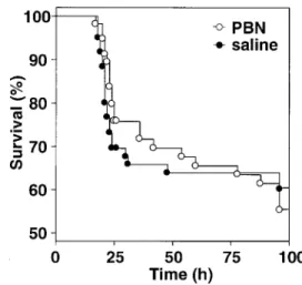
Figure 1. Survival curves for rats with Streptococcus pneumoniae meningitis, treated with a-phenyl-tert-butyl nitrone (PBN) or saline. the extent of hippocampal apoptosis observed in the acute
dis-ease correlates with learning function in meningitis survivors treated with PBN or saline.
Materials and Methods
Model of meningitis. Nursing Sprague-Dawley rat pups (n p 117) were infected on postnatal day 11 by intracisternal in-jection with 10 mL of saline containing6.405 0.14log10cfu/mL
S. pneumoniae (mean5 SD) by means of a 32-gauge needle, as described elsewhere [23, 24]. Infected animals were randomized for treatment with PBN (Calbiochem), 100 mg/kg intraperitoneally every 8 h (n p 57), or an equal volume (0.3 mL) of saline intra-peritoneally every 8 h (n p 60), starting at the time of infection, for a duration of 5 days. Noninfected animals (n p 38) were in-jected intracisternally with 10 mL of sterile pyrogen-free saline and also were randomized to receive PBN (n p 8) or saline (n p 30), as described above. Eighteen hours after intracisternal inoculation, animals were weighed and assessed clinically. Clinical status was scored as follows: 5, normal motor activity and animal turned upright in!5 s when put on its back; 4, decreased spontaneous
activity but still turned upright in!5 s; 3, turned upright in15 s;
2, did not turn upright; and 1, did not move. To document men-ingitis, 10 mL of CSF was obtained by puncture of the cisterna magna and was cultured quantitatively. Infected animals then were treated with ceftriaxone (Roche Pharma), 100 mg/kg subcutane-ously every 12 h for 4 days.
Histopathology. For histopathologic examination, 52 animals (infected and PBN-treated, n p 18; infected and saline-treated, ; uninfected and PBN-treated, ; uninfected and
saline-n p 18 n p 8
treated,n p 8) were evaluated at22.55 2.0h (mean5 SD) after infection, as described elsewhere [23, 25]. The area of cortical brain damage was calculated as a percentage of the total cortex in each section and was expressed as the mean value per animal. Cells with morphologic changes compatible with apoptosis were counted in 3 visual fields (3400) in each of the 4 blades of the dentate gyrus, and the following scoring system was applied: 0–5cells p 0; 6–20 ; 120 . An average score per animal was
calcu-cells p 1 cells p 2
lated from all sections evaluated. All histopathologic evaluations were done by an investigator who was unaware of the clinical, microbiologic, and treatment data for the respective animal.
Assessment of learning function. For the water maze procedure, a round gray tank 1.8 m in diameter (surface, 2.54 m2
) and 0.7 m in height, filled with water at a temperature of 24–267C to a depth of 48 cm, was used. The water was darkened by the addition of nontoxic food coloring. A video camera was fastened to the ceiling above the center of the pool. Before the test, gross vestibulomotor dysfunction of animals to be assessed in the water maze was ex-cluded by use of a rotating rod. Rats were placed on a foam cylinder (circumference, 22 cm), which was fastened to a motor with ad-justable speed and was placed 20 cm above the table surface. Ani-mals had to stay on the cylinder at different speeds (4, 8, 12, and 16 rpm) for>10 s, to qualify for the water maze test. For assess-ment of learning function, swimming patterns of the rats were registered with the video tracking system (Ethovision; Noldus In-formation Technology). The water surface was virtually divided into 4 inner quadrants and a periphery with a width of 18 cm. An
adjustable platform, measuring163 13 cm and covered with a black, rough mat, was placed in the center of the first quadrant 0.5 cm below the water surface. Four entry zones, situated each between 2 quadrants, were marked outside the pool. Three posters of0.63 0.3m with different black-and-white patterns (horizontal stripes, diagonal stripes, and circles) were placed on 3 different walls 0.5 m from the edge of the pool, to serve as visual cues. The room was illuminated by indirect light from the ground.
Thirty-two-day-old survivors of meningitis (PBN-treated, n p ; saline-treated, ) and uninfected littermate controls (
27 n p 31 n p
) were transferred to the experiment room, where they were given 22
24 h to acclimatize in 12-h light/dark cycles; the light was switched on at 8 a.m. Animals were provided with water and food ad libitum. During days 1–4, animals performed 5 training trials per day, with the invisible platform in a fixed position throughout the test. Each rat was put into the water with its head directed toward the wall of the tank. If an animal found the platform within 90 s, it was allowed to stay on it for 15 s before it was returned to the cage. If the rat did not find the platform within 90 s, it was guided there by hand and was allowed to stay on it for 15 s. Between trials, animals rested for 45 min. Entry zones were randomized with a dice for each trial. Tracks were recorded by the video track-ing system, with 5 samples per second. The total distance moved and the time to reach the platform were documented for each trial.
Statistical methods. Bacterial titers in the CSF were compared with the unpaired Student’s t test. Survival curves were analyzed by use of the Kaplan-Meier method. Incidence of seizures, spon-taneous death, and cortical injury were compared with Fisher’s exact test. The score of hippocampal apoptosis was analyzed with the Mann-Whitney U test. The clinical activity score and weight at 18 h and before the water maze task were compared with 1-way analysis of variance and the Newman-Keuls post hoc test for mul-tiple comparison. Distance and time in the water maze task were calculated with repeated-measures analysis of variance, and pair-wise comparison was done with the Tukey-Kramer adjustment. SAS version 8.0 (SAS Institute) software was used.P< .05was considered to be significant.
Figure 2. Brain histopathology of infant rats with pneumococcal meningitis 22.5 h after infection (Nissl stain). A, Section of cortex and underlying hippocampus, at320 magnification. Cortex shows area of reduced neuronal density with wedge-shaped distribution (arrowheads). Bar, 0.5 mm. B, Border between areas of reduced (left) and normal (right) neuronal density, at3400 magnification. Bar, 25 mm. C, Section of dentate gyrus, at3200 magnification. Arrowheads, Apoptotic neurons in granule cell layer; bar, 100 mm. D, Apoptotic cells showing typical condensed, fragmented nuclei (arrowheads), at3400 magnification. Bar, 25 mm.
Results
Characteristics of disease. By 18 h after infection, all ani-mals had meningitis, as evidenced by lethargy and growth of
S. pneumoniae from CSF. Treatment with PBN, compared with
treatment with sterile saline, had no effect on bacterial titers in CSF (mean5 SD 7.42 5 1.06, vs.7.565 0.87log10cfu/mL;
not significant). The clinical score was a mean5 SD of for PBN-treated animals versus for
saline-4.15 0.6 3.95 0.6
treated animals (not significant). Uninfected animals weighed amean5 SDof30.35 3.7g; infected and PBN-treated ani-mals weighed25.75 3.6g; and infected and saline-treated ani-mals weighed 26.75 3.2 g (P!.001, each infected group vs. uninfected; not significant, infected and PBN-treated vs. in-fected and saline-treated). Survival curves were similar in the 2 infected groups (figure 1), and spontaneous death occurred in 23 of 57 PBN-treated animals and in 24 of 60 saline-treated
animals (not significant). Seizures were observed more fre-quently in saline- than in PBN-treated animals (15 of 60 vs. 3 of 57;P!.005). At the age of 32 days, uninfected control ani-mals weighed a mean5 SD of 138.05 12.7 g; infected and PBN-treated animals weighed116.65 22.0g; and infected and saline-treated animals weighed115.65 24.4 g (P!.001, each infected group vs. uninfected; not significant, infected and PBN-treated vs. infected and saline-PBN-treated).
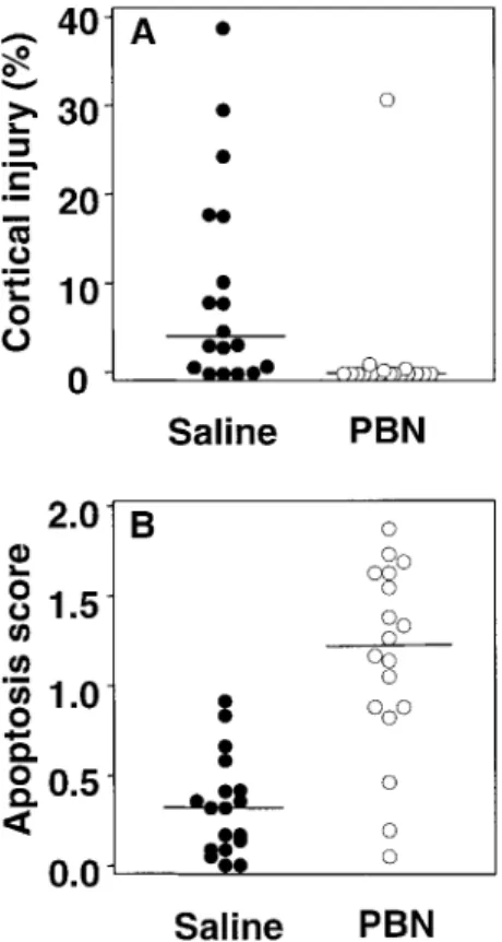
Histopathology. Histopathologic aspects of cortical and hippocampal injury are shown in figure 2. Cortical injury, de-fined as reduced neuronal density with morphologic features of necrosis, was seen in 83.3% of infected and saline-treated ani-mals (median, 3.97% [range, 0%–38.9%] of the cortex). This proportion was reduced to 22.2% of infected and PBN-treated animals (median, 0% [range, 0%–30.9%] of the cortex; P! ; figure 3A). In contrast, apoptosis in the dentate gyrus was .001
250 Loeffler et al. JID 2001;183 (15 January)
Figure 3. Effect of treatment with radical scavenger a-phenyl-tert-butyl nitrone (PBN) on neuronal injury in infant rats with pneumo-coccal meningitis. A, Histopathology at 22.55 2 h after infection showed extensive cortical injury in 83.3% of infected and saline-treated animals (median, 3.97% [range, 0%–38.9%] of cortex) but in only 22.2% of animals treated with PBN (median, 0% [range, 0%–30.9%] of cortex; ). B, In contrast, treatment with PBN increased apoptotic neu-P!.001
ronal injury in hippocampal dentate gyrus, compared with saline (me-dian score, 1.15 [range, 0.04–1.73] vs. 0.31 [0–0.92];P!.001). Histo-pathology of uninfected animals (not shown) revealed neither cortical nor hippocampal neuronal injury.
Figure 4. Assessment of learning function, by Morris water maze. Plots of distance and time to reach platform. Data aremean5 SE. Analysis of distance to reach platform showed decrease over time, which indicates that all animals had function to learn location of the platform. Uninfected control animals learned significantly more quickly than did survivors of meningitis (P!.001). Among these, treatment with a-phenyl-tert-butyl nitrone (PBN) led to inferior performance, compared with treatment with saline (P!.02).
more frequent in the infected and PBN-treated animals than in the infected, saline-treated group (median score, 1.15 [range, 0.04–1.73] vs. median score, 0.31 [range, 0–0.92];P!.001; fig-ure 3B). Uninfected animals, treated with PBN or saline, showed neither cortical nor hippocampal injury.
Water maze experiment. All animals had normal vestibu-lomotor function, as evidenced by their ability to stay on the rotating rod. The distance to reach the platform decreased in all 3 groups over time (figure 4), which indicated that all animals had the capacity to learn the location of the platform. Unin-fected control animals, however, learned significantly (P! ) more quickly than did the survivors of meningitis. Among .001
those, treatment with PBN, compared with treatment with sa-line, led to a significantly inferior performance (P!.02). The level of performance of uninfected animals on day 2 was ap-proximated by infected and saline-treated animals on day 3 and by infected and PBN-treated animals on day 4 only. Similar
results were obtained when we analyzed the time the animals needed to reach the platform (figure 4), with a significant dif-ference among all 3 groups (P!.001) and a trend for inferior performance of the infected and PBN-treated group, compared with that of the infected and saline-treated group (P p .061).
Discussion
The present study shows a detrimental effect of the radical scavenger PBN in experimental S. pneumoniae meningitis, lead-ing to an increase in hippocampal apoptosis and, in conse-quence, to impaired learning function. This is the first report to relate the severity of hippocampal apoptosis as a
conse-quence of bacterial meningitis to reduced learning function in the Morris water maze.
The histopathologic aspect of the cortical damage in men-ingitis models with S. pneumoniae or GBS is usually a well-demarcated wedge-shaped area of injury, which suggests an ischemic mechanism caused by vasculitis and intravascular mi-crothrombi. Production of ROS in the cortex during meningitis seems to be caused by ischemia and reperfusion, which is similar to the mechanisms in other brain diseases, such as brain con-cussion or stroke, in which ROS are thought to be mediators of injury [26–29]. Treatment with PBN, as well as other types of antioxidants, proved to be beneficial in preventing micro-vascular changes and cortical necrotic injury in various animal models of meningitis, independent of the infecting pathogen [20, 23, 30, 31].
The pathways that lead from bacterial invasion of the central nervous system to hippocampal neuronal apoptosis in bacterial meningitis are not well understood. The fact that PBN is neu-roprotective for the hippocampus in GBS meningitis, whereas it is neurotoxic in the present study of S. pneumoniae meningitis, suggests that pathogens may interact via different pathways at the level of cellular signaling and that, depending on the precise nature of that interaction, scavenging of ROS by PBN favors or attenuates apoptosis.
Two hypotheses may explain our observations. First, bac-terial pathogens seem to interact differently with ROS. In our present study, CSF titers of S. pneumoniae were similar in the PBN and saline treatment groups, which indicates that their growth remains unaffected by the presence of a potent anti-oxidant in vivo. In contrast, the CSF titer of GBS in saline-treated rats was 100-fold lower than that in PBN-saline-treated ani-mals at 18 h after infection, which suggests that ROS play a role as a host defense mechanism against GBS [10]. Recently it was shown that S. pneumoniae possess a distinct mechanism that decreases measurable superoxide production during in-cubation with stimulated neutrophils [32, 33]. The preservation of a nonoxidative environment, for example, through produc-tion of superoxide dismutase, might prevent the deactivaproduc-tion of the S. pneumoniae–specific virulence factor pneumolysin [34, 35]. Moreover, apoptotic neuronal death involves several types of cellular signaling mechanisms—such as glutamate receptors, protein kinases, and transcription factors—which are all sen-sitive to the redox status of the cell [36, 37]. Thus, combination of antioxidant treatment and the presence of S. pneumoniae in our model may create a reductive environment that favors vir-ulent properties of S. pneumoniae and can directly lead to neu-ronal death itself [37].
The second hypothesis relies on a direct pathogen-specific signaling pathway via the recently discovered human toll-like receptor (TLR). This is suggested by the finding that the human TLR-2 is a signaling receptor for S. pneumoniae but not for GBS [38, 39]. Human TLRs are transmembrane proteins that, among other functions, are known to mediate translocation
and activation of NF-kB by gram-positive bacteria. PBN blocks the predominantly antiapoptotic effect of NF-kB activation [40, 41] and may hence shift the balance from antiapoptotic to pro-apoptotic signaling. Apart from NF-kB activation, TLR-2– gated discrimination among different pathogens may account for alternate signaling pathways with different susceptibility to ROS. Discrimination among specific pathogens has been shown elsewhere by the documentation of pathogen-dependent pat-terns of cytokine activation in bacterial meningitis [42]. Thus, the TLR pathway may ultimately determine the pro- or anti-apoptotic effect of therapeutic intervention with antioxidants. These opposite effects of the same agent depending on the pathogen strongly suggest a critical interplay between microbial and host factors, which are currently under investigation. We suspect that the elucidation of these processes will lead to the identification of novel factors important in the pathogenesis of bacterial infections.
Neither increased nor decreased apoptosis has been observed with other antioxidants given during S. pneumoniae meningitis [23], which can be explained by the shorter circulatory half-life and lower blood-brain barrier passage of these drugs, compared with that of PBN.
In conclusion, we found that treatment with the radical scav-enger PBN during S. pneumoniae meningitis in rat pups atten-uated cortical necrosis but increased apoptotic neuronal death in the hippocampus. This is in contrast to what was seen in GBS meningitis, in which PBN protected neurons from both forms of injury. Increased hippocampal damage by PBN led to further decrease in learning function of survivors in the Morris water maze. Antioxidant treatment is therefore not uniformly beneficial in bacterial meningitis. Pathogen- and host-related mechanisms that influence the redox status and transcription factors of neuronal cells may be involved.
Acknowledgments
We thank Philipp Joss and Oliver Schu¨tz (Institute for Infectious Diseases, University of Bern, Bern, Switzerland) for technical support.
References
1. Grimwood K, Anderson VA, Bond L, et al. Adverse outcomes of bacterial meningitis in school-age survivors. Pediatrics 1995; 95:646–56. 2. Bohr VA, Rasmussen N. Neurological sequelae and fatality as prognostic
mea-sures in 875 cases of bacterial meningitis. Dan Med Bull 1988; 35:92–5. 3. Baraff LJ, Lee SI, Schriger DL. Outcomes of bacterial meningitis in children:
a meta-analysis. Pediatr Infect Dis J 1993; 12:389–94.
4. Grimwood K, Nolan TM, Bond L, Anderson VA, Catroppa C, Keir EH. Risk factors for adverse outcomes of bacterial meningitis. J Paediatr Child Health 1996; 32:457–62.
5. Morris RG, Garrud P, Rawlins JN, O’Keefe J. Place navigation impaired in rats with hippocampal lesions. Nature 1982; 297:681–3.
6. Sutherland RJ, Whishaw IQ, Kolb B. A behavioural analysis of spatial lo-calization following electrolytic, kainate-, or colchicine-induced damage to the hippocampal formation in the rat. Behav Brain Res 1983; 7:133–53. 7. Maguire EA, Burgess N, Donnett JG, Frackowiak RS, Frith CD, O’Keefe
252 Loeffler et al. JID 2001;183 (15 January)
J. Knowing where and getting there: a human navigation network. Science
1998; 280:921–4.
8. Nau R, Soto A, Bruck W. Apoptosis of neurons in the dentate gyrus in humans suffering from bacterial meningitis. J Neuropathol Exp Neurol
1999; 58:265–74.
9. Free SL, Li LM, Fish DR, Shorvon SD, Stevens JM. Bilateral hippocampal volume loss in patients with a history of encephalitis or meningitis. Epi-lepsia 1996; 37:400–5.
10. Leib SL, Kim YS, Chow LL, Sheldon RA, Ta¨uber MG. Reactive oxygen intermediates contribute to necrotic and apoptotic neuronal injury in an infant rat model of bacterial meningitis due to group B streptococci. J Clin Invest 1996; 98:2632–9.
11. Bogdan I, Leib SL, Bergeron M, Chow L, Ta¨uber MG. Tumor necrosis factor–a contributes to apoptosis in hippocampal neurons during experi-mental group B streptococcal meningitis. J Infect Dis 1997; 176:693–7. 12. Pfister LA, Leib SL, Michelet C, Ta¨uber MG. Neuronal injury in
experi-mental meningo-encephalitis due to Listeria monocytogenes: early apop-tosis of neurons in the hippocampus [abstract]. Schweiz Med Wochenschr Suppl 1998; 100:S9.
13. Pfister HW, Koedel U, Lorenzl S, Tomasz A. Antioxidants attenuate micro-vascular changes in the early phase of experimental pneumococcal men-ingitis in rats. Stroke 1992; 23:1798–804.
14. Leib SL, Kim YS, Ferriero DM, Ta¨uber MG. Neuroprotective effect of excitatory amino acid antagonist kynurenic acid in experimental bacterial meningitis. J Infect Dis 1996; 173:166–71.
15. Kastenbauer S, Koedel U, Pfister HW. Role of peroxynitrite as a mediator of pathophysiological alterations in experimental pneumococcal menin-gitis. J Infect Dis 1999; 180:1164–70.
16. Spellerberg B, Cundell DR, Sandros J, et al. Pyruvate oxidase as a deter-minant of virulence in Streptococcus pneumoniae. Mol Microbiol 1996; 19:803–13.
17. Schreck R, Rieber P, Baeuerle PA. Reactive oxygen intermediates as appar-ently widely used messengers in the activation of the NF-kappa B tran-scription factor and HIV-1. EMBO J 1991; 10:2247–58.
18. Schmidt KN, Amstad P, Cerutti P, Baeuerle PA. The roles of hydrogen per-oxide and superper-oxide as messengers in the activation of transcription factor NF-kappa B. Chem Biol 1995; 2:13–22.
19. Clemens JA, Stephenson DT, Smalstig EB, Dixon EP, Little SP. Global is-chemia activates nuclear factor–kappa B in forebrain neurons of rats. Stroke 1997; 28:1073–80.
20. McKnight AA, Keyes WG, Hudak ML, Jones MD Jr. Oxygen free radicals and the cerebral arteriolar response to group B streptococci. Pediatr Res
1992; 31:640–4.
21. Chen G, Griffin M, Poyer JL, McCay PB. HPLC procedure for the phar-macokinetic study of the spin-trapping agent, alpha-phenyl-N-tert-butyl nitrone (PBN). Free Radic Biol Med 1990; 9:93–8.
22. Schaefer CF, Janzen EG, West MS, Poyer JL, Kosanke SD. Blood chemistry changes in the rat induced by high doses of nitronyl free radical spin traps. Free Radic Biol Med 1996; 21:427–36.
23. Auer M, Pfister LA, Leppert D, Ta¨uber MG, Leib SL. Effects of clinically used antioxidants in experimental pneumococcal meningitis. J Infect Dis
2000; 182:347–50.
24. Leib SL, Leppert D, Clements J, Ta¨uber MG. Matrix metalloproteinases contribute to brain damage in experimental pneumococcal meningitis. Infect Immun 2000; 68:615–20.
25. Pfister LA, Tureen JH, Shaw S, et al. Endothelin inhibition improves cerebral blood flow and is neuroprotective in pneumococcal meningitis. Ann Neu-rol 2000; 47:329–35.
26. Kontos CD, Wei EP, Williams JI, Kontos HA, Povlishock JT. Cytochemical detection of superoxide in cerebral inflammation and ischemia in vivo. Am J Physiol 1992; 263:H1234–42.
27. Defraigne JO, Detry O, Pincemail J, et al. Direct evidence of free radical production after ischaemia and reperfusion and protective effect of des-ferrioxamine: ESR and vitamin E studies. Eur J Vasc Surg 1994; 8:537–43. 28. Sen S, Goldman H, Morehead M, Murphy S, Phillis JW. a-phenyl-tert-butyl nitrone inhibits free radical release in brain concussion. Free Radic Biol Med 1994; 16:685–91.
29. Schulz JB, Henshaw DR, Siwek D, et al. Involvement of free radicals in excitotoxicity in vivo. J Neurochem 1995; 64:2239–47.
30. Berkowitz ID, Traystman RJ. Oxygen radical scavengers prevent impairment of microvascular autoregulation in H. influenzae type b meningitis in rats [abstract]. FASEB J 1993; 7:A530.
31. Koedel U, Bernatowicz A, Paul R, Frei K, Fontana A, Pfister HW. Exper-imental pneumococcal meningitis: cerebrovascular alterations, brain edema, and meningeal inflammation are linked to the production of nitric oxide. Ann Neurol 1995; 37:313–23.
32. Perry FE, Elson CJ, Greenham LW, Catterall JR. Interference with the oxidative response of neutrophils by Streptococcus pneumoniae. Thorax 1993; 48:364–9. 33. Perry FE, Elson CJ, Mitchell TJ, Andrew PW, Catterall JR. Characterisation of an oxidative response inhibitor produced by Streptococcus pneumoniae. Thorax 1994; 49:676–83.
34. Clark RA. Oxidative inactivation of pneumolysin by the myeloperoxidase system and stimulated human neutrophils. J Immunol 1986; 136:4617–22. 35. Yesilkaya H, Kadioglu A, Gingles N, Alexander JE, Mitchell TJ, Andrew PW. Role of manganese-containing superoxide dismutase in oxidative stress and virulence of Streptococcus pneumoniae. Infect Immun 2000; 68: 2819–26.
36. Arrigo AP. Gene expression and the thiol redox state. Free Radic Biol Med
1999; 27:936–44.
37. Castagne V, Gautschi M, Lefevre K, Posada A, Clarke PG. Relationships between neuronal death and the cellular redox status. Focus on the de-veloping nervous system. Prog Neurobiol 1999; 59:397–423.
38. Yoshimura A, Lien E, Ingalls RR, Tuomanen E, Dziarski R, Golenbock D. Cutting edge: recognition of gram-positive bacterial cell wall components by the innate immune system occurs via toll-like receptor 2. J Immunol
1999; 163:1–5.
39. Flo TH, Halaas O, Lien E, et al. Human toll-like receptor 2 mediates mono-cyte activation by Listeria monocytogenes, but not by group B strepto-cocci or lipopolysaccharide. J Immunol 2000; 164:2064–9.
40. Pogrebniak HW, Merino MJ, Hahn SM, Mitchell JB, Pass HI. Spin trap salvage from endotoxemia: the role of cytokine down-regulation. Surgery
1992; 112:130–9.
41. Sang H, Wallis GL, Stewart CA, Kotake Y. Expression of cytokines and activation of transcription factors in lipopolysaccharide-administered rats and their inhibition by phenyl-N-tert-butylnitrone (PBN). Arch Biochem Biophys 1999; 363:341–8.
42. Diab A, Zhu J, Lindquist L, Wretlind B, Bakhiet M, Link H. Haemophilus
influenzae and Streptococcus pneumoniae induce different intracerebral
mRNA cytokine patterns during the course of experimental bacterial men-ingitis. Clin Exp Immunol 1997; 109:233–41.