Fat intake modifies vascular responsiveness and receptor expression of
vasoconstrictors: Implications for diet-induced obesity
Alexa L. Mundy
a, Elvira Haas
a, Indranil Bhattacharya
a, Corinne C. Widmer
a, Martin Kretz
a,
Regina Hofmann-Lehmann
b, Roberta Minotti
a, Matthias Barton
a,⁎
aDepartment of Medicine, Internal Medicine I, Medical Policlinic, University Hospital, CH-8091 Zürich, Switzerland bClinical Laboratory, Vetsuisse Faculty, University of Zurich, Switzerland
Received 27 April 2006; received in revised form 27 October 2006; accepted 16 November 2006 Available online 18 November 2006
Time for primary review 18 days
Abstract
Objective: Angiotensin II (Ang II), endothelin-1 (ET-1) and reactive oxygen species (ROS) have been implicated in the development of pathologic changes associated with obesity including hypertension and atherosclerosis. The aim of this study was to investigate the effects of dietary fat content on vasoreactivity and receptor expression at the level of gene and protein expression.
Methods: C57BL/6 mice were fed diets of normal (Control, 12.3% kcal from fat), high (HF, 41% kcal from fat) and very high (VHF, 58% kcal from fat) fat content for 15 weeks. Glucose tolerance tests were performed, and aortic rings were exposed to ET-1 (0.01–300 nM) and Ang II (100 nM) in the presence ofL-nitro-arginine-methyl ester (L-NAME; 300μM). Gene and protein expressions of angiotensin and
endothelin receptors were examined by real-time PCR and immunoblotting, respectively. The effects of diet on responses to acetylcholine (ACh 0.1–300 μM), in the absence or presence ofL-NAME, and to exogenous ROS/
U
OH were also investigated.Results: Both high fat diets similarly impaired glucose tolerance (Pb0.05). Increasing dietary fat augmented contractions to Ang II in a step-wise manner (Pb0.05). Conversely, increasing dietary fat had no effect on contractions to ET-1. Exposure to ROS/
U
OH resulted in a rapid vasodilation that was markedly augmented in a step-wise manner with increasing dietary fat (Pb0.05). Endothelium-dependent relaxation to ACh was unaffected whereas vasoconstriction to high concentrations of ACh was enhanced in VHF animals (Pb0.05 vs. control). Gene expression of the AT1Breceptor was increased in the aorta of VHF mice, and aortic ETAreceptor protein expression was increased after bothhigh fat diets.
Conclusions: These findings demonstrate that changes in dietary fat intake modulate vascular reactivity in response to Ang II and ROS, as well as expression of vascular angiotensin and endothelin receptors. Dietary fat intake may thereby directly affect cardiovascular risk. © 2006 European Society of Cardiology. Published by B.V. All rights reserved.
Keywords: Hydroxyl; ROS; Artery; Comparative; Atherosclerosis; Vasoactive agents; Glucose tolerance; Vascular disease
1. Introduction
Dietary fat intake is a major determinant of the dramatic increase of obesity world-wide during the past two decades
[1]. Obesity increases cardiovascular risk, significantly alters metabolic and cardiovascular function, and is associated
with increased risk for diabetes, hypertension, and athero-sclerosis [2,3]. Inflammation, vascular remodeling, and changes in vascular reactivity play a central role in the pathogenesis of these diseases[4–6].
Angiotensin II (Ang II) has been implicated in the development of hypertension and atherosclerosis[7,8]. The effects of Ang II are mediated via two receptor subtypes, AT1 and AT2; the AT1receptor consists of two isoforms, the AT1A and the AT1Breceptor, which are functionally and pharma-cologically indistinguishable [8]. Angiotensin II activates intracellular signalling pathways, primarily through the AT1
⁎ Corresponding author. Department of Medicine, Internal Medicine I, Medical Policlinic, University Hospital Zürich, Rämistrasse 100, 8091 Zurich, Switzerland. Tel.: +41 44 255 56 63; fax: +41 44 255 87 47.
E-mail address:[email protected](M. Barton).
0008-6363/$ - see front matter © 2006 European Society of Cardiology. Published by B.V. All rights reserved. doi:10.1016/j.cardiores.2006.11.019
receptor, which promote atherosclerosis and hypertension through formation of reactive oxygen species (ROS)[9,10], inflammation[11,12], activation of cell growth[13], oxida-tive modification of lipoproteins [14], and by impairing endothelium-dependent vasodilation [8,15]. AT1 receptor expression is increased in vascular smooth muscle cells of hypertensive patients[10]. In addition to its direct effects on the vasculature, Ang II also stimulates production of other vasoactive factors including endothelin-1 (ET-1)[16], a po-tent vasoconstrictor and mitogen [17] that binds to endo-thelin A (ETA) and endothelin B (ETB) receptors [18]. In pathological conditions such as atherosclerosis, ETA recep-tors contribute to disease-progression[19,20]. Additionally, Ang II and ET-1 increase ROS generation [21,22], which include superoxide anion (O2−) and hydroxyl radical (
U
OH). Changes in the vascular expression and/or activity of Ang II, ET-1 and ROS have been demonstrated in obesity, and preceed pathological changes associated with obesity[23–26], which in humans is often due to excessive dietary fat intake[1]. The aim of the current study, therefore, was to investigate the effect of diets of different fat content (41% and 58% fat) on vascular activity of Ang II and ET-1 and the expression of their receptors in fat-fed mice, a commonly used model of human obesity[27]. Moreover, the effects of ROS/
U
OH, and acetylcholine in precontracted aortic rings were investigated.2. Methods
2.1. Animals and dietary treatments
Healthy male mice (C57BL/6, Charles River, Sulzfeld Germany) were housed in the institutional animal facilities on a 12:12-h light–dark cycle, and animals had free access to food and water. Housing facilities and experimental proto-cols were approved by the local authorities for animal research (Kommission für Tierversuche des Kantons Zürich, Switzerland) and conform to the Guide for the Care and Use of Laboratory Animals published by the US National Institutes of Health (NIH Publication No. 85-23, revised 1996). Mice were randomly assigned to one of the following diets (n = 10–12 mice/group): control (12.3% of total kcal from fat, Kliba Nafag 3430, Kaiseraugst, Switzerland), high fat (HF, 41% of total kcal from fat, Research Diets D12079B, New Brunswick, NJ), and a diet containing very high amounts of fat (VHF, 58% of total kcal from fat, Research Diets D12331) for 15 weeks. The macronutrient composi-tions of the three diets are reported inTable 1. At the end of the treatment, mice were anesthetized (xylazine: 100 mg/kg
body weight; ketamine: 23 mg/kg BW; and acepromazine: 3.0 mg/kg BW, i.p.), and exsanguinated via cardiac puncture. Blood was centrifuged at 5000 rpm at 4 °C for 15 min and plasma was stored at−80 °C.
2.2. Metabolic parameters and lipid measurements In the week of the experiment mice were fasted overnight for 14 h, weighed, and venous blood was obtained from the tail vein (0 min) for baseline glucose measurements. Mice were subsequently injected (i.p.) with 2 mg/g BWD-glucose
and blood was collected at 5, 10, 15, 30, 45, 60, 90, and 120 min. Blood glucose was determined with an AccuChek Advantage glucose meter (Roche Diagnostics, Switzerland). Plasma lipoproteins were determined enzymatically using a Cobas Integra 800 autoanalyzer (Roche Diagnostics, Rot-kreuz, Switzerland), as previously described[28].
2.3. Vascular function studies
The thoracic aorta was isolated and placed in cold Krebs Ringer bicarbonate solution (in mmol/L: NaCl 118.6; KCl 4.7; CaCl2 2.5; MgSO4 1.2; KH2PO4 1.2; NaHCO3 25.1; EDTANa2Ca0.026; glucose 10.1), dissected free of connec-tive tissue under a microscope (Olympus SZX9, Volketswil, Switzerland) and cut into rings 3 mm in length. Special care was taken not to damage the endothelium during this procedure. Experiments were performed as previously de-scribed [29]. Vascular rings were mounted onto two tungsten wires (100 μm) and transferred to water-jacketed organ baths containing Krebs solution (95% O2, 5% CO2at 37 °C, pH 7.4) and connected to force transducers (Hugo Sachs Elektronik, March-Hugstetten, Germany). Resting tension was gradually increased to the optimal level as previously determined in this laboratory (1.75 g) and aortic rings were repeatedly exposed to 100 mmol/L KCl until a stable response was achieved.
2.4. Vascular responses to angiotensin II and endothelin-1 Aortic rings were exposed to Ang II (100 nmol/L)[30]or ET-1 (0.01–300 nmol/L) [31] in the presence of the nitric oxide synthase inhibitor L-nitro-arginine methylester (L -NAME, 300μmol/L) preincubated for 30 min.
2.5. Effect of ROS/
U
OH on precontracted aortic rings Aortic rings were preincubated withL-NAME (300μmol/L) for 30 min. The vascular response to exogenously generated reactive oxygen species (ROS), predominantly consisting of hydroxyl radical (U
OH) was then investigated by simulta-neous addition of vitamin C and Fe2+(100μmol/L each)[32]to rings of aorta preconstricted with phenylephrine to 50% of the KCl-induced contraction, as previously described [33]. The generation of hydroxyl radicals in the bath was con-firmed by addition of terephthalic acid (TPA, 2.5 mmol/L).
Table 1
Major macronutrient constituents of diets given in percent of kcal Diet Control High fat Very high fat
Protein 22.4 17 16.4
Carbohydrate 65.4 43 25.5
Hydroxylation of TPA yields a stable and highly specific isomer (monohydroxyterephthalate, TPA-OH) not directly produced from superoxide or hydroperoxides[34]. Fluores-cence of TPA-OH was measured using a SpectraMax M2 (Molecular Devices, Basel, Switzerland) at 326 nM and 432 nM excitation and emission wavelength, respectively
[35]. Addition of TPA to Krebs solution containing vitamin C and Fe2+ yielded 1804 ± 178 versus 0.002 ± 6.6 relative fluorescence units for Krebs solution alone (Pb0.005). 2.6. Endothelium-dependent and -independent vasodilation
Rings were precontracted with phenylephrine (50% of KCl) and endothelium-dependent vasodilation was investi-gated using acetylcholine (ACh, 0.1 nmol/L–3 μmol/L) in the presence or absence of L-NAME (300 μmol/L). Endo-thelium-independent vasodilation was investigated using the nitric oxide donor sodium nitroprusside (SNP, 10μmol/L). 2.7. Real-time PCR
Aortic tissue was snap-frozen in liquid nitrogen and stored at −80 °C. Tissue was pulverized and total RNA was ex-tracted using the silica-based RNeasey Mini™ Kit (Qiagen, Hilden Germany). 150 ng RNA was reversed transcribed with Quantitect Reverse Transcription Kit™ (Qiagen, Hilden Germany), including a genomic DNA digestion step. Expres-sion levels of the murine genes encoding for ET-1 receptors (ETAand ETBreceptors) and AT1A, AT1Band AT2receptors were determined by real-time PCR as described[36]. Real-time PCR experiments were run on the iQ™ iCycler (Bio-Rad, Reinach, Switzerland) using specific cDNA primers (Microsynth, Balgach, Switzerland,Table 2). Murineβ-actin was used as a house-keeping control.
2.8. Western blot analyses
For protein expression analysis, three pieces of tissue from each group were pooled and homogenized in RIPA
lysis buffer. Equal amount of protein lysates were separated on an 8–16% SDS-PAGE gel and immunoblotted with anti-angiotensin receptor type I antibody and anti-ET-1 receptor antibody. Equal amount of protein loading was controlled by probing with an anti-p42/p44 antibody[37].
2.9. Materials and antibodies
ET-1 and L-NAME were supplied by Alexis Corp
(Lausanne, Switzerland). All other chemicals were supplied by Sigma Chemicals Co. (Buchs, Switzerland). Antibodies against angiotensin II type I receptor, against p42/p44 were obtained from Santa Cruz Biotechnology, Inc. (Santa Cruz, CA, USA) and anti-ET-1 receptor antibody from BD Transduction Laboratories (Franklin Lakes, NJ, USA). 2.10. Statistical analyses
Data are given as mean ± SEM and n denotes the number of animals used. Contraction is expressed as a percentage of contraction to100 mmol/L KCl, and dilations are given as a percentage of the maximal contraction. EC50 values (as negative logarithm pD2) were calculated with non-linear regression analysis and the area under the curve (AUC) was calculated for each individual curve using SigmaPlot (SPSS Inc. Chicago, IL). Data were analyzed using ANOVA for repeated measurements with Bonferroni correction, the un-paired Student's t-test or the Mann–Whitney U test, when appropriate. A P valueb0.05 was considered significant. 3. Results
3.1. Weight gain and metabolic studies
After 15 weeks mice fed a control diet had gained 11 ± 1 g of body weight, while the mice fed high fat and very high fat diets gained 17 ± 1 g and 21 ± 1 g, respectively (Pb0.004 C vs. HF, Pb0.001 C vs. VHF, Pb0.04 HF vs. VHF). While the feeding of high fat diets for 15 weeks did not alter fasting
Table 2
Primers used for amplification of a specific cDNA fragment encoding for ETAreceptor, ETBreceptor, AT1Areceptor, AT1Breceptor, AT2receptor and
β-actin Gene accession number Forward primer reverse primer Product size (bp) ETAreceptor 5′-GAAGGACTGGTGGCTCTTTG-3′ 149 BC_008277 5′-CTTCTCGACGCTGTTTGAGG-3′ ETBreceptor 5′-CGGTATGCAGATTGCTTTGA-3′ 189 NMU_32329 5′-CACCTGTGTGGATTGCTCTG-3′ AT1Areceptor 5′-AGCCTGCGTCTTGTTCTGAG-3′ 114 NM_177322 5′-ACTGGTCCTTTGGTCGTGAG-3′ AT1Breceptor 5′-GCCTGCTAGTGACATGATC-3′ 133 NM_175086 5′-GTACAGTCCAGAGTCCTTTC-3′ AT2receptor 5′-CGCAGTGTGTTTAGAGTTCCC-3′ 146 NM_007429 5′-AACCAATGGCTAGGCTGAC-3′ β−Actin 5′-CGTGCGTGACATCAAAGAGA-3′ 180 X_03672 5′-CCCAAGAAGGAAGGCTGGA-3′
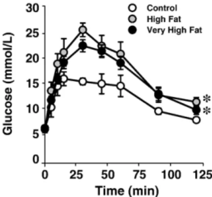
Fig. 1. Plasma glucose levels at base line (0 min) and at indicated time points after i.p. injection of 2 mg/g BWD-glucose (glucose tolerance test). Mice were fed diets containing control (C, 12.3%), high fat (HF, 41%) and very high fat (VHF, 58%) kilocalories from fat for 15 weeks. Data are means ± standard error (n = 5, C; n = 6, HF; n = 12, VHF). ⁎Pb0.04 vs. control diet.
glucose (in mmol/L, C = 5.5 ± 0.2, HF = 5.2 ± 0.4, VHF = 5.9 ± 0.3), glucose tolerance was significantly impaired in both the 41% (HF) and 58% fat (VHF) diet fed mice (Pb0.04 versus control,Fig. 1). Plasma cholesterol levels similarly increased after both high fat diets (in mmol/L, C = 2.1 ± 0.1, HF = 3.1 ± 0.2, VHF = 3.2 ± 0.2, Pb0.001 HF and VHF vs. C).
3.2. Contractility to angiotensin II and endothelin-1 In the aorta, increasing dietary fat content enhanced contractions to Ang II in a concentration-dependent manner (Pb0.04 HF vs. C; Pb0.002 VHF vs. C; Pb0.04 VHF vs. HF,Fig. 2A). Endothelin-1 caused concentration-dependent contractions that were unaffected by 15 weeks of high fat diets (Fig. 2B). The AUC values of contractions and sen-sitivity to ET-1 were also unaffected (Table 3).
3.3. Endothelium-dependent and -independent vasodilation Acetylcholine caused concentration-dependent relaxa-tions, which was unchanged with increasing dietary fat con-tent (Fig. 3A), and no difference in endothelium-independent
Fig. 2. Effects of dietary fat content on contraction to Ang II (100 nmol/L) in aorta (A). Dietary fat content increased vasoconstrictor responses to Ang II in aorta. n= 8–15/group. ⁎Pb0.04 vs. control. †Pb0.04 vs. high fat. Effects of dietary fat content on contractions to ET-1 in aorta (B). Increasing dietary fat and animal weight had no effect on ET-1-induced contractions in the aorta. n= 6–12/group.
Fig. 3. Endothelium-dependent vasodilation to acetylcholine (ACh) in the aorta, in the absence (A) or presence (B) ofL-NAME (300μmol/L). While increasing dietary fat had no effect on vasodilation to ACh, vasoconstriction at high concentrations of ACh (≥1 μmol/L) in the presence ofL-NAME was markedly increased in very high fat diet fed animals. n = 8–12/group. ⁎Pb0.05 vs. control, †Pb0.05 vs. high fat.
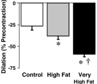
Fig. 4. Dilator responses to reactive oxygen species (ROS/
U
OH) in vascular rings precontracted with phenylephrine (to 50% of KCl). Increasing dietary fat content enhanced the vasodilation. n = 8–12/group. ⁎Pb0.05 vs. control. †Pb0.05 vs. high fat.Table 3
pD2values and area under the curve values (AUC, a measure of overall
contractility) were calculated for each dose–response curve to ET-1 and data were averaged
Diet Control High fat Very high fat Aorta
pD2 8.4 ± 0.04 8.5 ± 0.1 8.5 ± 0.1
vasodilation to SNP between groups was observed (data not shown). Inhibition of nitric oxide synthase with L-NAME completely abolished the vasodilator response to ACh (Fig. 3B). Interestingly, at high concentrations (≥1 μM, Fig. 3B) ACh caused contractions in the VHF group (Pb0.004 versus control) that were not seen in either the control or HF group.
3.4. Vascular responses to reactive oxygen species
Exogenously added ROS/
U
OH caused vasodilation in precontracted aortic rings (Pb0.0001 vs. untreated). In-creasing dietary fat augmented the vasodilator responses to ROS/U
OH independently of nitric oxide synthesis, whichwas inhibited by L-NAME (Pb0.05 C vs. HF and VHF,
Fig. 4).
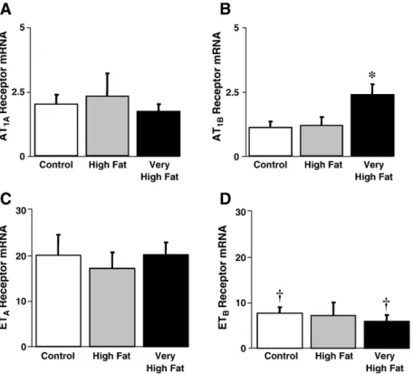
3.5. Gene expression of angiotensin and endothelin receptors In mice fed a VHF diet vascular AT1B receptor gene expression was increased compared to control diet fed mice (Pb0.03 VHF vs. C,Fig. 5B). AT1Areceptor gene expres-sion, however, was similar between groups (Fig. 5A). The AT2receptor gene could not be reliably quantified as it was expressed at very low levels close to the detection limit (amplification began around 34 PCR cycles, data not shown). Endothelin A (ETA) and endothelin B (ETB) receptors were expressed in all samples investigated, and gene ex-pression levels were similar between groups (Fig. 5C and D). ETA receptor gene expression was approximately 3-fold higher than that of the ETBreceptor in each group (Pb0.03 C and VHF, P = 0.067 HF).
3.6. Protein expression of angiotensin and endothelin receptors
Immunoblot analysis of AT1 receptor indicated no differences in expression between dietary groups (Fig. 6). However, protein expression analysis of ETA receptor indicated an increase in receptor expression in the HF- and VHF diet fed mice as compared to controls (Fig. 6).
Fig. 5. Effects of dietary fat content on relative Ang II receptor mRNA expression levels in aorta. Increasing dietary fat content had no effect on AT1Areceptor
expression, but increased the expression of AT1Breceptor expression in the very high fat diet group. n = 4–6/group. ⁎Pb0.05 vs. control. Effects of dietary fat
content on relative ET-1 receptor mRNA expression levels in aorta (B). Increasing dietary fat content had no effect on ETAor ETBreceptor expression. The
expression of ETAreceptors was 3-fold greater than that of ETBreceptors (Pb0.05). n=4–6/group. †Pb0.03 ETAvs. ETBwithin a treatment group.
Fig. 6. Effects of dietary fat content on AT1 and ETA receptor protein
expression in aorta. Tissue extracts were analyzed by immunoblotting with antibodies against AT1receptor, ETA receptor and ERK1/2 as a loading
4. Discussion
The current study demonstrates, for the first time, differential effects of fat intake on aortic vascular reactivity to and receptor expression of vasoactive factors despite similar changes in glucose tolerance and plasma cholesterol. Increasing fat intake caused a step-wise increase in the vasoconstriction to Ang II and acetylcholine, while the vasodilator response to ROS was enhanced. High dietary fat intake was associated with increased vascular AT1Band ETA receptor expression.
Angiotensin II plays an important role as a trophic factor in the development of hypertension [38], and enhanced vasoconstrictor effects to Ang II and increased Ang II plasma levels have been reported in obese patients and animal models[24,39,40]. In the present study we observed that fat intake dose-dependently augments aortic contractile responses to Ang II, which in young mice is largely mediated by cyclooxygenase-1[30], and similar changes were seen in the renal artery (Mundy and Barton, unpublished observa-tion, 2006). Additionally, aortic AT1Breceptor gene expres-sion increased in VHF mice as compared to controls, while AT1Areceptor gene expression remained unchanged. In mice, the AT1Breceptor mediates Ang II-induced vasoconstriction [41], and activation of the AT1receptor has been implicated in the development of atherosclerosis and hypertension[8]. The AT1receptor is also upregulated in leptin-deficient rats that spontaneously develop obesity[42]. The data of the present study suggest that enhanced sensitivity of the vasculature to Ang II and the increased receptor expression upon increasing dietary fat content are likely to facilitate the development of hypertension and vascular disease due to obesity.
Angiotensin II induces vascular ET-1 expression in vivo
[16], and circulating levels of ET-1 are increased in obese patients[23,24] and in the kidney of obese mice [24]; we have now investigated the effects of different amounts of dietary fat intake for 15 weeks on the vascular responses to ET-1. Vascular contractions to ET-1 were unchanged after 15 weeks with either of the high fat diets and no effect on the expression of both ETA and ETB receptor mRNA was observed. In contrast, at protein level, ETA receptor ex-pression was increased in HF and VHF groups as compared to the control group. This increase in protein expression is likely to be mediated by post-translational modifications and/or changes in protein stability. The results of other studies have shown variable results depending on a number of factors, especially the duration of dietary intervention and vascular bed studied. For example, while increased contrac-tions to ET-1 were observed in the aorta of mice fed a high fat diet for 30 weeks, contractions in the carotid artery were unaffected[29]. In the present study maximal contraction to ET-1 was also unaffected by 15 weeks of either the HF or VHF diet in both the renal and femoral arteries (Mundy and Barton, unpublished data, 2006). The shorter duration of dietary treatments used in the current study (15 vs. 30 weeks) and the vascular bed examined may explain the observed
differences [29]. When assessing contractile responses, pa-rameters such as receptor density, receptor affinity, signaling cascades mediating contraction (including calcium flux) could not be studied in our experimental set-up. However, endothelin is a potent trophic factor stimulating cell growth via the ETA receptor [43]. Thus, upregulation of the ETA receptor could promote accelerated myocardial and vascular hypertrophy, which are also known to occur in animals and patients with obesity[44].
Angiotensin II[9]and ET-1[22]are both known to induce vascular generation of ROS, which include superoxide anion (O2−) and hydroxyl radical (
U
OH). In the present study we investigated the effects of ROS/U
OH on the vasculature and changes of the responses by increasing fat intake. AlthoughU
OH is commonly perceived to be an “injurious” ROS[45,46], generated by the interaction of superoxide, hydrogen peroxide and iron (Fenton and Haber-Weiss Reactions), we have recently found that constitutively generated
U
OH also has vasodilatory effects, which are enhanced in the aorta of genetically obese mice[33]. In the current study, the dilatory effect of ROS/U
OH increased depending on dietary fat, suggesting that high fat intake and/or obesity, enhance vasodilating properties of ROS/U
OH. Remarkably, the vasodilator response to ROS/U
OH was unaffected by the inhibition of NO synthesis in all study groups. This may represent a novel vasodilator back-up mechanism in states associated with high fat intake and/or obesity, as well as low NO bioactivity. In most forms of vascular diseases such as atherosclerosis, diabetes, aging and particularly in obese patients, bioactivity of NO is reduced, which is mimicked in our experimental set-up by the presence ofL-NAME in the aortic rings[6,19].No differences in the endothelium-dependent vasodila-tion to acetylcholine were observed between groups after 15 weeks of feeding. Previous studies have demonstrated impaired endothelium-dependent vasodilation to ACh in aorta of mice after 30 weeks of high fat diet treatment[29], and in aorta of rats after 2 years[47]or 8 weeks[48]of high fat feeding. Thus, differences are possibly due to variations in duration of dietary treatment. In the absence of nitric oxide afterL-NAME treatment in vitro, however, a marked increase
in the vasoconstriction to ACh was noted in the VHF group as compared with the control group. The vasoconstrictor response to ACh in mice is known to be caused by cyclo-oxygenase-dependent prostanoids [29,49], which increase with obesity [29]. Given our previous observation that endothelium-dependent relaxation is impaired after 30 weeks of high fat diet, our data suggest the possibility that changes in prostanoid activity in the early stages of obesity deve-lopment precede overt impairment of endothelium-depen-dent vasodilation.
In conclusion, changes in fat intake specifically alter the reactivity to vasoconstrictor substances and ROS/
U
OH, accompanied by changes in angiotensin and endothelin receptor expression. As glucose tolerance and cholesterol levels were affected to a similar degree in mice fed either ofthe two high fat diets, the increase in the responses to Ang II and ROS/
U
OH are likely to be directly related to fat intake. The results suggest that, already at early stages of obesity development, the vasculature is sensitive to functional and expressional changes in response to modifications in dietary fat content. In view of the growth-promoting effect of Ang II and ET-1, and if applicable to human obesity, these results suggest important new roles for fat intake and obesity for vascular dysfunction and early development of cardiovas-cular disease[50,51].Acknowledgements
This work was supported by the Swiss National Foundation (SCORE 058426.99, 3232-058421.99, and 3200-108258/1), and the Hanne Liebermann Stiftung, Zürich. R. Hofmann-Lehmann holds a Swiss National Science Founda-tion Professorship (PP00B-102866) The authors gratefully acknowledge the excellent technical assistance of A. Perez-Dominguez and S. Abdulamin and the support of W. Vetter. References
[1] Mokdad AH, Serdula MK, Dietz WH, Bowman BA, Marks JS, Koplan JP. The spread of the obesity epidemic in the United States, 1991– 1998. JAMA 1999;282:1519–22.
[2] Ford ES, Giles WH, Dietz WH. Prevalence of the metabolic syndrome among US adults: findings from the third National Health and Nutrition Examination Survey. JAMA 2002;287:356–9.
[3] Barton M, Furrer J. Cardiovascular consequences of the obesity pandemic: need for action. Expert Opin Investig Drugs 2003;12: 1757–9.
[4] Duprez DA. Role of the renin–angiotensin–aldosterone system in vascular remodeling and inflammation: a clinical review. J Hypertens 2006;24:983–91.
[5] Dandona P, Aljada A, Chaudhuri A, Bandyopadhyay A. The potential influence of inflammation and insulin resistance on the pathogenesis and treatment of atherosclerosis-related complications in type 2 diabetes. J Clin Endocrinol Metab 2003;88:2422–9.
[6] Schulman IH, Zhou MS, Raij L. Interaction between nitric oxide and angiotensin II in the endothelium: role in atherosclerosis and hypertension. J Hypertens Suppl 2006;24:S45–50.
[7] Schulman IH, Zhou MS, Raij L. Nitric oxide, angiotensin II, and reactive oxygen species in hypertension and atherogenesis. Curr Hypertens Rep 2005;7:61–7.
[8] Mehta PK, Griendling KK. Angiotensin II cell signaling: physiological and pathological effects in the cardiovascular system. Am J Physiol Cell Physiol in press,doi:10.1152/ajpcell.00287.2006_________________________.
[9] Griendling KK, Minieri CA, Ollerenshaw JD, Alexander RW. Angiotensin II stimulates NADH and NADPH oxidase activity in cultured vascular smooth muscle cells. Circ Res 1994;74:1141–8. [10] Touyz RM, Tabet F, Schiffrin EL. Redox-dependent signalling by
angiotensin II and vascular remodelling in hypertension. Clin Exp Pharmacol Physiol 2003;30:860–6.
[11] Brasier AR, Recinos III A, Eledrisi MS. Vascular inflammation and the renin–angiotensin system. Arterioscler Thromb Vasc Biol 2002;22: 1257–66.
[12] Cheng ZJ, Vapaatalo H, Mervaala E. Angiotensin II and vascular inflammation. Med Sci Monit 2005;11:RA194–205.
[13] Itoh H, Mukoyama M, Pratt RE, Gibbons GH, Dzau VJ. Multiple autocrine growth factors modulate vascular smooth muscle cell growth response to angiotensin II. J Clin Invest 1993;91:2268–74.
[14] Keidar S, Kaplan M, Hoffman A, Aviram M. Angiotensin II stimulates macrophage-mediated oxidation of low density lipoproteins. Athero-sclerosis 1995;115:201–15.
[15] Mukai Y, Shimokawa H, Higashi M, Morikawa K, Matoba T, Hiroki J, et al. Inhibition of renin–angiotensin system ameliorates endothelial dysfunction associated with aging in rats. Arterioscler Thromb Vasc Biol 2002;22:1445–50.
[16] Barton M, Shaw S, d'Uscio LV, Moreau P, Luscher TF. Angiotensin II increases vascular and renal endothelin-1 and functional endothelin converting enzyme activity in vivo: role of ETA receptors for endothelin regulation. Biochem Biophys Res Commun 1997;238: 861–5.
[17] Yanagisawa M, Kurihara H, Kimura S, Tomobe Y, Kobayashi M, Mitsui Y, et al. A novel potent vasoconstrictor peptide produced by vascular endothelial cells. Nature 1988;332:411–5.
[18] Haynes WG, Webb DJ. Endothelin as a regulator of cardiovascular function in health and disease. J Hypertens 1998;16:1081–98. [19] Barton M, Haudenschild CC, d'Uscio LV, Shaw S, Munter K, Luscher
TF. Endothelin ETA receptor blockade restores NO-mediated endo-thelial function and inhibits atherosclerosis in apolipoprotein E-deficient mice. Proc Natl Acad Sci U S A 1998;95:14367–72. [20] Hafizi S, Allen SP, Goodwin AT, Chester AH, Yacoub MH.
Endothelin-1 stimulates proliferation of human coronary smooth muscle cells via the ET(A) receptor and is co-mitogenic with growth factors. Atherosclerosis 1999;146:351–9.
[21] Franco Mdo C, Akamine EH, Di Marco GS, Casarini DE, Fortes ZB, Tostes RC, et al. NADPH oxidase and enhanced superoxide generation in intrauterine undernourished rats: involvement of the renin– angiotensin system. Cardiovasc Res 2003;59:767–75.
[22] Li L, Fink GD, Watts SW, Northcott CA, Galligan JJ, Pagano PJ, et al. Endothelin-1 increases vascular superoxide via endothelin(A)-NADPH oxidase pathway in low-renin hypertension. Circulation 2003;107:1053–8.
[23] Barton M, Carmona R, Ortmann J, Krieger JE, Traupe T. Obesity-associated activation of angiotensin and endothelin in the cardiovas-cular system. Int J Biochem Cell Biol 2003;35:826–37.
[24] Barton M, Carmona R, Morawietz H, d'Uscio LV, Goettsch W, Hillen H, et al. Obesity is associated with tissue-specific activation of renal angiotensin-converting enzyme in vivo: evidence for a regulatory role of endothelin. Hypertension 2000;35:329–36.
[25] Ferri C, Bellini C, Desideri G, Baldoncini R, Properzi G, Santucci A, et al. Circulating endothelin-1 levels in obese patients with the metabolic syndrome. Exp Clin Endocrinol Diabetes 1997;105(Suppl 2):38–40.
[26] Furukawa S, Fujita T, Shimabukuro M, Iwaki M, Yamada Y, Nakajima Y, et al. Increased oxidative stress in obesity and its impact on metabolic syndrome. J Clin Invest 2004;114:1752–61.
[27] Petro AE, Cotter J, Cooper DA, Peters JC, Surwit SJ, Surwit RS. Fat, carbohydrate, and calories in the development of diabetes and obesity in the C57BL/6J mouse. Metabolism 2004;53:454–7.
[28] Stein EA, Myers GL. Lipids, lipoproteins, and apolipoproteins. In: Brutis CA, Ashwood ER, editors. Tietz textbook of clinical chemistry. Philadelphia, PA: W.B. Saunders; 1994. p. 1002–93.
[29] Traupe T, Lang M, Goettsch W, Munter K, Morawietz H, Vetter W, et al. Obesity increases prostanoid-mediated vasoconstriction and vascular thromboxane receptor gene expression. J Hypertens 2002;20:2239–45. [30] Kretz M, Mundy AL, Widmer CC, Barton M. Early aging and anatomic heterogeneity determine cyclooxygenase-mediated vasocon-striction to angiotensin II in mice. J Cardiovasc Pharmacol 2006;48:1–4.
[31] Widmer CC, Mundy AL, Kretz M, Barton M. Marked heterogeneity of endothelin-mediated contractility and contraction dynamics in mouse renal and femoral arteries. Exp Biol Med (Maywood) 2006;231: 777–81.
[32] Udenfriend S, Clark CT, Axelrod J, Brodie BB. Ascorbic acid in aromatic hydroxylation. I. A model system for aromatic hydroxylation. J Biol Chem 1954;208:731–9.
[33] Mundy AL, Widmer CC, Kretz M, Haas E, Barton M. Leptin deficiency unmasks an indirect vasodilator effect of hydroxyl radical on the vascular response to endothelin-1. Hypertension 2005;46:844 [abstract].
[34] Barreto JC, Smith GS, Strobel NH, McQuillin PA, Miller TA. Terephthalic acid: a dosimeter for the detection of hydroxyl radicals in vitro. Life Sci 1995;56:L89–96.
[35] Saran M, Summer KH. Assaying for hydroxyl radicals: hydroxylated terephthalate is a superior fluorescence marker than hydroxylated benzoate. Free Radic Res 1999;31:429–36.
[36] Emmanuele L, Ortmann J, Doerflinger T, Traupe T, Barton M. Lovastatin stimulates human vascular smooth muscle cell expression of bone morphogenetic protein-2, a potent inhibitor of low-density lipoprotein-stimulated cell growth. Biochem Biophys Res Commun 2003;302:67–72.
[37] Buschbeck M, Ullrich A. The unique C-terminal tail of the mitogen-activated protein kinase ERK5 regulates its activation and nuclear shuttling. J Biol Chem 2005;280:2659–67.
[38] Jeunemaitre X, Soubrier F, Kotelevtsev YV, Lifton RP, Williams CS, Charru A, et al. Molecular basis of human hypertension: role of angiotensinogen. Cell 1992;71:169–80.
[39] Nielsen S, Halliwill JR, Joyner MJ, Jensen MD. Vascular response to angiotensin II in upper body obesity. Hypertension 2004;44:435–41. [40] Zhang C, Knudson JD, Setty S, Araiza A, Dincer UD, Kuo L, et al.
Coronary arteriolar vasoconstriction to angiotensin II is augmented in prediabetic metabolic syndrome via activation of AT1 receptors. Am J Physiol Heart Circ Physiol 2005;288:H2154–62.
[41] Zhou Y, Chen Y, Dirksen WP, Morris M, Periasamy M. AT1b receptor predominantly mediates contractions in major mouse blood vessels. Circ Res 2003;93:1089–94.
[42] Vaziri ND, Xu ZG, Shahkarami A, Huang KT, Rodriguez-Iturbe B, Natarajan R. Role of AT-1 receptor in regulation of vascular MCP-1, IL-6, PAI-1, MAP kinase, and matrix expressions in obesity. Kidney Int 2005;68:2787–93.
[43] Luscher TF, Barton M. Endothelins and endothelin receptor antago-nists: therapeutic considerations for a novel class of cardiovascular drugs. Circulation 2000;102:2434–40.
[44] Peterson LR, Waggoner AD, Schechtman KB, Meyer T, Gropler RJ, Barzilai B, et al. Alterations in left ventricular structure and function in young healthy obese women: assessment by echocardiography and tissue Doppler imaging. J Am Coll Cardiol 2004;43:1399–404. [45] Chen SX, Schopfer P. Hydroxyl-radical production in physiological
reactions. A novel function of peroxidase. Eur J Biochem 1999;260: 726–35.
[46] Li J, Li W, Liu W, Altura BT, Altura BM. Mechanisms of hydroxyl radical-induced contraction of rat aorta. Eur J Pharmacol 2004;499: 171–8.
[47] Roberts CK, Barnard RJ, Sindhu RK, Jurczak M, Ehdaie A, Vaziri ND. A high-fat, refined-carbohydrate diet induces endothelial dysfunction and oxidant/antioxidant imbalance and depresses NOS protein expression. J Appl Physiol 2005;98:203–10.
[48] Noronha BT, Li JM, Wheatcroft SB, Shah AM, Kearney MT. Inducible nitric oxide synthase has divergent effects on vascular and metabolic function in obesity. Diabetes 2005;54:1082–9.
[49] Zhou Y, Varadharaj S, Zhao X, Parinandi N, Flavahan NA, Zweier JL. Acetylcholine causes endothelium-dependent contraction of mouse arteries. Am J Physiol Heart Circ Physiol 2005;289:H1027–32. [50] Weghuber D, Zaknun D, Nasel C, Willforth-Ehringer A, Muller T,
Boriss-Riedl M, et al. Early cerebrovascular disease in a 2-year-old with extreme obesity and complete metabolic syndrome due to feeding of excessively high amounts of energy. Eur J Pediatr in press, _________________________
doi:10.1007/s00431-006-0204-2.
[51] McGill Jr HC, McMahan CA, Herderick EE, Zieske AW, Malcom GT, Tracy RE, et al. Obesity accelerates the progression of coronary atherosclerosis in young men. Circulation 2002;105:2712–8.