Peripheral Facial Nerve Palsy in Severe Systemic
Hypertension: A Systematic Review
Rinaldo Jörg,
1Gregorio P Milani,
2Giacomo D. Simonetti,
3Mario G. Bianchetti,
1and
Barbara Goeggel Simonetti
4Background
Signs of nervous system dysfunction such as headache or convulsions often occur in severe systemic hypertension. Less recognized is the association between severe hypertension and peripheral facial nerve palsy. The aim of this study was to systematically review the literature on the association of peripheral facial palsy with severe hypertension. METHodS
Systematic review of Medline, Embase, Web of Science, and Google Scholar from 1960 through December 2011 and report of two cases. rESuLTS
The literature review revealed 24 cases to which we add two cases with severe hypertension and peripheral facial palsy. Twenty-three patients were children. Palsy was unilateral in 25 cases, bilateral in one case, and
recurred in nine. The time between the first facial symptoms and diag-nosis of hypertension was a median of 45 days (range, 0 days–2 years). In five case series addressing the complications of severe hypertension in children, 41 further cases of peripheral facial palsy were listed out of 860 patients (4.8%).
concLuSionS
The association between severe hypertension and peripheral facial palsy is mainly described in children. Arterial hypertension is diagnosed with a substantial delay. Outcome is favorable with adequate antihy-pertensive treatment. The pathophysiology is still debated.
Keywords: arterial hypertension; blood pressure; children; hypertension;
hypertensive emergency; peripheral facial paralysis; systematic review.
doi:10.1093/ajh/hps045
Peripheral facial nerve palsy typically presents with weak-ness of all muscles of the face: The facial creases and nasola-bial fold disappear, the forehead unfurrows, and the corner of the mouth droops. In more severe cases, the eyelid does not close and on attempted closure the Bell’s phenomenon with the eye rolling upward becomes noticeable. Acute peripheral facial nerve palsy may result from infectious conditions such as Lyme disease, otitis media, chickenpox, herpes zoster oticus, mumps, and mononucleosis; from noninfectious, presumably inflammatory disorders such as the Melkersson-Rosenthal syndrome; and from traumas or neoplasms. However most cases present without an appar-ent cause and are classified as Bell’s palsy.1 There is also an
association between peripheral facial nerve palsy and severe systemic hypertension, as was first described by Moxon more than a century ago.2–3 We systematically review the
lit-erature on this entity and report on two children in whom facial nerve palsy led to the detection of severe systemic hypertension.
METHodS
The US National Library of Medicine, Embase, Web of Science, and the web-based search engine Google Scholar were searched using the terms arterial hypertension, sys-temic hypertension, and malignant hypertension, each com-bined with peripheral facial nerve paralysis and Bell paralysis, limiting the search to publications in English, German, and Italian dating from January 1960 to December 2011. If the publication was available as a full-length article or a letter, if the patients included were individually described reveal-ing at least age, sex, and blood pressure (BP) measurement in the hypertensive range; if the facial palsy was specified as being of the peripheral type, the publication was included in this study. From each case of severe systemic hypertension complicated by peripheral facial nerve palsy, two authors (R.J. and G.P.M.) independently excerpted the patients’ clinical characteristics including information on the under-lying cause of systemic hypertension, family and previous
1division of Pediatrics, Mendrisio and Bellinzona Hospitals and university
of Berne, Switzerland; 2Pediatric Emergency unit, clinica Pediatrica de
Marchi, Fondazione irccS ca’ granda ospedale Maggiore Policlinico, Milan, italy; 3Pediatric nephrology, university children’s Hospital Berne
and university of Berne, Switzerland; 4Pediatric neurology, university
children’s Hospital Berne and department of neurology, university Hospital Berne and university of Berne, Switzerland.
Correspondence: Giacomo D. Simonetti ([email protected]). Initially submitted July 23, 2012; date of first revision October 24, 2012; accepted for publication October 29, 2012.
© American Journal of Hypertension, Ltd 2013. All rights reserved. For Permissions, please email: [email protected]
352 American Journal of Hypertension 26(3) March 2013
personal history, further investigations performed, such as neuroimaging, treatment modalities applied, and critically appraised the publications.
The time to diagnosis was defined as the time elapsed between the first facial symptoms and the diagnosis of arte-rial hypertension. Severe hypertension was defined as a BP exceeding the 99th percentile by >5 mm Hg in children or 180/110 mm Hg in adults.4 For each patient the extent of
severe hypertension was expressed by means of the modified BP severity index, which is calculated dividing the measured BP (in mm Hg) by either the calculated 99th percentile BP value specific for that patient plus 5 mm Hg in children or by 180/110 mm Hg in adults. The facial nerve dysfunction was graded as mild in subjects with preserved eye closure and severe in those with impaired eye closure.
Written informed consent for publication of information regarding the two described cases was obtained from both patients’ parents.
Continuous data are presented as median values with interquartile range (IQR) or minimum and maximum range, and categorical data as frequencies and percentages calcu-lated from the results reported in the original publications. Correlation analyses were performed with the Spearman test. Comparison of prevalence between the groups was per-formed using the Fisher exact test. All the statistical analy-ses were performed with GraphPad Prism, version 4.01 for Windows (GraphPad Software, San Diego, CA).
rESuLTS
Systematic review of the literature
The systematic review revealed 326 publications, of which 282 remained after excluding duplicates. Fifteen pub-lications met the inclusion criteria. The references of these 15 reports revealed three further publications meeting the inclusion criteria. In total, 18 reports were included in this study (17 in English, one in Italian) describing 24 cases.5–22
All publications included were reports of single cases or case series.
Finally, we found 41 additional cases of peripheral facial nerve palsy in five publications which specifically addressed the neurological signs of severe systemic hypertension in pedi-atric patients. These cases were only listed and not described in detail, permitting only analysis of prevalence.14, 23–26
case reports
Case 1. A previously healthy 5-year-old boy developed
left facial muscular weakness, which spontaneously recov-ered over two months. At this time BP was not measured. Three months later he was referred because of headache, nocturia, and a similar facial weakness on the same side. The physical examination disclosed left-sided peripheral facial nerve paresis with normal facial symmetry and tone at rest, four-limb BP of 220/160 mm Hg, and bilateral retinal hemorrhages and tortuous vessels on fundoscopy. Urinalysis disclosed pathological proteinuria (++) and mild erythrocy-turia. Electrocardiography and echocardiography revealed
left ventricular hypertrophy. Blood investigations showed a normal complete blood cell count and renal profile, a nor-mal circulating complement C3 level, and absent antinuclear and antineutrophil autoantibodies. The final diagnosis of segmental renal hypoplasia with systemic hypertension was made by means of renal ultrasound and, subsequently, con-trast-enhanced abdominal computed tomography, which revealed a segmental cortical thinning and dilated calices in the upper part of the right kidney. A voiding cystoure-thrography failed to detect a vesicoureteral reflux. The BP was gradually brought under control, initially with intrave-nous labetolol, later on medication with ramipril and chlor-talidon. After 2 weeks, BP values were slightly below the 95th percentile (108/69 mm Hg). The facial nerve paresis resolved after BP was brought under control and completely within 2 months. One year later, BP remained within normal range and further neurological complications were not observed.
Case 2. In a previously healthy 11-year-old girl with
sudden onset of left facial weakness and negative serologic testing for Lyme disease, the suspected diagnosis of idi-opathic peripheral facial nerve paresis was made and oral prednisone (1 mg per kg body weight per day for 7 days) prescribed. At this time BP was not measured. Ten days later the girl experienced a generalized tonic-clonic seizure and was admitted to hospital. Physical examination revealed a left-sided peripheral facial nerve paresis (with normal facial symmetry and tone at rest), four-limb BP of 230/170 mm Hg, bilateral retinal hemorrhages and tortuous vessels on fundoscopy, and an epigastric systolic bruit. Urinalysis dis-closed pathological proteinuria (+/++) and erythrocyturia. Electrocardiography and echocardiography revealed a left ventricular hypertrophy. Blood investigations showed a normal complete blood cell count and renal profile, and a negative autoimmune profile. The BP was gradually brought under better control (after 1 week BP was 123/84 mm Hg), initially with intravenous labetolol, later on combined medi-cation with amlodipine and chlortalidon. Renal angiography disclosed a proximal left renal artery stenosis secondary to a fibromuscular dysplasia that was successfully treated by angioplasty. The facial nerve paresis resolved within 6 weeks, after having brought BP under control (at that time BP was 115/74 mm Hg). On follow-up, after 6 months, antihyper-tensive treatment could be tapered off and BP remained within the normal range 1 year later.
analysis of all cases
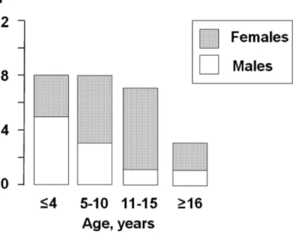
Demographics. Of the 24 patients reported in the
litera-ture and the presented two cases with severe systemic hyper-tension complicated by peripheral facial nerve palsy, 16 were female, 10 male. Median age was 9.5 years (IQR, 2.5– 12 years; range, 11 weeks–62 years). Twenty-three patients (88%) were children 15 years of age or younger (Table 1, Figure 1). The mean modified severity index of arterial BP was 1.67 (IQR, 1.39–1.73)/1.58 (IQR, 1.36–1.84).
Characteristics of peripheral facial palsy. At presentation the facial palsy was unilateral in 25 cases (left-sided, n = 14;
Table 1.
Characteristics of 26 patients
a with severe hypertension and peripheral facial nerve paresis
Reference number
Age
(years)
Gender
Additional presenting symptoms
Blood pressure (mm Hg) Underlying disease Treatment other than antihypertensive medication Neuroimaging Recurrence of facial palsy T ime to resolution of palsy Resolution of palsy Case I 5 M H 220/160 CAKUT No Ye s 2 months Complete Case II 11 F C 230/170
Renal artery stenosis
Renal angioplasty 1.5 months Complete 5 47 M A, O, V 216/142 Primary hypertension No
MRI: Ischemic infarct of dorsal pons
? ? 6 9 F A, C, H, O, V 200/150 CAKUT No
MRI: Pontine hemorrhage
Ye s 0.5 months Complete 7 5 M A 260/175 Pheochromocytoma No Ye s
Died after 4 months
— 8 62 F — 220/98
Renal artery stenosis
No
CT
: No acute changes, persistent chronic lesions
6 months Partial 9 11 F H, F , V 215/135 Primary hypertension No
MRI: Hypertensive encephalopathy
Ye s 0.5 months Almost complete 10 14 F H 230/150 Renal adenocarcinoma No Ye s 1 month Complete 11 10 M H 140/100 Guillain-Barré syndrome No 6 months Complete 12 42 F — 200/150 Primary hypertension No Ye s ? ? 13 0.9 F — 200/160
Mid aortic syndrome
No 6 months Complete 14 1 M — 240/130
Malignant hypertension of unknown origin
No 2 months Complete 15 – I 0.2 M — 133/94 Coarctation Surgical correction MRI: Normal Few days Complete 15 – II 13 M — 218/122 Coarctation Surgical correction Few days Complete 16 0.3 M — 200/-Coarctation No 3 weeks Complete 17 – I 10 F — 190/130 Primary hypertension No Ye s Several months Complete 17 – II 7 F — 192/120 CAKUT Nephrectomy 3 months Complete 17 – III 10 F O 205/153 CAKUT No
MRI: Multiple ischemic bilateral insular
, occipital
and basal ganglia infarcts
9 months Almost complete 18 4 M O 270/160
Renal artery stenosis
Surgical correction
MRI: Ischemic nucleus lenticularis and periven
-tr ic ular infarcts Ye s 12 months Complete 19 – I 5 F H, O 130/100 Guillain-Barré syndrome IVIG
MRI: Enhancement of the lumbosacral spinal nerve roots
4 months Partial 19 – II 3 F O 126/1 10 Guillain-Barré syndrome IVIG
MRI: Enhancement of the lumbosacral spinal nerve roots
3 months Complete 20 – I 12 F V 220/140 CAKUT No CT : Normal 6 months Complete 20 – II 13 F — 200/140 CAKUT No MRI: Normal 8 months Complete 21 – I 0.7 F C 220/170 CAKUT Nephrectomy and facial nerve decompression 1 months Complete 21 – II 12 F H, V 230/170 ? No Several weeks Complete 22 1 M C 220/120 ? No Ye s 1 months Complete Abbreviations:
A, altered mental status; C, convulsions; CAKUT
, congenital anomalies of the kidney and urinary tract; CT
, computed tomography; F
, fatigue; H, headache; IVIG, intrave
-nous immunoglobulines; MRI, magnetic resonance imaging; O, other central neurological manifestations; V
, vomiting.
354 American Journal of Hypertension 26(3) March 2013
right-sided, n = 11) and bilateral in one case. Recurrence of peripheral facial palsy was reported in nine patients, occur-ring ipsilaterally in five6–7,17,22 and contralaterally in another
four.9–10,12,18 Facial nerve dysfunction was mild in four and
severe in eight patients (no information on grading of the peripheral facial palsy is available for the remaining cases). No correlation was found in these patients between severity of facial palsy and the modified BP severity index.
Concomitant signs and symptoms. Additional signs of nervous system dysfunction were described in 16 patients, consisting of headache (n = 7), vomiting (n = 5), convulsions (n = 4), hyperactive reflexes (n = 2), positive Babinski sign (n = 1), altered level of consciousness (n = 3), visual field defect (n = 1), internuclear ophthalmoplegia (n = 1), and not further described transient double vision (n = 1), as depicted in Table 1. In three patients, peripheral facial nerve palsy and severe systemic hypertension were associated with Guillain-Barré syndrome.11,19
Diagnosis of arterial hypertension. The time elapsed
between the first facial symptoms and the diagnosis of severe systemic hypertension lasted a median of 45 days (range, 0 days–2 years) in 16 cases in which this information was provided. In nine patients, BP was not recorded initially and the preliminary diagnosis of idiopathic facial nerve palsy made, although in six of these patients additional neuro-logical signs and symptoms were present.6-7,9-10,17-18,21-22 Both
systolic and diastolic severe hypertension were disclosed in 24 and severe systolic hypertension in the remaining two patients (Table 1). In the three adult patients, facial palsy was found some years after the diagnosis of arterial hyper-tension. In these cases, facial palsy occurred when BP was not well controlled (nonadherence of antihypertensive treat-ment) and severe hypertension exacerbated.5,8,12 The
eti-ology of arterial hypertension was established in 19 patients, consisting of congenital anomalies of the kidney and urinary tract,6,17,20–21 aortic coarctation,13,15–16 renal artery stenosis,8,18
renal adenocarcinoma,10 and pheochromocytoma7(Table 1).
In the three children with Guillain-Barré syndrome,11,19 both
isolated peripheral facial nerve palsy and severe systemic
hypertension were the presenting signs before further fea-tures of the polyneuropathy were apparent. The cause of arterial hypertension remained unknown or classified as pri-mary hypertension in seven patients.5,9,12,14,17,21–22
Site of the lesion. Neuroimaging was described in 11 patients, nine of whom were children (Table 1).5–6,8–9,15,17–20
Computed tomography was performed in two and mag-netic resonance imaging (MRI) in nine patients. Cerebral MRI revealed a pontine hemorrhage at the site of the facial nucleus in a 9-year-old girl,6 and an ischemic stroke
in the dorsal pons affecting the post-nuclear facial fibers in a 47-year-old man.5 Four patients had signs of multiple
ischemic infarcts in the periventricular white matter; three showed involvement of the thalamus and the basal ganglia, one had additional lesions in the insular and occipital cor-tex.8–9,17–18 In two patients with Guillain-Barré syndrome
and gadolinium enhancement of spinal nerve roots on MRI, no cerebral or cranial nerve abnormalities were described.19
In three patients, one of whom had a computed tomography, neuroimaging was normal (Table 1).15,20
No electrophysiological findings specifically addressing the facial nerve were reported.
An edematous nerve sheath with vessel engorgement without hemorrhage was revealed in the child who under-went decompression of the facial nerve from the cochleari-form process to the stylomastoid foramen.21
Therapeutic approaches and outcome. In 23 patients, BP reduction after correction of the underlying disorder or antihypertensive drug management was followed by a grad-ual resolution of the facial nerve palsy (19 complete and four partial resolutions, median time to resolution was 2 [IQR, 0.9–6] months; Table 1). In an 8-month-old girl facial nerve palsy resolved following surgical removal of a nonfunction-ing kidney, medical stabilization of BP and surgical decom-pression of the facial nerve.21 The child with persisting facial
nerve palsy reported by Bialestock in 1961 died because of very severe, refractory hypertension.7
Steroids were prescribed in three cases reported in the lit-erature and in our case II.6,17–18 All four of these patients were
children in whom BP had not been measured at initial diagno-sis of peripheral facial nerve palsy, which had been interpreted as Bell’s palsy and subsequently treated with corticosteroids. No information on dosage and duration of steroid treatment was available in the three patients found in the literature.
Peripheral facial nerve palsy in publications dealing with nervous system involvement in the context of severe hypertension
Forty-one cases of peripheral facial nerve palsy were found in five publications which specifically addressed the neurological signs of systemic hypertension in a total of 860 severely hypertensive children (4.8%, Table 2).14,23–26
Headache was by far the most common neurological com-plication in children with severe arterial hypertension, fol-lowed in decreasing order by altered level of consciousness and vomiting, convulsions, focal central nervous system deficit, and peripheral facial nerve palsy.
Figure 1. Age distribution of the 26 patients with severe hypertension and peripheral facial nerve palsy. No significant difference was noted in the gender distribution between the age groups (Fisher exact test).
diScuSSion
The main finding of this study is the fact that peripheral facial nerve palsy associated with severe arterial hyperten-sion is primarily described in children and adolescents. The long time to diagnosis of arterial hypertension, lasting a median of 45 days, indicates that this association is not widely recognized. Particularly in children, BP measure-ment is not firmly considered as a critical component of the routine physical examination. However, recent clinical reviews addressing the management of peripheral facial palsy in childhood mention severe systemic hypertension as a specific cause.1, 27 Within the group of severely
hyper-tensive children and adolescents, peripheral facial palsy was described in 4.8% (41/860) ranging from 2.8% to 20% as indicated in five publications dealing with the complications of severe childhood hypertension.14, 23–26 Moreover, in a
sur-vey dealing with the different etiologies of peripheral facial nerve palsy, arterial hypertension was the cause in 7 of 87 (8%) children.14
Lower motor neuron facial palsy has previously been associated with the hypertensive disorders of pregnancy and puerperium,28 and a recent case-control study suggests that
chronic, nonsevere hypertension may increase the risk of lower motor neuron facial nerve palsy in subjects older than 40 years of age.29
A timely diagnosis of arterial hypertension in the case of peripheral facial palsy is crucial in order to start ade-quate antihypertensive treatment. The majority of the cases showed full recovery of the facial palsy some weeks after onset of treatment. It remains unclear whether medication directly affects speed and degree of resolution or acts only to prevent further recurrence, since some cases demonstrated spontaneous resolution prior to the diagnosis of arterial hypertension. In the three adult cases with known chronic arterial hypertension, facial palsy occurred during exacerba-tion of severe arterial hypertension due to nonadherence of medication and immediately resolved after better BP con-trol, suggesting a direct relationship between BP lowering and resolution of facial palsy.5,8,12 Corticosteroids, the
stand-ard treatment of Bell’s palsy, potentially exacerbate arterial hypertension and are therefore not indicated.
The putative mechanisms underlying the dysfunction of the facial nerve or its nucleus were addressed in a minority of the affected patients. These include a swelling of the facial nerve in its bony canal associated with vessel engorgement, a hemorrhage into the facial canal, as reported in two necrop-sies,3, 30 an ischemic stroke affecting the postnuclear fibers
of the nerve, or a bleeding in the facial nerve nucleus. These mechanisms may be implicated also in the development of facial palsy during preeclampsia.28
Both lower motor neuron facial nerve palsy and arterial hypertension have independently been described as features of Guillain-Barré syndrome.31 Whether the cases of facial
nerve palsy reported in three children with Guillain-Barré syndrome included in this analysis were caused by severe hypertension or were a first manifestation of the polyneu-ropathy remains uncertain.
The most important limitation of this review lies in the small number of reported patients affected by peripheral facial nerve palsy caused by severe systemic hypertension and the retrospective nature of the analysis. A second limita-tion is that adequate neuroimaging studies were rarely per-formed. Finally, the degree of facial palsy was not assessed using a facial nerve grading system.
In conclusion, this review provides evidence that there might be an association of uncontrolled hypertension and facial palsy that warrants additional evaluation. With effect-ive acute and chronic antihypertenseffect-ive therapy, the progno-sis of facial palsy is excellent. A gradual normalization of BP in 2–3 days is advised.4,32–33
diScLoSurE
The authors declared no conflict of interest.
rEFErEncES
1. Riordan M. Investigation and treatment of facial paralysis. Arch Dis
Child 2001;84:286–288.
2. Anonymous. Facial paralysis in hypertension. Br Med J 1966;2:1547. 3. Moxon W. Apoplexy Into the canal of fallopius in a case of Bright’s
dis-ease causing facial paralysis. Trans Path So 1869;20:420–421.
4. National high blood pressure education program working group on high blood pressure in children and adolescents. The fourth report on the diag-nosis, evaluation, and treatment of high blood pressure in children and adolescents. Pediatrics 2004;114:555–576.
5. Agarwal R, Manandhar L, Saluja P, Grandhi B. Pontine stroke present-ing as isolated facial nerve palsy mimickpresent-ing Bell’s palsy: a case report.
J Med Case Rep 2011;5:287.
6. Aynaci FM, Sen Y. Peripheral facial paralysis as initial manifestation of hypertension in a child. Turk J Pediatr 2002;44:73–75.
7. Bialestock D. Hyperplasia of the adrenal medulla in hypertension of children. Arch Dis Child 1961;36:465–473.
8. Ellis SL, Carter BL, Leehey MA, Conry CM. Bell’s palsy in an older patient with uncontrolled hypertension due to medication nonadher-ence. Ann Pharmacother 1999;33:1269–1273.
9. Harms MM, Rotteveel JJ, Kar NC, Gabreels FJ. Recurrent alternating facial paralysis and malignant hypertension. Neuropediatrics 2000;31:318–320. 10. Kumar S, Winearls CG, Owen DL. Facial palsy with severe
hyperten-sion due to renal tumour. Br Med J (Clin Res Ed) 1982;285:1266. 11. Laufer J, Passwell J, Keren G, Brandt N, Cohen BE. Raised plasma renin
activity in the hypertension of the Guillain-Barre syndrome. Br Med J
(Clin Res Ed) 1981;282:1272–1273.
Table 2. Peripheral facial nerve palsy noted in five pediatric case series with a total of 860 patients affected by severe hypertension
First Author Leumann23 Trompeter25 Deal26 Rance24 Lloyd14 Total
Total number of patients 600 45 110 70 35 860
Patients with facial nerve paralysis (%) 20 (3.3) 2 (4.4) 10 (9.1) 2 (2.8) 7 (20) 41 (4.8) These cases were only listed and not described in detail, permitting only analysis of prevalence.
356 American Journal of Hypertension 26(3) March 2013
12. Lavin PJ, Weissman BM. ‘Bell’s palsy’ in accelerated hypertension.
Postgrad Med 1985;77:165, 168.
13. Lewis VE, Peat DS, Tizard EJ. Hypertension and facial palsy in middle aortic syndrome. Arch Dis Child 2001;85:240–241.
14. Lloyd AV, Jewitt DE, Still JD. Facial paralysis in children with hyperten-sion. Arch Dis Child 1966;41:292–294.
15. Margabanthu G, Brooks J, Barron D, Miller P. Facial palsy as a pre-senting feature of coarctation of aorta. Interact Cardiovasc Thorac Surg 2003;2:91–93.
16. Moore P, Fiddler GI. Facial palsy in an infant with coarctation of the aorta and hypertension. Arch Dis Child 1980;55:315–316.
17. Siegler RL, Brewer ED, Corneli HM, Thompson JA. Hypertension first seen as facial paralysis: case reports and review of the literature.
Pediatrics 1991;87:387–389.
18. Smilari P, Incorpora C, Polizzi A, Sciacca P, Musi L, Petrillo G, Distefano G, Pavone L. [Recurrent facial paralysis in a child with renovascular hypertension]. Pediatr Med Chir 1995;17:461–463.
19. Smith N, Grattan-Smith P, Andrews IP, Kainer G. Acquired facial palsy with hypertension secondary to Guillain-Barre syndrome. J Paediatr
Child Health 2010;46:125–127.
20. Tirodker UH, Dabbagh S. Facial paralysis in childhood hypertension.
J Paediatr Child Health 2001;37:193–194.
21. Voorhees RL, Zeitzer LD, Ross M. Hypertension and associated periph-eral facial paralysis. Laryngoscope 1972;82:899–902.
22. Zeis PM, Rao S, John EG, Aschinberg LC. “Stress” polycythaemia and peripheral facial palsy complications of severe hypertension. Acta
Paediatr Scand 1979;68:287–289.
23. Leumann EP. Blood pressure and hypertension in childhood and ado-lescence. Ergeb Inn Med Kinderheilkd 1979;43:109–183.
24. Rance CP, Arbus GS, Balfe JW, Kooh SW. Persistent systemic hyperten-sion in infants and children. Pediatr Clin North Am 1974;21:801–824. 25. Trompeter RS, Smith RL, Hoare RD, Neville BG, Chantler C.
Neurological complications of arterial hypertension. Arch Dis Child 1982;57:913–917.
26. Deal JE, Barratt TM, Dillon MJ. Management of hypertensive emergen-cies. Arch Dis Child 1992;67:1089–1092.
27. Singhi P, Jain V. Bell’s palsy in children. Semin Pediatr Neurol 2003;10:289–297.
28. Shmorgun D, Chan WS, Ray JG. Association between Bell’s palsy in pregnancy and pre-eclampsia. QJM 2002;95:359–362.
29. Savadi-Oskouei D, Abedi A, Sadeghi-Bazargani H. Independent role of hypertension in Bell’s palsy: a case-control study. Eur Neurol 2008;60:253–257.
30. Monier-Vinard, Puech P. Nephrite Chronique et Paralysie Faciale. Bull
et Memoires de la Soc Med des Hopitaux de Paris 1930;20:977–980.
31. Shah DN. The spectrum of Guillain-Barre syndrome. Dis Mon 2010;56:262–265.
32. Chobanian AV, Bakris GL, Black HR, Cushman WC, Green LA, Izzo JL, Jr.,Jones DW, Materson BJ, Oparil S, Wright JT, Jr.,Roccella EJ. Seventh report of the Joint National Committee on Prevention, Detection, Evaluation, and Treatment of High Blood Pressure. Hypertension 2003;42:1206–1252.
33. Flynn JT, Tullus K. Severe hypertension in children and adolescents: pathophysiology and treatment. Pediatr Nephrol 2009;24:1101–1112.