Tissue engineering: A new approach in cardiovascular surgery; Seeding
of human fibroblasts followed by human endothelial cells on
resorbable mesh
1Gregor Zu¨nd *, Simon P. Hoerstrup, Andreina Schoeberlein, Mario Lachat, Georg Uhlschmid,
Paul R. Vogt, Marko Turina
Clinic for Cardio6ascular Surgery, Uni6ersity Hospital, Ra¨mistrasse100, CH-8091, Zurich, Switzerland
Received 30 September 1997; received in revised form 10 November 1997; accepted 19 November 1997
Abstract
Objective: In tissue engineering the material properties of synthetic compounds are chosen to enable delivery of dissociated cells
onto a scaffold in a manner that will result in in vitro formation of a new functional tissue. The seeding of human fibroblasts followed by human endothelial cells on resorbable mesh is a precondition of a successful creation of human tissues such as vessels or cardiac valves. Methods: Polymeric scaffolds (n = 18) composed of polyglycolic acid (PGA) with a fiber diameter of 12 – 15mm and a polymer density of 70 mg/ml were used as square sheets of 1 × 1 × 0.3 cm. Fibroblasts (passage 7) harvested from human foreskin were seeded (3.4 × 10⁶) and cultured over a 3 week period on a PGA-mesh, followed by seeding of endothelial cells
(passage 5, 2.8 × 10⁶) harvested from human ascending aorta. Thereafter the new tissue was stained for HE, van Gieson,
Trichrom – Masson, Factor VIII and CD 34 and proved by scanning electron microscopy. Results: Microscopic examination of the seeded mesh demonstrated that the human fibroblasts were attached to the polymeric fibers and had begun to spread out and divide. The scanning electron microscopic examination demonstrated a homogeneous scaffold resembling a solid sheet of tissue. The seeded endothelial cells formed a monolayer on the fibroblasts and no endothelial cell invasion or new formation of capillaris could be detected. Conclusions: These results are a first step to demonstrate that seeding of human fibroblasts and endothelial cells on PGA-mesh might be a feasible model to construct human tissues such as vessels or cardiac valves. © 1998 Elsevier Science B.V. All rights reserved.
Keywords: Tissue engineering; Synthetic biodegradable matrix; Endothelial cells
1. Introduction
Transplantation of endothelial cells onto the blood flow surface of a small-diameter vascular conduit, ho-mografts, or valve prosthesis to partially reproduce the antithrombogenic lining of a naturally occuring intima is a logical concept. In our experiences, endothelial cell transplantation has failed to make a significant
transi-tion from the animal laboratory to the clinical arena [1]. We believe nowadays that human endothelial cells need a living compound for normal growth and sur-vival in the human blood stream.
Tissue engineering is a multidisciplinary science that utilizes basic principles from engineering and life sci-ences to construct tissues from their cellular compo-nents [2]. For the creation of new functional tissue three general strategies have been adopted [3]. First, isolated cells or cell substitutes can be used; this technique allows the replacement of only those cells that supply the needed function. Second, tissue-inducing substances such as growth factors can be used to stimulate the * Corresponding author. Tel.: + 41 1 2551111; fax: + 41 1
2554369; e-mail [email protected]
1Presented at 11th Annual Meeting of EACTS, Copenhagen, September 28 – October 1, 1997.
1010-7940/98/$19.00 © 1998 Elsevier Science B.V. All rights reserved.
can be cultured on or within specific matrices. These days, our laboratory has focused on the cre-ation of new functional tissue substitutes utilizing hu-man cells attached to synthetic, biodegradable polymer scaffolds. This approach involves isolating cells from a donor and seeding these cells onto a polymeric scaffold, where they attach and grow and finally form a tissue-like structure. The cell-polymer construct is configured to serve as a template to guide this development. Preliminary animal studies have demonstrated the feasibility of this strategy with the successful formation of a tissue engineered valve leaflet and a successful implantation of this leaflet with good functional results [4 – 6]. The ability to isolate cells types is an important component to any tissue engineering project. Furthermore, the seed-ing of human fibroblast followed by human endothe-lial cells on resorbable mesh is a precondition of the creation of any tissue in the cardio-vascular field.
2. Material and methods 2.1. Scaffold
Polymeric non-woven scaffold, composed of polyg-lycolic acid (PGA) with a thickness of 3 mm, a fiber diameter of 12 – 15 mm and a polymer density of 70 mg/ml (Albany International Research, Mansfield, MA, USA; a kind gift from Dr. J. Vacanti, Chil-dren’s Hospital, Harvard Medical School, Boston, MA, USA) were used as square sheets of 0.3 × 1 × 1 cm. The PGA-polymer by hydrolysis has an in vitro resorption rate of 4 – 6 weeks.
2.2. Human fibroblasts
Human foreskins were obtained from phimosisop-erations performed at Children’s Hospital, University Zurich. The harvested foreskins were serially washed with phosphate buffered saline (Gibco BRL-Life Technologie, Grand Island, NY) supplemented with 1% antibiotic/antimycotic solution saline (Gibco BRL-Life Technologie, Grand Island, NY). Under a laminar flood hood (Forma Scientific, Marietta, OH), the tissue was minced into 1 – 2 mm pieces and evenly distributed in 15 × 60 mm tissue culture dishes (Becton Dickinson, Lincoln Park, NJ). Medium was gently added to the tissue culture dishes. Medium consisted of Dulbecco’s modified Eagle’s medium (Gibco BRL-Life Technologies) supplemented with 10% fetal bovine serum (Gibco BRL-Life Technolo-gies) and 1% antibiotic/antimycoic solution (Gibco BRL-Life Technologies). The explanted tissue was placed in a humidified incubator (Forma scientific) at
riod, the cells were migrated off the explants. There-after cells were serially passaged to obtain sufficient numbers of cells for cell seeding.
2.3. Human aortic endothelial cells
Human aortic endothelial cells were obtained from the ascending aorta of the explanted sick hearts of heart transplant patients. The harvested piece of tis-sue of ascending aorta was serially washed with phosphate buffered saline (Gibco BRL – Life Tech-nologies, Grand Island, NY) supplemented with 1% antibiotic/antimycotic solution (Gibco BRL-Life Technologies). Under a laminar flood hood (Forma Scientific, Marietta, OH), the intima surface was covered with Collagenase Typ I (75 U/ml in PBS pH 7.4, Worthington) and incubated for 30 min at room temperature. Cells were detached using a rub-ber cell scraper and cultured in 75 cm2 culture dishes (Becton Dickinson Lincoln Park, NJ). Medium 199 with EBS (Amimed, Allschwil, Switzer-land) supplemented with 10% fetal bovine serum, 16 U/ml Heparin, 20 mg/ml Endothelial cell growth supplement (Becton, Dickinson) and 1% antibiotic. The explanted cells were placed in a humidified in-cubator at 37°C with 5% CO2 for 2 – 3 weeks. Cells were serially passaged to obtain sufficient numbers of cells for cell seeding. The cells were then labeled with an acetylated low density lipoprotein (Ac-Dil-LDL) probe (Biomedical Technologies, Stoughton, MA) which selectively fluorescently tags endothelial cells through their LDL receptor. These cells popu-lations were then checked for their purity by fluores-cent microscopy.
2.4. Seeding
The PGA polymers (n = 18, 1 × 1 × 0.3 cm) were seeded with human fibroblasts (passage 7). There were 8 – 10 seeding procedures every 90 min. Each seeding procedure contained 3.4 × 10⁶ human fibrob-lasts. The seeded fibroblasts were cultured over a 21 days period. Media consisted of Dulbecco’s modified Eagle’s medium (Gibco BRL-Life Technologies) sup-plemented with 10% fetal bovine serum (Sigma, St. Louis, MO) and 1% antibiotic solution (Irvine Scien-tific, Santa Clara, CA). The medium was changed every 4 days. Thereafter this tissue-like structure was seeded with the pure human aortic endothelial cell culture (passage 5, 2.8 × 10⁶). The constructs were then fixed with 0.4% paraformaldehyde and stained by conventional histological methods (Hemalaun Eosin, Van Gieson, Trichrom-Masson) and examined by electron microscopy.
Fig. 1. Formation of fibroblast-polymer structure. Scanning electron microscopy demonstrates the formation of a continuous fibroblast tissue.
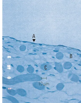
Fig. 3. Formation of endothelial monolayer. CD 34 stain demon-strates a monolayer of endothelial cells (A) on the surface of fibro-blasts core (B). After 4 weeks hydrolysis of polymer fibers (C) is in process.
3. Results
3.1. Con6entional microscopy
Conventional microscopy examination demonstrated that the seeded human fibroblasts were attached to the polymeric fibers and had begun to spread out and devide (Figs. 1 and 2). Furthermore the seeded cells were forming cell bridges and a homogeneous tissue.
The production of collagen matrices was demonstrated by staining the tissue-like structure with Van Gieson. There was evidence of beginning hydrolytic degradation of the polymeric fibers. The seeded endothelial cells formed a monolayer on the human fibroblasts as demonstrated by factor VIII and CD 34 (Fig. 3). In addition, no invasive growth of endothelial cells or new formation of capillaris could be detected.
3.2. Electron microscopy
The scanning electron microscopic examination 7 days before seeding with human aortic endothelial cells demonstrated attachment of fibroblasts to the poly-meric fibers and the formation of cell bridges between polymeric fibers. In addition a homogeneous distribu-tion of the seeded fibroblasts could be detected. There was the same densitiy of fibroblasts in the upper and lower part of the PGA mesh and no gravitiy gradient could be detected. Some of the polymeric fibers have started hydrolytic degradation.
4. Discussion
In tissue engineering, the material properties of syn-thetic compounds are chosen to enable delivery of Fig. 2. Continous formation of fibroblasts. Scanning electron
mi-croscopy shows a continuous distribution of fibroblasts throughout the ‘polymeric architecture’ and attachment of the fibroblasts (A) to the polymer fibers (B).
of new functional tissue. The advantage of such tissue engineered products is the ability to grow, repair and remodel, and no requirement for anticoagulation. Fur-thermore, if autologous cells are used for the construc-tion of such products, an increasing durability will be achieved with lack of risk of immunogenicity and rejec-tion. The new field of tissue engineering is emerging experimentally and tissue substitutes for liver, cartilage, bone, trachea, intestine and urologic tissue are created [7 – 10]. The first tissue engineered products in clinical use are skin replacements. In the field of cardiac surgery tissue engineering is not unknown. Marelli et al. demonstrated that skeletal muscle satellite cells can be harvested, grown in vitro, and autotransplanted into damaged heart muscle [11 – 13]. Preliminary results showed that muscle formation occured at 8 weeks, but not at 14 weeks after transplantation [12]. Apart from that, the group of Vacanti and Mayer at Children’s Hospital in Boston has become a leading group in cardiovascular tissue engineering [4 – 6]. The goal of their research is to create an autologous, tissue engi-neered aortic valve with the ability to grow, repair and remodel. They created an animal model in which a pulmonary valve leaflet was successfully replaced by an autologous tissue engineered valve leaflet. Furthermore at our institution autologous capillary endothelial cells have been harvested, grown in vitro and seeded onto artificial vascular grafts in order to promote hemocom-patibility [14 – 17]. In our experiences, endothelial cell transplantation has failed to make a significant transi-tion from the animal laboratory to the clinical area. We believe nowadays that human endothelial cells need a living compound for normal growth and survival in the human blood stream. Since the seeding of human fibroblasts on biodegradable mesh is a precondition of any tissue engineered product in cardiovascular surgery we first focused upon the development and evaluation of an adequate method. For this reason we started with creating a living compound of fibroblasts followed by seeding of endothelial cells. Only endothelial cells of the human ascending aorta were used suggesting that hu-man capillary endothelial cells thus be angiogenetic and might form new capillaris. For the development of a living compound we choose preputial fibroblasts from newborns. Since in contrast to adults this cells have a higher potential for growth and higher cell generations can be presumed [18]. The most important steps in this study are the attachment of human fibroblasts to the fibers and the formation of cell bridges and in addition the seeding of endothelial cells forming a monolayer with no evidence of invasion or formation of capillary structures into the core. The polymer templates that we utilize are both biocompatible and biodegradable; a rapid degradation is desirable in order to minimize any chronic foreign-body inflammatory response [19]. The
mixed cell population obtained from human ascending aorta to pure cell populations followed by serial passag-ing to obtain sufficient numbers of cells for cell seedpassag-ing. Cardiovascular tissue engineering has the potential to improve our ability to treat cardiovascular disease, one of the most important single cause of mortality in the industrial world. The preliminary results of this study represent a basic step on the way to construct human cardiovascular tissue and demonstrate that the applied technique of tissue engineering might be a feasible approach.
References
[1] Jarrwll BE, Williams SK. Microvessel derived endothelial cell isolation, adherence, and monolayer formation for vascular grafts. J Vasc Surg 1991;13:733 – 4.
[2] Vacanti CA, Vacanti JP. Bone and cartilage reconstruction with tissue engineering approaches. Otolaryngol Clin North Am 1994;27(1):263 – 76.
[3] Langer R, Vacanti JP. Tissue Eng Sci 1993;260:920 – 6. [4] Zu¨nd G, Breuer CK, Shinoka T, Ma PX, Langer R, Mayer JE,
Vacanti JP. The in vitro construction of a tissue engineered bioprosthetic heart valve. Eur J Cardio-thoracic Surg 1997;11:493 – 7.
[5] Shinoka T, Breuer CK, Tanel RE, Zu¨nd G, Miura T, Ma PX, Langer R, Vacanti JP, Mayer JE. Tissue engineering heart valves. Ann Thorac Surg 1995;60(6):513 – 6.
[6] Shinoka T, Ma X, Breuer CK, Cusick RA, Shum-Tim D, Zu¨nd G, Langer R, Vacanti JP, Mayer JE. Tissue engineering heart valves, autologous valve leaflet replacement study in a lamb model. Circulation 1996;94(suppl II):164 – 8.
[7] Cao Y, Vacanti JP, Ma X, Paige KT, Upton J, Chowanski Z, Schloo B, Langer R, Vacanti CA. Generation of neo-tendon using synthetic polymers seeded with tendocytes. Transplant Proc 1994;26(6):3390 – 1.
[8] Sakata J, Vacanti CA, Schloo B, Healy GB, Langer R, Vacanti JP. Tracheal composites tissue engineered from chondrocytes, trachel epithelial cells and synthetic degradable scaffolding. Transplant Proc 1994;26(6):3309 – 12.
[9] Vacanti JP, Morse MA, Saltzman WM, Domb AJ, Perez-Atayade A, Langer R. Selective cell transplantation using bioab-sorbable artificial polymers as matrices. J Pediatr Surg 1988;23(1):3 – 9.
[10] Vacanti JP. Beyond transplantation: Third annual Samuael Ja-son Mixter lecture. Arch Surg 1988;123:545 – 9.
[11] Greentreee D, Marelli D, Ma F, Chiu RC-J. Satellite cell trans-plantation for myocardial repair: labeling techniques. Transplant Proc 1994;26(6):3357.
[12] Marelli D, Desrosiers C, el-Alfy M, Kao RL, Chiu RC. Cell transplantation for myocardial repair: an experimental ap-proach. Cell Transplant 1992;1(6):383 – 90.
[13] Zibaitis A, Greentree D, Ma F, Marelli D, Duong M, Chiu RC-J. Myocardial regeneration with satellite cell implantation. Transplant Proc 1994;26(6):3294.
[14] Muller-Glauser W, Lehmann KH, Bittmann PO, et al. A compli-ant small-diameter vascular prosthesis lined with functional venous endothelial cells. ASAIO Trans 1988;34(3):528 – 31. [15] Pasic M, Muller-Glauser W, von Segesser L, Odermatt B,
Lachat M, Turina M. Endothelial cell seeding improves patency of synthetic vascular grafts: manual versus automatized method. Eur J Cardiothorac Surg 1996;10(5):372 – 9.
[16] Pasic M, Muller-Glauser W, Odermatt B, Lachat M, Seifert B, Turina M. Seeding with omental cells prevents late neointimal hyperplasia in small-diameter Dacron grafts. Circulation 1995;92(9):2605 – 16.
[17] Pasic M, Muller-Glauser W, von Segesser L, Lachat M, Mi-haljevic T, Turina M. Superior late patency of small-diameter Dacron grafts seeded with omental microvascular cells: An ex-perimental study. Ann Thorac Surg 1994;58:677.
[18] Matsouko H, Furusawa M, Kono A. Growth potential of human fibroblasts on contact sensitive confluent monolayer fixed with 3% glutaraldehyde. Gan To Kag R 1994;21:563 – 4. [19] Mooney DJ, Organ G, Vacanti JP, Langer R. Design and
fabrication of biodegradable polymer devices to engineer tubular tissues. Cell Transplant 1994;3(2):203 – 10.
Appendix A. Conference discussion
Dr T. Ferguson (St. Louis, USA): You caught me a little by
surprise because the abstract does not refer specifically to using this approach on valves, which to me is the most difficult experimental environment. I guess my question would be, is this technique
applica-ble to vascular grafts and other biologic situations?
Dr Zund: Yes. I do agree with you. In one of our animal studies we
were just replacing a pulmonary leaflet.
Dr Ferguson: Well, if it works in a valve, it will work anywhere in
the body, of that you can be sure.
Dr C. Muneretto (Padua, Italy): We have already developed a
research program on tissue engineering. And in our experience we use human vessel treated to suppress host-immune response, and we use this vessel as a geometrical and mechanical support for the seeding and growth of fibroblasts and endothelial cells. We found two main problems. The first one, the endothelial growth; and the second one, membrana basalis formation.
I have two questions for you. Did you use any method to increase speed in endothelial cell growth? And second question, did you find any formation of membrana basalis in you conduit?
Dr Zund: First of all, we don’t use any additional factors for
endothelial growth. We just are culturing them, and then, after some passages, we are seeding them on the fibroblast core. Second, we were not yet looking for the basilar membrane.
Dr Ferguson: Were you able to show by low microscopic scans, that
total coverage of the surface is achieved?
Dr Zund: Yes, sir. The fibroblasts were totally confluent and
formed a new tissue.