Pacing in hypertrophic obstructive cardiomyopathy
A randomized crossover study
L. Kappenberger*, C. Linde, C. Daubert, W. McKenna, E. Meisel, N. Sadoul,
L. Chojnowska, L. Guize, D. Gras, X. Jeanrenaud*, L. Ryden and the PIC
Study Group
*Division of Cardiology, CHUV, Lausanne, Switzerland
Background Uncontrolled studies have shown that short atrioventricular delay dual chamber pacing reduces outflow tract obstruction in hypertrophic obstructive cardiomyo-pathy. Although the exact mechanism of this beneficial effect is unclear, this seems a promising potential new treatment for hypertrophic obstructive cardiomyopathy. Method In order to evaluate the impact of pacing therapy, we performed a randomized multicentre double-blind cross-over (pacemaker activated vs non activated) study to inves-tigate modification of echocardiography, exercise tolerance, angina, dyspnoea and quality of life in 83 patients with a mean age of 53 (range 22-87) years with symptoms refractory or intolerant to classical drug treatment. Results After 12 weeks of activated or inactivated pacing, independent of which phase was first, the pressure gradient fell from 59 ± 36 mmHg to 30 ± 25 mmHg (P<0001) with active pacing.
Exercise tolerance improved by 21% in those patients who at baseline tolerated less than 10 min of Bruce protocol; symptoms of dyspnoea and angina also improved signifi-cantly from NYHA class 2-4 to 1-4 and 10 to 0-4, respect-ively (/"<0007). Quality of life assessment with a validated questionnaire objectivated the subjective improvement. Conclusion Pacemaker therapy is of clinical and haemo-dynamic benefit for patients with hypertrophic obstruc-tive cardiomyopathy, left ventricular outflow gradient at rest over 30 mmHg who are symptomatic despite drug treatment.
(Eur Heart J 1997; 18: 1249-1256)
Key Words: Hypertrophic obstructive cardiomyopathy, pacemaker treatment, quality of life assessment.
Introduction
Hypertrophic cardiomyopathy is a primary cardiac dis-ease genetically transmitted as an autosomal dominant trait with variable penetrance, and, additionally, there is a high proportion of sporadic non-familial cases'1"31. It is characterized by ventricular hypertrophy with myocyte and myofibrillar disarray. Molecular genetic studies have revealed the chromosomal locus and allowed iso-lation of several putative genes leading to mutations in the beta cardiac myosin heavy chain'4"6'. Penetration is heterogeneous, as are the phenotypic manifestations. Hypertrophic cardiomyopathy presents in various forms, but usually as an asymmetric hypertrophy which can be located in the subaortic, midventricular or apical regions and also elsewhere in the right or left ven-tricles'7'. The hypertrophy causes several pathophysio-logical features including a hypercontractile state, diastolic dysfunction'81, disturbed coronary flow with ischaemic'9""' and also abnormal responses of the peripheral vasculature to exercise1'2'. In the obstructive
form of hypertrophic cardiomyopathy, there are both septal thickening in the subaortic region and functional abnormalities of the mitral valve apparatus'13141 of which systolic anterior movement of the anterior cusp is the most prominent.
Two principal mechanisms for systolic anterior movement have been proposed, first the Venturi mech-anism, by which the hypertrophied septum induces acceleration of blood flow which sucks the anterior cusp towards it resulting in a further narrowing of left ventricular outflow. This phenomenon may be com-pounded by elongated tendons and a floppy valve. All this results in an obstruction and consequently pressure gradient across the outflow tract1'5"171. Second, the anatomical alterations in the mitral valve apparatus, particularly the anterior displacement of the papillary muscle, approximation of the anterior cusp to the septum exposing it to flow drag1'81, again results in obstruction. Inversion of the ventricular activation sequence by right apical pacing can, in part, eliminate these deleterious events'2'1.
Clinical observations have led to a haemo-dynamic classification of hypertrophic obstructive cardiomyopathy'71 where obstruction may be persistent (at rest) labile (variable) or latent (provocable). The symptoms of hypertrophic obstructive cardiomyopathy typically include dyspnoea which relates to diastolic dysfunction, angina due to ischaemia, and syncope or exertional dizziness caused by obstruction, arrhythmia and/or inappropriate behaviour of the peripheral circu-lation. The severity of symptoms does not necessarily correlate with the left ventricular outflow gradient. However, patients with non-obstructive hypertrophic cardiomyopathy usually present with fewer and milder symptoms'131.
Treatment of hypertrophic obstructive cardio-myopathy so far has been either pharmacological (beta-blockers, calcium antagonists, disopyramide)'19'20'221 with the goal of reducing outflow obstruction and improving ventricular relaxation and rhythm control, or surgery (myotomy, myectomy and mitral valve recon-struction) all offering an impressive gradient reduc-tion[2]. The observation that electrical stimulation of the heart has a beneficial effect on outflow obstruction was first reported from acute pacing studies123"251. Sympto-matic benefit and haemodynamic improvement was later achieved in long-term studies126"281.
In order to investigate prospectively subjective and objective effects of adding cardiac pacing to normal medical treatment in hypertrophic obstructive cardio-myopathy (PIC) we performed whenever possible a double blinded randomized crossover, pacemaker active vs non-active study in patients refractory or intolerant of drugs who were considered to be candidates for surgery. The following is a report of the crossover part of the PIC Study. To our knowledge, this is the first blinded randomized crossover study to investigate this effect of pacing and to study a device using drug-trial standards.
Patients and methods
This multicentre study is conducted according to the guidelines of the EN540, the European standard on clinical investigation of medical devices for human sub-jects, which includes compliance to the declaration of Helsinki. The study was co-ordinated by the European Working Group on Cardiac Pacing of the European Society of Cardiology, and was accepted by the ethical committees of each of the participating centres.
Patients with symptomatic hypertrophic obstruc-tive cardiomyopathy refractory or intolerant to drug treatment were evaluated. In general, they were referred to the study centres for haemodynamic evaluation with a view to surgical relief of obstruction. Inclusion criteria were therefore functional limitation (NYHA Class II or III), angina and/or dyspnoea, despite maximal tolerated drug therapy. These patients underwent a screening catheterization and/or an echocardiography study to evaluate the effect of dual-chamber pacing on the resting
gradient. Pacing was considered if, at rest, there was a subaortic obstruction with peak pressure gradient of >30 mmHg with or without provocation. When acute pacing was well tolerated (no fall in cardiac output or aortic pressure), implantation of a permanent dual-chamber pacing system was offered and the patient included in the study when informed written con-sent was obtained and the pacemaker implanted. The patients were classified into Group A when the acute study showed a peak gradient reduction of >30% or Group B if there was less or no gradient modification. The screening examinations also included graded tread-mill exercise tests using a modified Bruce protocol. Only patients with less than 85% of age-predicted VO2max
were considered suitable for the study. We excluded patients with symptoms at rest corresponding to NYHA Class IV, any severe valvular dysfunction not related to systolic anterior movement, documented coronary artery disease, age below 18 years, drug-refractory sys-temic hypertension, ejection fraction <50%, chronic atrial fibrillation and any patient where pacing was indicated for another reason, except drug-induced chronotropic incompetence.
Pacemakers
Any dual-chamber pulse generator could be used if it incorporated the following programmable characteris-tics: pacing modes including DDD and AAI lower rate at 30 beats . min~', atrioventricular delay of 30 ms or less to ensure complete ventricular capture, paced and sensed atrioventricular delay separately programmable, atrioventricular delay adaptation with increasing rate, VVIR fall back mode in the event of atrial tachyarrhyth-mias. Programming the pacemaker in DDD with opti-mal atrioventricular delay was considered to be pacemaker-activated; programming to AAI at 30 ppm was termed pacemaker non-activated.
Study design
Both Groups A and B were stratified by central ran-domization into two arms defining the sequence of therapies. Randomization was active after the discharge tests where the optimal atrioventricular delay was identified in each patient (full ventricular capture on the ECG without a drop in aortic pressure) and pacemaker function verified. Programming in the active or passive pacing arms of the study was not performed by the investigator, to allow a double-blind design as accurate as possible. After 12 weeks the two patient arms were similarly assessed and their pacing modes inverted (crossover). After the second 12-week period the two groups were again reassessed, and, finally, the pacing mode preferred by the patient was programmed for long-term pacing.
The following assessment methods were used: ECG, echocardiogram (left ventricular outflow tract
pressure gradient, left ventricular dimensions, left ventricular function, mitral valve movement and regur-gitation, etc), 24-h ECG, symptom-limited exercise test with measurement of spirometric and metabolic data. For quality of life analysis, the Karolinska questionnaire was used. Details on the questionnaire have previously been described'29'. It has been validated for patients paced for bradyarrhythmia and ischaemic heart disease. Prior to the investigation, translations of this document were validated against the original. The questionnaire was administered by the technician or nurse and com-pleted by the patient either at the clinic or at home at screening and after each study period. In case of prema-ture termination of one study arm a full evaluation including quality of life was carried out. Medical history focused on NYHA classification of global functional capacity and the presence of syncope, fatigue, angina and dyspnoea. Specific instructions on how to perform each test and which parameters to measure were given to each investigator. Programming of pacemakers was per-formed double blind; neither the patient nor the investi-gator were informed of the actual pacing mode. All complications had to be specified and reported.
Statistical methods
The statistical tests included comparison between the two arms (pacing vs non-pacing) as well as comparison within a patient when tested after each of the 3 month periods with pacing active vs non-active. All intra- and interpatient comparisons included testing of type of distribution (using Shapiro-Wilk or Kolmogorov-Smirnov tests), and testing of significance of observed differences (using the two-sided T-test, Wilcoxon Rank sum, or Chi-square tests). A P-value of less than 005 is considered significant. The quality of life was evalu-ated with analysis of variance calculevalu-ated by statistical analysis system (SAS).
Results
A total of 83 patients was recruited for this study in 12 centres over a period of 24 months. There were 33 females and 50 males; mean age was 53 years (range 32-87). Drug treatment consisted of beta-blockers taken by 42 patients, the average dose corresponding to 150mg.day~' of propanolol; 39 were on cal-cium antagonist (240 mg . day" ' of verapamil or equiv-alent), 12 patients were administered Amiodarone (200 mg . day"'), three disopyramide (300 mg) and three various others. Diuretics were given to 10 patients and three had ACE inhibitors. All drug prescriptions remained unchanged over the whole crossover period.
At screening, haemodynamic and/or Doppler echocardiographic measurements revealed that in 64 patients (77%) pacing induced an acute peak gradient reduction of more than 30% (785 ± 31-9 mmHg to
32-4 ± 220 mmHg, Group A) while in 18 patients (22%) it was less than 30% (853 ± 38-8 to 701 ± 351 mmHg, Group B). Group B patients were younger (43 ± 14 years vs 56± 14 years P=00007). One patient died in the screening phase due to right ventricular perforation and cardiac tamponade before detailed measurements could be made.
In order to confirm appropriate pacing, active as well as non-active, a 24 h ECG was performed in 48 and 29 patients, respectively, at the end of each study arm. Complete ventricular capture during the DDD phase was present in >98% of ventricular complexes and atrial pacing in either phase accounted for < 1 % of atrial complexes. The atrioventricular delay for the DDD phase was individually adapted and found optimal at 61 ± 23 ms for sensed atrial beats. No severe arrhyth-mias were registered and the range from mean mini-mal to maximini-mal heart rate was 44 ± 8 beats . min ~ ' and 119 ± 27 beats . min ~ ' in pacemaker non-active and 48 ± 6 beats . min ~ ' and 128 ± 20 beats . min ~ ' in pacemaker active.
No syncope was reported during the 24 h ECG, but two episodes occurred during the non-active and an equal number during the active pacing phase.
The evolution of individual pressure gradients in all patients is depicted in Fig. 1. The right panel shows that after 3 months of DDD pacing, 38 out of the 40 full documented patients had a decrease of the peak gradi-ent. The mean reduction was 51%, from 53 mmHg to 26 mmHg (/><00001). Switching the pacemaker off resulted in a re-increase of the gradient from 33 mmHg to 59 mmHg (/><00001) in 35 of 36 patients. In seven patients these measurements were not completed because one died, two had technical problems and in four the echocardiographic window was unreliable. In Group B, the gradient decreased from 69 ± 39 mmHg to 45 ± 27 mmHg (/>=0004). Interestingly, in Group B the gradient fell more than 30% in six patients (38%) in contrast to their acute studies. At the end of 3 months of activated pacing, the global ejection fraction in this population remained unchanged.
The result of exercise duration analysis reveals interesting findings (Fig. 2). Comparing the end of the inactivated with the end of the activated phase, there is a small but insignificant improvement. Subgroup analysis of the more severely limited patients who had less than 10 min exertional tolerance with the inactive pacemaker, however, showed that their walking time had signifi-cantly improved by 21% (/><0008) once activated. No difference in this respect could be demonstrated between Groups A and B.
Symptoms were assessed by clinical history and by a detailed double-blind validated quality of life questionnaire1291. The overall functional class and evol-ution of all patients is summarized in Fig. 3. Although several patients reported episodes of resting symptoms (NYHA Class IV), at the time of inclusion all corre-sponded to Class II or III. One patient experienced a brief deterioration and therefore entered Class IV with the inactive pacemaker. Of the 28 patients in Class III
140 120 100 -- 140 -.120 - 100 DDD AAI AAI ^¥40 20 0 DDD
Figure 1 Evolution of peak outflow gradient in the D D D to AAI (n=36, /»<0-001) and AAI to DDD (n=40
/><0-001) phases; it made no difference which phase was first.
during the inactivated phase, 24 (84%) improved with activated pacemakers and none deteriorated. Of the 37 in Class II, 17 (46%) improved with pacing activated and one deteriorated. When compared with the baseline findings at entry into the study, 20 of the 70 patients already had some improvement although the pacemaker was not activated. There was no difference with respect to the sequence pacemaker first active or non-active.
Overall
analysis subject analysisRetrospective
AAI<=10 min
Figure 2 Duration of exercise tolerance activated ( • ) vs
non-activated (") pacemaker in all (Group A and B) patients. There was no deterioration but patients with less than 10 min at baseline or AAI benefited significantly with a 21% increase of exercise duration.
Symptoms of dyspnoea and angina were similarly influ-enced as shown in Fig. 4 and again there was no difference between the sub-groups. Using intrapatient as well as group comparisons the modifications induced by the pacemaker active were highly significant on all reported parameters (.P^OOOl), while inactivating the pacemaker led to a significant deterioration. These sub-jective findings were further substantiated by the quality of life assessment with the validated quality of life questionnaire in 81 patients. The relevant results are summarised in Fig. 5. there was a greater improvement in quality of life when the pacemaker was switched on immediately than if it was turned on after 3 months, reflecting some placebo-effect of this intervention. Nevertheless, values of self-perceived symptomatology document that with the pacemaker activated there was a significant (/><0-05) improvement in the ability to self-autonomy and strenuous physical activity, alertness and cardiovascular symptomatology. Cognitive and sexual functioning as well as emotional state were, however, not significantly affected by the pacing mode.
Of the 83 patients included; 81 completed the study, one died and one had surgery. Of the 41 ran-domized to pacemaker active first, all completed a 12-week period. After inactivation of the pacemaker, however, 14 returned prematurely to the clinic (1-83 days, mean 26 days) because of severe deterioration in symptoms and self-perceived health status. At the end of the crossover, 79 patients preferred pacing and two more went to surgery. Of the 41 in pacemaker non-active first, three returned to the clinic early because of no
Baseline AAI DDD
II
III
IV
Figure 3 Pre-implant (baseline), non-activated (AAI) and activated
(DDD) limitation of functional capacity according to the NYHA classification. Class III or IV were 60% at baseline, 41% with non-activated and 7% with activated pacemaker.
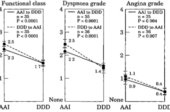
Functional class Dyspnoea grade Angina grade
AAI to DDD n = 35 P < 0.0001 DDDtoAAI - n = 35 P < 0 0001 ±2.5 2 -, 3 i < 1 None — 2 5 ! ^ 22 — AAI to DDD P = 0.0001 DDDtoAAI n = 36 P < 0.0001 AAI to DDD n = 3 5 P = 0 004 DDDtoAAI n = 36 P < 0.007 AAI DDD AAI None DDD AAI DDD
Figure 4 Overall modification of main symptoms according
to the NYHA classification of HOCM comparing the activated (DDD) to the non-activated (AAI) period of the patient.
improvement of symptoms, but when switching the pacemaker to the active mode during a procedure blinded for the patient and the investigating physician (crossover), all completed their second 12-week arm of the study. The majority of the 30 reported adverse events were related to pacemaker treatment: infection and local irritation (n = 8) or electrode displacements (7 atrial and 3 ventricular). Despite these problems, correct pace-maker function was finally achieved in all patients. No other minor event was related to pacing; one patient was operated on during crossover phase for failure of therapy.
Discussion
This study documents that pacing is beneficial in patients with hypertrophic obstructive cardiomyopathy and a resting gradient of more than 30 mmHg with
l -p p p - l **
n
n
***
n
***
*** ***
*
n
Alertness Self Strenuous Self- Chest Dyspnoea autonomy activity perceived pain
health
Figure 5 Comparison of grading quality of life and
cardiovascular symptomatology parameters between the activated (DDD) and non-activated (AAI) period of the study. Note in all parameters a significant preference for the paced mode (* = 0 0 5 , ** 0 0 1 , *** 0001).
symptoms refractory to conventional medical treatment. Although we were able to document a placebo effect of implanting a pacemaker and not activating it, the sub-sequent effect of switching it on in a blinded manner significantly improved symptoms, quality of life, and exercise tolerance in those who are severely limited. The reverse, inactivating the pacemaker, is even more im-pressive as return of symptoms is more strongly felt by the patients.
The haemodynamic benefit of dual chamber short atrioventricular delay pacing has been documented in many previous studies127'28-301 and these results have been confirmed. As we investigated patients with and without an acute gradient reduction (Groups A and B) and found no difference between them, the relationship between maximal gradient and severity of symptoms must be held in doubt. It would seem that pressure gradient reduction by pacing is not the only factor which explains the efficacy of pacing in hypertrophic obstructive cardiomyopathy. This is in agreement with earlier analysis of this disease where symptoms could not be related to obstruction'3'1.
The experimental evidence of modification of contractile behaviour'321, of improved capillary coronary flow'331 and remodelling of the ventricle'341 with ectopic stimulation may, in part, explain our results. Reduction of wall-stress, of mitral regurgitation and prolongation of systolic ejection time all contribute to the benefit of pacing, although not analysed in detail in this report. As hypertrophic obstructive cardiomyopathy causes important diastolic dysfunction1351, pacing might also influence this. However, only detailed subgroup analysis of our echocardiographic findings will elucidate this aspect. Currently only the segmental wall motion changes are an evident feature'361. The population investigated in this study was considered primarily to be candidates for surgery. However, they were not extremely symptomatic as no Class IV patients were included. It can therefore be concluded that pacing relieves symptoms in most Class II and III patients who are already on maximal tolerated drug therapy. As pacing allows an increase in beta-blocking and calcium-blocking therapy without the risk of extreme brady-cardia, a further benefit of pacing to be considered might be by further optimization of drug treatment; however, the opposite may also be true. Symptomatic relief induced by cardiac pacing may allow patients to be cared for without pharmacological support. Some of the drugs used to treat hypertrophic obstructive cardiomy-opathy, and in particular at dosages frequently admin-istered, may be badly tolerated. Patients may suffer side effects of the drugs rather than symptoms of the cardiac disease itself. Further investigations should look at this possibility.
The effect of pacing depends on full ventricular capture together with appropriately timed left atrial contribution to left ventricular filling'271. Achievement of an atrioventricular delay which both optimizes atrio-ventricular synchrony and provides complete ven-tricular capture in some hypertrophic obstructive
cardiomyopathy patients can be difficult and one might consider atrioventricular nodal ablation. In fact, further improvement in pacemaker benefit may be achieved by atrioventricular node ablation in patients with a short intrinsic atrioventricular delay'37381. One of the advan-tages of pacing is that it does not prevent use of other available treatments and is reversible as leads and pace-makers can be removed; this is in contrast to chemical ablation'391 where there is inevitably an irreversible infarcted area. Failure of pacing still allows surgery to be considered, and three of the study patients finally underwent this intervention. Although our patient population seems slightly less symptomatic than recently published surgical series'40'4'1, the results compare favourably with these non-randomized retrospective analyses. However, pacemaker treatment is not without complications and the high incidence reported in this series is at variance with generally accepted stan-dards. The relatively high level of complications may reflect this, being the first prospective multi-centre study investigating pacemaker treatment, or that the hypercontractile ventricles in hypertrophic obstruc-tive cardiomyopathy hearts are more likely to be associ-ated with electrode displacements. The one fatality reported was due to right ventricular perforation, a recognised but rare complication of pacing that was diagnosed too late and therefore led to cardiac tamponade.
Limitations of the study
Hypertrophic obstructive cardiomyopathy is character-ized by symptoms and objective parameters that are variable and, with the exception of the gradient, difficult to assess accurately. This study only investigated a subgroup of hypertrophic cardiomyopathy patients which comprises approximately 5-10% of all patients with this disease'42', but might nevertheless contribute to judgement. Although symptomatic, many patients showed quite good exercise tolerance despite their V02rnax being reduced but further analysis on these
investigations will follow. Quality of life assessments with a standardized questionnaire seemed the appropri-ate technique to judge objectively subjective criteria, as it has been validated for other diseases, such as cancer'43'. The study design was double-blind, but when program-ming a pacemaker, looking at an echocardiogram and at an ECG during exercise testing complete blinding of the medical personnel might be incomplete. The quality of life questionnaire was filled out by nurses who were not informed of the pacing mode in operation; thus this aspect of the study was also double blind. At this point the study cannot answer questions about recurrent syncope, arrhythmias or sudden death but the patients will be followed and more information will be reported with a follow-up period long enough to address these topics as well as echocardiographic substudies.
Conclusions
We have shown that dual-chamber pacing is a useful complement to drug treatment in symptomatic hyper-trophic obstructive cardiomyopathy. It significantly reduces outflow obstruction in all and improved symptoms in 84% of patients while increasing exercise tolerance by 21% in those severely limited.
The study design allowed the device and invasive procedure to be tested blindly for the first time. Short atrioventricular delay (below 100 ms) pacing, therefore, can be recommended in patients corresponding to our selection criteria. However, further critical investigations are needed to identify the major underlying mechanisms of the beneficial effect of pacing in order to best define the patient who will have the greatest benefit from this new treatment.
We thank Dr R. Sutton and I. Bourgeois for assistance in the preparation of this manuscript, G Werlen for secretariat work and typing and H. Dols for statistical and graphic advice.
The following centres and investigators have collaborated in this study: Stockholm (Sweden): L. Ryden, F. Gadler, C. Linde;
Lausanne (Switzerland): L. Kappenberger, X. Jeanrenaud, N.
Aebischer; Rennes (France): C. Daubert, P. Mabo, D. Gras;
London (U.K.): W. McKenna, A. Slade; Dresden (Germany): E.
Meisel; Nancy (France): E. Allot, N. Sadoul; Warsaw (Poland): L. Chojnowska, L. Malecka; Pans (France): L. Guize, T. Lavergne;
Dublin (Ireland). B. Maurer; Liege (Belgium): H. Kulbertus, V.
Mahaux; London (U.K.): R. Sutton; Bad Rothenfeld (Germany): W. Kranich.
The study was supported by grants from the Swiss national foundation for scientific research N° 32—37275-93, the Swedish heart and lung foundation and the Bakken research center, Medtronic, Maastricht, NL and the European working group of Cardiac pacing, European society of cardiology (ESC).
References
[1] Teare D. Asymmetrical hypertrophy of the heart in young adults. Br Heart J 1958; 20: 1-8.
[2] Maron BJ, Bonow RO, Cannon RO, Leon MB, Epstein SE. Hypertrophic cardiomyopathy: interrelations of clinical mani-festations, pathophysiology and therapy. N Engl J Med 1987; 316: 780-9,844-52.
[3] McKenna WJ, Cann AJ. Sudden death in hypertrophic car-diomyopathy. Assessment of patients at high risk. Circulation
1989; 80: 1489-92.
[4] Hejtmancik JF, Brink PA, Towbin J et al. Localisation of gene for familial hypertrophic cardiomyopathy to chromosome 14q 1 in a diverse US population. Circulation 1991; 83: 1592-7. [5] Solomon SD, Gelsterfer-Lowrance AAT, Vosberg HP et al. A
locus for familial hypertrophic cardiomyopathies closely linked to the cardiac myosin heavy genes CR ± L436, and CR ± L329 on chromosome 14 at ql 1-912. Am J Hum Genet
1990; 47: 389-94.
[6] Solomon SC, Jarcho JA, McKenna WJ et al. Familial hyper-trophic cardiomyopathy is a genetically heterogeneous dis-ease. J Clin Invest 1990; 86: 993-9.
[7] Wigle ED, Rakowsky H, Kimball BP, Williams WG. Hyper-trophic cardiomyopathy, clinical spectrum and treatment. Circulation 1995; 92: 1680-92.
[8] Wigle ED. Hypertrophic cardiomyopathy. Mod Cone Cardio-vasc Dis 1988; 57: 1-6.
[9] Nienaber CA, Gambhir SS, Mody FV et al. Regional myo-cardial blood flow and glucose utilisation in symptomatic patients with hypertrophic cardiomyopathy. Circulation 1993; 87: 1580-90.
[10] Maron BJ. Wolfson JK, Epstein SE, Roberts WC. Intramural 'small vessel' coronary artery disease in hypertrophic cardio-myopathy. J Am Coll Cardiol 1986; 8: 545-57.
[11] Cannon RJ III. Rosing DR, Maron BJ et al. Myocardial ischemia in patients with hypertrophic cardiomyopathy: Con-tribution of inadequate vasodilator reserve and elevated left ventricular filling pressures. Circulation 1985: 71: 234-43. [12] Frenneaux MP. Counihan PJ, Caforio ALP, Chimakori T.
McKenna WJ. Abnormal blood pressure response during exercise in hypertrophic cardiomyopathy. Circulation 1990: 82: 1995-2002.
[13] Wigle ED, Sasson Z, Henderson MA et al. Hypertrophic cardiomyopathy: The importance of the site and extent of hypertrophy: a review. Prog Cardiovasc Dis 1985: 28: 1-83. [14] Wigle ED, Adelmann AG, Auger P, Marquis Y. Mitral
regurgitation in hypertrophic subaortic stenosis. Am J Cardiol 1969; 24: 698-706.
[15] Klues HG. Dollar AL, ProScan MA. Echocardiographic assessment of mitral valve size in obstructive hyper-trophic cardiomyopathy: anatomic validation for mitral valve specimens Circulation 1993; 88: 548-55.
[16] Jebara VA, Mihaileanu SA, Acar C. Left ventricular outflow obstruction after mitral valve repair: results of the sliding leaflet technique. Circulation 1993; 88-2: 3 0 ^
[17] Lee KS, Stewart WJ, Lever HM, Underwood PL. Cosgrove DM. Mechanism of outflow tract obstruction causing failed mitral valve repair: anterior displacement of leaflet coaptation. Circulation 1993; 88-2: 24-9.
[18] Yoganathan AP, Lemmon JD, Kim YH, Levine RA, Vesier CC. A three dimensional computational investigation of intraventricular fluid dynamics: examination into the initiation of systolic anterior motion of the mitral valve leaflets. J Biomech Eng 1995; 117: 94-102.
[19] Kuhn H, Loogen F. Die Anwendung von Beta-Rezeptorenblockern bei hypertropher obstruktiver Kardio-myopathe. Internist 1978; 19: 527-31.
[20] Hopf R, Kaltenbach M. 10-year results and survival of patients hypertrophic cardiomyopathy treated with calcium-antagonists. Z Kardiol 1987; 76 (Suppl 3): 137^4.
[21] Kappenberger L, Jeanrenaud X. Le traitement de la myo-cardiopathie obstructive par stimulation cardiaque (Abstr). Arch Mai Coeur 1991; 84: 293.
[22] Sherrid M, Delia E, Dwyer ED. Oral disopyramid therapy for obstructive hypertrophic cardiomyopathy. Am J Cardiol 1988; 62: 1085-8.
[23] Gilgenkrantz JM, Cherrier F, Petitier H, Dodinot B. Cardio-myopathie obstructive du ventricule gauche avec bloc auriculo-ventriculaire complet. Arch Mai Coeur 1968; 60: 439-53.
[24] Hassenstein P, Wolter HH. Therapeutische Beherrschung einer bedrohlichen Situation bei der idiopathischen hyper-trophischen Subaortenstenose. Verh Dtsch Ges Kreisl 1967; 33: 242-6.
[25] Rothlin M, Moccetti T. Beeinflussung der muskularen Sub-aortenstenose durch intraventrikulare Reizausbreitung. Verh Dtsch Ges Kreisl 1967; 37: 411-5.
[26] McDonald K, McWilliams E, O'Keeffe B, Maurer B. Functional assessment of patients treated with permanent dual-chamber pacing as a primary treatment for hypertrophic cardiomyopathy. Eur Heart J 1988; 9: 893-8.
[27] Jeanrenaud X. Goy JJ, Kappenberger L. Effects of dual-chamber pacing in hypertrophic obstructive cardiomyopathy. Lancet 1992:339: 1318-23.
[28] Fananapazir L, Cannon RO, Tripodi D, Panza JA. Impact of dual-chamber pacing in patients with hypertrophic cardio-myopathy with symptoms refractory to verapamil and /?-adrenergic blocker therapy. Circulation 1992; 85: 2149-61. [29] Linde-Edelstam C, Nordlander R, Unden AL, Orth-Gomer
K, Ryden L. Quality of life in patients treated with atrio-ventricular synchronous pacing compared to rate modulated ventricular pacing: a long-term, double blind, crossover study. PACE 1992; 15: 1467-76.
[30] Duck HJ, Hutschenreiter W, Paneau H, Trenckmann H. Vorhofsynchrone Ventrikelstimulation mit Verkiirzter AV Verzogerungzeit als Therapieprinzip der hypertrophischen obstruktiven Kardiomyopathie. Z Gesamte Inn Med 1984; 39: 437^17.
[31] Braunwald E, Lambrew CT, Rockoff SD, Foss J, Morrow AG. Idiopathic hypertrophic subaortic stenosis. A description of the disease based upon an analysis of 64 patients. Circu-lation 1964; 29:30 (Suppl IV): 3-213.
[32] Prinzen FW, Augustijn CH, Arts T, Allessie MA, Reneman RS. Redistribution of myocardial fiber strain and blood flow by asynchronous activation. Am J Physiol 1990; 259: H300-H308.
[33] Amitzur G, Manor D, Pressman A et al. Modulation of the arterial coronary blood flow by asynchronous activation with ventricular pacing. PACE 1995; 18-1: 697-710.
[34] Delhaas T, Arts T, Prinzen FW, Reneman RS. Relation between regional electrical activation time and subepicardial fiber strain in the canine left ventricle. Pfliigers Arch (Eur J Physiol) 1993; 423: 78-87.
[35] Nishimura RA, Hayes DL, Ilstrup DM, Holmes DR, Tajik AJ. Effect of dual-chamber pacing on systolic and diastolic function in patients with hypertrophic cardiomyopathy. J Am Coll Cardiol 1996, 27: 421-30.
[36] Jeanrenaud X. Left ventricular wall motion changes during excentric ventricular activation in HOCM. J Interven Cardiol 1996; 9: 327-33.
[37] Gras D, De Place C, Varin C, Leclercq C, Mabo P, Daubert C. Radiofrequency catheter ablation of AV junction to improve AV synchrony in obstructive hypertrophic cardio-myopathy treated by DDD pacing (Abstract). PACE 1994; 17: 745.
[38] Sadoul N, Dodinot B, Beurrier D, de Chillou C, Aliot E. AV-Node ablation for optimisation of pacemaker treat-ment in hypertropic obstructive cardiomyopathy. J Interven Cardiol 1996, 9: 347-53.
[39] Sigwart U. Non-surgical myocardial reduction for hyper-trophic obstructive cardiomyopathy. Lancet 1995; 346: 211-3. [40] Seiler C, Hess OM, Schoenbeck M et al. Long-term follow up
of medical versus surgical therapy for hypertrophic cardio-myopathy: a retrospective study. J Am Coll Cardiol 1991; 17-634-^2.
[41] McCully RB, Nishimura RA, Bailey K.R, Schaff HV, Danielson GK, Tajik AJ. Hypertrophic obstructive cardiomy-opathy: preoperative echocardiographic predictors of out-come after septal myectomy. J Am Coll Cardiol 1996; 27: 1491-6.
[42] Maron BJ. Appraisal of dual-chamber pacing therapy in hypertrophic cardiomyopathy: too soon for a rush to judgment? J Am Coll Cardiol 1996; 27: 431-2.
[43] Spitzer O, Dobson A, Hall J et al. Measuring the quality of life of cancer patients. J Chron Dis 1981; 34: 585-97.