Structure prediction of subtilisin BPN' mutants using molecular
dynamics methods
Andreas P.Heiner1, Herman J.C.Berendsen3 and
Wilfred F.van Gunsteren2
Laboratory of Physical Chemistry and BIOSON Institute, University of Groningen, The Netherlands
'Present address: VTT/Bio, PO Box 202, SF-02151 Espoo, Finland
2Present address: Laboratorium fur Physikalische Chemie, ETH-Zentrum,
CH-8092 Zurich, Switzerland
3To whom correspondeace should be addressed
In this paper we describe the achievements and pitfalls encountered in doing structure predictions of protein mutants using molecular dynamics simulation techniques in which properties of atoms are slowly changed as a function of time. Basically the method consists of a thermodynamic integration (slow growth) calculation used for free energy determination, but aimed at structure prediction; this allows for a fast determination of the mutant structure. We compared the calculated structure of the mutants Met222Ala, Met222Phe and Met222Gln of subtilisin BPN' with the respective X-ray structures and found good agreement between predicted and X-ray structure. The conformation of the residue subject to the mutation is relatively easy to predict and is mainly determined by packing criteria. When the side chain has polar groups its exact orientation may pose problems; long-range Coulomb interactions may generate a polarization feedback involving system relaxation times beyond the simulation time. Changes induced in the environment are harder to predict using this method. In particular, rearrangement of the hydration structure was difficult to predict correctly, probably because of the long relaxation times. In all conversions made the changes observed in the environment were found to be history-dependent and in particular the hydrogen bonding patterns provided evidence for metastable substates. In all cases the structure predicted was compared with available kinetic data and the reduced activity could be explained in terms of changes in the configuration of the active site.
Key words: computer simulation/mutant structure/molecular dynamics/serine protease/solvent structure/subtilisin
Introduction
One of the main objectives of current protein chemistry is the design of proteins with specific functional properties. This 'protein engineering' (Craik et al., 1985; Thomas et al., 1985) requires, amongst others, computational prediction of both the structure of mutated proteins and their functional properties on the basis of these structural data. The reliability of the first step can be tested independently of the second step if crystallographic data for the mutants are available. Once both steps are mastered, protein engineering in die true sense will become possible.
In this article we address the question of how the first step (i.e. prediction of the structure of the mutant enzyme) can be made using molecular dynamics (MD) simulations. [For recent © Oxford University Press
reviews on the methods and applications of MD we refer to van Gunsteren and Berendsen (1990a) or Karplus and Petsko (1990) and references therein.] The mutation can, in principle, be made abruptly. The conformation of the mutated residue may be modelled using a database of preferred side chain conformations (Janin et al., 1978; Ponder and Richards, 1987), using sequence homologies (Doolittle, 1981), structural data derived from the database of known crystal structures (Bernstein et al., 1977) or packing criteria (Ponder and Richards, 1987; Lee and Subbiah, 1991). To remove bad contacts and local strain, the mutation may be followed by some kind of regularization scheme such as energy minimization (EM) with a subsequent MD equilibration. Using such a protocol, the resulting configuration may well be trapped in a metastable substate, as the local solvent structure is not considered when the side chain conformation is modelled. A similar problem may be encountered when systematic search procedures are employed.
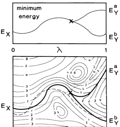
Another approach to obtaining the structure of a mutant enzyme originates from the free energy perturbation approach. In this procedure the system is described by a mixture of the potential functions of the wild type and mutant enzyme and by slowly changing the balance between the two potential functions the structure of the mutant enzyme is obtained from the structure of the wild type enzyme. This technique is known as the slow growth procedure (Berendsen et al., 1985) and is illustrated in Figure 1 for a process in which the final state Y has two distinct minimum energy conformations. Which of the two final conformations is obtained depends on the behaviour of the system near the branching point. The slower the balance between the two potential functions is changed, the higher the probability that the branch leading to the low-energy conformation (B-branch) is found, as the barrier between the A-branch and die B-branch is still small. When the system is changed more rapidly, it might get stuck in the high-energy state. This assumes that some of the information of the final state is present at the branching point, hence die steepest gradient with respect to X at uiis point is likely to be directed towards the padi leading to the B-branch. The rate of change of me system should be so slow that each intermediate state can be considered as a small perturbation of the equilibrated previous structure. In general uiis requirement is tested by comparing the free energy differences of die forward change (system X — system Y) and backward change (system Y — system X). When the difference is small in all intermediate states, the conversion is assumed to be made reversibly. Unfortunately, the determination of a free energy difference is computationally much more expensive, since the underlying distribution function has to be determined. In order to determine me free energy difference widiin 10% for the process sketched in Figure 1, the complete area within contours of 2 kT has to be sampled. A more accurate determination requires diat bodi branches have to be sampled (contours within, e.g. 4 kT). In the simple case of the hydration of medianol, sufficient sampling took at least 50 ps per intermediate state; die change itself could be made much faster (unpublished data). Thus, if one is able to convert the system from state X into state Y in a 'near 397
A.P.Heiiier et al.
Fig. 1. Minimum energy curve (top) and energy contour plot (bottom) of
the change of a hypothetical process as a function of the coupling parameter X. The y-axis of the contour plot represents the IN conjugate coordinates. Bold lines represent the local energy minimum of a cluster of configurations as a function of \ , thin lines are contours with energy 1 kT, 2 kT etc., relative to the absolute minimum energy at a given value of X. The final state Y has two stable minima EY' >EYb separated by an energy barrier > 4 kT. The cross denotes the branching point; for this value of X, the final conformation has been mixed into the system such that one distinct conformation (X-like system) splits into two distinct conformations (Y-like system). Near the branching point the energy barrier between the two distinct conformations is small. The broken and dotted lines represent two possible paths from the minimum energy conformation of the initial stage X into one of the local minimum energy conformation of the initial slate Y.
equilibrium' manner without reference to the free energy difference, the change may be made much faster.
A validation of the predicted mutant structure can be obtained by comparing the average structure with available X-ray data. Furthermore, structural changes of the mutant enzyme with respect to the wild type enzyme may be used to explain altered kinetic activity. When the mutant enzyme is changed back into the wild type enzyme, the average structures before mutation and after backward mutation may be compared, provided that the total change forth-and-back is carried out in a time exceeding the intrinsic relaxation. Estimates of this correlation time range from 0.06 ps (Hermans et al., 1992) to 3 ps (Berendsen, 1991). These correlation times are typical for arrangements within a local structure. There is much experimental evidence that the structural effects of point mutations in a protein are limited to such local structures (Bott et al., 1985; Wells, 1990). Hydrogen bonding patterns during the initial equilibration period and the equilibra-tion period after mutating forth-and-back may give clues whether the condition of a 'near equilibrium conversion' is met.
Another point that deserves some attention is the reliability of the force field and the computational procedure used. In a previous paper (Heiner et al., 1992) we addressed this by analysing an MD run of native subtilisin BPN' in a crystal unit cell (referred to as the crystal simulation) and found good agreement with X-ray data. The largest structural deviations were 398
Table I. Crystallographic data and kinetic constants of the wild type enzyme
subtilisin BPN' and three mutants at position Met222 Enzyme Crystallographjc data* Kinetic dateb
Met Ala Gin Phe Resolution (A) 1.8 2.0 2.0 2.0 fl-factor 0.144 0.159 0.147 0.146 I " - ) 50 40 8 3 6 KM (10~4 M) 1 4 7.3 8.6 5.6 (s/M) 3.6 x 5.0 x 9 0 x 6.4 x 105 104 103 103
"Crystallographic data for Met from Bon et al. (1988) and Ala, Phe and Gin from Boa and Ultsch (1986).
bActivity with respect to A'-succinyl-L-Ala-L-Ala-L-Pro-L-Phe-/>-nitroanilkie
(sAAPF-pna). Data for Met222 and Ala222 from Estell et al. (1985), for Gln222 from Boa et al. (1988) and for Phe222 from Bonneau et al. (1991).
found in the vicinity of Ca2+-ions. Since the ion binding sites
are rather distant from the active site, we do not expect that this will influence the results obtained here.
In this paper we will use the approach sketched above to predict the structure of a few mutants of subtilisin BPN' at the Met222 site. Upon oxidation of methionine the enzyme loses about 90% of its proteolytic activity (Stauffer and Etson, 1969). The enzyme can be made resistant to oxidation, but in general it loses activity in the process (Estell etai, 1985). The Genentech/Genencor group has made all 19 possible variants of subtilisin BPN' at this site and the highly refined X-ray structures of a number of these mutants are known (Bott, 1985; Bott et al., 1985, 1988). Kinetic and crystallographic data of mutants discussed are summarized in Table I. It should be noted that at the time of the calculations none of the mutant structures were available to us.
Method and computational procedure
As an initial molecular model we used the structure of native subtilisin BPN' at 1.8 A resolution with an ^-factor of 0.144 (Bott etal., 1988). A periodic unit cell with four protein molecules and solvent molecules as described in an earlier paper (Heiner et al., 1992) was used as a starting model. In order to reduce the computational effort, a sphere around the active site of one molecule was selected. All atoms belonging to charge groups whose centre of geometry was beyond 2.5 nm from this active site centre rc.
rc = 1/t(r(His64:Ne2) + r(Ser221:OJ) (1)
were removed from the system. Atoms belonging to charge groups of which the centre of geometry was in the outer 0.8 nm shell of the sphere were positionally restrained by a quadratic position restraining potential of which the force constant was given by = 0 rK < 1.7 nm 1 + sin rK ~ 1.9 2.1 - 1.7 1.7 nm < rK < 2.1 nm Ks(rK) = Ks(<x>) 2.1 nm < rK < 1.5 nm (2) with A"s(oo) = 9000 kJ/mol/nm2
and rK = | r, — rc |
This way a smooth change from unrestrained to strongly positionally restrained atomic motion is achieved.
1987) for hydrated systems was used. Non-bonded interactions were calculated using the twin range concept (Berendsen et al., 1986) with a short-range cut-off ^.shon = 0-8 nm and a
long-range cut-off R^ long = 1.2 nm. Both radii applied to the centres of geometry of charge groups to prevent the breaking of charge neutrality. The long-range Coulomb force was updated every 10 MD steps, as was the pair list used to calculate the short-range force. The spherical system was energy minimized for 1500 steps using the steepest descent method. Initial velocities were taken from a Maxwellian distribution at 298 K. The temperature was controlled by weak coupling of the system to a temperature bath (Berendsen et al., 1984) (7" = 298 K, T = 0.1 ps, separate scaling for solute and solvent atoms). A leap-frog variant of the multiple time-step formalism (Teleman and Jonsson, 1986) was used for solute atoms only, with five intermediate steps per time step for the bond-stretching force. Time steps of At = 2.0 ps and At = 1.0 ps were employed. The system was equilibrated for 25 ps [During the first 20 ps of the equilibration, the bond lengths were constrained using the SHAKE-procedure (Ryckaert et al., 1977; van Gunsteren and Berendsen, 1977)]. During the first 5 ps of the equilibration all atoms were positionally restrained to their initial positions by a quadratic restraining potential with a force constant K, = 9000 kJ/mol/nm2 that decreased linearly
in time. For the shell atoms, the maximum of the two force constants K, and k^r) was taken.
Conversion of the native protein (system X) into a mutant protein (system Y) is done by making the atomic interaction function V(r\-rN) a function of the so-called coupling parameter
X as well. The boundary conditions are given by K(X = 0) = Vx, the potential energy function of the native
protein and V(K = 1) = VY, the potential energy function of the
mutant enzyme. The path of conversion is determined by two quantities.
(i) The atomic map, i.e. the list of atoms whose properties are chosen to depend on the coupling parameter X.
(ii) The parameterization of the potential K(X).
Atoms of the atomic map that have to be eliminated (e.g. the methyl group Ce of Met222 in the conversion from native
enzyme to the Ala-mutant) are made dummy atoms (i.e. atoms with no van der Waals or Coulomb interaction and a mass of 1 a.m.u.) in the final state. Atomic masses were scaled linearly in X. Bonded interaction parameters involving one or more dummy atoms were kept at their values in the native system, but with zero force constant. When atoms had to be created (e.g. the Cj-atom in the conversion from the native protein into the Phe-mutant), dummy atoms were added to the methionine side chain such that a reasonable chemical structure (correct bond lengths, reasonable bond angles) of the mutant was generated, but without interactions with the environment. The conversion of native protein into the Gin-mutant allows for two atomic maps due to the asymmetric side chain of glutamine; both paths were used. A detailed description of the atomic map is given in Table U.
The parameterization of the hybrid potential V(\) was fixed during all conversions made and is given by (van Gunsteren,
1988, 1989).
V{rx . . . rN,\) = £ V(b,,\) + £ V(0,,\) + bonds / angles i
£ V&,D + £ v(^,\) + £ vinj.X) (3) improper dihedrals i pairs ij
dihedrals i V(bh\) = (b, - ((1 -
+
(0, ((1 -£/.X) = fc(0 «, - (d " v{rijt\) = (i -(4) - 6,x)) cos(/i,-Y 0,-where q, is a force field parameter (bond length, bond angle etc.) q, o the equilibrium value of qh and K* and tfY the force
constant of interaction <?,- in systems X and Y respectively. For dihedrals, n^ and n^ are the multiplicities of dihederal i in systems X and Y and 6* and 6Y( are the phase shifts of the
dihedrals in the respective systems.
Once the atomic map and the X-parameterization of the atomic interaction function are chosen, the time dependence of the coupling parameter X determines the rate of change of system X into system Y. To quantify the rate of change as a function of X is difficult, due to the complexity of the hybrid potential [equations (3) and (4)]. Thus, a linear time dependency is the most unbiased choice. However, it has been argued that a change in the van der Waals radius is the predominant factor in the system change. In order to keep the rate of change of the system constant in such a case, an nth power [n = 5 (van Gunsteren, 1990b), n = 6 (Postma, 1985), n = 11 (Mazor and Pettitt, 1991)] time dependence for X has been suggested.
The quality of the predicted structure is assessed by comparing the average structure of the mutant protein with the mutant X-ray structure (process X — Y). Next the mutant protein is equilibrated for some time (usually 10 ps) before it is converted back into the native enzyme (process Y — X); average structures are determined. The latter allows for a test whether the changes were made in a reversible way (i.e. the substates along the path are 'near equilibrium'). The exact protocols are given in Tables II and IE and Figure 2.
Comparison of the structures was done by calculating the root mean square positional difference (r.m.s.PD) of residues closest to the methionine side chain, i.e. residues His64, His67, Tyr217, Ser221 - Ser224 and the backbone of residues Asn218 - Thr220 (Figure 3a). These residues partly shield the methionine from the solvent. Residues Met222 and Tyr217 are part of the S,' binding site [nomenclature of Schechter and Berger (1967) is used]. The r.m.s.PD was calculated using a translational/ rotational fit of the Ca-atoms onto the reference structure; the
fit applied to all residues whose Ca-atom was within 1.5 nm of
the active site centre [equation (1)] at the start of the equilibration period.
The root mean square positional fluctuation (r.m.s.PF) was calculated with respect to the average structure; no fit procedures were applied.
The definition of a hydrogen bond is in accordance with Kohler et al. (1987); when the acceptor-hydrogen distance is less than 0.25 nm and the donor-hydrogen-acceptor angle is larger than 135°, a hydrogen bond is observed. The donor is the first atom mentioned.
A.P.Heiner et al.
Table D. Atomic map of the three conversions of Met222 into one of its mutants
Met Gin, Phe
Mutant X-state Y-state Atom
X-state Atom Y-state Ala Phe" Met Phe Ala Met Glnlb Gin Met Gln2b Gin Met Ht22 Dummy Dummy Dummy Dummy Dummy C, Dummy Dummy Dummy Dummy C« Dummy Dummy "Dummy atoms are inserted such that the phenyl group is coplanar.
bDummy atoms are inserted such that C^, Sj, C4 and dummy atoms are
coplanar.
A dummy atom has zero van der Waals/Coulomb interaction parameters and a mass of 1 a.m.u. Bonded interactions involving one or more dummy atoms have zero force constant.
Atomic map of the three mutants. V(k = 0) is defined as the X-state, H[X = 1) is defined as the Y-state. Bonds involving dummy atoms are of correct length, angles involving dummy atoms are distorted.
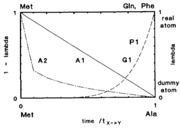
Table m . Perturbation protocol used in the five conversion simulations Mutant Code" M (fs) (PS) 'Y - x (ps) 'eq.X X-time-(ps) dependency Ala Phe Glnl Gln2 Al A2 PI Gl G2 1.0 2.0 2.0 2.0 2.0 40.0 10.0 12.0 10.0 10.0 25.0 10.0 10.0 10.0 -10.0 12.0 10 0 -10.0 10.0 10.0 -Lineai* Sixth roof Sixth power11 Sixth power1 Sixth power* "Code as used in article.
bAX linear over full conversion time (Figure 2, curve Al)
CAX linear over intervals of 1 ps; interval size determined from sixth root of
'/'x - Y> where / is the time since the conversion started (Figure 2, curve A2).
dAX determined from sixth power of f//x _ Y, where r is the time since the
conversion started (Figure 2, curve PI).
eAX linear over intervals of 1 ps; interval size determined from sixth power
of tltx _ Y, where / is the time since the conversion started (Figure 2, curve
Gl).
Results
Wild type enzyme Met222
Since the structural differences between the native enzyme and the mutant enzymes are expected to be small (Bott et al., 1986; Alber et al., 1987), it is of major importance to first analyse structural differences between the average native structure and the X-ray structure, induced by force field or methodology. Hereto the wild type enzyme was simulated for 10 ps following the equilibration period.
The r.m.s.PD of the average structure with respect to the
X-T3
Met
time / tx_> Y
Fig. 2. Time dependence of the coupling parameter X and the amount of real/dummy atoms as a function of time. The conversion time tx _ Y is in
Table in. For the Met — Ala conversion (marked Al and A2; left abscissa, 1 1 X) real atoms are slowly changed into dummy atoms as a function of time. For the Met — Phe/Gln conversions (marked PI and Gl; right abscissa X) dummy atoms are slowly changed into real atoms as a function of time.
ray structure of the backbone atoms near the mutant residue (Met222) is small (0.032 run), indicating a similar backbone folding (Figure 3b). The r.m.s.PD of the side chain atoms is considerably larger, 0.105 nm (Table TV). The major contribution (0.27 nm) comes from the Tyr217-phenol group, which shows at 44° rotation around the Cp-Cy bond. The phenol group is
pushed away by a solvent molecule in the S^ binding crevice and a hydrogen bond between Tyr217:O^ and a water molecule in a water channel (Bott et al., 1988; McPhalen and James, 1988) is formed. The water molecule in the binding crevice is not found in the X-ray structure, but existence of a solvent molecule in this area is well documented (Sielecki et al., 1979). The average X2-angle found in the simulation (100°) is in good agreement with values found in the database of preferred side chain conformations (Janin et al., 1978; Ponder and Richards, 1987). The X2"angle found in the X-ray structure (147°) represents a
high-energy conformation that may be selected due to thermal disorder of the phenol group (mean B-factor 0.13 nm2). Another
difference (r.m.s.PD 0.06 nm) between X-ray structure and average conformation concerns the xi-angle of the active site serine Ser221: - 5 9 ° in the simulated structure, and - 2 3 ° in the X-ray structure. The latter is a high-energy conformation that is scarcely found in the database of preferred side chain conformations. Finally, a minor difference for the Met222:x3 angle is observed: -119° in the X-ray structure compared to -156° in the average simulated structure. No preferred orienta-tion for this angle could be found. Together with the rotaorienta-tion of the Tyr217-phenol group these changes in structure cause an enlargement of the volume accessible to the P]' leaving group. While MD structures may deviate from the X-ray structure as a result of force field errors, the deviations of the dihedral angles near the active site may well be caused by the presence of the inhibitor phenylmethylsulphonylfluoride (PMSF) in the X-ray structure.
The hydrogen bonding patterns in the simulated structure are similar to those observed in the crystal simulation. The water molecule in the oxy anion hole and the hydrogen bonding network near the active site are well preserved.
Whether the system could be considered as equilibrated was tested by a consecutive simulation of 10 ps. The hydrogen
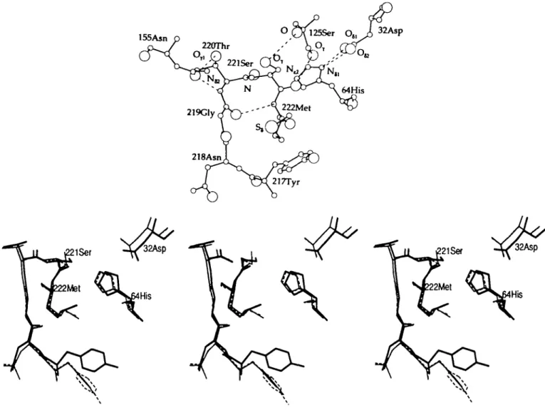
155Aan
Fig. 3(a). Environment of residue Met222 in native subtilisin BPN' in the X-ray structure. Residues and atoms discussed throughout the paper are labelled;
broken lines denote hydrogen bonds, (b) Environment of the residue Met222 in native subtilisin BPN' in the X-ray structure (bold lines) and the two lOps average structures (20-30 ps: thin-dashed lines; 30-40 ps: thin lines). The active site residues His64, Ser221 and the residue subject to mutation Met222 are labelled.
Table IV. Quality of the predicted structure of Met222 mutants after conversion into Ala, Phe or Gin
X-ray Met Ala(A2) Phe(Pl) Gln(Gl)
r m.s.PD (nm) Backbone Side chain r.m.s.PF (nm) Backbone Side chain Dihedral (°) Ser221:Xl Met222:xi Met222:x2 Met222:*3 Distance (nm) His64:Ne2-Ser221:O7 Asnl55:nH-Ser221:N Scr221:OT 0.032* 0 105" 23 75 73 119 0.380 0.470 0.472 0.729 0.01lb 0.024b 0.036 0.046 - 60(9) - 72(9) 70 (10) - 1 5 6 (22) 0.397 0.466 0.440 0.713 0.018 0.034 0.032 0.045 5(41) - 73 (10) 71 613) -167 (22) 0.384 0.324 0.428 0.628 0.022 0.097 0.030 0.035 - 61 (8) - 72 (13) 69 (10) -164(22) 0.364 0.603 0.507 0.673 0.016 0.047 0.036 0.046 62(11) 68(6) 64(10) -152 (31) 0.320 0.526 0.514 0.741 Ym.s.PD of the average structure from 20 to 30 ps with respect to the X-ray structure.
Vm.s.PD of the average structure from 20 to 30 ps with respect to the average structure of the next 10 ps.
Indicators of the quality of the predicted structures of the MetOYOMet mutants, i.e. after conversion of Met222 into Y222 into Met222. The r.m.s.PD is taken with respect to the average native structure from 20 to 30 ps. Averages over 10 ps.
A.P.Hdner et al.
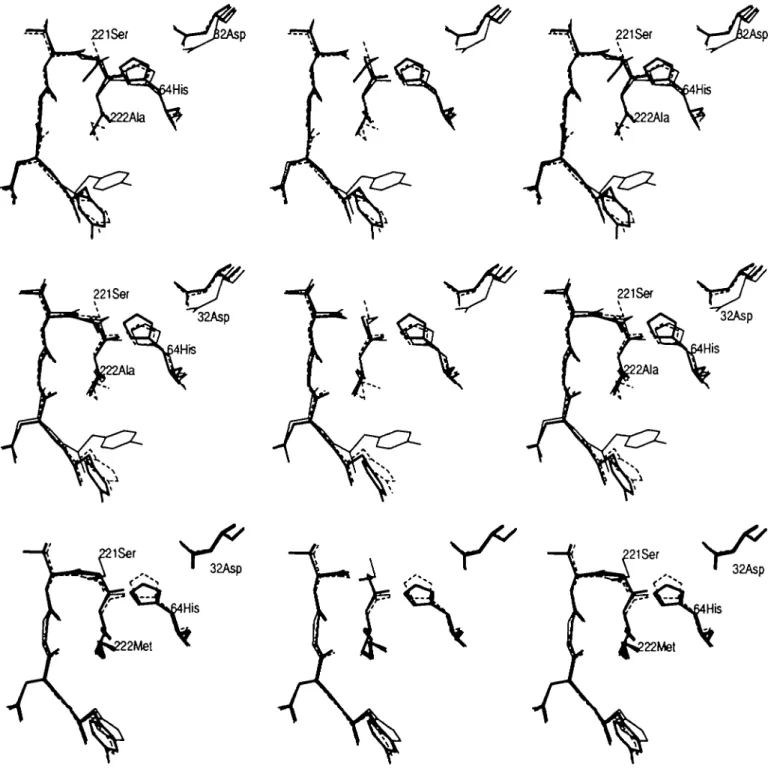
Fig. 4.(a). The average structure of the Ala-mutant using protocol Al is shown (bold lines, <Met(Al)Ala>). The average structure of the native enzyme (thin-dashed lines, < Met > ) and the X-ray structure of the Ala-mutant (thin lines) are shown as a reference The residues His64 and Ala222 as well as the hydroxyl group of Ser221 are labelled. Comparison of the solvent positions is not possible due to the diffusive behaviour of the molecules, (b) The average structure of the Ala-mutant (bold lines, < Met(A2)Ala>) using protocol A2. The average structure of the native enzyme (thin-dashed lines, <Met>) and the X-ray structure of the Ala-mutant (thin lines) are also shown. The residues His64 and Ala222 as well as the hydroxyl group of Ser221 are labelled, (c) The average Met-structure after mutating residue Met222 into Ala222 using protocol A2 (bold lines, <Met(A2)Ala(A2)Met>). In thin-dashed lines the average starting configuration <Met(A2)Ala> and in thin lines the average native structure <Met> are shown for comparison. The residues His64 and Met222 as well as the hydroxyl group of Ser221 are labelled.
bonding patterns and occupancies of die two simulations were almost identical; die r.m.s.PF of die two simulations of 10 ps were of similar magnitude (0.03 nm for 2 0 - 3 0 ps, 0.036 run for 3 0 - 4 0 ps for backbone atoms; 0.046 nm for 2 0 - 3 0 ps, 0.046 nm for 3 0 - 4 0 ps for side chain atom). The r.m.s.PD of die average structures wim respect to one anodier was low (0.011 nm for backbone atoms, 0.024 nm for side chain atoms). This indicates that die system may be considered equilibrated;
die respective values for die r.m.s.PD express die statistical variation between two consecutive 10 ps-averaged structures. Mutant enzyme Met222 — Ala222
The average structure of die Ala222-variant obtained using the first perturbation scheme < Met(A l)Ala > is almost identical to die average structure of die native enzyme (Figure 4a). [Average structures are denoted by ' < ' and ' > ' . The various mutants are
coded as follows: initial state—protocol of conversion—final state. Thus, <X(P1)Y(P2)X> denotes the average structure with residue Z at position 222. This structure is obtained by converting residue X222 into residue Y222 using protocol pi. After equilibration of the structure having residue Y222, the conversion from Y222 into Z222 is made using protocol P2. When P2 and PI are identical, the second part of the conversion protocol PI is to be taken (see also Table HI).] The r.m.s.PD of the backbone atoms is 0.028 nm and that of the side chain atoms is 0.055 nm, both with respect to the average native structure. The backbone deviations are evenly distributed over the molecule and are only marginally larger than expected from statistical fluctuations. The side-chain of Ser221 yields the largest contribution, 0.165 nm. This is caused by a 112° rotation of the xi-angte with respect to the average native conformation. The gauche"1"/gauche"
difference [the definition of gauche"/gauche"1" ( + 6 0 ° / - 6 0 ° ,
also g~/g+) is in agreement with Janin et al. (1978)] between
the wild type enzyme and the mutant enzyme is also observed in the X-ray structure, but the Ser221:xi is only 29° in the mutant (Table V).
The mechanism of the transition is sketched in Figure 5a. The nucleophilic hydroxyl group forms a hydrogen bond to a water molecule (SI) that is also hydrogen bonded to His64:Nt2. The
water molecule S2 is partly located in the oxy anion hole and also forms a hydrogen bond to the imidazole. Due to the thermal motion the water molecules move upwards and the hydrogen bond from Ser221:OT to SI is replaced by a hydrogen bond to S2
(Figure 5b). When solvent molecule S2 moves back to the original site, the xi-angle flips over (Figure 5C). In the native enzyme this process is also observed, but the gauche" conformation is less stable due to the proximity of the methionine side chain. During the conversion from Met into Ala the bonded force constants reduce, thereby increasing the flexibility of the methionine side chain. Also the van der Waals radii of the
29 52 (10) - 5 4 (14)
Table V. Quality of the predicted structure of the Met222 Ala mutants Angle (°) Ser221:Xl Distance (nm) H i ^ i N ^ - S e r 2 2 1 : 0 Asnl55:Nj2-Ser221:N Ser221:OT 0.380 0.487 0.474 0.716 0.409 0.454 0.492 0.636 0.311 0.430 0.356 0.600 Indicators of the quality of the predicted structure for the Met22Ala mutant using two different pathways. Averages over 10 ps.
methionine side chain atoms decrease (0.37 of the original values at the moment the transition occurs). This may render the gauche" conformation as stable as the gauche"1" conformation.
Due to the reorientation of the side chain the hydrogen bonding pattern is slightly different. In the wild type enzyme Ser221:O0
serves as a donor to Serl25:O; in the Ala-mutant this hydrogen bond is completely lacking.
Another difference between the crystal structures is a very mobile water (B-factor 0.38 nm2) in the Ala-mutant at the
position where the methyl group is located in the wild type structure. When the conversion has been completed the site is still unoccupied; it is only after 15 ps equilibration that a solvent molecule diffuses in from the solvent channel near Tyr216:O,. We tested whether the protein structure changed due to the reorganization of the solvent. The protein hydrogen bonding pattern occupancies of the first 10 ps (empty Met222:Ce-site)
and the next 10 ps (occupied Met222:Ce-site) were very much
the same. The same holds for the respective r.m.s.PF values (backbone atoms, 0.036/0.035; side chain atoms, 0.045/0.045) and the r.m.s.PD of the two average structures with respect to the average native structure (backbone atoms, 0.030/0.023; side chain atoms, 0.070/0.066).
To reduce the computational effort we performed the conversion from Met to Ala in 5 ps. Using a linear time dependency for X, the dihedral transition was not observed. Using a sixth-root time dependency (X oc r1/6), the dihedral transition
was observed, though at a larger X-value (0.95, compared to 0.15 in protocol Al). When the mutation was reversed, the gauche"1"
conformation was not restored; additional equilibration did not accomplish this either. In both cases the conformation of the methionine side chain was restored very well.
Increasing the time step to At = 2 fs while keeping the number of steps the same (protocol A2) should allow for a better equilibration of the system. However, the expected dihedral angle rotation did not occur, either during the conversion or during the equilibration (Figure 4b). Also, no solvent molecule diffused into the empty Met222:Ce-site. The r.m.s.PD of the average
structure < Met(A2)Ala > (backbone atoms 0.031 nm, side chain 0.054 nm) was similar to the r.m.s.PD of <Met(Al)Ala>, both with respect to the average native structure.
After converting the alanine side chain back into the methionine side chain, the conformation of the latter was restored almost perfectly (Table IV, Figure 4c). The r.m.s.PD of the average structure < Met(A2)Ala(A2)met > with respect to the average native conformation was small (backbone 0.018 nm, side chain
MUm—O. Itlhr —O.
81 \ llifcr —O ••' V -— Cf \ \ [Hf MHl »118« — l t U M / \ ; *
-Fig. 5.(a). The predominant side chain conformation of the active site serine Ser221 is gauche"1" . In this conformation the nucleophilic oxygen is hydrogen
bonded to Serl25:O and a water molecule SI. The latter is also hydrogen bonded to Hiso^iN^. S2 is, mutually exclusive, hydrogen bonded to the oxy anion hole and Hiso^iN^. (Thin solid line, covalent bond; dotted line, hydrogen bond.) (b) Due to thermal motion the solvent molecule S2 replaces SI in forming the hydrogen bond from Ser2U:OT to Hiso^iN^. (c) Solvent molecule S2 moves back to the original position. Due to the hydrogen bond Ser211.Oy-S2 the
dihedral Ser21 l:xi flips over to gauche". In the wild type enzyme the proximity of the methionine side chain makes this conformation less stable compared to the gauche"1" conformation. In the absence of the bulky side chain, the gauche" conformation may be as stable as the gauche"1" conformation sketched in Figure 5a.
A.P.Hdner et al.
E*+ S
s
AGd+AGb+AGa
Fig. 6. Rale equations of the enzyme assuming a two-site model for the
enzyme. The active form of the enzyme is the gauche"1" conformation for
Ser221:x|. The difference in catalytic activity is caused by a difference in population of the gauche"1" and gauche" for the Ser221:xi °f *"* t *0
enzymes (oc tx^—AG^/RT)). An additional energy difference AC, is introduced to allow different gauche"1"/gauche" ratios in the free and bound
enzyme Using this model, the binding affinity AGj, for sAAPF-pna for the wild type enzyme and the Ala-mutant is almost the same.
0.034 run); only the Ser221:xi-angle is for 50% of the equilibration period in the gauche"1" conformation, the other 50%
bring in the gauche" conformation.
The hydrogen bonding pattern in both the <Met(A2)Ala> and the < Met(A2)Ala(A2)Met > configurations are similar to the patterns found in the wild type enzyme; only the pattern near the active site is distorted. In particular, the nucleophilic oxygen is hydrogen bonded to HisMiN^ (69% occurrence) rather than to Serl25:O. The oxy anion hole is partly distorted due to a hydrogen bond from Asn 155:1^2 to Ser221:Or The hydrogen
bond from His67:Ne2 to Ile205:O is observed in the
configurations having an alanine side chain at position 222 only. The gauche" conformation of the active site serine is rather peculiar, since in almost all serine proteases the x r ^ g l e of this residue is gauche+. James et al. (1980) propose a two-site
model for the nucleophilic oxygen in the unbound enzyme. Using this hypothesis, the X-ray data yield a gauche+/gauche" ratio
of 3:1 for the wild type enzyme and a ratio of 1:3 for the Ala-mutant. Large B-factors for the hdyroxyl group in both crystal structures support the hypothesis. For Streptomyces griseus protease A (SGPA) a gauche"1"/gauche" ratio of 4 was found
(James et al., 1980). Data on porcine pancreatic elastase (Meyer et al., 1988) show large B-factors for the hydroxyl group, but not for the C^-atom; this also indicates two possible orientations for the active serine side chain. In the crystal simulation (Heiner et al., 1992) we found a gauche"1"/gauche" ratio of 6.
To test this two-site model against (the few) kinetic data available, we propose that only the gauche"1" conformation is
active; the gauche" conformation is not capable of forming a tetrahedral intermediate. This assumption is reasonable, since in almost all complexed enzymes a gauche"1" conformation is
observed. In the bound enzyme the water molecule S2 is replaced by the substrate and the mechanism for the g+/g" dihedral
transition is blocked. Hence, it is reasonable to assume an additional energy difference AGa in the gauche" complex
(Figure 6). The catalytic rate fc3 now becomes 51.6s"1, and the
model also implies a higher fraction gauche+ in the complex
than in the free enzyme (wild type, fraction g+:
~ 0.75, Pbound ~ 0.97; Ala-mutant, fraction g +^
~ 0.25, Abound ~ 0.77). Assuming steady state, the Kb for the
wild type enzyme is 1.1 x 10~4 M and for the Ala-mutant
1.8 x 10"4 M; the difference in binding energy of the two
enzymes is only 0.5 kT (— 1.2 kJ/mol). This is consistent with the experimental finding of Estell et al. (1986) that when the difference in volume of the Pi-residue and the Si-site is large,
Table VI. Quality of the predicted
Dihedral (°) Ser221:x, Phe222:Xl Phe222:x2 Distance (nm) His64:Nt2-Ser221:OT Asnl55 NH-Ser221:N Ser221:O7 His64:Nt2 structure of the X-ray - 7 3 - 3 9 - 6 9 0.329 0.481 0.504 0.727 Met222Phe mutants <Met(Pl)Phe> - 6 1 (8) - 5 4 ( 7 ) - 5 6 ( 1 1 ) 0.369 0.592 0.486 0.686 Indicators of the quality of the predicted structure of the Met222Phe mutants. Averages over 10 ps.
the binding between substrate and enzyme is strong (Estell et al., 1986; Graycar, 1991). The mobile solvent at the Met222:C{-site
may be the reason that in the Ala-mutant the g"-conformation is favoured over the g+-conformation.
Using protocol A2, the dihedral rotation was not observed. The water molecule S2 (Figure 5b) has moved from the oxy anion hole into the (partially) empty Met222:Cf-site before the
hydrogen bond to Ser221:OT is made. Using the Al or A2 lfs
protocol the solvent molecule S2 is closer to the imidazole than to the oxy anion hole. Apparently very minor structural details are important when the conversion is made fast.
The rotation of Ser221:xi during the equilibration of Met(A2)Ala(A2)Met is not according to the scheme sketched earlier (Figure 5a-c). Another mechanism could not be identified. During the equilibrium simulation of the final Met structure a hydrogen bond is observed from Ser221:O7 to
Hiso^iN^. Although this bond occurs in the complexed enzyme, its presence in the free enzyme is unlikely. The absence of a hydrogen bond from Ser221:O7 to Ser 125:0 indicates a
strained structure as well. In the wild type enzyme, the carbonyl of a nearby serine serves as a 'parking place' for the nucleophilic oxygen (Meyer et al., 1988; Geller etal., 1990, 1991). Mutant enzyme Met222 — Phe222
The r.m.s.PD of the average structure of the Phe222-mutant <Met(Pl)Phe> with respect to the average native structure is moderate (0.023 nm for backbone atoms, 0.039 nm for side chain atoms) considering the large group that is inserted (Figure 7a). The largest r.m.s.PD for the backbone atoms is found for the mutated residue (0.034 nm); they are pushed away from the centre of the Si'-pocket. The <p-\p angles of the peptide plane Ser221 -Phe222 differ by 15° with respect to the average native structure. The axes of the 3)0-helix and the central a-helix are
less aligned. The largest r.m.s.PD for the side chain atoms is observed for residue Tyr217 (0.089 nm); the X2-angte rotates
over —38° relative to the average native structure. The net effect of these structural changes is an enlargement of the Sj' pocket to accommodate the phenyl group. The conformation of the mutated residue Phe222 compares very well with the X-ray structure of this mutant (Table VI). The dihedral angles differ by 14° only, which is of the order of the fluctuations of these angles.
The methionine side chain conformation in the <Met(Pl)Phe(Pl)Met> structure is close to the conformation in the average native conformation; the average dihedral difference is only 2° (Table IV). The average structure <Met(Pl)Phe(Pl)Met> compares very well with the average native structure (Figure 7b; r.m.s.PD of backbone atoms 0.021 nm, r.m.s.PD of side chain atoms 0.042 nm). The
Fig. 7. (a) The average structure of the Phe mutant (bold lines, <Met(Pl)Phe>). The average structure of the native enzyme (thin-dashed lines, < M e t > )
and the X-ray structure of the Phe-mutant (think lines) are shown for comparison. The residues His64, Ser221 and Phe222 are labelled, (b) The average structure of the Met mutant (bold lines, <Mtt(Pl)Phe(Pl)Met>) generated from the Phe-mutant is shown. The average structure Phe mutant
(<Met(Pl)Phe>) is shown in thin-dashed lines and the average native structure < M e t > in thin lines. The residues His64, Ser221 and Met222 are labelled.
Tyr217-phenol group rotates back to the original position, but at the end of the Phe — Met conversion the rotation is not completed yet; this explains the difference of 13° for Tyr217:x2 in the average structure <Met(Pl)Phe(Pl)met> relative to the average native structure.
The hydrogen bonding pattern in the mutant structure is somewhat different from the pattern in the native conformation. In the wild type enzyme, Met222:MN or Ala223:N forms a hydrogen bond to Gly219:O in a ratio 1:1. In the mutant enzyme the ratio of the two hydrogen bonds is 10:3. This may be induced by the increased bend between /t310-helix and a-helix, which
makes the distance from Ala223:N to Gly219:O longer. In the wild type enzyme the oxy anion hole (Wells et al., 1985) is oriented by a hydrogen bond from Thr220:N to Asnl55:Oai, in
the mutant enzyme the oxy anion hole is weakly oriented by a hydrogen bond from Thr220:O7l to a solvent molecule that has
in turn a hydrogen bond to Asnl55:O41. When changing the
phenylalanine into methionine again, the fixating hydrogen bond is not recovered. Though the original relative orientation of the peptide planes of Ser221 and Met222 is recovered (Aa ~ 0, A\p ~ 0), the ratio of the hydrogen bond Met222:N - Gly219:Oto Ala223:N - Gly219:O is 8:3, which indicates a Phe-like structure for the calculated Met-enzyme.
The predicted structural changes in this mutant are, as far as the main chain is concerned, in agreement with the X-ray structure. The rotation of die Tyr217 side chain is not observed in the X-ray structure. Considering the compactness of the
Table VTI. Quality of the predicted structure of the Met222Gln mutants
X-ray <Met(Gl)Gln> Dihedral (°) Scr221:X, Gln222:xi Gln222:x2 CHn222:x3 Distance (ran) His64:Nt2-Ser221:Or Asnl55:Na2-Ser221:N His64:Nt2 51 79 65 130 0.327 0.454 0.443 0.700 -11 (11) -81 (8) 51 (13) -86 (14) 0.430 0.440 0.430 0.672 Indicators of the quality of the predicted structure of the Met222Gln mutants. Averages over 10 ps.
Si'-site in me mutant (B-factors of side chain of Tyr217 are 0.09 nm2 in the mutant, 0.12 nm2 in the wild type enzyme;
B-factors of side chain of Phe222 are 0.03 nm2) and the water
channel located on the opposite side of the phenol group (Bott et al., 1988; McPhalen and James, 1988), the change of the Tyr217 side chain conformation is plausible. The Phe-like hydrogen bonding patterns in the <Met(Pl)Phe(Pl)Met> structure (oxy anion hole fixation, hydrogen bonding ratio) is a neat demonstration of a situation in which the system relaxation time is considerably longer than the simulation period.
In a modelling study of this mutant (Bott, 1985), the shift of 405
A.P.Heiner et al.
32Asp 32Asp
F ^ . 8. (a). The average structure of the Gin-mutant (bold lines, <Met(Gl)Gln>), the average structure of the native enzyme (thin-dashed lines, < M e t > ) and the X-ray structure of the Gin-mutant (thin lines) are shown. The residues His64 and Ser221 are labelled, as well as the oxygen of the amide group Gln222:Ne2. (b) The average structure of the Met-mutam (bold lines, <Met(Gl)Gln(Gl)Met>) generated from the Gin-mutant is shown. The average structure of the Gin-mutant <Met(Gl)Gln> is shown in thin-dashed lines and the average native structure < M e t > is shown in thin lines. The residues His64, Ser221 and M«222 are labelled.
the backbone was not predicted and the modelled orientation of the phenyl plane was incorrect by 45°. Instead, a structural change of the active site was suggested because of the low specific activity of the mutant towards hydrolysis of sAAPF-pna. Recent studies revealed that the reduction of specific activity is due to the reduction of the space available to the P, '-leaving group (Bonneau et al., 1991). In the same study it was also found that for esters the catalytic rate is higher for die Phe-mutant enzyme than for the wild type enzyme. Since for this group of compounds deacylation is the rate-determining step, one may suggest that the observed angle between the 3io-helix and a-helix reduces the ability to transfer protons (Hoi et al., 1978; Hoi, 1985), thereby increasing the stability of the Michaelis complex over the tetrahedral intermediate in the deacylation step. Also, the distorted oxy anion hole may reduce the stability of the acylenzyme; both mechanisms may account for an increased overall catalytic rate. However, the catalytic rate for esters of the mutant enzyme is only twice as fast as for the wild type enzyme and the structural explanations given are based on minor differences. As shown by Dijkman (1989), relatively small changes in the active site geometry can have a considerable effect on the calculated activation and stabilization energies. Structural changes of the activated enzyme relative to the free enzyme were estabbshed experimentally as well (James et al., 1980). Hence, relating kinetic data to observed structural changes in the unbound enzyme is extremely hazardous; the best one can say is that the structural changes observed are not contradictory with the kinetic data available.
406
Mutant enzyme Met222 - Gln222
The average structure <Met(Gl)Gln> obtained using the Gl-protocol (Met222:Ce — Gln222:O£l, create Ne2, He2i, He22
from dummy atoms) is very much the same as the average native structure (Figure 8a; r.m.s.PD of the backbone atoms 0.025 nm, r.m.s.PD of the side chain atoms 0.038 nm). The largest difference is found for the active site serine; the xr&ngle has
rotated over - 2 8 ° (cf. Tables IV and VTT). The side chain dihedral angles of the glutamine agree well with the values derived from the X-ray structure, except for the x3-angle; a difference of -144° is observed (Table VII, Figure 8a). The path chosen may force the terminal amide group into a conformation with higher energy than one of its rotamers. To see whether a different path would give better agreement with the experimental structure, we used the alternative atomic map G2 (Meatt-.Ca - G l r ^ : ^ , create Oe{, Ha, H ^ from
dummy atoms). After 2 ps simulation the temperature had in-creased by a factor 3 and after 8 ps simulation the leap-frog algorithm did not converge any more. This indicates that using the G2 atomic map the Gln222 side chain amide dipole is oriented against the local electric field; steric hindrance prevents the amide dipole orienting parallel to the electric field.
Performing the backward conversion, the conformation of the methionine side chain in the <Met(Gl)Gln(Gl)Met> structure agrees well with the conformation in the native structure (Figure 8b, Table IV). The r.m.s.PD of both the backbone atoms and side chain atoms with respect to the average native structure is small (0.018 nm and 0.036 nm respectively). The major
difference is the Ser221:xi conformation that is gauche ; the dominant wild type conformation is gauche+ (Table IV).
Since glutamine is a polar residue, the hydrogen bonding pattern near the active site is expected to be somewhat different from the wild type enzyme. The side chain amide nitrogen is fixed by hydrogen bonds to Asn218:O and His64:Nj2 and replaces water molecule S2 (Figure 5a) in the wild type enzyme. This water molecule hops to the bottom of the St' binding site
where it becomes hydrogen bonded to Gln22:O{1 and His67:Nd.
The structure and fixation of the oxy anion hole remains intact. The nucleophilic hydroxyl group loses the hydrogen bond Serl25:O and forms one with AsnlSSiN^.
In the average structure <Met(Gl)Gln(Gl)Met> the wild type hydrogen bonding pattern is not restored; in addition, a hydrogen bond from Ser221:O7 to the imidazole group is observed.
Whether the predicted conformation of the glutamine side chain is correct is hard to judge. The orientation of the amide plane is off by 36°; the assignment of the atoms in the X-ray structure makes the difference increase to 144°, but the calculated orientation of the angle between the electric field due to the protein atoms and amide dipole is rather favourable (8°) in the MD structure.
The assignment of the respective positions in the X-ray structure is based on a minor (structural) shift of the imidazole group away from the glutamine side chain (Bott and Ultsch, 1986) and the reduction of the specific activity of the enzyme for hydrolysis of sAAPF-pna. In this model it is assumed that the electrostatic field of Gln222:Otl destabilizes the transition state.
This destabilization of die transition state will also occur when the dipolar field is reversed. Another explanation to account for die reduced activity of die Gin -enzyme is the formation of the hydrogen bond from Gln222:Ne2, which reduces the rotational
freedom of the imidazole. It has been argued that during the acylation step (rate-determining for hydrolysis of sAAPF-pna) the imidazole rotates to form the tetrahedral intermediate (Drenth et al., 1975; van Duynen et al., 1979). The motional freedom of the latter needed in die formation of the tetrahedral intermediate is restricted by the Gln222 side chain dipole field. A definite assignment may be obtained from, e.g. neutron diffraction data. The insertion of the polar side chain causes a massive solvent rearrangement; the water molecule bound to the oxy anion hole moves over several A, even during the equilibration period. This is probably due to die chosen functional form of X(t). During die last ps of the conversion die driving force for diis process (die Coulomb interaction) increases from 0.53 to 1.0. Another example of slow electrostatic equilibration is observed in die <Met(Gl)Gln(Gl)Met> structure; the hydrogen bond from Ser221:O7 to Hiso^N^ is typical for die Michaelis complex,
not for die free enzyme. The origin of diis hydrogen bond is probably diat die 0111222:1^ drags die imidazole towards die nucleophilic hydroxyl. When die glutamine side chain is converted into mediionine die hydrogen bond is transferred to Ser221:Or The absence of the hydrogen bond from Ser221.O7
to Ser 125:0 also indicates diat die <Met(Gl)Gln(Gl)Met> structure is a strained structure.
Conclusions
In diis study a modelling technique derived from die free energy perturbation mediodology is used to predict die structure of mutants of subtilisin BPN' at position Met222. The mediod is based on the slow growth procedure. The emphasis is on 'near equilibrium conversion' radier dian on sufficient sampling of die
conformational space along die padi of conversion. This allows for a relatively fast change of one mutant into die odier. Unfortunately die test for a 'near equilibrium conversion' is now much harder, since a check on a zero hysteresis of die free energy difference is made impossible. Instead, we used a combination of comparison of calculated structure widi the X-ray structure, hydrogen bonding patterns and changes in activity to assess die validity of the structural prediction.
The structural changes can be divided into two groups; structural changes of atoms for which interaction parameters change (first order changes) and structural changes induced in the environment (second order changes). The latter may range from tiny rotations due to van der Waals repulsion to diffusion of solvent molecules into sites left empty by first order changes. The first class of changes is relatively easy to predict; a simple substitution of die residue may already reveal die preferred conformation; die conformation is mainly determined by packing criteria. However, details of die conformation, as compared to die X-ray structure, may be better predicted by die slow growtii procedure. When changing die mutant residue back into die wild type mediionine, die conformation of the latter was correct in all cases, no matter how tense die conformation of nearby residues was.
Induced changes are much harder to predict using replacement techniques only. As was illustrated in the conversion of mediionine into phenylalanine, die conformational change of die tyrosine side chain was not anticipated in die modelling study, tiiough it is a realistic option. Also die gauche" conformation in die Ala-mutant was rather unexpected. The latter conversion also illustrates that a certain protocol may yield die correct mutant structure, but diat a successful prediction depends very much on die initial configuration. Only when me conversion is made very slowly, do we expect that diis configurational dependency of die predicted structure will vanish.
Whether or not the conversion was made 'near equilibrium' was tested by comparing the average hydrogen bonding pattern in die native simulation and die MetOYOMet simulation. This appeared to be a very sensitive tool: in almost all cases die pattern depended on die history of die simulation, whereas the r.m.s.PD did not reveal diis. This may be caused by die 'polarization feedback' of me Coulomb interaction. Also, since die functional form of X(r) was made optimal for die van der Waals interaction, die Coulomb interaction changed 50% during the last 2 ps of die conversion. A linear time dependency for diis interaction would be more optimal. Anodier strategy to reduce die dependency on the history is to reduce the overall interaction along die path of slow growth (Mark et al., 1991). In order to obtain reliable free energy differences (i.e. to sample correctly), die conversion should be made orders of magnitude slower.
Finally, die structural changes were compared widi die kinetic data available. In all cases the changes were compatible widi die data, almough we had to make a few assumptions to explain die activity of die Ala-mutant relative to die wild type enzyme. However, the kinetic activity is very much dependent on minor structural details, which makes it hazardous to rely on structure—activity relations too heavily.
Acknowledgements
The authors gratefully thank DrR.R.Bottof Genencorlnc. for providing us with the coordinates of native subtilisin BPN' as well as the mutants prior to publication and thank ton for helpful discussions and a reading of the manuscript. The financial support of Genencor Inc. is gratefully acknowledged.
A.P.Hetoer et al.
References
Alber.T., Doa-Pin,S., Wilson,K., WozniakJ.A., Cook.S.P. and Matthews.B W. (1980) Nature, 330, 4 1 - 4 1 .
Berendsen.H.J.C., PosanaJ.P.M., van Gunsteren,W F., DiNoia,A and HaakJ.R (1984)7. Chem. Phys., 81, 3684-3690.
Berendsen.HJ.C, Postma^.P.M. and van Gunsleren.W.F. (1985) In HermansJ. (ed.), Molecular Dynamics and Protein Structure. PolyCrystal Book Service, Western Springs, IL, USA, pp. 4 3 - 4 6 .
Berendsen.H.J.C., van Gunsteren.W.F., Zwinderman.H.R.J. and Geurtsen,R.G (1986) Ann. NY Acad. Sri., 482, 269-285.
Berendsen.H.J.C. (1991) In Renugopalakrishnan.V., Cary.P.R., Smhh.I C.P., Huang.S.G and Slorer.A.C. (eds), Proteins: Structure, Dynamics, and Design. Escom, Leiden, The Netherlands, pp 384-392.
Bemstein.F.C, Koetzle.T.F., Williams.G.J.B., Meyer.E.F., Bnce.M.D., RodgerJ.R., Kcrmard.O., Shimanouchi.T. andTasumi.M. (1977)7. Moi BioL, 112, 535-542.
Bonneau,P.R , Graycar,T.P., Estell.D.A. and Jones.J.B. (1991) 7. Am. Oiem.
Soc., 113, 1026-1030.
Bott.R.R. (1985) World Biotech. Rep.., 2, 5 1 - 5 9 .
Bott.R.R., Ultsch.M., WeUsJ., Powers.D., Brudick.D , Strubte.M., BumierJ., EsteU,D., Miller,J., Graycar.T., Adams.R. and Power.S. (1985) In LeBaron.H.M., Mummfl.R.O., Honeycott.R.C. and DuesingJ.H. (eds),
Biotechnology in Agricultural Chemistry. ACS Symposium Series 334,
Washington DC, USA, pp. 139-147.
Bott,R.R. and Ultsch.M. (1986) In Alacevic.M., Hranudi.D. and Toman.Z. (eds),
Proc. Fifth Int. Syrup. Genetics of Industrial Microorganisms, Zagreb, pp.
375-385.
Bott.R.R., Ultsch.M., Kossiakoff.A., Graycar,T., Kaiz.B. and Power.S (1988) 7. Biol. Chem., 263, 7895-7906.
Craik.C.S., Largman.C, Fletcher.T., Roczniak.S., Barr.P.J., Fletterick.R. and Rutter.W.J. (1985) Science, 228, 291 - 2 9 7 .
Dijkman.J.P. (1989) PhD Thesis, University of Groningen, The Netherlands. Doolittle.R.F. (1981) Science, 214, 149-159.
DrenthJ., Swen.H.M., Hoogenstraaten.W and Sluyterman.L.E.A.E. (1975)
Proc. Koninkt. Akad. Wetensch., C78, 104-110.
EsteU.D.A., Graycar.T.P. and Wells J . A. (1985) 7. Biol. Chem.. 262, 6518-6521.
EsteU.D.A., Graycar.T.P., Miller.J.V., Powers.D.B., Burnier.J.P., Ng.P.G. and Wells.J.A. (1986) Science, 233, 659-662.
Gefler.M., Swanson.S.M. and Meyer,E.F.,Jr (1990) 7. BiomoL Struct. Dynamics, 7, 1043-1052.
Gdler.M., Swanson.S.M. and Meyer.E.F. J r (1991) J. Am. Chem. Soc, 112, 8925-8931.
Graycar.T.P. (1991) In Dordick,J.S. (ed.) Biocatatysis in Industry. Plenum, New York, NY, pp. 237-283.
Heiner.A.P., Berendsen.H.J.C. and van Gunsteren.W.F. (1992) Proteins: Struct.
Funct. Genet., 14, 451-464.
Hermans,J., Yun.R.H. and Arjderson.A.G. (1992) 7. Comput. Chem., 13, 429-442.
Hol.W.G.J., Van Duynen.P.T. and Berendsen.H.J.C. (1978) Nature, 273, 443-446.
Hol.W.G.J. (1985) Prog. Biophys. MoL BioL, 45, 149-195.
James.M.N.G., Sielecki.A.R., Braycr.G.D., Delbaere.L.T.J. and Bauer,C.-A. (1990) 7. MoL Biol., 144, 4 3 - 8 8 .
JaninJ., Wodak.S., Levitt,M. and Maigret.B. (1978) 7. MoL Biol., 125,
357-386.
Karplus.M. and Petsko.G.A. (1990) Nature, 347, 631-637.
KohlerJ., SSnger.W. and van Gunsteren.W.F. (1987) Eur. Biophys. J., 15, 197-210.
Lee.C. and Subbiah.S. (1991) / MoL Biol., 217, 373-388.
Mark.A.E., van Gunsteren.W.F. and Berendsen.H.J.C. (1991)7. Chem. Phys., 94, 3808-3816.
Mazor.M. and Pettitt.B.M. (1991) MoL Sim., 6, 1-4.
McPhakn.C.A. and James,M.H.G. (1988) Biochemistry, 27, 6582-6598. Meyer.E.F., Cole.G.R., Radhatrishnan.R. and Epp.O. (1988) Aaa Crystallogr.,
B44, 26-38.
Ponder^.W. and Richards.F.M. (1987) J. MoL Biol., 193, 775-791. Postma,J.P.M. (1985) PhD Thesis, University of Groningen, The Netherlands. RyckaerU.-P., CiccotrJ.G. and Berendsen.H.J.C. (1977)7. Comput. Phys., 23,
327-341.
Schechter.I. and Berger.A. (1967) Biochem. Biophys. Res. Commun , 27, 157-162.
Sielecla.A R . Hendrickson.W.A . Borughton.C.G.. Delbaere.L.T.J.. Brayer.G.D. and James.M.N.G. (1979) 7 Mol. Biol., 134. 781 -804 Stauffer.C.E. and Etson.D. (1969) 7. Biol. Chem., 244, 5333-5338. Teleman.O. and J6nsson,B. (1986) 7. Comput. Chem., 7, 5 8 - 6 6 .
Thomas.P.G., Russle.A.J. and Fersht,A.R. (1985) Nature, 318, 375-376. van Duynen.P.Th., Thole,B.T. and Hol.W.G. (1979) 7. Biophys. Chem., 9,
273-280.
van Gunsteren.W.F. and Berendsen.H.J.C. (1977) Mol. Phys., 34, 1311-1327. van Gunsteren.W.F. and Berendsen.H.J.C. (1987) In van Gunsteren.W.F. and
Berendsen.H.J.C. (eds), Groningen Molecular Simulation (GROMOS) Library
Manual. Biomos, Nijenborgh 16, Groningen, The Netherlands,
van Gunsteren.W F. (1988) Protein Engng, 2, 5 - 1 3 .
van Gunsteren.W.F. (1989) In van Gunsteren.W.F. and Weiner.P (eds).
Computer Simulation of Biological Systems. Escom, Leiden, pp. 27-59.
van Gunsteren.W.F. and Berendsen.H.J.C. (1990a) Angew. Chem. Int. Ed. EngL, 29, 992-1023.
van Gunsteren.W.F. (1990b) In Troyanowsky.C. (ed.), Modelling of
Macromolecular Structures and Properties. Elsevier, Amsterdam.
WeUsJ.A., Cunningham.B.C, Graycar.T P. and Estell.D.A. (1985) Phil. Trans.
R. Soc. London A, 317, 415-423.
WeUsJ.A. (1990) Biochemistry, 29, 8509-8517.
Received on June 23, 1992; revised on October 27, 1992; accepted on January 30, 1993