© Crown copyright 2013.
A Comparative Study of Different In Vitro Lung Cell Culture Systems to
Assess the Most Beneficial Tool for Screening the Potential Adverse
Effects of Carbon Nanotubes
Martin J. D. Clift,
*
,1Carola Endes,
*
Dimitri Vanhecke,
*
Peter Wick,
†
Peter Gehr,
‡
Roel P. F. Schins,
§
Alke Petri-Fink,
*
,¶
and
Barbara Rothen-Rutishauser
*
,||
*Bio-Nanomaterials, Adolphe Merkle Institute, University of Fribourg, 1723 Fribourg, Switzerland; †Empa, Swiss Federal Laboratories for Material Science and Technology, Materials-Biology Interactions Laboratory, CH-9014 St. Gallen, Switzerland; ‡Institute of Anatomy, University of Bern, 3012 Bern,
Switzerland; §Institute for Umweltmedizinische Forschung (IUF), Heinrich-Heine-Universitat Düsseldorf, D-40225 Duesseldorf, Germany; ¶Department of Chemistry, University of Fribourg, 1700 Fribourg, Switzerland; and ||Respiratory Medicine, Bern University Hospital, 3010 Bern, Switzerland. 1To whom correspondence should be addressed at Bio-Nanomaterials, Adolphe Merkle Institute, University of Fribourg, Rte de l’Ancienne Papeterie,
1723 Marly 1, Fribourg, Switzerland. Fax: +0041 (0)26 300 9624. E-mail: [email protected].
Received June 24, 2013; accepted September 9, 2013
To determine the potential inhalatory risk posed by carbon nanotubes (CNTs), a tier-based approach beginning with an in vitro assessment must be adopted. The purpose of this study therefore was to compare 4 commonly used in vitro systems of the human lung (human blood monocyte-derived macrophages [MDM] and monocyte-derived dendritic cells [MDDC], 16HBE14o- epithelial cells, and a sophisticated triple cell co-culture model [TCC-C]) via assessment of the biological impact of different CNTs (single-walled CNTs [SWCNTs] and multi-walled CNTs [MWCNTs]) over 24 h. No significant cytotoxicity was observed with any of the cell types tested, although a signifi-cant (p < .05), dose-dependent increase in tumor necrosis factor (TNF)-α following SWCNT and MWCNT exposure at concen-trations up to 0.02 mg/ml to MDM, MDDC, and the TCC-C was found. The concentration of TNF-α released by the MDM and MDDC was significantly higher (p < .05) than the TCC-C. Significant increases (p < .05) in interleukin (IL)-8 were also found for both 16HBE14o- epithelial cells and the TCC-C after SWCNTs and MWCNTs exposure up to 0.02 mg/ml. The TCC-C, however, elicited a significantly (p < .05) higher IL-8 release than the epithelial cells. The oxidative potential of both SWCNTs and MWCNTs (0.005–0.02 mg/ml) measured by reduced glu-tathione (GSH) content showed a significant difference (p < .05) between each monoculture and the TCC-C. It was concluded that because only the co-culture system could assess each end-point adequately, that, in comparison with monoculture systems, multicellular systems that take into consideration important cell type-to-cell type interactions could be used as predictive in vitro screening tools for determining the potential deleterious effects associated with CNTs.
Key Words: in vitro lung systems; carbon nanotubes; nanotoxi-cology; oxidative stress; inflammation; risk assessment.
Carbon nanotubes (CNTs) have unique and novel
physico-chemical characteristics (
Donaldson et al., 2006
), which may
be extremely advantageous to a plethora of applications (eg,
medicine and sporting equipment) (
Robertson, 2004
). Despite
this, the potential use of CNTs in many of these
applica-tions has raised extreme concerns regarding their inevitable
human interaction (
Maynard, 2007
). Recently, understanding
the paradox posed by CNTs has gained increased emphasis
due to their potential correlation to “asbestos-like” effects
(
Donaldson et al., 2010
;
van Berlo et al., 2012
). Although
the length and stiffness of CNTs clearly play a central role
in CNT-associated biological effects (
Donaldson et al., 2010
;
Murphy et al., 2011, 2012
), many CNT samples are bundled/
entangled and it is these that are most likely to be used
(com-monly within a polymer matrix;
Robert et al., 2012
) in their
associated application(s) (
Robertson, 2004
). Therefore, in
these forms, CNTs could (potentially) be exposed to humans
during their life cycle (either through accidental or
occu-pational exposure) (
Donaldson et al., 2006
;
Nowack et al.,
2012
).
Exposure routes of CNTs to the human body potentially
consist of the epidermis, via the bloodstream and also through
the gastrointestinal tract (
Oberdörster et al., 2005
). Although
the extent of CNT exposure to humans via these routes is
debateable, especially both skin and bloodstream exposure,
inhalation exposure is widely accepted as being the primary
route of exposure for aerosolized nanofibers (
Mueller et al.
2011
;
Maynard et al., 2004
). Many studies have therefore
focused upon understanding how CNTs may interact with lung
cells (
Johnston et al., 2010
). Despite this, there is a current
lack of knowledge regarding the CNT-lung cell interaction.
This is due to inconsistencies in (1) the concentrations used
(
Oberdörster, 2010
), (2) the specific physicochemical
charac-teristics of the CNT samples (
Bouwmeester et al., 2011
), (3)
the dispersion state (
Bihari et al., 2008
) and the dispersant
used (
Gasser et al., 2012
;
Wick et al., 2007
), as well as (4) the
biological system and biochemical endpoint tests employed
(
Clift et al., 2011a
,
c
).
Although numerous in vivo studies have focused upon
determining CNT-associated effects upon the lung (
Johnston
et al.
, 2010
), in order to initiate the refinement, reduction and
replacement of invasive animal experimentation (
Hartung,
2010, 2011
) in vitro systems have been used to gain an insight
into the mechanistic effects of CNTs with lung cells (
Brown
et al.
, 2007
;
Rothen-Rutishauser et al., 2010
). The differences
between in vivo and in vitro systems are well documented
(
Han et al., 2012
). Yet, in order to understand NP-cell
interac-tions, as well as the potential risk posed by these materials,
it is essential to adopt a tiered approach, beginning with an
in vitro
analytical screening strategy (
Hartung, 2010, 2011
).
Commonly in vitro monoculture cell-based systems have
been chosen to study the nanofiber-cell interaction. With this
approach, however, there are both advantages and
disadvan-tages. Although primary monocultures can provide relative
heterogeneous populations, they are not readily available.
Cell lines negate such an issue, however, are limited as to their
phenotypic differentiation. Furthermore, monocultures do
not allow intercellular (cell type-to-cell type) interactions to
occur (
Rothen-Rutishauser et al., 2008
). Therefore, to bridge
the extensive divide between in vitro monoculture systems
and complex in vivo models, in vitro co-culture models, that
enable the important cell-to-cell interplay, pose a valuable
and realistic alternative (
Rothen-Rutishauser et al., 2008
).
Many in vitro co-culture systems have been highlighted
recently, representing reliable and realistic models to study
the NP-cell interaction (
Alfaro-Moreno et al., 2008
;
Bhabra
et al.
, 2009
;
Kasper et al., 2011
;
Rothen-Rutishauser et al.,
2005
). Evidence supporting the use of such in vitro systems
as efficient screening tools for assessing the potential risk of
nano-based materials however (ie, CNTs) remains equivocal
and limited.
The aim of this study, therefore, was to assess the
biologi-cal impact of CNTs, dispersed in different biologibiologi-cally relevant
dispersants, at perceived realistic concentrations, using a series
of reliable, efficient, and relative biochemical endpoint tests to
assess the applicability of monoculture and co-culture in vitro
systems in order to determine the most efficient in vitro tool to
screen, mechanistically, the potential adverse effects posed by
CNTs to human health.
MaTErIaLS aND METHoDS Chemicals and Reagents
All chemicals and reagents were purchased from Sigma-Aldrich (Switzerland), unless otherwise stated.
Cell Culture
Monocultures. Monocultures of human monocyte-derived macrophages (MDM) and monocyte-derived dendritic cells (MDDC) were isolated from human whole blood as previously described in Rothen-Rutishauser et al. (2005). In addition, the human bronchial epithelial cell line 16HBE14o- (a generous gift from Dr D. Gruenert [Cardiovascular Research Institute, University of California, San Francisco]) was used as previously described in Blank et al. (2007). Both MDM and MDDC monocultures were cultured at a density of 1 × 10⁵ cells/ml in Roswell Park Memorial Institute (RPMI) 1640 cell culture medium supplemented with 10% fetal calf serum (FCS), 1% L-glutamine (L-G), and 1% penicillin/streptomycin (P/S) for 7 days at 37°C, 5% CO2. To aid the maturation of the MDM and MDDC monocultures, the growth factors M-CSF (for MDM) and GM-CSF + interleukin (IL)-4 (for MDDC) were added to the culture medium for the maturation period. The 16HBE14o- epithelial cell mono-cultures were cultured at 0.5 × 10⁶ cells/ml in minimum essential media (MEM) supplemented with 10% FCS, 1% L-G, and 1% P/S for 7 days at 37°C, 5% CO2.
3D Triple Cell Co-culture Model of the Epithelial Airway Barrier. The 3D in vitro triple cell co-culture model (TCC-C) of the epithelial airway bar-rier, consisting of a layer of 16HBE14o- cells with MDM and MDDC present on the apical (upper section) and basolateral (lower section) sides, respectively, was cultured as previously described and characterized by Rothen-Rutishauser
et al. (2005, 2008). The cell culture densities of both MDM and MDDC, as well
as the 16HBE14o- were kept precisely the same as studied with the monocul-ture versions of each cell type; MDM and MDDC = 1 × 10⁵ cells/ml in RPMI 1640 supplemented with 10% FCS + 1% L-G + 1% P/S for 7 days at 37°C, 5% CO2; 16HBE14o- epithelial cells = 0.5 × 10⁶ cells/ml in MEM supplemented with 10% FCS + 1% L-G + 1% P/S for 7 days at 37°C, 5% CO2. Again, both MDM and MDDC cells were exposed to macrophage-colony stimulating factor (M-CSF) and granulocyte macrophage-colony stimulating factor (GM-CSF) + IL-4, respectively, in order to promote their maturation state following isolation from human whole blood. It is important to note that although there are over 40 types of cells present within the human alveolar region (Ochs and Weibel, 2008), the present co-culture system takes into consideration 3 important cell types of the alveolar epithelial barrier. Both macrophages, which are profes-sional phagocytes, and dendritic cells, which are essential antigen-presenting cells, are the 2 important immune cells present at the epithelial airway barrier (Nicod, 2007). The 16HBE14o- epithelial cells represent the epithelial layer that is important for the structure and function of the barrier system. All 3 of these cell types are important aspects of lung clearing mechanisms of xenobiot-ics (Rothen-Rutishauser et al., 2008).
Nanofibers and Positive Control Samples
Single-walled CNTs (SWCNTs) (Yangtze Nanotechnology, China) dis-persed in Tween 80 (0.04 mg/ml) and multiwalled CNTs (MWCNTs) (Cheap Tubes Inc.) dispersed in Pluronic F127 (160 ppm) were used as test samples in this study. The dispersion protocol for both SWCNTs and MWCNTs in both Pluronic F127 and Tween 80, respectively, is described in Wick et al. (2007)
and Thurnherr et al. (2009). Both crocidolite asbestos fibers (CAFs) (National Research Institute for Occupational Diseases, Johannesburg, South Africa) and standard diesel exhaust particles (DEPs) from the National Institute of Standards and Technology (SRM No. 2975) were also employed as positive fiber and (nano)particle controls, respectively. Furthermore, because it has been highlighted that the biological impact of CNTs can be attributed to the elemen-tal contaminants (ie, Fe) (Kagan et al., 2006), a SWCNT “pellet” (SWCNTs P) sample, previously described by Wick et al. (2007), was also assessed. Briefly, the term “pellet” defines all nonfibrous material of the SWCNT suspension (ie, trace elements). CAFs, DEPs, and SWCNTs P were used as reference materials for the biochemical testing strategy of this study only. All samples were exposed to each different cell culture system at a concentration of 0.005, 0.01, 0.02, 0.03, and 0.04 mg/ml for 24 h at 37°C, 5% CO2, unless otherwise stated. In addition, to control for the effects of the dispersants used for both SWCNTs and MWCNTs on the biochemical factors investigated because these have also been highlighted as potentially being the driving factor contribut-ing to the (adverse) biological effect of CNTs, both Pluronic F127 and Tween
80 were also assessed independently at concentrations of 50–20 000 ppm and 0.01–0.1 mg/ml, respectively.
Particle Characterization
The length, diameter, chemical content, and endotoxin levels were pre-viously assessed and are reported in Wick et al. (2007), Thurnherr et al. (2009), and Clift et al. (2011b). All key physicochemical characteristics of the SWCNTs and MWCNTs in addition to the SWCNTs P, DEPs, and CAFs used in the present study are summarized in Supplementary Table 1.
Assessment of the Nanofiber-Cell Interaction
Electron Tomography. To investigate the nanofiber-lung cell interaction, the TCC-C was exposed to SWCNTs, MWCNTs, and CAFs, as a positive con-trol, in suspension at a particle concentration of 0.03 mg/ml for 24 h at 37°C, 5% CO2. This particle concentration was used because it was found to be the optimal concentration allowing for the visualization of the nanofibers via elec-tron microscopy. Samples were then prepared for transmission elecelec-tron micros-copy (TEM) as previously described by Brandenberger et al. (2010). Briefly, ultrathin sections were formed through a series of dehydration steps and cut to 300 nm thick before being mounted onto 100 mesh copper grids. The samples were then screened for a suitable area for electron tomography using a CM12 TEM (FEI Co. Philips Electron Optics, Zurich, Switzerland). Electron tomog-raphy was performed as previously reported by Clift et al. (2011b). Briefly, tomography imaging was recorded on a selected area with a Tecnai F20 TEM (FEI, Eindhoven, The Netherlands) equipped with a GIF Tridiem energy filter and Ultrascan 1000 CCD camera (Gatan, Pleasanton, California). The tomo-gram was recorded at a magnification of ×34 000 while performing a continu-ous tilt angle shift from −70° to +70° with a dual tilt Fischione specimen holder (Fischione Instruments). To correct for the missing wedge (−90° to −70° and +90° to +70°), dual tilt axis acquisition was performed with an angle difference of 90°. Image processing and 3D stack reconstruction was performed with the Inspect 3D software V.3.0. (FEI Company). Further image processing and the reconstruction of the different (nano)fibers was performed using AMIRA 5.2.2 (Visage Imaging, Berlin, Germany) (Clift et al., 2011b).
Cytotoxicity
Lactate dehydrogenase release. The ability for CAFs, DEPs, SWCNTs, MWCNTs, and SWCNTs P, as well as Pluronic F127 and Tween 80 to cause lactate dehydrogenase (LDH) release from MDM, MDDC, and 16HBE14o- monocultures as well as the TCC-C (both upper and lower sections) was measured using the method previously described in Brown et al. (2001). As a positive control, 0.2% Triton X-100 diluted in PBS was used. It is important to note that although the LDH assay is principally considered the fundamen-tal test to determine the cytotoxicity of any nanosized material (Clift et al., 2011c), in order to gain an insight into the viability of the cells, further testing is necessary, such as the Annexin V assay via flow cytometry and/or confocal laser scanning microscopy. In the present study, however, the limited cytotoxic levels observed were supported by both conventional light and TEM, which eluded no signs of apoptosis or necrosis within the different cellular systems (Clift et al., 2013).
LDH adsorption. The potential for the LDH enzyme to adsorb to the surface of CAFs, DEPs, SWCNTs, MWCNTs, and SWCNTs P as well as Pluronic F127 and Tween 80 in the lysate of MDM, MDDC, and 16HBE14o- epithelial cell monocultures or the TCC-C, thus eliciting a false negative toxicity was determined by using the protocol previously described by Clift
et al. (2008). It is important to note that for the TCC-C, both the upper
and lower sections were combined in order to assess the adsorption patterns of the system as 1 entity. All data are presented within the Supplementary Information.
Cytokines and Chemokines
Tumor necrosis factor-α. In MDM and MDDC monocultures as well as
the TCC-C, the ability for CAFs, DEPs, SWCNTs, MWCNTs, and SWCNTs
P as well as Pluronic F127 and Tween 80 to elicit a release of the proinflam-matory cytokine tumor necrosis factor (TNF)-α was assessed via the use of an ELISA diagnostic kit (R&D Systems, Switzerland). As a positive control, lipopolysaccharide (LPS) at a concentration of 0.1 mg/ml was used. It is impor-tant to note that the 16HBE14o- epithelial cell monocultures were not tested for their TNF-α release because these exact cells do not readily elicit this pro-inflammatory cytokine, as recently shown by Lehmann et al. (2010).
Interleukin-8. In 16HBE14o- epithelial cell monocultures and the TCC-C, the ability for CAFs, DEPs, SWCNTs, MWCNTs, and SWCNTs P as well as Pluronic F127 and Tween 80 to stimulate the release of the (pro)inflamma-tory chemokine IL-8 was assessed via the use of an ELISA diagnostic kit (R&D Systems, Switzerland). As a positive control, TNF-α at a concentration of 0.1 mg/ ml was used for the 16HBE14o- epithelial cell monocultures, whereas LPS at a concentration of 0.1 mg/ml was used for the TCC-C. It is important to note that neither the MDM or the MDDC monocultures were assessed for their IL-8 release because it has previously been shown that these cell types do not readily produce this (pro)inflammatory chemokine (Lane et al., 2002; Müller et al. 2010).
Protein adsorption. The potential for the protein of either TNF-α or IL-8 to adsorb to the surface of CAFs, DEPs, SWCNTs, MWCNTs, and SWCNTs P as well as Pluronic F127 and Tween 80, thus eliciting a false negative toxicity, was also assessed. All nano-objects were initially incubated with the specific protein at 10 ng/ml (diluted in PBS) for TNF-α or IL-8, respectively, for 1 h at 37°C, 5% CO2. Samples were then centrifuged at 2000 rpm to remove all debris prior to being assessed using each specific ELISA test. All data are presented within the Supplementary Information.
Oxidative Stress
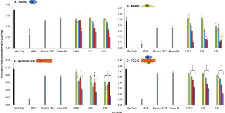
Reduced glutathione content. The intracellular reduced glutathione (GSH) content of MDM, MDDC, 16HBE14o-, and TCC-C after exposure to CAFs, DEPs, SWCNTs, MWCNTs, or SWCNTs P at 0.005, 0.01, and 0.02 mg/ml, as well as Pluronic F127 (160 ppm) and Tween 80 (0.04 mg/ml) was determined using a diagnostic kit (Cayman Chemical), as previously described in Steiner
et al. (2012). It was not possible to gain information pertaining to the oxidative
GSH component (GSSG); therefore, all GSH values are presented relative to the protein expression in the sample (GSH:protein [µM/mg]). tert-Butyl hydro-gen peroxide at a concentration of 0.04 mg/ml was used as a positive control. Statistical Analysis
All results are presented as the mean ± SEM. All data sets were observed to be normally distributed (data not shown). Statistical significance was deter-mined via a parametric 1-way ANOVA, followed by, when appropriate, a Tukey’s pairwise comparisons post hoc test (SPSS, IBM). The result was con-sidered significant if p ≤ .05.
rESuLTS Nanofiber-Cell Interaction
The lung-cell interactions of the MWCNTs and CAFs with the TCC-C have previously been reported in Clift et al. (2011b). Focusing upon the SWCNTs, these CNTs were specifically found within the MDM of the TCC-C after 24 h exposure at 0.03 mg/ml (Fig. 1). Furthermore, the SWCNTs were found to be fragmented and not present within a vesicular body (Fig. 1). Although no clear signs of frustrated phagocytosis were seen with the SWCNTs because their characteristics would not indicate such a biological response, the SWCNTs were, however, found to be protruding from the MDM after 24 h exposure (Fig. 1). For comparison, a negative control image of a MDM is provided in
Supplementary Figure 1. Cytotoxicity
No significant (p > .05) cytotoxicity was observed for the SWCNTs, MWCNTs, CAFs, or DEPs in any of the monoculture systems tested (MDM,
MDDC, 16HBE14o- epithelial cells) after 24 h exposure at concentrations rang-ing from 0.005 to 0.04 mg/ml (Supplementary Figure 2). Both the SWCNTs and MWCNTs were only noncytotoxic (p > .05) in the TCC-C up to 0.02 mg/ ml. A complete description of the cytotoxic nature of the panel of each CNT and standard samples with each different biological system tested is given in
Supplementary Figures 2 and 3.
(Pro)inflammatory Response
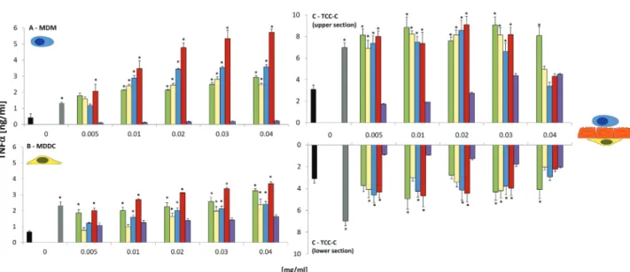
TNF-α release. A significant (p < .05) dose-dependent increase in TNF-α release was observed from MDM and MDDC following exposure to MWCNTs for 24 h at all concentrations tested (Fig. 2). A similar trend was also found for SWCNTs, CAFs, and DEPs in both MDM and MDDC monocultures at con-centrations 0.01–0.04 mg/ml (p < .05) (Fig. 2). No significant (p > .05) TNF-α release was noted from either MDM or MDDC monocultures after exposure to SWCNTs P (Fig. 2). The latter finding was associated with the increased cytotox-icity previously shown (Supplementary Figure 2). Significant adsorption patterns (p < .05) were found with all 5 samples at concentrations of 0.03 and 0.04 mg/ ml, suggesting false positive events recorded for both MDM and MDDC at these concentrations (Supplementary Figure 3). Similar results were also observed following exposure of SWCNTs, MWCNTs, CAFs, DEPs, and SWCNTs P to the TCC-C.
Following exposure to the TCC-C (upper section only), CAFS, DEPs, SWCNTs, and MWCNTs all elicited a significant dose-dependent increase in TNF-α release, at similar TNF-α concentrations produced by both MDM and MDDC monocultures, from baseline levels (medium only) to 0.03 mg/ml (Fig. 2). In the lower section of the TCC-C, only the SWCNTs and MWCNTs elicited a dose-dependent increase in TNF-α release up to 0.03 mg/ml. At 0.04 mg/ml, no significant biological effect (p > .05) was noted for either SWCNTs or MWCNTs (the TNF-α concentration for both CNTs was shown to decrease from 0.03 mg/ml) (Fig. 2). This was attributed to a significant adsorption (p < .05) of the TNF-α protein to the SWCNTs and MWCNTs at 0.04 mg/ml (Supplementary Figure 4). A similar effect was also observed at a concentration of 0.03 mg/ml (Supplementary Figure 4). DEPs and CAFs were both found to stimulate an intermittent significant TNF-α release from 0.005 to 0.04 mg/ml (Fig. 2). Significant adsorption patterns (p < .05) at both 0.03 and 0.04 mg/ml for both samples were found (Supplementary Figure 4), addi-tionally due to the variance observed within each sample concentration; these results were not considered significant. Similar to the MDM and MDDC monocultures, no effect was observed for the SWCNTs P at any concentra-tion tested (Fig. 2), although again at both 0.03 and 0.04 mg/ml, a significant (p < .05) adsorption was observed for SWCNTs P (Supplementary Figure 4).
IL-8 release. A significant dose-dependent increase in the release of the (pro)inflammatory chemokine IL-8 was observed for CAFs, DEPs, SWCNTs, and MWCNTs in 16HBE14o- monocultures up to 0.02 mg/ml after 24 h
suspension exposure (Fig. 3). At concentrations 0.03 and 0.04 mg/ml, similar adsorption patterns with these samples and the IL-8 protein were observed as for the TNF-α protein (Supplementary Figure 5). SWCNTs P showed a significant, dose-dependent increase up to 0.04 mg/ml although this was attrib-uted to the significant cytotoxicity elicited by this nonfibrous fraction of the SWCNT sample (Fig. 3). In the TCC-C, the IL-8 release caused by CAFs, DEPs, SWCNTs, and MWCNTs is significant (p < .05), 4-fold lower than that produced by the 16HBE14o- epithelial cells. In the upper and lower sec-tions of TCC-C, a significant dose-dependent increase (p < .05) was found for CAFs, DEPs, SWCNTs, and MWCNTs up to 0.04 mg/ml (Fig. 3), although these findings should have careful consideration due to the significant adsorption patterns (p < .05) for all 4 of these materials at 0.03 and 0.04 mg/ ml (Supplementary Figure 5). No significant effects were found following exposure of SWCNTs P to the TCC-C (in either the upper or lower sections) (Fig. 3).
Reduced GSH Content
Because a significant interference was observed at both 0.03 and 0.04 mg/ ml (Supplementary Figures 3 and 4), concentrations ranging from 0.005 to 0.02 mg/ml were used. Both MDM and MDDC monocultures only showed a significant loss (p < .05) in intracellular reduced GSH following exposure to MWCNTs at 0.01 and 0.02 mg/ml (Fig. 4). In the 16HBE14o- epithelial monocultures, the DEPs, SWCNTs, and MWCNTs, at all concentrations tested (0.005, 0.01, and 0.02 mg/ml), showed a significant loss (p < .05) in reduced GSH content (Fig. 4). CAFs also caused a significant loss in reduced GSH at a 0.02 mg/ml in 16HBE14o- cell monocultures (Fig. 4). Analysis following exposure of CAFs, DEPs, SWCNTs, and MWCNTs to the TCC-C showed a 10-fold, significant increase (p < .05) in the intracellular reduced GSH con-tent compared with each monoculture assessed (Fig. 4). Interestingly though, a similar trend for each sample was observed (Fig. 4), with a dose-dependent significant (p < .05) loss in reduced GSH content for DEPs, SWCNTs, and MWCNTs from 0.005 to 0.02 mg/ml after 24 h exposure (Fig. 4). Due to the heightened cytotoxicity of the SWCNT P (Supplementary Figure 1), a signifi-cant loss in the reduced GSH of MDM, MDDC, 16HBE14o- as well as the TCC-C was recorded.
DISCuSSIoN
The aim of this study was to compare 4 commonly used in
vitro
lung cell cultures to determine which may be most apt in
the hazard assessment of nanomaterials. The findings of this
comparison are summarized in
Table 1
.
FIG. 1. Electron tomography still images of the triple cell co-culture system (TCC-C) after exposure to single-walled carbon nanotubes (SWCNTs) after
submerged culture exposure at 0.03 mg/ml for 24 h at 37°C, 5% CO2. Image (A) is a still 2D image of SWCNTs present in the monocyte-derived macrophages of the TCC-C, whereas image (B) is the projected image (inset) of image (A). Image (C) shows representative tomogram slices of image (B), whereas image (D) shows the rendered 3D electron tomogram (SWCNTs are colored blue, whereas the cell membrane is yellow). Scale bar in image (A) represents 1 µm. Scale bar in images (B and C) represents 0.2 µm.
Observation that the SWCNTs, as well as the MWCNTs and
CAFs (
Clift et al., 2011b
) only interacted with the MDM of
the TCC-C suggests that the macrophages are performing their
primary function following a xenobiotic insult. It is important
to note, however, that although none of the (nano)fibers were
observed to be located within the epithelial layer or the MDDC
of the co-culture system, it does not discount the possibility
that they were present or interacting with these cell types of the
FIG. 2. Release of the proinflammatory cytokine tumor necrosis factor-α from (A) monocyte-derived macrophages (MDM), (B) monocyte-derived dendritic cells (MDDC), and (C) 3D triple cell co-culture model (TCC-C) of the epithelial airway barrier (upper and lower sections represented on the graph) following exposure to crocidolite asbestos fibers (CAFs), diesel exhaust particles (DEPs), single-walled carbon nanotubes (SWCNTs), multiwalled carbon nanotubes (MWCNTs), and single-walled carbon nanotube “pellet” (SWCNTs P) at 0.005, 0.01, 0.02, 0.03, and 0.04 mg/ml after 24 h at 37°C, 5% CO2 (n = 3). Data pre-sented are the mean ± SEM. Lipopolysaccharide at a concentration of 0.1 mg/ml was employed as a positive control (solid grey bar). Cell culture media only (Med Only) represents the negative control (solid black bar). *Relates to a significant difference from baseline (p < .05).FIG. 3. Release of the (pro)inflammatory chemokine interleukin-8 from (A) 16HBE14o- epithelial cells and (B) 3D triple cell co-culture model (TCC-C) of
the epithelial airway barrier (upper and lower sections represented on the graph) following exposure to crocidolite asbestos fibers (CAFs), diesel exhaust particles (DEPs), single-walled carbon nanotubes (SWCNTs), multiwalled carbon nanotubes (MWCNTs), and single-walled carbon nanotube “pellet” (SWCNTs P) at 0.005, 0.01, 0.02, 0.03, and 0.04 mg/ml after 24 h at 37°C, 5% CO2 (n = 3). Data presented are the mean ± SEM. Both tumor necrosis factor (16HBE14o- cells) (solid white bar) and lipopolysaccharide (TCC-C) (solid grey bar) at concentrations of 0.1 mg/ml were employed as positive controls. Cell culture media only (Med Only) represents the negative control (solid black bar). *Relates to a significant difference from baseline (p < .05).
TCC-C at this time point. Of interest is the observation that the
interaction of the SWCNTs and MWCNTs differed with the
MDM. SWCNTs showed 2 “bundles” within the cytosol of the
MDM, suggesting a possible, preferential entry route into the
MDM via a nonendocytotic mechanism. The MWCNTs,
how-ever, were found to be present within a vesicular body (
Clift
et al.
, 2011b
). Furthermore, the presence of proposed SWCNT
“fragments” in the cytosol of the cell suggests that there is a
possibility that they have undergone degradation by
intracel-lular enzymes (
Kagan et al., 2010
). This aspect requires further
research. Despite the differences in the interaction observed,
in the present study, the overall biochemical response was not
found to be significantly different between the 2 different CNT
types, suggesting a limited influence of the specific CNT-cell
“interaction” as regards their cellular effects.
Observation of the biochemical response of each in vitro
system following exposure to the SWCNTs and MWCNTs, in
addition to the CAFs and DEPs tested, found that in all in vitro
culture systems, no significant cytotoxicity (p > .05) occurred.
These findings support those previously reported when
com-paring the effects of cellulose nanowhiskers to MWCNTs at
concentrations up to 0.03 mg/ml using the same 4 in vitro
sys-tems (
Clift et al., 2011b
). It was further shown that despite the
SWCNTs and the MWCNTs being dispersed using different
surfactants (Tween 80 and Pluronic F127, respectively) that
no cytotoxic effect was evident in either MDM, MDDC,
16HBE14o- epithelial monocultures or the TCC-C, and
there-fore supporting that, depending on the concentration used,
sur-factants may be advantageous in obtaining a well-dispersed and
characterized CNT sample (
Wick et al., 2007
). The lack of any
cytotoxic response also supports
Thurnherr et al. (2009)
who
reported the same MWCNTs used in the present study to cause
no apoptosis or necrosis in the Jurkat A3 human leukemic T
cell line after 24 h exposure to 0.03 mg/ml.
The SWCNTs pellet sample showed a significant LDH
release over 24 h at the highest concentrations in all 4
differ-ent cell culture systems. These findings support those of
Kagan
et al.
(2006)
who reported that the catalyst metals used to
pro-duce CNTs (ie, Fe) are directly responsible for the adverse
cellular effects noted and that any biological effects observed
are not due to a fibrous effect (
Kagan et al., 2006
). Thus, the
present study highlights that if a correct and specific dispersion
method is used (
Wick et al., 2007
), then the proposed
“cyto-toxic” component of the CNT sample (ie, contaminant metals)
(
Kagan et al., 2006
) can be extracted and allow for a thorough
investigation of CNT effects upon normal cellular homeostasis
(ie, the effects noted for the SWCNTs can be attributed to their
fibrous characteristics).
FIG. 4. Reduced glutathione (GSH) content for (A) human blood monocyte-derived macrophages (MDM), (B) human blood monocyte-derived dendritic cells
(MDDC), (C) 16HBE14o- epithelial cells, and (D) 3D triple cell co-culture model (TCC-C) of the epithelial airway barrier (upper and lower sections represented on the graph) following exposure to crocidolite asbestos fibers (CAFs), diesel exhaust particles (DEPs), single-walled carbon nanotubes (SWCNTs), multiwalled carbon nanotubes (MWCNTs), and single-walled carbon nanotube “pellet” (SWCNTs P) at 0.005, 0.01, and 0.02 mg/ml after 24 h at 37°C, 5% CO2 (n = 3). Effects of Pluronic F127 (160 ppm) and Tween 80 (0.04 mg/ml) are also shown in graphs (A and D). Data presented are the mean ± SEM. tBHP, at a concentration of 0.04 mg/ml, was employed as a positive control. Cell culture media only (Med Only) represents the negative control. *Relates to a significant difference from baseline (p < .05).
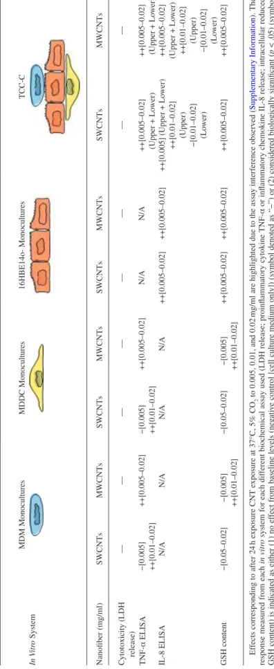
T
a
BLE
1
a
Summary of the Biological
r
esponse of Human Blood MDM, Human Blood MDDC, 16HBE14o- Epithelial Cells, and the 3D
TCC-C of the Epithelial
a
irway Barrier
(r
esponse Is Designated Between “
u
pper” and “Lo
wer” Sections Wher e a ppr opriate) a
fter Suspension Exposur
e to SWCNTs and MWCNTs In V itr o System MDM Monocultures MDDC Monocultures 16HBE14o- Monocultures TCC-C Nanofiber (mg/ml) SWCNTs MWCNTs SWCNTs MWCNTs SWCNTs MWCNTs SWCNTs MWCNTs Cytotoxicity (LDH release) — — — — — — — — TNF-α ELISA −[0.005] ++[0.01–0.02] ++[0.005–0.02] −[0.005] ++[0.01–0.02] ++[0.005–0.02] N/A N/A ++[0.005–0.02] (Upper + Lo wer) ++[0.005–0.02] (Upper + Lo wer) IL-8 ELISA N/A N/A N/A N/A ++[0.005–0.02] ++[0.005–0.02] ++[0.005] (Upper + Lo wer) ++[0.01–0.02] (Upper) −[0.01–0.02] (Lo wer) ++[0.005–0.02] (Upper + Lo wer) ++[0.01–0.02] (Upper) −[0.01–0.02] (Lo wer) GSH content −[0.05–0.02] −[0.005] ++[0.01–0.02] −[0.05–0.02] −[0.005] ++[0.01–0.02] ++[0.005–0.02] ++[0.005–0.02] ++[0.005–0.02] ++[0.005–0.02] Ef
fects corresponding to after 24
h e
xposure CNT e
xposure at 37°C, 5% CO
2
to 0.005, 0.01, and 0.02
mg/ml are highlighted due to the assay interference observ
ed (
Supplementary Information
). The
response measured from each
in vitr
o system for each dif
ferent biochemical assay used (LDH release; proinfl
ammatory c
ytokine
TNF-α
or infl
ammatory chemokine IL-8 release;
intracellular reduced
GSH content) is indicated as either (1)
no ef
fect from baseline le
vels (ne
gati
ve control [cell culture
medium only]) (symbol denoted
as “−”) or (2) considered biologically significant (
p < .05) (symbol
denoted as “++”).
The ef
fect at each concentration measured is gi
The findings that both SWCNTs and MWCNTs can cause a
(pro)inflammogenic response in vitro (TNF-α and IL-8 release)
support previous literature using cells representing the lung
in vitro
(
Brown et al., 2007
;
Donaldson et al., 2006
;
Johnston
et al.
, 2010
). It is important to note the cell-specific
proinflam-mogenic assessment performed in the present study. In regard to
the lack of TNF-α analysis performed on the 16HBE14o- cells,
as described in the Materials and Methods section, these cells,
as with epithelial cells in general, do not readily produce this
proinflammatory cytokine. This has previously been shown in
another comparison study of the TCCC and with its respective
monocultures following exposure to iron oxide hybrid
nanopar-ticles (
Lehmann et al. (2010)
. Similar results were also found
in another study by
Müller et al. (2010)
, where SWCNTs were
shown not to produce a detectable level of the proinflammatory
chemokine IL-8 from either MDM or MDDC.
The (pro)inflammogenic effects suggestive of mediation via
oxidative stress (loss in GSH) confirm that CNTs can be highly
reactive in vitro (
Rothen-Rutishauser et al., 2010
). Similar
find-ings were also found for both particle and fiber controls (DEPs
[found to be present within each cell type of the TCC-C;
Müller
et al.
, 2010
] and CAFs [causing frustrated phagocytosis in the
MDM of the TCC-C;
Clift et al., 2011b
]), further suggesting
that the biochemical response recorded is not dependent upon
the specific cellular “interaction.” Furthermore, the findings that
both SWCNTs and MWCNTs can elicit similar biochemical
reactions as both DEPs and CAFs, both human class 1
carcino-gens as recognized by the International Agency for Research
on Cancer, highlight the potential hazardous nature that CNTs
may pose toward human health (
Donaldson et al., 2010
).
In respect to which in vitro system is most apt for hazard
assessment of nanomaterials,
Müller et al. (2010)
showed
that the SWCNTs, as used in the present study, cause no
sig-nificant difference between mono- and co-cultures following
24 h exposure up to 0.03 mg/ml when assessing markers of
oxidative stress (eg, the fluorescent probe
2',7'-dichlorodihy-drofluorescein diacetate [H
2DCFDA]) and (pro)inflammatory
cytokine release (TNF-α and IL-8). Although
Müller et al.
(2010)
investigated A549 epithelial cells and not 16HBE14o-,
it is due to high variability shown by
Müller et al. (2010)
in all
data presented that no significant differences were observed.
A recent study by
Gasser et al. (2012)
, however, in which
dif-ferent surface charged MWCNTs, either coated with or
with-out lung surfactant (Curosurf), were assessed in regard to their
interaction with MDM and the TCC-C (using 16HBE14o-
cells), did report significant differences between mono- and
co-cultures. Interestingly, it was shown that the trend in the
decrease of the intracellular thiol GSH was a significant
100×-fold difference between each respective in vitro system (GSH
decrease = TCC-C 10× > MDM), evident for all the different
surface charged MWCNTs. It was also shown that the TNF-α
response between MDM and the TCC-C was 3× decreased in
the TCC-C than in the MDM (
Gasser et al., 2012
). The trends
shown by
Gasser et al. (2012)
, at least those of the GSH
analysis, are comparable with the present study. Although,
considering the findings of
Müller et al. (2010)
, as well as
those of
Lehmann et al. (2010)
and
Clift et al. (2011b)
, in
which iron oxide NPs and cellulose nanowhiskers,
respec-tively, were shown to cause significant differences in the
response (eg, proinflammatory) between mono- and
co-cul-tures, it is apparent that the biochemical endpoint and
expo-sure period tested are essential determinants regarding the
resultant effect between the different in vitro systems. Yet,
none of these studies categorically show that in vitro
co-cul-tures are better, or worse, than monoculco-cul-tures, and vice versa.
Although the advantages and disadvantages of both types of
in vitro
systems must be weighed (
Rothen-Rutishauser et al.,
2008
), this argument does not allow for a clear conclusion
to be met. Comparison with the in vivo response is therefore
essential to gain an understanding of which in vitro system
may elicit a similar effect. Although it is possible to compare
the CNT effects of the present study with the in vivo
litera-ture (eg,
Johnston et al., 2010
), this comparison is futile due
to the many, innate differences such as the characteristics of
the samples used and the exposure methods employed,
high-lighting a further important issue of specificity in
compar-ing the biological response of nanomaterials across different
systems. Therefore, a clear, defined, comparative in vitro
(mono- and co-cultures) versus in vivo study must be
con-ducted that considers these aspects in addition to many others
(eg, standardized concentrations [doses], exposure methods,
and times) in order to define which in vitro system is optimal
in assessing the (potential) hazard posed by nanomaterials
(eg, CNTs).
In conclusion, the findings presented from the current study
show that there are significant differences between the
bio-chemical responses monitored between mono- and co-culture
in vitro
systems that are used to mimic the human lung. It
is not possible to state from these findings alone that either
mono- or co-cultures are prevalent over the other in
determin-ing the (potential) hazard posed by nanomaterials. Although
it is possible to highlight that while monocultures suffice to
determine a simple live/dead assessment following
nanomate-rial exposure, multicellular systems additionally provide the
ability to determine the mechanistic, molecular pathology of
nanomaterials in vitro because they take into consideration the
important cell-to-cell interplay as occurs in vivo. Therefore,
by adopting the approach of using multicellular systems
instead of monocultures, it might be possible to truly
under-take an adequate in vitro study that may holistically assess the
(potential) risk of nanomaterials and that may be sufficient
enough to refine, reduce, and replace animal experimentation.
SuppLEMENTary DaTa
Supplementary data are available online at
http://toxsci.
FuNDING
The authors would like to acknowledge the support of the
European Respiratory Society, Fellowship LTRF-MC1572-2010
to M.J.D.C., as well as the Swiss National Science Foundation
(#3100A0_118420, 406440_131264/1), the German Research
Foundation (DFG SPP 1313), the Animal Free Research
Foundation, the Doerenkamp-Zbinden Foundation as well as
the Adolphe Merkle Foundation for their generous financial
support. The Dr Alfred Bretscher fund and the Microscopy
Imaging Center (University of Bern) are also acknowledged for
the use of the Tecnai F20 TEM.
aCkNoWLEDGMENTS
The authors would like to acknowledge the essential
labo-ratory technical assistance from Barbara Tschirren and Yuki
Umehara in regard to all cell culture, as well as both Mohammed
Ouanella and Andrea Stokes for the preparation of the samples
for electron microscopy. The authors would like to express no
conflicts of interest for the above study. The authors are entirely
responsible for the preparation of the manuscript as well as all
of the data contained within it.
rEFErENCES
Alfaro-Moreno, E., Nawrot, T. S., Vanaudenaerde, B. M., Hoylaerts, M. F., Vanoirbeek, J. A., Nemery, B., and Hoet, P. H. (2008). Co-cultures of mul-tiple cell types mimic pulmonary cell communication in response to urban PM10. Eur. Respir. J. 32, 1184–1194.
Bhabra, G., Sood, A., Fisher, B., Cartwright, L., Saunders, M., Evans, W. H., Surprenant, A., Lopez-Castejon, G., Mann, S., Davis, S. A., et al. (2009). Nanoparticles can cause DNA damage across a cellular barrier. Nat. Nanotechnol. 4, 876–883.
Bihari, P., Vippola, M., Schultes, S., Praetner, M., Khandoga, A. G., Reichel, C. A., Coester, C., Tuomi, T., Rehberg, M., and Krombach, F. (2008). Optimized dispersion of nanoparticles for biological in vitro and in vivo studies. Part. Fibre Toxicol. 5, 14.
Blank, F., Rothen-Rutishauser, B., and Gehr, P. (2007). Dendritic cells and macrophages form a transepithelial network against foreign particulate anti-gens. Am. J. Respir. Cell Mol. Biol. 36, 669–677.
Bouwmeester, H., Lynch, I., Marvin, H. J., Dawson, K. A., Berges, M., Braguer, D., Byrne, H. J., Casey, A., Chambers, G., Clift, M. J. D., et al. (2011). Minimal analytical characterization of engineered nanomaterials needed for hazard assessment in biological matrices. Nanotoxicology 5, 1–11.
Brandenberger, C., Clift, M. J. D., Vanhecke, D., Mühlfeld, C., Stone, V., Gehr, P., and Rothen-Rutishauser, B. (2010). Intracellular imaging of nano-particles: Is it an elemental mistake to believe what you see? Part. Fibre Toxicol. 7, 15.
Brown, D. M., Kinloch, I. A., Bangert, U., Windle, A. H., Walter, D. M., Walker, G. S., Scotchford, C. A., Donaldson, K., and Stone, V. (2007). An in vitro study of the potential of carbon nanotubes and nanofibres to induce inflam-matory mediators and frustrated phagocytosis. Carbon 45, 1743–1756. Brown, D. M., Wilson, M. R., MacNee, W., Stone, V., and Donaldson, K.
(2001). Size-dependent proinflammatory effects of ultrafine polystyrene
particles: a role for surface area and oxidative stress in the enhanced activity of ultrafines. Tox. and Appl. Pharm. 175, 191–199.
Clift, M. J. D., Blank, F., Gehr, P., and Rothen-Rutishauser, B. (2011a). Nanotoxicology: A brief overview, discussion of the current toxicological testing in vitro and suggestions for future research. In Systems Toxicology – A Handbook (S. C. Sahu and D. A. Casciano, Eds.), pp. 761–786. John Wiley & Sons Publishers, London, UK.
Clift, M. J. D., Foster, E. J., Vanhecke, D., Studer, D., Wick, P., Gehr, P., Rothen-Rutishauser, B., and Weder, C. (2011b). Investigating the interaction of cellulose nanofibers derived from cotton with a sophisticated 3D human lung cell coculture. Biomacromolecules 12, 3666–3673.
Clift, M. J. D., Frey, S., Endes, C., Hirsch, V., Kuhn, D. A., Johnston, B. D., Wick, P., Petri-Fink, A., and Rothen-Rutishauser, B. (2013). Assessing the impact of the physical properties of industrially produced carbon nano-tubes upon their interaction with human primary macrophages in vitro. Bionanomaterials. DOI 10.1515/bnm-2013-0013.
Clift, M. J. D., Gehr, P., and Rothen-Rutishauser, B. (2011c). In vitro testing for nanotoxicology: A valid alternative? Arch. Toxicol. 85, 723–731.
Clift, M. J. D., Rothen-Rutishauser, B., Brown, D. M., Duffin, R., Donaldson, K., Proudfoot, L., Guy, K., and Stone, V. (2008). The impact of different nanoparticle surface chemistry and size on uptake and toxicity in a murine macrophage cell line. Toxicol. Appl. Pharm. 232, 418–427.
Donaldson, K., Aitken, R., Tran, L., Stone, V., Duffin, R., Forrest, G., and Alexander, A. (2006). Carbon nanotubes: A review of their properties in relation to pulmonary toxicology and workplace safety. Toxicol. Sci. 92, 5–22.
Donaldson, K., Murphy, F. A., Duffin, R., and Poland, C. A. (2010). Asbestos, carbon nanotubes and the pleural mesothelium: A review of the hypothesis regarding the role of long fibre retention in the parietal pleura, inflammation and mesothelioma. Part. Fibre Toxicol. 7, 5.
Gasser, M., Wick, P., Clift, M. J., Blank, F., Diener, L., Yan, B., Gehr, P., Krug, H. F., and Rothen-Rutishauser, B. (2012). Pulmonary surfactant coating of multi-walled carbon nanotubes (MWCNTs) influences their oxidative and pro-inflammatory potential in vitro. Part. Fibre Toxicol. 9, 17.
Han, X., Corson, N., Wade-Mercer, P., Gelein, R., Jiang, J., Sahu, M., Biswas, P., Finkelstein, J. N., Elder, A., and Oberdörster, G. (2012). Assessing the relevance of in vitro studies in nanotoxicology by examining correlations between in vitro and in vivo data. Toxicology 297, 1–9.
Hartung, T. (2010). Lessons learned from alternative methods and their valida-tion for a new toxicology in the 21st century. J. Toxicol. Environ. Health B Crit. Rev. 13, 277–290.
Hartung, T. (2011). From alternative methods to a new toxicology. Eur. J. Pharm. Biopharm. 77, 338–349.
Johnston, H. J., Hutchison, G. R., Christensen, F. M., Peters, S., Hankin, S., Aschberger, K., and Stone, V. (2010). A critical review of the biological mechanisms underlying the in vivo and in vitro toxicity of carbon nanotubes: The contribution of physico-chemical characteristics. Nanotoxicology 4, 207–246.
Kagan, V. E., Konduru, N. V., Feng, W., Allen, B. L., Conroy, J., Volkov, Y., Vlasova, I. I., Belikova, N. A., Yanamala, N., Kapralov, A., et al. (2010). Carbon nanotubes degraded by neutrophil myeloperoxidase induce less pul-monary inflammation. Nat. Nanotechnol. 5, 354–359.
Kagan, V. E., Tyurina, Y. Y., Tyurin, V. A., Konduru, N. V., Potapovich, A. I., Osipov, A. N., Kisin, E. R., Schwegler-Berry, D., Mercer, R., Castranova, V., et al. (2006). Direct and indirect effects of single walled carbon nanotubes on RAW 264.7 macrophages: Role of iron. Toxicol. Lett. 165, 88–100. Kasper, J., Hermanns, M. I., Bantz, C., Maskos, M., Stauber, R., Pohl, C.,
Unger, R. E., and Kirkpatrick, J. C. (2011). Inflammatory and cytotoxic responses of an alveolar-capillary coculture model to silica nanoparticles: Comparison with conventional monocultures. Part. Fibre Toxicol. 8, 6.
Lane, B. R., Liu, J., Bock, P. J., Schols, D., Coffey, M. J., Strieter, R. M., Polverini, P. J., and Markovitz, D. M. (2002). Interleukin-8 and growth-regulated oncogene alpha mediate angiogenesis in Kaposi’s sarcoma. J. Virol. 76, 11570–11583. Lehmann, A. D., Parak, W. J., Zhang, F., Ali, Z., Röcker, C., Nienhaus, G.
U., Gehr, P., and Rothen-Rutishauser, B. (2010). Fluorescent-magnetic hybrid nanoparticles induce a dose-dependent increase in proinflammatory response in lung cells in vitro correlated with intracellular localization. Small
6, 753–762.
Maynard, A.D. (2007). Nanotechnology: the next big thing, or much ado about nothing? Ann. Occ Hyg. 51, 1–12.
Maynard, A. D., Baron, P. A., Foley, M., Shvedova, A. A., Kisin, E. R., and Castranova, V. (2004). Exposure to carbon nanotube material: Aerosol release during the handling of unrefined single-walled carbon nanotube material. J. Toxicol. Environ. Health A 67, 87–107.
Müller, L., Gasser, M., Raemy, D. O., Herzog, F., Brandenberger, C., Schmid, O., Gehr, P., Rothen-Rutishauser, B., and Clift, M. J. D. (2011). Realistic exposure methods for investigating the interaction of nanoparticles with the lung at the air-liquid interface in vitro. InSci. J. (Nanotech.) 1, 30–64. Müller, L., Riediker, M., Wick, P., Mohr, M., Gehr, P., and Rothen-Rutishauser,
B. (2010). Oxidative stress and inflammation response after nanoparti-cle exposure: Differences between human lung cell monocultures and an advanced three-dimensional model of the human epithelial airways. J. R. Soc. Interface 7(Suppl. 1), S27–S40.
Murphy, F. A., Poland, C. A., Duffin, R., Al-Jamal, K. T., Ali-Boucetta, H., Nunes, A., Byrne, F., Prina-Mello, A., Volkov, Y., Li, S., et al. (2011). Length-dependent retention of carbon nanotubes in the pleural space of mice initiates sustained inflammation and progressive fibrosis on the parietal pleura. Am. J. Path. 178, 2587–2600.
Murphy, F. A., Schinwald, A., Poland, C. A., and Donaldson, K. (2012). The mechanism of pleural inflammation by long carbon nanotubes: Interaction of long fibres with macrophages stimulates them to amplify pro-inflammatory responses in mesothelial cells. Part. Fibre Toxicol. 9, 8.
Nicod, L. P. (2007). Immunology of tuberculosis. Swiss Med. Wkly. 137, 357–362. Nowack, B., Brouwer, C., Geertsma, R. E., Heugens, E. H. W., Ross, B. L.,
Toufektsian, M.-C., Wijnhoven, S. W. P., and Aitken, R. J. (2012). Analysis of the occupational, consumer and environmental exposure to engineered nanomaterials used in 10 technology sectors. Nanotox. 7, 1152–1156. Oberdörster, G. (2010). Safety assessment for nanotechnology and
nanomedi-cine: concepts of nanotoxicology. J. Int. Med. 267, 89–105.
Oberdörster, G., Oberdörster, E., and Oberdörster, J. (2005). Nanotoxicology: An emerging discipline evolving from studies of ultrafine particles. Environ. Health Perspect. 113, 823–839.
Ochs, M., and Weibel, E. R. (2008). Functional design of the human lung for gas exchange. In Fishman’s Pulmonary Diseases and Disorders (A. P. Fishman, J. A. Elias, J. A. Fishman, M. A. Grippi, R. M. Senior, and A. I. Pack, Eds.), pp. 23–69. McGraw Hill, New York, NY.
Robert, C., Feller, J. F., and Castro, M. (2012). Sensing skin for strain monitor-ing made of PC-CNT conductive polymer nanocomposite sprayed layer by layer. Appl. Mater. Inter. 4, 3508–3516.
Robertson, J. (2004). Realistic applications of CNTs. Mater. Today 7, 46–52. Rothen-Rutishauser, B., Blank, F., Mühlfeld, C., and Gehr, P. (2008). In vitro
models of the human epithelial airway barrier to study the toxic potential of particulate matter. Expert Opin. Drug Metab. Toxicol. 4, 1075–1089. Rothen-Rutishauser, B., Brown, D. M., Piallier-Boyles, M., Kinloch, I. A.,
Windle, A. H., Gehr, P., and Stone, V. (2010). Relating the physicochemical characteristics and dispersion of multiwalled carbon nanotubes in different suspension media to their oxidative reacitivity in vitro and inflammation in vivo. Nanotox. 4, 331–342.
Rothen-Rutishauser, B. M., Kiama, S. G., and Gehr, P. (2005). A three-dimen-sional cellular model of the human respiratory tract to study the interaction with particles. Am. J. Respir. Cell Mol. Biol. 32, 281–289.
Steiner, S., Muller, L., Popovicheva, O. B., Raemy, D. O., Czerwinski, J., Comte, P., Mayer, A., Gehr, P., Rothen-Rutishauser, B., and Clift, M. J. D. (2012). Cerium dioxide nanoparticles can interfere with the associated cel-lular mechanistic response to diesel exhaust exposure. Toxicol. Lett.; 214, 218–225.
Thurnherr, T., Su, D. S., Diener, L., Weinberg, G., Manser, P., Pfaender, N., Arrigo, R., Schuster, M. E., Wick, P., and Krug, H. F. (2009). Comprehensive evaluation of in vitro toxicity of three large-scale produced carbon nanotubes on human Jurkat T cells and a comparison to crocidolite asbestos. Nanotox.
3, 319–338.
van Berlo, D., Clift, M. J. D., Albrecht, C., and Schins, R. P. (2012). Carbon nanotubes: An insight into the mechanisms of their potential genotoxicity. Swiss Med. Wkly. 142, w13698.
Wick, P., Manser, P., Limbach, L. K., Dettlaff-Weglikowska, U., Krumeich, F., Roth, S., Stark, W. J., and Bruinink, A. (2007). The degree and kind of agglomeration affect carbon nanotube cytotoxicity. Toxicol. Lett. 168, 121–131.