HAL Id: hal-02414326
https://hal.archives-ouvertes.fr/hal-02414326
Submitted on 4 Jan 2021
HAL is a multi-disciplinary open access
archive for the deposit and dissemination of
sci-entific research documents, whether they are
pub-lished or not. The documents may come from
teaching and research institutions in France or
abroad, or from public or private research centers.
L’archive ouverte pluridisciplinaire HAL, est
destinée au dépôt et à la diffusion de documents
scientifiques de niveau recherche, publiés ou non,
émanant des établissements d’enseignement et de
recherche français ou étrangers, des laboratoires
publics ou privés.
Arabidopsis thaliana alternative dehydrogenases: a
potential therapy for mitochondrial complex I
deficiency? Perspectives and pitfalls
Alessia Catania, Arcangela Iuso, Juliette Bouchereau, Laura Kremer, Marina
Paviolo, Caterina Terrile, Paule Bénit, Allan Rasmusson, Thomas
Schwarzmayr, Valeria Tiranti, et al.
To cite this version:
Alessia Catania, Arcangela Iuso, Juliette Bouchereau, Laura Kremer, Marina Paviolo, et al..
Arabidop-sis thaliana alternative dehydrogenases: a potential therapy for mitochondrial complex I deficiency?
Perspectives and pitfalls. Orphanet Journal of Rare Diseases, BioMed Central, 2019, 14 (1), pp.236.
�10.1186/s13023-019-1185-3�. �hal-02414326�
mitochondrial complex I deficiency? Perspectives and pitfalls
Article in Orphanet Journal of Rare Diseases · December 2019DOI: 10.1186/s13023-019-1185-3 CITATIONS 2 READS 94 14 authors, including:
Some of the authors of this publication are also working on these related projects:
mitochondria and neurodegenerative deseaseView project
MitochondriaView project Alessia Catania
12PUBLICATIONS 104CITATIONS
SEE PROFILE
Arcangela Iuso
Helmholtz Zentrum München 59PUBLICATIONS 1,551CITATIONS
SEE PROFILE
Juliette Bouchereau
Hôpital Universitaire Robert Debré 12PUBLICATIONS 88CITATIONS
SEE PROFILE
Laura S Kremer
Helmholtz Zentrum München 66PUBLICATIONS 1,144CITATIONS
SEE PROFILE
All content following this page was uploaded by Paule Bénit on 03 November 2019.
R E S E A R C H
Open Access
Arabidopsis thaliana alternative
dehydrogenases: a potential therapy for
mitochondrial complex I deficiency?
Perspectives and pitfalls
Alessia Catania
1,2†, Arcangela Iuso
3,4†, Juliette Bouchereau
1,5†, Laura S. Kremer
3,4, Marina Paviolo
1,5,
Caterina Terrile
3, Paule Bénit
1, Allan G. Rasmusson
6, Thomas Schwarzmayr
3,7, Valeria Tiranti
2, Pierre Rustin
1†,
Malgorzata Rak
1†, Holger Prokisch
3,4*†and Manuel Schiff
1,5†Abstract
Background: Complex I (CI or NADH:ubiquinone oxidoreductase) deficiency is the most frequent cause of mitochondrial respiratory chain defect. Successful attempts to rescue CI function by introducing an exogenous NADH dehydrogenase, such as the NDI1 from Saccharomyces cerevisiae (ScNDI1), have been reported although with drawbacks related to competition with CI. In contrast to ScNDI1, which is permanently active in yeast naturally devoid of CI, plant alternative NADH dehydrogenases (NDH-2) support the oxidation of NADH only when the CI is metabolically inactive and conceivably when the concentration of matrix NADH exceeds a certain threshold. We therefore explored the feasibility of CI rescue by NDH-2 from Arabidopsis thaliana (At) in human CI defective fibroblasts.
Results: We showed that, other than ScNDI1, two different NDH-2 (AtNDA2 and AtNDB4) targeted to the mitochondria were able to rescue CI deficiency and decrease oxidative stress as indicated by a normalization of SOD activity in human CI-defective fibroblasts. We further demonstrated that when expressed in human control fibroblasts, AtNDA2 shows an affinity for NADH oxidation similar to that of CI, thus competing with CI for the oxidation of NADH as opposed to our initial hypothesis. This competition reduced the amount of ATP produced per oxygen atom reduced to water by half in control cells.
Conclusions: In conclusion, despite their promising potential to rescue CI defects, due to a possible
competition with remaining CI activity, plant NDH-2 should be regarded with caution as potential therapeutic tools for human mitochondrial diseases.
Keywords: Mitochondria, Mitochondrial diseases, Complex I, Alternative dehydrogenases, Arabidopsis thaliana, AtNDA2 Introduction
Human NADH:ubiquinone oxidoreductase or complex I (CI) is the largest complex of the respiratory chain, with
a mass of 980 kDa and 44 different subunits encoded by
both mitochondrial and nuclear genomes [1].
CI catalyzes the consecutive transfer of two electrons, one per time, to a ubiquinone pool for each molecule of NADH oxidized. NADH oxidizing activity of CI is tightly controlling intra-mitochondrial metabolism, and electron transfer is coupled to both heat and ATP generation. Elec-tron transfer is associated with the pumping of 4H+ across the inner mitochondrial membrane, which sustains part of
the mitochondrial membrane potential [2]. The 44
sub-units are arranged in three functional modules: the N © The Author(s). 2019 Open Access This article is distributed under the terms of the Creative Commons Attribution 4.0
International License (http://creativecommons.org/licenses/by/4.0/), which permits unrestricted use, distribution, and reproduction in any medium, provided you give appropriate credit to the original author(s) and the source, provide a link to the Creative Commons license, and indicate if changes were made. The Creative Commons Public Domain Dedication waiver
(http://creativecommons.org/publicdomain/zero/1.0/) applies to the data made available in this article, unless otherwise stated.
* Correspondence:[email protected]
†Alessia Catania, Arcangela Iuso, Juliette Bouchereau, Pierre Rustin, Malgorzata Rak, Holger Prokisch and Manuel Schiff contributed equally to this work.
3
Institute of Human Genetics, Helmholtz Zentrum München, German Research Center for Environmental Health, Neuherberg, Germany 4Institute of Human Genetics, Technische Universität München, Munich, Germany
Full list of author information is available at the end of the article Catania et al. Orphanet Journal of Rare Diseases (2019) 14:236 https://doi.org/10.1186/s13023-019-1185-3
module involved in oxidizing NADH, the Q module involved in reducing ubiquinone, and the P module
dedi-cating proton translocation [3]. A number of mutations in
nuclear and in mitochondrial genes coding for many of the 44 subunits, as well as in genes coding for assembly or regulatory factors, have been shown to result in CI
defi-ciency [4]. Therefore, CI deficiency can result in a
com-bination of abnormalities: impaired oxidation of NADH to NAD+, which alters the NADH/NAD+ ratio and leads to intra-mitochondrial metabolic disequilibrium and ultim-ately to lactic accumulation, release of electrons that are not correctly channeled to the ubiquinone subsequently generating radical oxygen species (ROS), and loss of proton pumping activity, which reduces mitochondrial potential, hence lowering ATP synthesis.
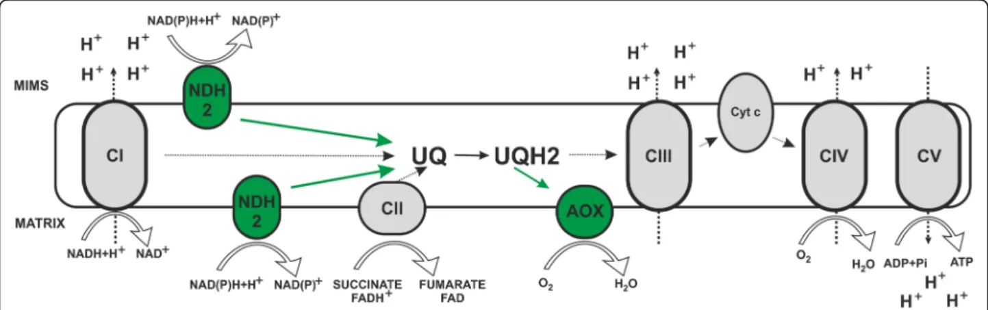
In microbes, fungi, plants, and also in some metazoan phyla (but not in arthropods or vertebrates), two key steps of the mitochondrial respiratory chain, namely ubiquinone reduction and ubiquinol oxidation, differ from mammals as they involve bypassing enzymes: alternative NADH dehy-drogenases (NDH-2) and alternative oxidases (AOXs). NDH-2 can functionally replace NADH oxidizing activity of CI, transferring electrons from NADH directly to ubiquin-one, while AOXs can be a functional substitute of com-plexes III and IV (AOXs being able to transfer electrons
from a ubiquinol pool directly to oxygen, see Fig.1) [5].
These alternative enzymes possess some key properties that distinguish them from other mitochondrial complexes: they are single or oligo subunit, non-proton pumping en-zymes, as the energy they convey during their activation does not support mitochondrial potential; they are not inhibited by cytochrome pathway inhibitors (e.g. rotenone and cyanide) and, in contrast to other mitochondrial
com-plexes, they are not transmembrane proteins but
are associated to either the inner or the outer surface of the
inner mitochondrial membrane [6,7].
In Saccharomyces cerevisiae CI is absent and replaced by the ScNDI1 protein. In an attempt to rescue CI deficiency, Yagi and collaborators introduced this type II NAD(P) H dehydrogenase from yeast, ScNDI1, into mammalian cells with impaired CI. This resulted in the recovery of NADH oxidation and reduction of ROS production in a variety of CI defective cell cultures harbouring mutations in either ND4, ND5 or NDUFA1
[8, 9]. Cells with CI deficiency acquired the ability to
grow in a non-fermentable medium, such as galactose, upon transfection with ScNDI1. Moreover, ScNDI1 has
proven beneficial in fly models of CI deficiency [10].
This concept was further developed in gene therapy ap-proaches in mice and rats. Bypassing CI by expression of
ScNDI1 was demonstrated to be well tolerated.
Furthermore, ScNDI1 protected rat neurons against the CI
specific inhibitor rotenone, rescued CI deficiency [11–13],
and showed potential therapeutic effects in a mouse model
of Parkinson disease [14].
However, when introduced into control HEK293 cells, ScNDI1 caused a decrease in the amount of ATP produced per oxygen reduced (P/O ratio) for the CI-dependent
respiration from a value of 2.5 to 1.8 [15], showing that
ScNDI1 is active even in the presence of a fully functional CI, therefore competing with CI for the oxidation of NADH. Such competition could compromise energy pro-duction and lower mitochondrial potential, thus potentially leading to unpredictable metabolic consequences.
Unlike Saccharomyces cerevisiae, which lacks CI, many plants have NDH-2, which naturally coexist with CI. They oxidize NADH only in specific physiologic condi-tions, depending on the nature of the available organic
Fig. 1 Mitochondrial respiratory chain and alternative enzymes. Schematic representation of the canonical mitochondrial respiratory chain (in black and white) characterized by four multi-subunit complexes (Complex I, Complex II, Complex III and Complex IV) and two intermediary substrates (ubiquinone and cytochrome c) generating an electrochemical gradient through the inner mitochondrial membrane. Protons flow back to the matrix via Complex V to produce ATP. The figure also illustrates alternative pathways of NAD(P)H and ubiquinol oxidation (in green) represented by alternative dehydrogenases (NDH2) and alternative oxidases (AOX), respectively. CI to CV, complexes I to V; UQ, ubiquinone; UQH2, ubiquinol; Cyt c, cytochrome c; MIMS, mitochondrial intermembrane space
acids, considering that some alternative dehydrogenases from plants were shown to have a 3 to 10 folds higher
KMfor NADH than plant CI in native conditions [16–18],
or hypothetically on the presence of specific matrix com-partmentalized NADH pools.
Arabidopsis thaliana, in particular, expresses different isoforms of NDH-2 associated either to the inner or to the outer mitochondrial membrane. The intrinsic role of these alternative systems could be to maintain a redox balance and a proper turn-over of mitochondrial metab-olism, continuing to oxidize substrates when the meta-bolic demand is modified. This particularly manifests during daylight exposition of plants, when OXPHOS is inhibited by the extensive mobilization of cytosolic ADP by the photosynthetic process: and indeed, it was shown that activation/expression of NDH-2 takes place in
physiologic conditions lowering CI activity [19].
Thus, the plant enzyme is expected to naturally take over for NADH oxidation only when CI is prevented from working, providing a potential mechanism to mitigate the redox imbalance in cells with defective CI, without com-peting with its endogenous residual activity.
A very similar strategy based on the expression of the tunicate Ciona intestinalis alternative oxidase (AOX) has previously been shown to exert beneficial effects in counteracting the consequences of complexes III or IV respiratory chain deficiency in human cells and animal
models [20], even though relevant constraints deriving
from a profound influence on energy production and other biological processes have been recently reported
after transfection in Drosophila [5].
Taken together, all these considerations open a way to forecast xenotopic transfection of genes encoding for plant’s NDH-2 as a conceivable treatment for CI defi-ciency, as these enzymes should be active only when electron transfer from NADH through CI is impaired. Therefore, we evaluated the potential benefit of introdu-cing alternative dehydrogenases AtNDA2 and AtNDB4 from Arabidopsis thaliana into a CI defective patient fibroblast cell line carrying a homozygous mutation in NDUFS4 and compared it to ScNDI1 from Saccharomy-ces cerevisiae. Moreover, we assessed kinetic and bio-chemical effects of one of these proteins (AtNDA2) in control fibroblasts.
Materials and methods Cell transfection and selection
For the evaluation of the above-described therapeutic strategy on cellular models, we focused on control and CI defective human fibroblasts.
The control fibroblasts (NDHF) were purchased from Lonza (Cat. No. CC-2509). Patients’ fibroblasts were obtained from skin biopsies of patients with signed informed consent. The CI defective cell line (79787) belongs to a
patient affected with Leigh syndrome carrying the homozy-gous frameshift mutation c.462delA (p.Lys154fs) within NDUFS4, located in 5q11 and encoding for a CI subunit close to the catalytic region of NADH-quinone oxidoreduc-tase. The mutation is predicted to result in the synthesis of a truncated protein. Indeed, the absence of NDUFS4 protein was previously reported in fibroblasts derived from patients
harbouring the same NDUFS4 homozygous mutation [21].
Skin fibroblast cells were grown in Dulbecco’s modi-fied Eagle’s medium (DMEM) with Glutamax +/− 4.5 g/L Glucose, supplemented with 10% fetal calf serum (FBS), 2.5 mM Pyruvate and maintained in a 5% CO2 incubator at 37 °C. Patients’ fibroblasts were obtained from skin bi-opsies of patients and signed informed consent. Selective growth of transfected cells was maintained by adding
blasticidin 5μg/ml to DMEM.
Control and patient fibroblasts were transfected with constructs containing the four NDH-2 genes of interest (AtNDA1, AtNDA2, AtNDB4 and ScNDI1) fused with human mitochondrial targeting signal (MTS) and a
blas-ticidin resistance sequence (Additional file 1:
Supplementary Methods). Transfection has been per-formed using a lentiviral vector from Invitrogen™ (Vira-Power™ HiPerform™) according to Kremer and Prokisch
[22]. Assessment of transduction efficacy and selection
of transfected cell lines were performed using the results of qPCR (not shown) and oxygen consumption analysis
(Fig.2) as previously described [22].
Enzymatic activity assay and determination of kinetic parameters
Collection and permeabilization of fibroblasts were
performed as previously described [23].
Spectrophotometric analysis of NADH:quinone oxido-reductase specific activity was performed on a Cary 60 spectrophotometer equipped with a18-cell holder main-tained at 37 °C.
Measures of NADH:quinone oxidoreductase specific activity were performed in buffer A containing 10
mM KH2PO4, pH 7.2 and 1 mg/mL BSA at
wave-lengths of 340 nm–380 nm to assess NADH oxidation using an extinction coefficient of 4.87 as previously
described [23, 24].
The sample compartment was kept open in order to allow manual stirring of the cuvette content after each
addition. For KM determination, samples (8–20 μL) were
added to water, incubated for 1 min before mixing with
buffer A. Rotenone (8μM), KCN (650 μM), DCQ (50 μM)
were sequentially added to the cuvettes before starting the reaction with the substrate NADH (at concentrations
ranging from 0.3 to 150μM) and following the reaction
kinetics. A comparative assay was performed without rotenone in order to quantify the amount of rotenone
resistant NADH:quinone oxidoreductase activity. All the measurements were performed at least in triplicates.
KM and Vmax were estimated using an online available
tool (http://www.ic50.tk/KMvmax.html) using the
Michaelis-Menten model.
Proteins were measured according to Bradford [25].
Superoxide dismutase (SOD) activity assessment
SOD activity was measured according to Stefan L. Marklund following the described method of pyrogallol auto-oxidation inhibition. One unit of SOD inhibits 50%
of pyrogallol autoxidation, measured at 420 nm [26].
P/O assay
Subconfluent fibroblasts (75 cm2 flask) were trypsinized
and the pellet was washed once with 1 mL PBS. The oxygen uptake was measured with an optic fiber equipped with an oxygen-sensitive fluorescent terminal
sensor (Optode device: FireSting O2, Bionef, Paris,
France). The optic fiber was fitted to a printed cap ensuring the closure of the quartz-cell yet allowing micro-injections (0.6 mm hole diameter) for concurrent measurement of oxygen uptake with mitochondrial
potential (determined by the fluorescence change of 100
nM rhodamine). Cells were added to 750μL of buffer
consisting of 0.25 M sucrose, 15 mM KCl, 30 mM
KH2PO4, 5 mM MgCl2, 1 mM EGTA, pH 7.4, followed
by the addition of rhodamine (100 nM), BSA 1 mg /ml and 0.01% w/v digitonin. The permeabilized cells were successively added followed by the addition of mito-chondrial substrates (6.25 mM glutamate/malate or 6.25 mM succinate) and two consecutive injections of ADP (40 nmol each) to ensure state 3 (phosphorylating) conditions or ATP (40 nmol) in order to estimate ATP recycling due to ATPases activity. The reaction was followed until state 4 (the respiratory rate after all the ADP has been phosphorylated to form ATP) was reached back and maintained. Respiratory rates during state 3 and state 4 were estimated as the speed of oxygen consumption (nmol/min) adjusted to protein concentra-tion (μg) in each cuvette. Respiratory control index was later calculated as the ratio between state 3 to state 4 respiratory rates. P/O values (corresponding to the number of ATP molecules produced for every oxygen atom consumed) were also measured as the ratio be-tween concentration (in nmol) of ADP (or ATP) added
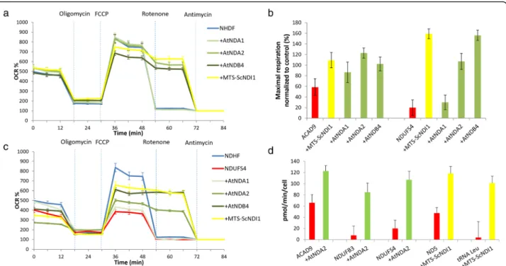
Fig. 2 Oxygen consumption analysis: Oxygen consumption was evaluated using the Seahorse XF Analyzer; a Oxygen consumption rate (OCR) expressed as percent (%) of rate measurement 13 in control cells (NDHF) and in control cells transduced with alternative dehydrogenases from A. thaliana (+AtNDA1, +AtNDA2, +AtNDB4) and yeast (+MTS-ScNDI1); b OCR expressed as % of rate measurement 13 in NDHF, in NDUFS4-deficient cells (NDUFS4) and in patient cells transduced with alternative dehydrogenases from A. thaliana (+AtNDA1,+AtNDA2, +AtNDB4) and yeast (+MTS-ScNDI1); c Maximal respiration rate in CI deficient cells (carrying pathogenic variants in ACAD9 and NDUFS4), before and after transduction with AtNDA1, AtNDA2, AtNDB4 and MTS-ScNDI1. Values were normalized to maximal respiration of untransduced control cells; d Oxygen consumption rate (OCR) expressed as pmol O2/min/cell in cell lines presenting with CI-defect due to mutations in ACAD9, NDUFB3, NDUFS4, ND5, tRNALeu
before and after transduction with alternative dehydrogenases from A. thaliana and yeast. Each cell line was measured at least twice in independent experiments. During experiment, four technical replicates were run for each cell line. Values are expressed as mean ± SD
to the cuvette and the amount of oxygen atoms (nmol of molecular oxygen*2) consumed during state 3 to state 4 transition. All the assays were repeated at least three times. Protein content was measured according to
Bradford [25].
RNA sequencing
RNA sequencing was performed as described [27].
Briefly, RNA was isolated from whole-cell lysates using the AllPrep RNA Kit (Qiagen) and RNA integrity num-ber (RIN) was determined with the Agilent 2100 BioaAnalyzer (RNA 6000 Nano Kit, Agilent). For library
preparation, 1μg of RNA was poly(A)- selected,
fragmented and reverse transcribed with the Elute, Prime and Fragment Mix (Illumina). End repair, A-tailing, adaptor ligation and library enrichment were performed as described in the Low Throughput protocol of the TruSeq Stranded mRNA Sample Prep Guide (Illu-mina). RNAcDNA libraries were assessed for quality and quantity with the Agilent 2100 BioaAnalyzer and quan-tity using the Quant-iT PicoGreen dsDNA Assay Kit (Life Technologies). RNA libraries were sequenced as 150 bp paired-end runs on an Illumina HiSeq4000 platform. The STAR aligner* (v 2.4.2a) with modified parameter settings (−-twopassMode = Basic) was used for split-read alignment against the human genome assembly hg19 (GRCh37) and UCSC knownGene anno-tation. Prior alignment, the reference genome sequence was augmented by two new contigs, one for each plant gene (NDA2 and NDB4, respectively). The nucleotide sequences of these two genes corresponded to the transgenic constructs cloned in the lentiviral vector (see
Additional file 1: Supplementary Methods). To quantify
the number of reads mapping to annotated genes we used HTseq-count (v0.6.0). FPKM (Fragments Per Kilobase of transcript per Million fragments mapped) values were calculated using custom scripts.
Statistical analysis
All data are expressed as the mean ± SD, and compari-sons between groups performed using Student’s t test. Results
Proof of concept that NDH-2 dehydrogenases counteract CI deficiency
Preliminary assays on different CI defective fibroblast cell lines had showed the ability of several NDH-2 to
rescue respiration defect (Fig. 2b-d). We decided to
focus our subsequent analysis on three NDH-2: ScNDI1, the internal NDH-2 of Saccharomyces cerevi-siae; AtNDB4, an Arabidopsis thaliana NDH-2 local-ized to the external side of inner mitochondrial
membrane (IMM); AtNDA2, another Arabidopsis thali-ana NDH-2 localized to the internal side of IMM.
As expected, transfection of control fibroblasts with AtNDA2, AtNDB4, and ScNDI1 led to rotenone-resistance without any significant effect on overall
res-piration rate (Fig. 2a). In order to examine the rescuing
efficiency of plant NDH-2 in more depth, we chose to focus on fibroblasts carrying a pathogenic homozygous mutation in the nuclear gene NDUFS4, as a well-established cellular model of complex I deficiency. In-deed, consequences of deleterious mutations affecting NDUFS4 have been thoroughly studied on several pa-tient cell lines and on whole-body and tissue specific
knockout mice [28].
Therefore, we verified and confirmed that besides conferring rotenone resistance, all the aforemen-tioned NDH-2 (ScNDI1, AtNDA2 and AtNDB4) were able to restore respiration when expressed in NDUFS4 deficient fibroblasts, nearly reaching control
levels. (Fig. 2c).
Expression of NDH-2 dehydrogenases does not affect growth of cultured human fibroblasts
ScNDI1, AtNDA2, and AtNDB4 transfected control
fibroblasts (NHDF) and CI-defective fibroblasts
(NDUFS4) exhibited comparable growth rates when compared to the corresponding untransfected control, both in glucose (4.5 g/L) and glucose-deprived media (not shown).
At-NDA2 and at-NDB4 rescue NADH:quinone
oxidoreductase activity of NDUFS4 mutant fibroblasts
We further confirmed the observed rescue by measur-ing NADH:quinone oxidoreductase specific activity by
spectrophotometry in control cells and NDUFS4
mutated fibroblasts before and after transfection by ScNDI1, AtNDA2, and AtNDB4, show that all three dehydrogenases were able to rescue the CI defect
(Table 1). We could also observe that, while AtNDA2
and AtNDB4 restored CI activity to levels comparable to those observed in control cells, ScNDI1 transfected cells exhibited levels of NADH:quinone oxidoreduc-tase activity much higher than untransfected cells
(Table 1).
SOD activity in NDUFS4 deficient cells
As a direct effect of defective CI activity and consequent increase of ROS production, SOD activity is shown to be significantly higher in NDUFS4 mutated patient’s cell
(Fig. 3). Transfection with AtNDA2 and AtNDB4 but
not with ScNDI1 was almost able to decrease SOD
activ-ity to levels observed in control fibroblasts (Fig.3).
AtNDA2 and AtNDB4 expression in control cell lines
We evaluated by RNA sequencing the expression levels of CI subunits, AtNDA2, and AtNDB4 in control cells before and after the stable transduction with the plant genes AtNDA2 and AtNDB4 (RNA sequencing was not performed on NDUFS4 deficient fibroblasts due to scar-city of material). We evaluated the FPKM values for CI subunits before and after transduction. The median FPKM of CI subunits was similar in all cell lines, indicat-ing that the transduction with the plant genes did not affect the expression level of CI subunits (median FPKM
between 30 and 35, Table 2). AtNDA2 had an
expres-sion level of 25 FPKM, which falls in the range of CI subunit expression, while AtNDB4 had an expression level of 127 FPKM, much higher than the median
ex-pression level of CI subunits (Table 2). In A. thaliana
the endogenous expression of NDA2 and NDB4 is sig-nificantly lower than the expression of CI subunits, in all
parts of the plant (flower, root, leaf, and fruit). The expression of NDA2 is 10 times lower than the median expression of CI subunits, while NDB4 is almost 500 times lower than the median expression of CI subunits
[29] (Additional file1: Table S1).
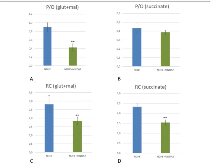
AtNDA2 competes with CI when expressed in human fibroblasts
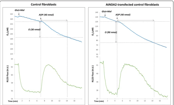
Thereafter, in view of its expression profile in A. thali-ana (Add file 1), we selected the AtNDA2 to verify the lack of competition between CI and plant NDH-2 when expressed in control fibroblasts. We first studied the P/ O ratio with different substrates, the latter being sup-posedly decreased if NADH, normally oxidized by the proton-motive CI, is diverted to AtNDA2. P/O calcula-tion is usually performed on isolated mitochondria in order to eliminate cytosolic ATPases activity. ATPases increase ADP recycling, allowing for a continuous stimu-lation of the mitochondrial ATP synthase and respir-ation, thus affecting state 4 establishment. However, taking into consideration the scarcity of material and the slow growth rate of fibroblasts, we performed the assays using permeabilized cells. As expected, the observed P/ O values were underestimated comparing to the ones measured on purified mitochondria (about 2.5 for NADH related substrates and 1.5 for succinate,
respect-ively)-see Hinkle et al. [30] for a complete review on this
topic. Nevertheless, using this approach, we were able to
measure the P/O ratio (Fig. 4). Unexpectedly, we
evi-denced a competition between AtNDA2 and functional mitochondrial respiratory chain CI during glutamate/ malate oxidation in the control cell line expressing
AtNDA2 (Fig. 4). Transfected cells showed P/O values
lowered by half (0.43 ± 0.08) in comparison to untrans-fected cells (0.9 ± 0.1). Moreover, respiratory control index, calculated as the ratio between state 3 and state 4, which represents a numeric estimation of mitochondrial coupling efficiency, was also clearly lowered in
trans-fected cells under glutamate/malate stimulation (Fig.5).
A further validation of these results came from P/O data obtained with succinate, the substrate of CII. Indeed, although P/O ratios for succinate were not
Table 1 NADH:quinone oxidoreductase activity in control and transfected cells
Cell line NADH:quinone oxidoreductase specific activity w/o rotenone (nmol/min/mg Prot)
NADH:quinone oxidoreductase specific activity with rotenone (nmol/min/mg Prot) Inhibition (%) NHDF 7.7 (±0.5) 1.7 (±0.2) 78.3% 79,787 2.5 (±0.2) 1.4 (±0.1) 42.6% 79,787-T-ScNDI1 24.0 (±8.7) 20.7 (±8.3) 13.7% 79,787-T-AtNDB4 11.8 (±2.4) 10.6 (±1.9) 10.0% 79,787-T-ATNDA2 5.3 (±0.9) 5.3 (±0.9) 2.7%
Spectrophotometric assessment of NADH:quinone oxidoreductase specific activity in control cells (NDHF) and NDUFS4 cell line untransfected (79787) and transfected with alternative NADH oxidases (ScNDI1, AtNDA2 and AtNDB4). Values are expressed as mean ± SD
Fig. 3 Assessment of SOD activity: Spectrophotometric assessment of SOD activity according to the pyrogallol autoxidation method. NDUFS4 mutated cell line (79787) displays higher SOD activity when compared to control fibroblasts (NHDF). Transfection with AtNDB4 and AtNDA2 (79787-AtNDB4 and 79,787-AtNDA2) significantly decreases SOD activity, which is nearly restored to normal levels. Values are expressed as means ± SD (ns: not significant; *p < 0.05; **p < 0.01)
significantly lower in transfected cells, 0.43 ± 0.05 and 0.39 ± 0.02, respectively, we observed a reduction of
re-spiratory control index in transfected cells (Fig. 5). This
is likely due to the metabolic conversion of a fraction of succinate to glutamate, which later enters the oxidation machinery proceeding through CI and AtNDA2.
When ATP instead of ADP was used, we observed only a very low OXPHOS stimulation in both transfected and untransfected cell lines, providing an additional confirm-ation that our measurments were not significantly affected by ATPase mediated ATP recycling to ADP (not shown).
A further evidence of the likely competition between
CI and AtNDA2, came from calculation of KM for
NADH in both transfected and untransfected cell lines
(Fig. 6). CI affinity for NADH was estimated in
untrans-fected control cells considering only rotenone sensitive internal NADH:quinone oxidoreductase activity, while, to evaluate AtNDA2 affinity for NADH, we exclusively analyzed rotenone insensitive activity in AtNDA2-transfected control cells. Our estimation of CI and
AtNDA2 KM gave values of 2.7 ± 0.4μM and 9.7 ±
3.3μM, respectively. Hence, when transfected in human
cells, KMof AtNDA2 for NADH appear to be about only
3 folds higher than KMof CI for NADH, i.e. in the same
order of magnitude; this gap is probably insufficient to prevent competition for the substrate within physio-logical range of NADH concentration inside the mito-chondria, therefore indirectly confirming that upon these experimental conditions, it seems likely that CI and AtNDA2 compete for the oxidation of NADH. Discussion
CI is the largest complex of the respiratory chain, con-sisting of 44 different subunits encoded by both nDNA and mtDNA. These subunits are assembled in a precise
order by numerous assembly factors [1]. Thus,
patho-genic mutations in genes encoding either for structural subunits or for assembly factors, can result in the enzymatic impairment of CI, often with a yet poorly understood tissue-specificity and time-dependency. Be-sides these mechanisms, CI deficiency may arise as a consequence of mutations in genes encoding proteins involved in mitochondrial translation, in iron-sulphur cluster assembly, and mtDNA-depletion associated genes
[31,32]. This could explain why CI deficiency is the far
most common finding in mitochondrial disorders. In terms of therapeutic approach, having a unique treatment applicable to all CI deficiencies, regardless of the genetic cause, would be desirable. A bypassing strat-egy using alternative dehydrogenase proteins seems to offer such possibility. Indeed the monomeric NADH de-hydrogenase of yeast, ScNDI1, inserted in CI-deficient
cells by Yagi and colleagues [14], presented an apparent
beneficial effect on several experimental models in vitro
Table 2 Expression level of AtNDA2, AtNDB4, and
NADH:quinone oxidoreductase (CI) subunits in control cell lines (NDHF) before and after transduction with AtNDA2 and AtNDB4
Expression levels are indicated in FPKM. In gray are labelled the NADH:quinone oxidoreductase subunits, in green the two NDH-2 genes. Median FPKM for CI subunits in both cell lines is given in red
and in vivo [8–13]. Nevertheless, it lowered P/O values for CI-dependent oxidation of NADH indicating reduced ATP synthesis upon ScNDI1 transfection in control
mammalian cells [14] raising questions about its
feasibil-ity as treatment for patients with impaired but residual CI activity, where prevalence of ScNDI1 over residual CI activity could worsen metabolic disturbances and lower OXPHOS energetic yield. The consequences on cellular homeostasis could be potentially deleterious, since ATP synthesis decline is one of the major pathomechanism involved in the CI deficiency-related phenotype.
A. thaliana NDH-2 naturally coexist with CI: their activity is stimulated when electron flux through mito-chondrial OXPHOS is slowed down, most likely
depend-ing on their intrinsic enzymatic properties [15–18, 33].
Hence, they represent valuable candidates for comple-menting a defective CI activity without competing with it. AtNDA2, and AtNDB4, in particular, show a substrate preference for NADH over NADPH and their catalytic
activity is Ca2+ independent, similarly to CI. AtNDA2 is
typically detected in the mitochondrial inner membrane,
facing the matrix [15, 29], but there is evidence of an
additional peroxisomal location [34]. AtNDB4 instead
faces the intermembrane space. To target these proteins
specifically to the mitochondrial matrix of mammalian fibroblasts, the plant-specific mitochondrial targeting sequence (MTS) was replaced with the human MTS.
AtNDA2, and AtNDB4 were both able to rescue the biochemical defect when expressed in CI-deficient cells, as indicated by increased respiration determined by oxy-gen consumption determination and lowered SOD
activ-ity, a surrogate of ROS production. Functional
expression of plant NDH-2 was further indicated by complementation of defective CI on spectrophotometric assays, since both enzymes were able to re-establish NADH:quinone oxidoreductase activity close to control values. Besides, they did not affect cell growth both in standard culture conditions and in the case of glucose deprivation, when cells are forced to switch on OXPHOS for energy production. This observation suggested a lack of competition with CI in standard culture conditions. However, the absence of any apparent effect on cellular growth could be also due to inadequate strength of com-petition or low level of NDA2 in proportion to CI prevent-ing from detection of such a competition.
We decided to focus on AtNDA2, which represents the most promising candidate to replace CI in deficient cells based on its location and activity profile within
Fig. 4 Evaluation of mitochondrial respiration: Mitochondrial membrane potential variations assessed by rhodamine 123 fluorescence and oxygen uptake measured with optode device in digitonine permeabilized fibroblasts (representative graphs for the control fibroblasts - left panel and AtNDA2-transfected control fibroblasts - right panel). The reaction was started by the addition of glutamate/malate, followed by injections of ADP (see text). Note that the amount of oxygen reduced during ADP phosphorylation is significantly higher in AtNDA2 transfected cells comparing to control
Fig. 5 RC (respiratory control) and P/O coupling ratios: Comparison of P/O values (a and b) and Respiratory Control Index (c and d) with Glutamate/Malate (a,c) and Succinate (b, d) in non-transfected control and AtNDA2 transfected control cells
Fig. 6 KMevaluation: Plots of NADH:quinone oxidoreductase activity (y) as a function of NADH concentration (μM) (x): a rotenone-sensitive
NADH:quinone oxidoreductase activity in control cells; b rotenone-resistant NADH:quinone oxidoreductase activity in AtNDA2 transfected control cells
plant mitochondria [16,18,35] To verify this possibility, we expressed AtNDA2 in human control fibroblasts and evaluated the effect on the ADP phosphorylation levels under different respiratory substrates. Using an NADH related substrate (glutamate/malate), the P/O ratio of transfected cells was lowered by half when compared to control cells. This indicates that AtNDA2 is active when expressed in control cells and competes with CI for electron transfer from NADH to quinone. Additionally, rotenone resistant NADH oxidase activity in
AtNDA2-control cells has an apparent KM of 9.7μM for NADH
which is slightly over 3-fold higher than affinity of CI for
NADH evaluated in control cells (2.7μM). Hence, in our
experimental model AtNDA2 and CI affinity for the same substrate seem to fall within a similar order of magnitude, thus supporting the existence of a competi-tion in human cells.
However, there are few important limitations linked to this measurement that need to be considered.
Previous studies on plant mitochondria had calcu-lated rotenone-resistant NADH oxidase activity of the
inner membrane to be up to 10-fold higher than KM of
CI [17, 29, 36], although other authors later reported a
considerably lower value of 13.9μM [37], which is
closer to our results.
Likewise, reported apparent KM values of CI for
NADH are quite heterogeneous, ranging from 2μM to
20μM [38–41].
There are few important considerations to explain the observed intergroup variability. First of all, the develop-ment of a specific method to evaluate kinetic properties of CI has notoriously been a recurrent challenge for
researchers [23,42,43].
Besides this, we should also consider methodological heterogeneity (e. g. sample preparation, category of quinone-analogues employed as electron acceptors, difficulty to accurately estimate enzymatic activity when dealing with extremely low concentration of the sub-strate etc). Indeed, kinetic properties of these enzymes have been mainly estimated on isolated mitochondria/ submitochondrial preparations and on different cell line-ages, while we studied permeabilised cell preparations, which are unavoidably contaminated to some extent by soluble NADH dehydrogenases activities. Moreover, AtNDA2 compartmentation within the inner surface of mitochondrial membrane or its association to a supra-molecular complex (enzyme malic/specific quinone pool/AOXs) under native conditions, could contribute to its distinctive kinetic properties for NADH and to prevent competition with CI, thus ensuring AtNDA2
ac-tivity only in specific physiological circumstances [44].
Most reasonably, the apparent competition with the endogenous OXPHOS system could depend on the plant enzyme concentration within human mitochondria. In
our experimental system we used a strong promoter and reached an overall AtNDA2 RNA expression level (25 FPKM) falling in the range of complex I subunit expres-sion (median 29 FPKM). Interpreting these data as a rough measure to approximate the protein levels (unfortu-nately, we lack information about the post-transcriptional effects for both, AtNDA2 and complex I), would indicate quite high AtNDA2 levels when compared to A. thaliana, where AtNDA2 expression has been reported to be up to
10-fold lower than complex I [45]. This very high level of
plant enzyme could thus result in the observed competi-tion between AtNDA2 and complex I in our cellular model.
Therefore, our data suggest to test experimental sys-tems exhibiting lower expression levels of NDH-2 for further studies.
Conclusions
In conclusion, we showed that transfection of plant NDH-2 was able to rescue CI defect in vitro. However, AtNDA2, the most promising candidate based on its properties in plants, exhibits competing activity with human CI when expressed at high levels, thus raising concerns which need to be considered in case of its application to human therapy. In cells with substantial CI activity, a balance in terms of energy production and metabolic dysfunctions needs to be determined, in which the gain of additional NADH oxidation is more benefi-cial than the reduced ATP production through competi-tion with CI. If not controlled, consequences of this uncoupling effect are unpredictable in vivo, and risk to be deleterious in affected patients. A substantial amount of translational work still has to be done in the near future, from genetic manipulation of the transfected plant product to possible modification of its enzymatic properties, to the generation of an animal model to test its effects in vivo.
Nevertheless, we have moved an important step to-wards a deeper comprehension of potential advantages and drawbacks of trans-kingdom replacing therapy for respiratory chain defects.
Additional file
Additional file 1:Supplementary methods. Supplementary results.
Table S1. Expression level of AtNDA2, AtNDB4, and NADH:quinone oxidoreductase (CI) subunits in various compartment of A.thaliana. (DOCX 105 kb)
Acknowledgements
We thank Dr. Dejana Mokranjac for providing genomic DNA of the yeast, and Prof. Takao Yagi for providing the antibody against the yeast NDI1. The authors declare no conflicts of interest.
Authors’ contributions
AC performed ADP/O essays and evaluation of KM, analyzed data and wrote
the manuscript. AI generated all transgenic constructs, performed transduction experiment and Western Blotting, analyzed Seahorse data and wrote the manuscript. CT cultured cells and isolated the RNA. LSK performed Seahorse experiments. TS processed and analyzed the RNA-seq data. JB, MP and PB performed spectrophotometry and SOD experiments. AGR provided expertise regarding Western blot experiments. VT, PR, MR, HP and MS designed the experiments and wrote the manuscript. All authors read and approved the final manuscript.
Author’s information Not applicable.
Funding
This study was supported by the German BMBF and Horizon2020 through E-Rare project GENOMIT (01GM1603 and 01GM1906B to H.P.); Horizon2020 Project SOUND (633974 to H.P.); German Network for Mitochondrial Disorders (mitoNET 01GM1113C to H.P.); AMMi (Association contre les Maladies Mitochondriales) grants to M.P., P.R., P.B. and M.S.; E-Rare project GENOMIT (ANR-15-RAR3–0012 to P.R., A.C. and M.S.); Italian Ministry of Health (RF-2016-02361495 to A.C.); Mitocon (Grant no. 2018–01 to V.T.); Mariani Foundation (V.T.). Availability of data and materials
The datasets used and/or analysed during the current study are available from the corresponding author on reasonable request.
Ethics approval and consent to participate
All procedures performed in studies involving human participants were in accordance with the ethical standards of the institutional and/or national research committee and with the 1964 Helsinki declaration and its later amendments or comparable ethical standards.
Informed consent was obtained from all individual participants (or their legal tutors) included in the study prior to performing cutaneous biopsies.
Consent for publication Not applicable.
Competing interests
The authors declare that they have no competing interests.
Author details
1UMR1141, PROTECT, INSERM, Université de Paris, Paris, France.2Unit of Medical Genetics and Neurogenetics, Fondazione IRCCS Istituto Neurologico Carlo Besta, Milan, Italy.3Institute of Human Genetics, Helmholtz Zentrum München, German Research Center for Environmental Health, Neuherberg, Germany.4Institute of Human Genetics, Technische Universität München, Munich, Germany.5Reference Center for Inborn Errors of Metabolism, Hôpital Universitaire Robert Debré, APHP, Paris, France.6Department of Biology, Lund University, Biology building A, Sölvegatan 35, SE-22362 Lund, Sweden.7Dr. von Hauner Children’s Hospital, Department of Pediatrics, University Hospital, Ludwig-Maximilians-Universität (LMU), Munich, Germany.
Received: 14 June 2019 Accepted: 30 August 2019
References
1. Sánchez-Caballero L, Guerrero-Castillo S, Nijtmans L. Unraveling the complexity of mitochondrial complex I assembly: a dynamic process. Biochim Biophys Acta. 2016;1857(7):980–90.https://doi.org/10.1016/j.bbabio. 2016.03.031.
2. Ripple MO, Kim N, Springett R. Mammalian complex I pumps 4 protons per 2 electrons at high and physiological proton motive force in living cells. J Biol Chem. 2013;288(8):5374–80.
3. Brandt U. Energy converting NADH:quinone oxidoreductase (complex I). Annu Rev Biochem. 2006;75:69–92.
4. Haack TB, Haberberger B, Frisch EM, Wieland T, Iuso A, Gorza M, et al. Molecular diagnosis in mitochondrial complex I deficiency using exome sequencing. J Med Genet. 2012;49(4):277–83.https://doi.org/10.1136/ jmedgenet-2012-100846.
5. Saari S, Garcia GS, Bremer K, Chioda MM, Andjelković A, Debes PV, et al. Alternative respiratory chain enzymes: therapeutic potential and possible pitfalls. Biochim Biophys Acta Mol Basis Dis. 2019;1865(4):854–66.https://doi. org/10.1016/j.bbadis.2018.10.012.
6. Cannino G, El-Khoury R, Pirinen M, Hutz B, Rustin P, Jacobs HT, et al. Glucose modulates respiratory complex I activity in response to acute mitochondrial dysfunction. J Biol Chem. 2012;287(46):38729–40.https://doi.org/10.1074/jbc. M112.386060.
7. McDonald AE, Gospodaryov DV. Alternative NAD(P) H dehydrogenase and alternative oxidase: proposed physiological roles in animals. Mitochondrion. 2019;45:7–17.https://doi.org/10.1016/j.mito.2018.01.009.
8. Bai Y, Hajek P, Chomyn A, Chan E, Seo BB, Matsuno-Yagi A, et al. Lack of complex I activity in human cells carrying a mutation in MtDNA-encoded ND4 subunit is corrected by the Saccharomyces cerevisiae NADH-quinone oxidoreductase (NDI1) gene. J Biol Chem. 2001;276(42):38808–13. 9. Bai Y, Hu P, Park JS, Deng JH, Song X, Chomyn A, et al. Genetic and
functional analysis of mitochondrial DNA-encoded complex I genes. Ann N Y Acad Sci. 2004;1011:272–83.
10. Cho J, Hur JH, Graniel J, Benzer S, Walker DW. Expression of yeast NDI1 rescues a Drosophila complex I assembly defect. PLoS One. 2012;7(11): e50644.https://doi.org/10.1371/journal.pone.0050644.
11. Marella M, Seo BB, Thomas BB, Matsuno-Yagi A, Yagi T. Successful amelioration of mitochondrial optic neuropathy using the yeast NDI1 gene in a rat animal model. PLoS One. 2010;5(7):e11472.
12. Marella M, Seo BB, Flotte TR, Matsuno-Yagi A, Yagi T. No immune responses by the expression of the yeast Ndi1 protein in rats. PLoS One. 2011;6(10):e25910. 13. Chadderton N, Palfi A, Millington-Ward S, Gobbo O, Overlack N, Carrigan M,
et al. Intravitreal delivery of AAV-NDI1 provides functional benefit in a murine model of Leber hereditary optic neuropathy. Eur J Hum Genet. 2013;21(1):62–8. 14. Barber-Singh J, Seo BB, Matsuno-Yagi A, Yagi T. Protective role of
rAAV-NDI1, serotype 5, in an acute MPTP mouse Parkinson's model. Parkinsons Dis. 2010;2011:438370.https://doi.org/10.4061/2011/438370.
15. Seo BB, Matsuno-Yagi A, Yagi T. Modulation of oxidative phosphorylation of human kidney 293 cells by transfection with the internal rotenone-insensitive NADH-quinone oxidoreductase (NDI1) gene of Saccharomyces cerevisiae. Biochim Biophys Acta. 1999;1412(1):56–65.
16. Michalecka AM, Svensson AS, Johansson FI, Agius SC, Johanson U, Brennicke A, et al. Arabidopsis genes encoding mitochondrial type II NAD(P) H dehydrogenases have different evolutionary origin and show distinct responses to light. Plant Physiol. 2003;133(2):642–52.
17. Rasmusson AG, Soole KL, Elthon TE. Alternative NAD(P) H dehydrogenases of plant mitochondria. Annu Rev Plant Biol. 2004;55:23–39.
18. Moller IM, Palmer JM. Direct evidence for the presence of a rotenone-resistant NADH dehydrogenase on the inner surface of the inner membrane of plant mitochondria. Physiol Plant. 1982;54(3):267–74. 19. Rasmusson AG, Geisler DA, Møller IM. The multiplicity of dehydrogenases in
the electron transport chain of plant mitochondria. Mitochondrion. 2008; 8(1):47–60 Review.
20. El-Khoury R, Kemppainen KK, Dufour E, Szibor M, Jacobs HT, Rustin P. Engineering the alternative oxidase gene to better understand and counteract mitochondrial defects: state of the art and perspectives. Br J Pharmacol. 2014;171(8):2243–9.https://doi.org/10.1111/bph.12570. 21. Anderson SL, Chung WK, Frezzo J, Papp JC, Ekstein J, DiMauro S, et al. A
novel mutation in NDUFS4 causes Leigh syndrome in an Ashkenazi Jewish family. J Inherit Metab Dis. 2008;31(Suppl 2):S461–7.https://doi.org/10.1007/ s10545-008-1049-9.
22. Kremer LS, Prokisch H. Identification of disease-causing mutations by functional complementation of patient-derived fibroblast cell lines. Methods Mol Biol. 2017;1567:391–406.https://doi.org/10.1007/978-1-4939-6824-4_24. 23. Bénit P, Goncalves S, Philippe Dassa E, Brière JJ, Martin G, Rustin P. Three
spectrophotometric assays for the measurement of the five respiratory chain complexes in minuscule biological samples. Clin Chim Acta. 2006; 374(1–2):81–6.
24. Chretien D, Benit P, Chol M, Lebon S, Rötig A, Munnich A, et al. Assay of mitochondrial respiratory chain complex I in human lymphocytes and cultured skin fibroblasts. Biochem Biophys Res Commun. 2003;301:222–4. 25. Bradford MM. A rapid and sensitive method for the quantitation of
microgram quantities of protein utilizing the principle of protein-dye binding. Anal Biochem. 1976;72:248–54.
26. Geromel V, Kadhom N, Cebalos-Picot I, Ouari O, Polidori A, Munnich A, et al. Superoxide-induced massive apoptosis in cultured skin fibroblasts harboring Catania et al. Orphanet Journal of Rare Diseases (2019) 14:236 Page 11 of 12
the neurogenic ataxia retinitis pigmentosa (NARP) mutation in the ATPase-6 gene of the mitochondrial DNA. Hum Mol Genet. 2001;10(11):1221–8.49. 27. Kremer LS, Bader DM, Mertes C, Kopajtich R, Pichler G, Iuso A, et al. Genetic
diagnosis of Mendelian disorders via RNA sequencing. Nat Commun. 2017;8: 15824.https://doi.org/10.1038/ncomms15824.
28. Breuer ME, Willems PH, Smeitink JA, Koopman WJ, Nooteboom M. Cellular and animal models for mitochondrial complex I deficiency: a focus on the NDUFS4 subunit. IUBMB Life. 2013;65(3):202–8.https://doi. org/10.1002/iub.1127.
29. Geisler DA, Broselid C, Hederstedt L, Rasmusson AG. Ca2+−binding and Ca2+−independent respiratory NADH and NADPH dehydrogenases of Arabidopsis thaliana. J Biol Chem. 2007;282(39):28455–64.
30. Hinkle PC. P/O ratios of mitochondrial oxidative phosphorylation. Biochim Biophys Acta. 2005;1706(1–2):1–11.
31. Scheffler IE. Mitochondrial disease associated with complex I (NADH-CoQ oxidoreductase) deficiency. J Inherit Metab Dis. 2015;38(3):405–15.https:// doi.org/10.1007/s10545-014-9768-6.
32. Emmanuele V, López LC, Berardo A, Naini A, Tadesse S, Wen B, et al. Heterogeneity of coenzyme Q10 deficiency: patient study and literature review. Arch Neurol. 2012;69(8):978–83.https://doi.org/10.1001/archneurol. 2012.206Review. Erratum in: Arch Neurol. 2012 Jul;69(7):886.
33. Lee CP, Eubel H, Millar AH. Diurnal changes in mitochondrial function reveal daily optimization of light and dark respiratory metabolism in Arabidopsis. Mol Cell Proteomics. 2010;9(10):2125–39.https://doi.org/10. 1074/mcp.M110.001214.
34. Carrie C, Murcha MW, Kuehn K, Duncan O, Barthet M, Smith PM, et al. Type II NAD(P) H dehydrogenases are targeted to mitochondria and chloroplasts or peroxisomes in Arabidopsis thaliana. FEBS Lett. 2008;582(20):3073–9.
https://doi.org/10.1016/j.febslet.2008.07.061.
35. Elhafez D, Murcha MW, Clifton R, Soole KL, Day DA, Whelan J.
Characterization of mitochondrial alternative NAD(P) H dehydrogenases in Arabidopsis: intraorganelle location and expression. Plant Cell Physiol. 2006; 47(1):43–54.
36. Soole KL, Dry IB, James AT, Wiskich JT. The kinetics of NADH oxidation by complex I of plant mitochondria. Physiol Plant. 1990;80:75–82.
37. Rasmusson AG, Møller IM. NAD(P) H dehydrogenases on the inner surface of the inner mitochondrial membrane studied using inside-out particles. Physiol Plant. 1991;83:357–65.
38. Hatefi Y, Stiggal DL. In: Boyer PD, editor. The enzymes. 3rd ed. New York: Academic; 1976. p. 175–297.
39. Vinogradov AD. Kinetics, control, and mechanism of ubiquinone reduction by the mammalian respiratory chain-linked NADH-ubiquinone reductase. J Bioenerg Biomembr. 1993;25(4):367–75.
40. Majander A, Huoponen K, Savontaus ML, Nikoskelainen E, Wikström M. Electron transfer properties of NADH:ubiquinone reductase in the ND1/3460 and the ND4/11778 mutations of the Leber hereditary optic
neuroretinopathy (LHON). FEBS Lett. 1991;292(1–2):289–92.
41. Barrientos A, Kenyon L, Moraes CT. Human xenomitochondrial cybrids. Cellular models of mitochondrial complex I deficiency. J Biol Chem. 1998; 273(23):14210–7.
42. de Wit LE, Scholte HR, Sluiter W. Correct assay of complex I activity in human skin fibroblasts by timely addition of rotenone. Clin Chem. 2008; 54(11):1921–2; author reply 1922-4.https://doi.org/10.1373/clinchem.2008. 104802.
43. Yano T. The energy-transducing NADH: quinone oxidoreductase, complex I. Mol Aspects Med. 2002;23(5):345–68.
44. Rustin P, Moreau F. Malic enzyme activity and cyanide-insensitive electron transport in plant mitochondria. Biochem Biophys Res Commun. 1979;88(3): 1125–31.
45. Klodmann J, Sunderhaus S, Nimtz M, Jänsch L, Braun HP. Internal architecture of mitochondrial complex I from Arabidopsis thaliana. Plant Cell. 2010;22(3):797–810.https://doi.org/10.1105/tpc.109.073726.
Publisher’s Note
Springer Nature remains neutral with regard to jurisdictional claims in published maps and institutional affiliations.
View publication stats View publication stats