Coordination of Lower Limb Movement Utilizing
the Agonist-Antagonist Myoneural Interface
by
Tony Shu
B.Sc., Georgia Institute
of
Technology (2016)
Submitted to the Program in Media Arts and Sciences
School of Architecture and Planning
in partial fulfillment of the requirements for the degree of
Master of Science in Media Arts and Sciences
at the
MASSACHUSETTS INSTITUTE OF TECHNOLOGY
September 2019
©Massachusetts
Institute of Technology 2019. All rights reserved.
Signature redacted
A uthor ... ...
...
Program in Media Arts and Sciences
August 9, 2019
Signature redacted
C ertified
by ...
...
Hugh Herr, Ph.D.
Professor of Media Arts and Sciences
Thesis Superv
Acceptedby....Signature
redacted..
MASSACHUSETTS INSTITUTE
Tod Machover, Ph.D.
cademic
Head, Program in Media Arts and Sciences
Coordination of Lower Limb Movement Utilizing the
Agonist-Antagonist Myoneural Interface
by
Tony Shu
Submitted to the Program in Media Arts and Sciences, School of Architecture and Planning
on August 9, 2019, in partial fulfillment of the requirements for the degree of
Master of Science in Media Arts and Sciences
Abstract
The agonist-antagonist myoneural interface is a novel surgical construct that shows promise as a method of providing persons with amputation proprioceptive sensation of movement and force.
This thesis aims to quantify the volitional coordination capabilities of the agonist-antagonist myoneural interface for applications related to control of active prostheses. In the first section, bilateral rhythmic coordination of ankle and subtalar joint move-ments is investigated in a control group of physically intact human subjects to char-acterize stereotypical kinematics of volitional lower limb movement. Subsequently, neuromusculoskeletal modeling techniques are developed to directly map estimated neural excitations from agonist-antagonist myoneural interface musculature to in-tended subtalar inversion and eversion kinematics.
In a case study, the developed neuromusculoskeletal modeling techniques are ap-plied to optimize a dynamic subtalar model for use by a unilateral subject with amputation possessing the agonist-antagonist myoneural interface. The subject's sub-sequent performance in bilateral rhythmic coordination utilizing the model and her own intact subtalar demonstrates the capacity of the agonist-antagonist myoneural interface to coordinate with intact anatomy in a biomimetic manner.
Thesis Supervisor: Hugh Herr, Ph.D. Title: Professor of Media Arts and Sciences
Coordination of Lower Limb Movement Utilizing the
Agonist-Antagonist Myoneural Interface
by
Tony Shu
The following people served as readers for this thesis:
Signature redacted
Thesis Reader ...
Edward Boyden, Ph.D.
Y. Eva Tan Professor in Neurotechnology
McGovern Institute for Brain Research
Signature redacted
Thesis Reader . ..
-**...
-...
. .
Te
Neville Hogan, Ph.D.
Sun Jae Professor of Mechanical Engineering
Department of Mechanical Engineering
Acknowledgments
I want to offer my deepest gratitude to Professor Hugh Herr, who gave me the chance to become in ways I will appreciate for many years into the future. Thank you for the opportunity and the vision which I could not possibly get from anyone else.
I also cannot fully express my appreciation for Professor Neville Hogan and his insight into the very fabric of being. I now know that it is just energy and abstraction all the way down. Or is it up? It is everywhere and everything, and so, so beautiful. To Professor Ed Boyden, when I find myself wondering about what a mind is, I always end up wondering about yours. Thank you for the work you do, the interesting things you say. Every sentence is a new inspiration.
To Tsung-Han and Seong Ho, I truly appreciate the laughs, the collective stum-bling, and the unanimous victories.
To Hyungeun, I enjoy all of our conversations, but especially those about motor control theory. It feels like I sneak away some piece of knowledge every time we talk. To Biomech, thank you for teaching me all of the hands-on lessons. I would say growth requires at least two points for comparison, and certainly you all have given me many.
To Shan Shan, Chris, and Austin: I quite literally could not have done it without you. You can take that to the bank.
Contents
1 Background
1.1 M otivation . . . . 1.2 Neuromusculoskeletal Modeling . . . . 1.2.1 Anatomy of the Lower Leg . . . . 1.2.2 Biomechanics of Movement . . . .
1.2.3 Proprioception . . . .
Spinal Reflexes . . . . 1.2.4 Hill-type Muscle-Tendon Units . . . .
1.3 The Agonist-Antagonist Myoneural Interface . . . .
1.3.1 Transtibial Amputation . . . . 1.3.2 Restoration of Relevant Proprioception . . . .
1.3.3 Dynamic Model of the AMI . . . .
1.4 Kinematic Characteristics of Volitional Motor Control . . . 1.4.1 Stereotypical Velocity Profiles . . . . Kinematics-oriented Models . . . . Dynamics-oriented Models . . . . 1.4.2 Speed-Accuracy Tradeoff . . . . 1.4.3 Coordination of Bimanually Symmetric Movement .
1.5 Background Summary . . . .
2 Coordination and Control Utilizing the Agonist-Antagonist
Myoneu-ral Interface 45 2.1 Contributions . . . . 46 17 . . . . 18 . . . . 19 . . . . 19 . . . . 20 . . . . 22 . . . . 24 . . . . 24 . . . . 26 . . . . 26 . . . . 28 . . . . 29 . . . . 31 . . . . 31 . . . . 31 . . . . 35 . . . . 39 . . . . 40 . . . . 43
Characteristics of Intact Lower Limb Movement . . . . 46
AMI Control of the Optimized Neuromusculoskeletal Subtalar M odel . . . . 46
2.2 M ethodology . . . . 47
2.2.1 Subjects . . . . 47
2.2.2 Apparatus and Signals Processing . . . . 47
sEMG... 47
Ankle-foot Kinematics . . . . 49
2.2.3 Bilateral and Unilateral Coordination Tasks . . . . 49
Relative Phase . . . . 51
Velocity Profile Analysis . . . . 52
Human Control Bandwidth . . . . 53
2.2.4 NMS Model Parameter Optimization . . . . 53
Dynamic Model of Subtalar Inversion-Eversion . . . . 53
Genetic Algorithm Implementation . . . . 56
Generated Trajectory Analysis . . . . 58
2.3 R esults . . . . 59
2.3.1 Optimal Model Parameters . . . . 59
2.3.2 Characteristics of Bilateral Movement . . . . 60
Relative Phase Comparisons . . . . 60
Averaged Velocity Profiles . . . . 63
2.3.3 Bandwidth Limitations . . . . 66
2.4 D iscussion . . . . 69
2.4.1 Characterization of Volitional Lower Limb Coordination . . . 69
2.4.2 Volitional AMI Coordination . . . . 70
Validity of Associating AMI Excitations with Intact Kinematics 70 Analysis of Optimized Model Output . . . . 71
Stereotypical Velocity Profiles as a Result of Mechanical Impedance 73 2.4.3 Future Directions . . . . 75
Appendices 77
List of Figures
1-1 Musculature of the lower leg . . . . 20
1-2 Anatomical planes and ankle-foot rotational directions . . . . 21
1-3 Type Ia muscle spindle . . . . 23
1-4 Thelen's Hill-type muscle model . . . . 25
1-5 Standard transtibial amputation procedure . . . . 27
1-6 Transtibial amputation with two agonist-antagonist myoneural interfaces 29 1-7 Linear mechanical model of the agonist-antagonist myoneural interface 30 1-8 Reaching experiment apparatus . . . . 32
1-9 Reaching experiment with velocity-dependent forcefields . . . . 34
1-10 Dynamic motion primitives explain reaching . . . . 36
1-11 Plamondon's muscle synergy model . . . . 37
1-12 Log-normal support-bounded velocity profile . . . . 38
1-13 Bimanual coordination of finger adduction/abduction . . . . 41
1-14 Bimanual coordination of rotational patterns . . . . 42
2-1 Electrode placement over AMI muscles . . . . 48
2-2 Relative phase unit circles . . . . 51
2-3 Simplified inversion-eversion dynamic model . . . . 54
2-4 Effective muscle-tendon unit lever arm lengths . . . . 55
2-5 Bond graph of dynamic inversion-eversion model . . . . 55
2-6 Neural excitation and trajectory used for model optimization . . . . . 57
2-7 Optimization flow chart . . . . 58
2-9 Optimized dynamic model impulse responses . . . . 61
2-10 Subject BKA rhythmic coordination results generated from dynamic model ... .... . . .. ... .. ... . . . .. . . . . 61
2-11 Control group bilateral rhythmic coordination results . . . . 62
2-12 Selected averaged velocity profiles . . . . 64
2-13 Movement amplitudes and frequency distributions of inversion-eversion
cycles . . . . 67
2-14 Fitts's Law assessment of inversion-eversion . . . . 68 A-1 Mechsner's bimanual coordination results . . . . 80
List of Tables
2.1 Experimental Conditions for Ankle-foot Coordination Tasks . . . . . 50
2.2 Optimization Parameter Ranges and Initial Values . . . . 59
2.3 Optimized Model Parameters . . . . 59
Chapter 1
Background
This chapter provides a relevant overview of hu-man motor function, biomechanics, and ampu-tation to explain how residual musculature can be used to volitionally control an active pros-thesis. Much of this thesis is based on extant literature regarding upper limb control, and ad-ditional background is given on the methodolo-gies adapted within for characterizing volitional control of physically intact and amputated lower
limbs.
Nu descendant un escalier n' 2, 1912 Marcel Duchamp (1887-1968) Oil on canvas; 147 x 89.2 cm The Philadelphia Museum of Art
1.1
Motivation
Passive lower limb prostheses are fundamentally incapable of replicating the dynamics of human locomotion. The advent of microelectronics and high power density electric motors has allowed researchers to develop active prostheses that seek to recover this functionality for persons with lower limb amputations [1]. However, while locomotion is essential, the purpose of a prosthesis should not be limited to an explicit function
of this single ability.
Ramachandran and Rogers-Ramachandran [2] famously demonstrated the power of embodiment, or limb ownership, through what is now known as mirror therapy. As persons with unilateral upper limb amputation had their intact limb visually superimposed over the spatial location of their phantom limb, perceived kinesthetic sensations were elicited from the phantom related to manipulation of the intact limb. Similarly, the capacity to express volitional lower limb behaviors such as foot tapping, leg crossing, and even simple fidgeting through an active prosthesis may grant the owner with an even greater sense of embodiment, yet these remain unrealized on devices that have already satisfied the power and mechanical bandwidth requirements. The agonist-antagonist myoneural interface is a novel surgical construct for per-sons with amputation that physically links two residual muscles, causing contraction of one to pull the other. This mechanical linkage should establish a way to perceive the movement and force levels of both muscles via preexisting embedded mechanore-ceptors. What remains is to create a mapping for this construct so that control of an external device matches exactly what the user perceives internally. Accuracy of both internal feedback and external actuation may not only restore embodiment to persons with amputation, but may also produce new paradigms for control of non-anthropomorphic machinery by physically intact humans.
1.2
Neuromusculoskeletal Modeling
Before characterizing a system, it is worthwhile to determine the scope and scale of interest. This section details how neural excitations cause muscle contraction to produce forces and motion in the lower leg.
1.2.1
Anatomy of the Lower Leg
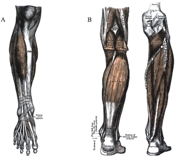
The lower leg, as it is referred to in this thesis, is skeletally comprised of the tibia, fibula, and all bones relatively distal. It includes all muscles and immediate structures that affect the ankle and joints of the foot. Figure 1-1 shows the general region of interest. Defining coordinates intrinsic to the body is also helpful when describing human anatomy and motion. Relative anatomical orientations and planes are shown
in Figure 1-2.
The ankle is truly a single joint known scientifically as the talocrural joint. The talocrural joint forms the axis of rotation for ankle dorsiflexion and plantarflexion (DP). It is plantarflexion that propels the body forward in the sagittal plane during level-ground walking. Tiptoeing and jumping also involve extensive rotation of this joint. The subtalar joint, located closely to the talocrural, forms the axis of rotation for foot inversion and eversion (IE). Actuation in these directions promotes stability in the coronal plane during quiet standing and locomotion.
The muscles which actuate the talocrural and subtalar are numerous, but only 12 are responsible for the majority of motion [4]. These muscles are extrinsic; they insert into the foot but originate proximally to the knee. The major plantarflexors include the medial gastrocnemius, lateral gastrocnemius, and soleus. The primary dorsiflexor is the tibialis anterior. The major inverter is the tibialis posterior, though the tibialis anterior and soleus also contribute to this motion, and the major everter is the peroneus longus (PL). It is necessary to state that the muscles of the lower leg do not contribute purely to a single degree of freedom. The sophisticated skeletal structure and varying muscle wrapping surfaces of the ankle-foot remain a challenge to fully model, but making a few modeling assumptions generally allows researchers
A B
Figure 1-1: (A) View of the anterior lower leg and (B) view of the posterior lower leg through
various muscle layers from Gray's Anatomy of the Human Body.
to capture the significant qualitative characteristics of ankle-foot motion.
1.2.2
Biomechanics of Movement
Upon initial consideration, movement is simple. Humans do it "without thinking" all the time, every day. However, even a passing interest in movement raises difficult questions. Why do babies require around a full year before learning how to walk while hoofed animals can do so almost immediately after birth [5]? How do jugglers keep several balls in the air without even opening their eyes? Explanations vary widely in scope and detail.
0-- Sgittal plane Superior Transvere plane - Frontal plan Inferior
K
Figure 1-2: (A) General anatomical orientations and (B) rotational directions for the
ankle-foot. Reproduced with permission from Elsevier Science [3].
can be generated by skeletal muscles to produce motion (or lack thereof). Muscles are actuators which only contract. Skeletal muscles are always found in series with tendons that anchor to bone. By contracting, muscles can generate moments of force about a joint at some distance away from the axis of rotation, producing a net torque trajectory according to
n2
(1.1)
Tnet (0, t)= ri(6)Fi(6, t)
where n is the total number of muscles acting about a joint, ri(6) is the effec-tive moment arm length of muscle i dependent on joint angle, and F(O, t) is the instantaneous force of muscle i at time t. The torques generated by all muscles and other contributors such as passive ligaments, sliding fascia, joint capsules, and ex-ternal forces like gravity and environmental contact can be summed to determine the dynamics of a moving biological system through the Newton-Euler equations of motion.
As a result of skeletal muscles being strictly contractile actuators, they are always paired with at least one other muscle so that contraction of one can be counteracted
by contraction of the other to produce full range of motion at the joint. The biceps
B Inversion Everalon N_. 1_ A Dorsfledon Plantarflexion
and triceps muscles of the upper arm are classical examples of this agonist-antagonist configuration. While seeming like a limitation at first, the implications are actually quite useful for animal movement. By varying the muscle activation levels of the
agonist and antagonist, equivalent to scaling their force production, the net torque generated about a joint can be independently modulated from its mechanical stiffness, or torque generated per unit displacement [6]. Stiffness is a result of simultaneous agonist and antagonist coactivation, also known as cocontraction, and may be intu-itively understood by holding your hand out and squeezing all of your muscles without actually moving your arm. Modulation of joint stiffness is vital for successful com-pletion of all tasks including walking without tripping, using a screwdriver without the tip slipping, and typing up thesis sections on a keyboard.
1.2.3
Proprioception
Muscles and tendons are embedded with a diverse set of biological mechanoreceptors which inform us of our body's internal dynamics in addition to the dynamics of our limbs in space. This sensory awareness, called proprioception, is a critical contrib-utor to our ability to generate robust motion in unknown environments and in the presence of disturbances, even without visual feedback. Proprioceptive signals are af-ferent, meaning they conduct information toward the central nervous system. This is opposite of efferent signals which conduct information away from the central nervous system. Though a variety of mechanoreceptors exists, only the three most closely related to the muscles and tendons are discussed here.
Muscle spindles are organs embedded within intrafusal muscle fibers in parallel that provide the central nervous system with muscle length and velocity information.
There are two types:
- Type Ia: responds to rate of change of muscle length and is somewhat sensitive to static muscle length
Combined, these two signals are enough to calculate the position and velocity of a limb in free space.
Muscie Muscle
fires
Tendon
Sensory ftres
Motor neuron
Figure 1-3: Type Ia muscle spindles spiraling around intrafusal muscle fibers [7].
Golgi tendon organs are separate mechanoreceptors that are embedded in the tendons at both the origin and insertion ends of a muscle. As they are in series with the muscle, they provide afferent information about the forces generated by the muscle.
Referring back to the agonist-antagonist configuration of muscles, it can now be understood how activation of the agonist produces rotation at the joint and corre-sponding stretch of the antagonist to provide a sensation of position and motion from both muscles. However, this is only true when impedances are sufficiently low to allow motion in the first place. When the limb is loaded against an obstacle like a wall, agonist activation produces no afferent feedback from the antagonist corresponding to motion. Instead, the increasing afferent signals from the agonist's Golgi tendon organs and muscle spindles inform the central nervous system of mounting levels of tension and contraction.
Spinal Reflexes
Spinal reflexes rely heavily on afferent feedback from proprioceptive sensation. Briefly, spinal reflexes are involuntary excitatory or inhibitional modulations of alpha motor neuron activity that can either elicit or inhibit muscle activation. As the name implies, the interneuronal networks responsible for these reflexes are unique in that they are located solely in the spinal cord. Well-known reflexes include the stretch or myotactic reflex which prevents a muscle from overlengthening and damaging itself. When this occurs in the knee, it is known as the knee-jerk or patellar reflex and causes the subject to kick out. Spinal reflexes activate quickly due to the relatively short distances required for nerve action potentials to travel (compared to a round trip to and from the brain).
While spinal reflexes are important for dynamic tasks such as walking and running, they are assumed to be largely inactive during the experiments performed in this thesis. Instead, this work focuses on volitional control of movement which is informed
by the brain's motor cortices.
1.2.4
Hill-type Muscle-Tendon Units
To mathematically model muscles for the purposes of computational simulation, dy-namic models based on the work of A.V. Hill [8] are constructed to represent the known dynamic features of real muscles. These modeled muscle-tendon units (MTUs) are formulated by a transfer function modulated by both physiological parameter constants and muscle state variables to relate muscle activation, usually derived from electromyography in practice, to output force. The cumulative effect of all MTU forces imposed on a modeled skeleton generates joint torques appropriate for forward simulation as described by Equation 1.1.
LCosa', L
muscleteadon
a) Muscle F-L
b) Muscle F-V
c)Tendon
&Ce ." --- aw.0~
:2 - 0 .
4-0
1 1+64' -1 we- -~ 1 0
Normalized Length Normalized Velocity Tendon Strain
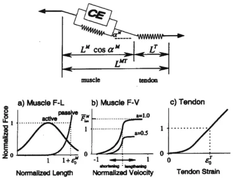
Figure 1-4: Thelen's Hill-type muscle model with associated parameter curves. Reproduced with permisson from ASME [9].
In this work, Thelen's Hill-type muscle model is utilized. The nonlinear differential equation describing the force balance of the massless MTU is given by
fmax(a(t)fAL(IM)fv(iM) - fPL(IM))coS(O) - fmaxfSE(IT)= 0 (1.2)
where fmax is the maximum force at lt or optimal fiber length , a(t) is the activation value at time t, fAL(M) is the force-length scalar based on the relative length of the MTU's contractile element as seen in Figure 1-4a,
f,(iM)
is the scalar based on the contractile element's current velocity as seen in Figure 1-4b, fpL(lM) is the contribution from the parallel elastic element, a is the pennation angle of the muscle fibers, andfsE(IT)
is the force of the series elastic element as seen in Figure1-4c.
To summarize the transfer function terms physiologically, the force-length scalar describes the ability of the MTU to generate force when it is at lengths that are not its optimal. Muscles have a finite stroke length due to the finite area of molecular actin and myosin interactions which generate tension in the contractile element, and
changes in length reduce this interaction area. The force-velocity scalar describes the velocity-sensitive rate of cross-bridge attachment for actin and myosin. The parallel elastic element describes the contributions from the structural components of muscle membranes, and the series elastic element represents the tendon elasticity. A thorough physiological explanation for these values can be found in [9].
Importantly for this work, a distinction is made between muscle activation a(t) and neural excitation u(t). The transformation from u(t) to a(t) describes cellular calcium release and diffusion dynamics when u(t) > a(t), and calcium uptake dynamics when u(t) < a(t). For the experiments in this work, surface electromyography was processed into an estimated u(t) signal to allow for adjustment of the activation and deactivation time constants associated with the transformation.
1.3
The Agonist-Antagonist Myoneural Interface
The development of the agonist-antagonist myoneural interface (AMI) marks one of the few major lower limb amputation paradigm shifts since the Civil War era
[10]. While amputation surgery is a broad area covering a large number of limbs
and conditions, the potential advantages of the AMI can be understood through comparison with standard transtibial amputation as it is most commonly performed today.
1.3.1
Transtibial Amputation
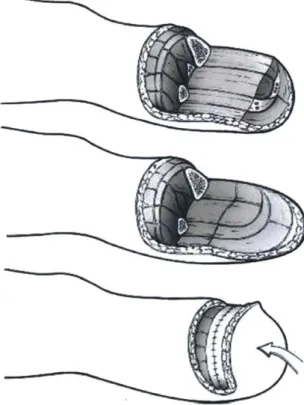
Transtibial amputation is any limb removal between the knee and ankle which tran-sects the tibia. In the standard procedure starting with an intact limb (not always guaranteed), a primary incision creates a posterior flap of tissue from the gastroc-nemius and soleus muscles as seen in Figure 1-5. This flap is cut to maximize skin length to prevent suture tension when it is eventually closed over the distal end of the residuum. While the flap is still open, the tibia and fibula are cut to preserve as much length as possible, a factor that is known to improve long-term outcomes associated with gait symmetry and musculoskeletal health. The bone ends are filed
to remove sharp protrusions, and the fibula is shortened another 1 to 2 cm proximally to avoid creating a pressure point when seated into a prosthetic socket. Nerve ends are pulled distally before being further transected and placed in a location with min-imal expected tension and scarring. Muscle ends are shaped, folded over, and tied or otherwise anchored to connective tissue and bone before the posterior tissue flap is sutured to the anterior incision line [11].
Figure 1-5: The standard transtibial amputation restructures residual lower leg musculature and tissue to provide protection to distal bone ends and padding for prosthetic socket use. Reproduced with permission from Springer Nature.
Because alpha motor neuron innervation is generally preserved, the person with amputation is able to activate and contract residual musculature. However, percep-tion of afferent feedback varies from pain and numbness to phantom limb sensapercep-tion and is decidedly dissimilar from intact physiology [12]. From the mechanical perspec-tive, the activated agonist is in series with the effectively infinite impedance of the anchored sutured end, and no antagonist feedback can be generated in this arrange-ment. Admittedly, though studies have been performed to characterize the cutaneous
and proprioceptive sensation of the traditional transtibial residuum [13, there are no known studies that have quantitatively measured isolated afferent signal generation to verify if the previous mechanical argument is responsible for the erroneous afferent sensations. The following section proposes a method which restores a physiologi-cally relevant agonist-antagonist configuration in the residual musculature that may also alleviate some of these symptoms through generation of natural proprioception related to agonist-antagonist stretch.
1.3.2
Restoration of Relevant Proprioception
The AMI is a surgical construct that distally connects portions of two natural agonist and antagonist muscles in series through a harvested synovial canal. The agonist-antagonist pair is able to move freely through the anchored canal which acts as a fixed pulley. When one muscle in the pair is activated, its contraction creates tension and corresponding stretch in the antagonist. Because the AMI is joined in the center
by residual tendons from both muscles, muscle spindle and golgi tendon afferents
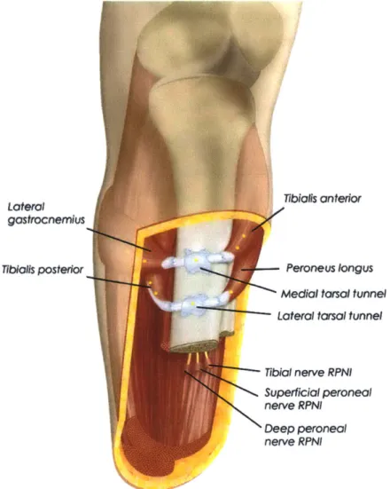
are simultaneously generated from both agonist and antagonist [14]. This contrasts significantly with the infinite impedance encountered by muscles in the standard transtibial amputation. Two AMIs constructed within a transtibial residuum can be seen in Figure 1-6.
The AMI was first conceptualized as a method for bidirectional communication with active prostheses. Because it is possible to simultaneously read electromyo-graphic activity from the interface while the user receives proprioceptive feedback, it was hypothesized that a reasonable mapping could be achieved between phantom limb perception and forward control of an active prosthesis. Case studies have shown early evidence that after some training, AMI users can effectively understand AMI proprioceptive force, length, and velocity feedback signals to more accurately modu-late their muscle activations for a given task compared to a control group of persons with standard amputation [15]. In the other direction, because AMIs remain me-chanically uncoupled from the outside environment, artificially providing feedback is necessary for conveying contact dynamics in a biomimetic manner. To this end,
Lateral gastrocnemius TibIolls posterior
I
fi
I
Tibial nerve RPNI Superficial peroneal
nerve RPNI
Deep peroneal
nerve RPNI
Figure 1-6: Transtibial amputation with two AMIs constructed from primary DP and IE agonist-antagonist muscles. Regenerative peripheral nerve interfaces are created for other nerve endings during the reconstruction procedure [15].
there is promising evidence showing relevant differentiation of force feedback through functional electrical stimulation of AMI pairs [16]. Additionally, optogenetic stimu-lation of muscle activation triggered by single wavelengths of light has been proven in animal models [17].
1.3.3
Dynamic Model of the AMI
Just as physically intact agonist and antagonist muscles are able to independently modulate net torque and stiffness about a rotational axis by varying activation and
Tibialls anterior Peroneus longus
Medial torsal tunnel
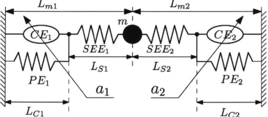
coactivation, AMI pairs should able to independently modulate net force and stiffness in a linear domain. The mechanical model of Figure 1-7 representing major AMI components demonstrates the similarity in structure.
-2 Lmi-1,, Lm2 m i C 2 SEE1 SEE-V2 Ls1 'Ls2 PE1 i PE2 I
1
a,2
I Lc1 LC2Figure 1-7: Agonist-antagonist configuration of two Hill-type muscles with parallel elasticity
and point mass. Reproduced with permission from John Wiley and Sons [18].
However, it is currently uncertain whether users truly perceive proprioception from the AMI as representative of intact limb movement. Even if all the proprioceptive pieces can be accounted for by the model, nothing can be said about what values the model parameters must be in a real AMI to convince the user it is anything but two formerly intact muscles tied together. An intact limb also possesses much greater dimensionality in terms of number of muscles and number of states necessary to describe its internal physical system. It is uncertain if this level of complexity must be matched for the AMI user to acquire a full sense of embodiment when controlling an active prosthesis.
1.4
Kinematic Characteristics of Volitional Motor
Control
In the early 1980s when Japan's precision automation made it the most efficient au-tomobile manufacturer in the world [19], there was still no bipedal robot that could walk convincingly at even normal speeds [20]. It was perhaps this superior dexter-ity and robustness of human motion compared to even state-of-the-art robots of the era which encouraged biomechanics researchers to explore a prominent question re-garding human movement, namely: How can control and organization of movement be explained from the interactions between human perception and the neuromuscu-loskeletal system [21]?
1.4.1
Stereotypical Velocity Profiles
To elucidate this connection between wetware and kinematics, biomechanicists have given special attention the stereotypical bell-shaped velocity profiles which result from volitional motions. Morasso, Abend, and Bizzi first reported velocity profiles of the human hand as being generally invariable to starting position and joint angle configuration for various straight reaching motions [22],[23]. These three researchers proposed possible explanations for this stereotypical velocity profile based on high level organizing principles decided by the central nervous system or the anatomical arrangement of muscles and joints, but did not investigate in detail at the time. Today, there are two main opposing model types, referred to as kinematics-oriented models and dynamics-oriented models, that are used to explain volitional motor behavior and velocity profile observations [24]. Key arguments for both are detailed below.
Kinematics-oriented Models
In kinematics-oriented models, human motor control is a three-tiered system com-posed of trajectory formulation, dynamic primitives mapping, and muscle activation in descending heirarchical order. Under this paradigm, trajectories are planned
cor-responding to task space variables external to our bodies while force disturbances experienced during realization of those trajectories are compensated for via joint space variables. The significance of the previous discoveries lies in the fact that com-plex human movement was sufficiently summarized by a reduced order model that was not only descriptive, but also generative.
This class of kinematics-oriented models developed from early analysis of hand ve-locity profiles when it was suggested that planar horizontal reaching motions involving the shoulder and elbow joints follow trajectories which can be described as maximally smooth. In other words, for point-to-point motions of the hand, the time integral of its mean-squared jerk (time derivative of acceleration) trajectory is minimal for the functionals over the duration of the movement according to
C(X(t),y(t)) = 2
f
(dt) + 2(dt3t)
dt (1.3)where C is the cost to be minimized, tf is the movement end time, and x(t) and y(t) are the Cartesian coordinates of the position trajectory.
T4
Figure 1-8: Reaching experiment setup used by [21],[221. Copyright 2019 Society for Neuro-science.
Generation of such aminimum-jerk trajectory given time interval ofmovement, start position, and end position can be done by separately finding x(t) and y(t) which satisfy the Euler-Poisson equation
SL d{8L) (\ d" 3L
__ -.cit .I+±(-1)" (T =)0 (1.4)
6x di 6x dt" (x
where L is the integrand of 1.3, n is the greatest degree of differentiation in L, and x in this case is any function of time that is continuously differentiable at least 2n times in the specified time interval. A thorough treatment has been provided by Flash and Hogan [21] which, after assuming static kinematic boundary conditions, yields a minimum-jerk trajectory per coordinate of the form
x(t) = xi + (xf - Xi)
(15
+6() (1.5)where xi is initial position, xf is final position, and t is current time point within the movement of duration d.
In the same publication, Flash and Hogan extended the minimum-jerk criterion to explain curved trajectories for reaching motions which include an intermediate position target. Because smoothness is an invariable feature of both straight and multiple-point hand trajectories affine relative to the global axes of the workspace
[22],[23],[25], planning for reaching motions in terms of end effector variables as
op-posed to joint variables remains a strong hypothesis.
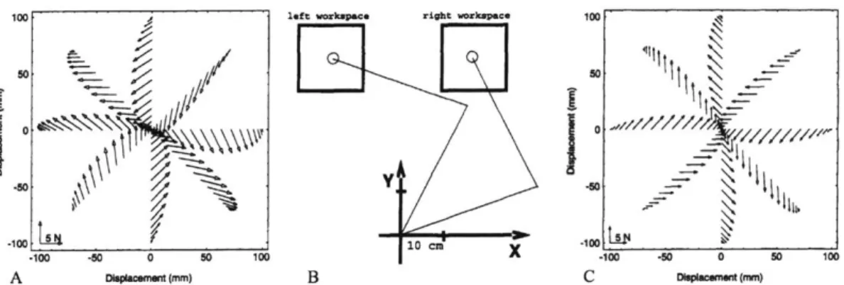
However, knowledge of joint variables is necessary to compensate for external force disturbances when realizing minimum-jerk reaching motions. Shadmehr and Mussa-Ivaldi [26] demonstrated this elegantly with another planar horizontal reach-ing experiment, now with imposed velocity-dependent force fields that were either invariant to the global position of the workspace or to the relative angular positions of the elbow and shoulder. The latter setup is shown via Figure 1-9.
Shadmehr and Mussa-Ivaldi highlighted several important phenomena regarding disturbed reaching motions. A non-exhaustive list follows:
1. Subjects produced minimum-jerk trajectories with no force disturbances
ap-plied, but demonstrated significant deviations from these trajectories when dis-turbed while still eventually reaching the target positions.
-- 1
100 left workspace right workspace 100
-X
j0
0
410 L5 40 .10410 CID____100 .100 -M 0 50 100 X -10 .50 0 so 100 A pa~cemtmm) B C 0hpswomemm)Figure 1-9: Reaching experiment with disturbance force vectors generated for point-to-point reaching motions assuming minimum-jerk trajectories in joint angle invariant forcefields.
A) Disturbance forces anticipated in the left workspace of B) the experimental setup. C)
Disturbance forces anticipated in the right workspace. Copyright 2019 Society for Neuro-science.
2. Subjects were able to compensate for invariant force disturbances after train-ing over hundreds of trials, returntrain-ing to trajectories closely resembltrain-ing the minimum-jerk trajectories in the undisturbed condition. The learning rate re-mained positive but monotically decreased with increasing numbers of trials.
3. Subjects demonstrated residual compensatory behavior from repeated exposure
to force disturbances when attempting to move in the same workspace without disturbances.
4. When trained in the right workspace of Figure 1-9B with force disturbances gen-erated from the underlying velocity-dependent forcefield of Figure 1-9C, subjects demonstrated completely dissimilar and non-smooth trajectories when suddenly moved to the left workspace and subjected to the same forcefield.
5. When trained in the right workspace of Figure 1-9B with force disturbances
gen-erated from the underlying velocity-dependent forcefield of Figure 1-9C, subjects demonstrated similar minimum-jerk trajectories when suddenly moved to the left workspace and subjected to the forcefield underlying Figure 1-9A.
6. Subjects with no visual feedback during these experiments demonstrated similar
Consideration of points 4 and 5 together provided significant evidence of com-pensatory motor learning behavior based on joint variables rather than task space variables. In other words, a roughly 45 counterclockwise rotation at the shoulder and a similar rotation of the forcefield in the translated workspace allowed subjects to reproduce similarly smooth trajectories. If instead compensatory behavior were task space dependent, similarly smooth trajectories would be generated with colo-cated translation of the work space and unrotated forcefield.
While the minimum-jerk hypothesis is remarkable for mathematically generat-ing these results which agree with observed reachgenerat-ing kinematics in a dimensionally-reduced manner, it alone does not sufficiently explain how the body as a biological entity produces these trajectories.
In 2000, Mussa-Ivaldi and Bizzi proposed a model of motor learning and actuation using the idea of dynamic motion primitives [27]. These motion primitives are distinct spatially varying forcefields that are uniquely activated via discrete modules formed
by premotor circuits in the spinal cord. They were shown to be capable of being
vectorially added to produce virtual trajectories consistent with Hogan's [28] well-known paradigm of impedance control. As a demonstration of the explanatory ability of dynamic motion primitives, the authors were also able to recreate the undisturbed and disturbed reaching trajectories of Shadmehr and Mussa-Ivaldi [26] using only four primitives, generating bell-shaped velocity profiles in the process. Details are shown
in Figure 1-10.
Importantly, the notion of dynamic motion primitives was supported by previous work observing the hindlimb forcefields generated by selective spinal stimulation in rats and frogs [29],[30], providing a holistic view of organizing principles as a result of biological processes.
Dynamics-oriented Models
In dynamics-oriented models, human motor control is instead sufficiently described
by coded patterns based on neural biology, muscle state, and mechanical properties [31]. These codes have been presented as any number of piecewise or continuous
A 0.5 - 0--0.5 -0 0 -0.5 0 0.5 I I I -0.5 0 0.5 I I t I -0.5 0 0.5
.
0
0
-0.5 0 0.5 B 0.5 0.4 0.3 0.2 0.1 0 - I -0.2 0 x (M) 0.2 C ... - I -0.2 0 0.2 x (m) D -'7..: ~ a a -0.2 0 x (M) 0.2Figure 1-10: A) Four motion primitives with temporally variable magnitude which can be vectorially added B) Target positions in right workspace of Figure 1-9B C) Undisturbed reaching trajectories generated from dynamic motion primitives D) Disturbed reaching tra-jectories after the forcefield underlying Figure 1-9C was applied to C. Reproduced with
permission from The Royal Society.
mathematical functions, linear and nonlinear, that generate speed, force, or oscillation
[24].
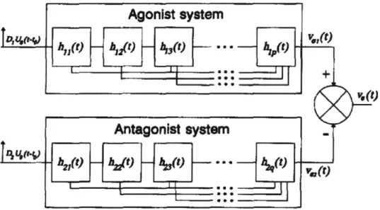
Plamondon's support-bounded log-normal function (lgnb) comes from one such model that has demonstrated superior velocity profiling abilities compared to many other kinematics- and dynamics-oriented models [32]. Its form is derived from the interactions of a synergistic agonist-antagonist neuromuscular system where each mus-cle's individual motor components have both highly parallel and highly hierarchical shared activation inputs, as shown in Figure 1-11.
Plamondon states that if the individual impulse responses from each module of type h(t) can be linearly superimposed, and if each module experiences an activation time delay relative to its immediately superior module, then the time delay T" for the nth module in the sequence can be expressed as
hy(t) h1 t) h,(t) • -
t)
(t)Antagonist system.
Figure 1-11: Agonist-antagonist muscle system which accepts delayed activation impulses
Uo(t - to) of some magnitude D and outputs end effector velocity V,(t) after filtering through cascaded modules of type h(t). Reproduced with permission from Springer Nature
[33].
T, = (1 + Ci)(1 + 2)(1 + 3)...(1 + Ce)T (1.6)
where To is an arbitrary time reference and {j
j
CN,j < n} is the set of allprevious random time delays. If additionally the impulse responses are non-negative functions, n is sufficiently large, and each response has finite variance, then the central limit theorem predicts V,(t) will be log-normal in shape. Bounding the support of this log-normal function ensures velocity at the end of movement is zero, and the resultant lgnb function is defined as
D(ti - to) 1 (t -t ) (1.7)
EOt) = -exp - 2 [n 1 - p (1.7
o-7(t - to)(ti - t) 2. 2
L(t
1- t)where D is total displacement of the movement, to the time of the impulse com-mand, t1 the end time of the movement, y and o.2 the mean and variance of ln(t - to).
A thorough derivation may be found in [32].
Determination of the descriptive parameters D, to, ti, y, and o- for a given move-ment can be performed computationally through iterative nonlinear least squares
gression, resulting in close agreement with recorded velocity profiles as seen in Figure 1-12. Compared to minimum-jerk, the form of lgnb is capable of describing the skew observed in many wrist velocity profiles while the objective function of minimum-jerk limits it to symmetric solutions. However, the generative form of minimum-minimum-jerk seen in Equation 1.5 allows trajectories to be planned in advance while lgnb is purely descriptive. This fact alone raises questions about the sufficiency of similar dynamics-oriented models in describing formulation of movement. A parallel line of questioning regarding sufficiency of velocity profile analysis itself is raised when considering the uniqueness of handwriting, even between identical twins [34]. Though invariances between individuals should exist for any given task on some level, what is the limit of their ability to explain motor control even one level up the cognitive hierarchy?
SUpport-bounded
ft-l0ima
30 AMPitude (o1)9
I
1
IIs
0
0
0,61i0,666
0.7150,766
Ti.
(a)
Figure 1-12: Reconstructed velocity profile (solid line) with parameters D = 1.990, to= 0.6195, ti = 0.7735, L = -0.162, o- = 0.8325. Reproduced with permission from Springer
Differences in modeled stereotypical velocity profiles of human motion and the bi-ological processes underlying their generation may also depend on the specific joints and limbs involved. It is critical to note that the experiments which preceded formula-tion of the kinematics-oriented minimum-jerk hypothesis and the dynamics-oriented lgnb function were fundamentally different. Upper limb reaching experiments per-formed by Flash and Hogan [21] involved multiple joints connecting body segments with significant mass moving the hand over distances of 20 - 40 cm while the trajec-tories analyzed by Plamondon et al. [35] involved only the wrist and fingers moving
distances of 4 cm.
1.4.2
Speed-Accuracy Tradeoff
Another hallmark of human motion is the speed-accuracy tradeoff. It is a relationship first quantified by Paul Morris Fitts [36] and described mathematically as
ID= 1og2 )(1.8)
(W
where ID is the index of difficulty in bits, D is the linear distance from starting point to target, and W is the width of the target along the axis of motion. ID can be related to movement time in terms of throughput (information per unit time) by
TP =
(fID)(1.9)
MT
where MT is average time to complete the movement.
When 1.8 and 1.9 are taken together, average time required to complete a move-ment can be directly related to target size and distance to target
MT= a+b log22 j)(1.10)
where a and b are empirically determined constants based on regression analysis of a given task's data. This basic form of Fitts's Law has been successfully adapted to account for observed limitations in human motor performance across many tasks,
even serving as the prime reason for selection of the computer mouse as the preferred method of interaction with a digital display [37]. Though the explanatory power of Fitts's Law does not scale as any one of the temporal or physical parameters is tested at extreme ranges, it remains an easily interpreted metric used to quantify certain em-pirical observations of motor behavior without expounding underlying biomechanical causes.
Michmizos and Krebs [38] have investigated discrete ankle-foot pointing with anal-ysis based on Fitts's Law, concluding that movement time was highly correlated with
ID in both DP and IE directions while peak velocity remained relatively constant.
Additionally, while increasing ID did not change the value of maximum velocities observed, it did reduce the average velocity for the movement due to the presence of slower, corrective submovements near the target to correct for overshoot and under-shoot.
A study on rhythmic upper limb motions by Park et al. [39] also demonstrated
the existence of characteristic preferred movement times, indicating a limit where humans no longer generate smooth motion with further reductions of velocity. In the study, movements of a given displacement which were slower than the preferred time showed evidence of being composed of multiple submovements while movements at the preferred time and faster demonstrated single lgnb velocity profiles. It was the fact that submovements themselves could also be described by multiple overlapping lgnb functions which provided evidence of dynamic motion primitives which are linearly composable.
1.4.3
Coordination of Bimanually Symmetric Movement
A study by Mechsner et al. [40] investigated subtleties of human motor control during bimanual coordination tasks. The researchers discovered that coordination of upper limb movement involving both sides of the body was largely dependent on visual perception rather than any sort of variables related to internal body state. They demonstrated this phenomenon primarily through three tests, two of which are described here.
The first test revisited the bimanual finger coordination studies well-known in the field from a slightly different perspective [41], [42]. The test conditions are shown in Figure 1-13.
A
-QfV7
B
F
Figure 1-13: A) Bimanual coordination symmetric condition B) Parallel condition C-F) Orientations of Mechsner et al.'s coordination tests. Reproduced with permission from Springer Nature [40].
The original test requires participants to adduct and abduct their index fingers in rhythm with a metrononome while the tempo increases. Participants are instructed to maintain their hands in the orientation of Figure 1-13C while performing periodic symmetric (Figure 1-13A) or parallel (Figure 1-13B) motions. As the tempo increases, participants generally converge on the symmetric pattern regardless of the pattern with which they begin. Early hypotheses proposed the results indicated preference for activation of homologous muscles [41], while variations of the test involving flexion and extension of ipsilateral limbs suggested easier coordination when moving in the
C6
E6
same direction regardless of actual muscles used [43].
It was Mechsner et al. who performed the test with all four orientations shown in Figure 1-13. For all conditions, symmetry was either maintained or adapted as tempo increased. This definitively disproved the proposal that symmetry arose from preferred activation of homologous muscles. To emphasize the importance of visual perception of task space variables, they designed another test shown in Figure 1-14.
A B
Figure 1-14: A) Experimental setup requiring separate instrument rotation through differ-ent transmission ratios B) In-phase condition C) Anti-phase condition. Reproduced with permission from Springer Nature [40].
In the setup above, the left flag above the divider circles at a 1:1 ratio with the left hand while the right flag circles at a 4:3 ratio with the right hand. Earlier tests demonstrated that explicitly attempting to rotate left and right hands at a 4:3 ratio was nearly impossible. With the experiment, it was found that participants were able to easily maintain the in-phase condition by observing the flags, thereby achieving a 4:3 rotational frequency between left and right hands.
1.5
Background Summary
With the introduction of the AMI comes a new research platform ready for explo-ration. However, it remains to be determined if this construct is intuitive and ad-vantageous for control of a modeled limb or active prosthesis beyond the capability of persons with standard amputation. To provide hope for the endeavor, numerous studies on upper limb coordination and volitional movement have suggested the hu-man ability of reducing complex trajectories into simple task space representations which are then carried out by lower levels of muscle control. From the mechanical perspective, the AMI should generate a descriptive set of sensory feedback through relevant proprioceptive signals which more accurately informs these lower level con-trollers of its state in time. Though each AMI is only one muscle pair which does not compare to the redundancy of intact human musculature, it is perhaps just enough to draw the map between intent and actuation.
Chapter 2
Coordination and Control Utilizing
the Agonist-Antagonist Myoneural
Interface
This thesis aims to quantify the volitional coordination capabilities of the agonist-antagonist myoneural interface (AMI) for applications related to control of active prostheses. In the first section, bilateral rhythmic coordination of ankle and subta-lar movements is investigated in a control group of physically intact human subjects to characterize stereotypical kinematics of volitional lower limb movement. Subse-quently, neuromusculoskeletal (NMS) modeling techniques are developed to directly map estimated neural excitations from agonist-antagonist myoneural interface mus-culature to intended subtalar inversion and eversion kinematics.
In a case study, the developed neuromusculoskeletal modeling techniques are ap-plied to optimize a dynamic subtalar model for use by a unilateral subject with amputation possessing the agonist-antagonist myoneural interface. The subject's sub-sequent performance in bilateral rhythmic coordination utilizing the model and her own intact subtalar demonstrates the capacity of the agonist-antagonist myoneural interface to coordinate with intact anatomy in a biomimetic manner.
2.1
Contributions
Characteristics of Intact Lower Limb Movement
Preliminary evidence supports the following hypotheses regarding characteristics of intact lower limb movement:
• Bilateral rhythmic coordination of subtalar inversion-eversion (IE) demonstrates greater stability in the symmetric rather than the parallel pattern as frequency increases, agreeing with findings on bimanual coordination.
•
Symmetric rhythmic coordination of ankle dorsiflexion-plantarflexion (DP) is less variable than subtalar IE with respect to relative phase within a given frequency, suggesting greater volitional control of DP over IE.• Rhythmic IE motions demonstrate stereotypical velocity profiles, agreeing with findings on coordination of upper limb movements.
* There are no significant differences observed between sighted and blind condi-tions in rhythmic coordination of IE.
• Evidence is shown of movement amplitude and effective target width varying in accordance with Fitts's Law as frequency of rhythmically coordinated move-ments increases.
AMI Control of the Optimized Neuromusculoskeletal Subtalar Model
Preliminary evidence supports the following statements regarding AMI control of the optimized neuromusculoskeletal subtalar model:
• Bilateral rhythmic coordination of IE involving the optimized NMS model and intact subtalar also demonstrates greater stability in the symmetric rather than parallel pattern.
* The optimized NMS model is general enough to achieve movement frequen-cies beyond the one used for optimization while respecting human bandwidth limitations predicted by Fitts's Law.
9 The optimized NMS model generates velocity profiles that differ significantly from those of an intact subtalar, though subsequent analysis suggests pathways for improvement.
2.2
Methodology
2.2.1
Subjects
Physically intact female subjects (n = 4) were recruited for the control group,
aver-aging 21 ± 1 years old, 1.62± 0.05 m, and 73± 12 kg (mean ± standard deviation). A single female, Subject BKA (below knee AMI), who posseses two AMI pairs through elective unilateral transtibial amputation was also recruited (42 years old, 1.68 m, 85
kg). One pair was constructed from her tibialis anterior (TA) and lateral
gastrocne-mius (LGAS) for primary control over ankle DP. A second pair was constructed from her tibialis posterior (TP) and peroneus longus (PL) for primary control over subta-lar IE. All subjects were right foot dominant and had normal vision. In a precheck questionnaire, all subjects indicated no physiological disorders which would affect their ability to control movement of their intact ankle-foot joints. Experiments were conducted with informed consent at the Massachusetts Institute of Technology (MIT) Media Laboratory under the approval of the MIT Committee on the Use of Humans as Experimental Subjects.
2.2.2
Apparatus and Signals Processing
For all experiments, surface electromyography (sEMG) signals and ankle-foot kine-matics were collected from subjects. An electromechanical relay was used to synchro-nize recorded data in time.
sEMG
A commercial Refa (TMSi, Oldenzaal, Netherlands) 128-channel amplifier was used
alcohol, and adhesive wet surface electrodes were placed on the dominant leg in redundant pairs over exposed locations nearest to the bellies of the following muscles: tibialis anterior (TA), medial gastrocnemius (MGAS), lateral gastrocnemius (LGAS), peroneus longus (PL), and tibialis posterior (TP). A ground reference electrode was placed on the patella of the dominant leg. For Subject BKA, additional electrodes were placed in a gridlike pattern over the estimated locations of the four muscles which compose her two AMI pairs. 1.5 m shielded cables connected each electrode terminal to the Refa amplifier.
4b
20
1
Figure 2-1: Clusters of four wet surface electrodes were placed over each AMI muscle. Pic-tured are the PL cluster (left) and TA cluster (right). Antagonist muscles for both are located medially on the residuum.
For each trial, monopolar sEMG signals were recorded at a sampling frequency of 2,048 Hz. Bipolar signals were reconstructed for each muscle after data collection.
A fourth order 10 - 500 Hz band pass infinite impulse response (IIR) Butterworth
filter was designed in MATLAB 2019a and applied forward and backward over each recording. Filtered data were then rectified and normalized against the maximum voltage within each recording, assumed to represent maximum voluntary contraction (MVC). Then, a fourth order 10 Hz low pass IIR Butterworth filter was applied forward and backward to produce a record of neural excitation p(t). See Buchanan et al. [44] for details on this sEMG processing procedure. For experiments involving
NMS model optimization, sEMG recordings were level shifted so that the minimum
excitations across the entire trial were 0.01 to satisfy Thelen Hill-type muscle tendon unit (MTU) constraints [9].
Though sEMG was recorded from all intact subjects, the data are not reported here.
Ankle-foot Kinematics
A pair of commercial two-axis goniometers (Biometrics Ltd., Newport, UK) was used
to measure ankle-foot kinematics. Subjects were required to wear well-fitted athletic shoes. For each goniometer, one end was adhered to the heel of the shoe while the other was adhered to skin superior the Achilles tendon. Goniometers were calibrated for measurement axis and motion axis parallelism by instructing subjects to perform
slow, controlled motions while adjusting the positioning of the sensors. Subjects were
carefully guided to compensate for the naturally oblique DP axis of rotation during this portion. Afterward, goniometer ends were further secured to the shoe and shank using porous medical tape.
All ankle-foot position trajectories were recorded at a sampling frequency of 1,000
Hz. A fourth-order 5 Hz low pass IIR Butterworth filter was applied forward and backward over the raw trajectory data. Velocity trajectories were generated from the filtered position trajectories by two-point forward finite differentiation.
2.2.3
Bilateral and Unilateral Coordination Tasks
With inspiration from Mechsner et al.'s bimanual coordination [40] and Park et al.'s rhythmic reaching studies [39], all subjects were asked to complete a series of rhythmic tasks involving ankle DP and subtalar IE to determine characteristics and limitations of lower limb coordination. Subjects were seated on the edge of a patient bed with knees at 90 flexion. sEMG electrodes and goniometers were attached as described in the previous section. Each trial required subjects to perform one cyclical motion per downbeat of a pre-recorded metronome soundtrack. The one minute soundtrack
![Figure 1-3: Type Ia muscle spindles spiraling around intrafusal muscle fibers [7].](https://thumb-eu.123doks.com/thumbv2/123doknet/14492851.526200/23.917.267.620.212.481/figure-type-muscle-spindles-spiraling-intrafusal-muscle-fibers.webp)
![Figure 1-8: Reaching experiment setup used by [21],[221. Copyright 2019 Society for Neuro- Neuro-science.](https://thumb-eu.123doks.com/thumbv2/123doknet/14492851.526200/32.917.282.594.640.890/figure-reaching-experiment-copyright-society-neuro-neuro-science.webp)