HAL Id: inserm-03090395
https://www.hal.inserm.fr/inserm-03090395
Submitted on 29 Dec 2020HAL is a multi-disciplinary open access archive for the deposit and dissemination of sci-entific research documents, whether they are pub-lished or not. The documents may come from teaching and research institutions in France or abroad, or from public or private research centers.
L’archive ouverte pluridisciplinaire HAL, est destinée au dépôt et à la diffusion de documents scientifiques de niveau recherche, publiés ou non, émanant des établissements d’enseignement et de recherche français ou étrangers, des laboratoires publics ou privés.
Vector competence of anthropophilic mosquitoes for a
new mesonivirus in Senegal
Alioune Gaye, Moussa Moïse Diagne, El Hadji Ndiaye, Marie Henriette Dior
Ndione, Martin Faye, Cheikh Talla, Gamou Fall, Yamar Ba, Diawo Diallo,
Ibrahima Dia, et al.
To cite this version:
Alioune Gaye, Moussa Moïse Diagne, El Hadji Ndiaye, Marie Henriette Dior Ndione, Martin Faye, et al.. Vector competence of anthropophilic mosquitoes for a new mesonivirus in Senegal. Emerging microbes & infections, Earliest : Springer-Nature ; Latest : Taylor & Francis, 2020, 9 (1), pp.496-504. �10.1080/22221751.2020.1730710�. �inserm-03090395�
Full Terms & Conditions of access and use can be found at
https://www.tandfonline.com/action/journalInformation?journalCode=temi20
Emerging Microbes & Infections
ISSN: (Print) 2222-1751 (Online) Journal homepage: https://www.tandfonline.com/loi/temi20
Vector competence of anthropophilic mosquitoes
for a new mesonivirus in Senegal
Alioune Gaye, Moussa Moïse Diagne, El Hadji Ndiaye, Marie Henriette Dior
Ndione, Martin Faye, Cheikh Talla, Gamou Fall, Yamar Ba, Diawo Diallo,
Ibrahima Dia, Pascal Handschumacher, Ousmane Faye, Amadou Alpha Sall
& Mawlouth Diallo
To cite this article: Alioune Gaye, Moussa Moïse Diagne, El Hadji Ndiaye, Marie Henriette Dior Ndione, Martin Faye, Cheikh Talla, Gamou Fall, Yamar Ba, Diawo Diallo, Ibrahima Dia, Pascal Handschumacher, Ousmane Faye, Amadou Alpha Sall & Mawlouth Diallo (2020) Vector competence of anthropophilic mosquitoes for a new mesonivirus in Senegal, Emerging Microbes & Infections, 9:1, 496-504, DOI: 10.1080/22221751.2020.1730710
To link to this article: https://doi.org/10.1080/22221751.2020.1730710
© 2020 The Author(s). Published by Informa UK Limited, trading as Taylor & Francis Group, on behalf of Shanghai Shangyixun Cultural Communication Co., Ltd
Published online: 28 Feb 2020.
Submit your article to this journal
Article views: 551
View related articles
Vector competence of anthropophilic mosquitoes for a new mesonivirus in
Senegal
Alioune Gayea, Moussa Moïse Diagneb, El Hadji Ndiayea, Marie Henriette Dior Ndioneb,c, Martin Fayeb, Cheikh Tallae, Gamou Fallb, Yamar Baa, Diawo Dialloa, Ibrahima Diaa, Pascal Handschumacherd, Ousmane Fayeb, Amadou Alpha Sallband Mawlouth Dialloa
a
Unité d’Entomologie Médicale, Institut Pasteur de Dakar, Dakar, Sénégal;bPole de virologie, Unité des Arbovirus et virus de Fièvres Hémorragiques, Institut Pasteur de Dakar, Dakar, Sénégal;cUniversité Cheikh Anta Diop de Dakar, Dakar, Sénégal;dAix Marseille Univ, INSERM, IRD, UMR SESSTIM, Sciences Economiques & Sociales de la Santé & Traitement de l’Information Médicale, Marseille, France;
e
Epidemiology of infectious diseases unit, Institut Pasteur de Dakar, Dakar, Senegal
ABSTRACT
The mesoniviruses (MESOVs) belong to the newly described Mesoniviridae family (Order: Nidovirales). They have never been reported in Senegal until recently during a study in arbovirus emergence with the detection of a new species of MESOV named Dianke virus (DKV) from common mosquitoes from eastern Senegal. Actually, their vector competence for this newly described DKV is unknown. We, therefore, estimated the vector competence of Culex tritaeniorhynchus, Culex quinquefasciatus, Aedes aegypti, and Anopheles gambiae mosquitoes collected in Senegal for DKV using oral infection. Whole bodies, legs/wings, and saliva samples were tested for DKV by RT–PCR to estimate infection, dissemination, and transmission rates. The infectivity of virus particles in the saliva was confirmed by infecting C6/36 cells. Virus transmission rates were up to 95.45% in Culex tritaeniorhynchus, 28% in Cx. quinquefasciatus and 9.09% in Aedes aegypti. Viral particles in the saliva were confirmed infectious by C6/36 cell culture. An. gambiae was able to disseminate DKV only at 20 days post-infection. This study shows that Culex mosquitoes are more competent than Ae. aegypti for DKV, while Anopheles gambiae is not likely a competent vector.
ARTICLE HISTORY Received 12 December 2019; Revised 11 February 2020; Accepted 12 February 2020 KEYWORDS Vector competence; Dianke virus; Culex; Aedes; Anopheles
Introduction
Until 2011, the Nidovirales was known as an order of positive-sense single-stranded RNA (ssRNA+) viruses that included three families: the Arteriviridae, consisting of small-genome nidoviruses (12.7–15.7 kb), the Corona-viridae and the RoniCorona-viridae, both consisting of large-gen-ome nidoviruses (26.3–31.7 kb). The discovery of Nam Dinh [1] and Cavally [2] viruses in Vietnam and Côte d’Ivoire, respectively, resulted in the addition of a fourth family named the Mesoniviridae, in reference to their medium-sized genomes (about 20 kb) [3]. The viral par-ticles are enveloped, 60–80 nm diameter spheres with club-shaped surface spikes [4]. Their 20 kb genome is generally organized as ORF1a-ORF1b-ORF2a-ORF2b-ORF3a-ORF3b-ORF4 [5]. ORF1a and ORF1b are pre-dicted to encode two polymerase polyproteins (pp), while the ORFs in the 3’-end encode structural proteins such as the spike (S) glycoprotein (ORF2a), the nucleo-capsid (N) protein (ORF2b), and two proteins with mem-brane-spanning regions (ORF3a and -3b) [1,2].
Mesoniviruses are considered as insect-specific viruses (ISVs) as they were isolated only from mosquito
pools or cell lines with no detection in vertebrates [1,2,5,6]. ISVs were also found in sandflies [7] and chir-onomids, indicating that they can also infect other dip-terans. An increasing number of arthropod-specific viruses (ASVs) are being discovered in haematophagous arthropods over the world [8], and there is a growing interest on mesoniviruses interactions with viruses of public health concern to develop strategies that could curb arbovirus transmission through dual infection and competition, as implemented with the Wolbachia mediated strategy [9]. Some studies suggested for instance that ISVs negatively affect the fitness of West Nile (WNV) [10–12] and dengue (DENV) [13,14] viruses in the mosquito, while other studies demon-strated that there is no effect [15] or even an enhancing effect [11]. However, there are still few studies on meso-niviruses phenotypic behaviour in mosquito vectors.
A new mesonivirus species was recently reported in Senegal [16]. The so-called Dianke virus (DKV) exhib-ited a wide host range within mosquito populations. Some of them are important vectors for arboviruses or Plasmodium species.
© 2020 The Author(s). Published by Informa UK Limited, trading as Taylor & Francis Group, on behalf of Shanghai Shangyixun Cultural Communication Co., Ltd
This is an Open Access article distributed under the terms of the Creative Commons Attribution License (http://creativecommons.org/licenses/by/4.0/), which permits unrestricted use, distribution, and reproduction in any medium, provided the original work is properly cited.
CONTACT Alioune Gaye alioune.gaye@pasteur.sn Unité d’Entomologie Médicale, Institut Pasteur de Dakar, 36, Avenue Pasteur, Dakar BP 220, Sénégal
Emerging Microbes & Infections 2020, VOL. 9
The aim of this study was to characterize DKV fitness in four different anthropophilic mosquito species of public health concern. This work paves the way toward future investigations on potential inter-actions between DKV and other vector transmitted pathogens and brings forward the possibility of a second host implied in the DKV life cycle.
Materials and methods
Mosquito species
We used four mosquito species representing Culex, Aedes, and Anopheles genus. Cx. quinquefasciatus and Cx. tritaeniorhynchus were collected in Barkedji (15° 16′50.242” N, 14°51′54.751” W). Aedes aegypti and An. gambiae were collected in Dakar (14°43’29” N, 17°28’24” W). These species were chosen because of their abundance, anthropophilic behaviour, and associ-ation with DKV and other previously described meso-niviruses in thefield [2,16].
Larvae and pupae were collected from the field. Adults were reared in the laboratory at 27 ± 1°C and relative humidity of 70–75%, with a 12 h photoperiod. Females (F0) were fed several times on guinea pigs to obtain F1-generation eggs. These were hatched, and the larvae were reared under the conditions described above to obtain the F1 adults used in this study. Mos-quitoes were fed on a 10% sucrose solution.
Virus strain and preparation of the stock
The DKV isolate (Genbank accession number: MN622133) was passaged twice in C6/36 cell cultures (Table 1). An additional passage into monolayers C6/ 36 cells was performed as previously described [16] to obtain the viral stock used to infect mosquitoes. Briefly, Continuous cells lines, initially provided by the American Type Culture Collection (ATCC), were cultured in Leibovitz-15 (L-15) medium (GibcoBRL, Grand Island, NY, USA) supplemented with 10% foetal bovine serum (FBS) (Gibco BRL, Grand Island, NY, USA) and penicillin- streptomycin (Sigma, GmBh, Germany) and 10% of Bacto™ Tryptose Phosphate Broth (Becton, USA) and maintained in 25-cm2tissue culture flasks. The medium was removed from the flasks before adding of 150 µl of the supernatant of the virus directly into monolayers C6/36 cells. After 1 h of incubation at room temperature, infected cells were recovered by 5 ml of L-15 medium supplemented with 5% FBS and incubated at 28°C until cytopathic effect (CPE). After harvesting, infected cells
supernatants were aliquoted, frozen at −80°C, and used as viral stocks for mosquito infection. The virus stock was prepared and titrated using C6/36 cell lines. As plaque formation could not be observed on C6/36 cells, viral titres were evaluated using a pan Mesonivirus RT-qPCR assay previously used for the DKV detection infield samples [16].
After preparation of stocks on cells, viral titre of 1.6 × 10⁷ RNA copies/ml was obtained and used for mosquito infections.
Mosquito oral infections procedure
Three- to five-day-old F1 generation female mosqui-toes were placed into 0.45 L cardboard cages and starved for 48 h before being allowed to take an infec-tious blood meal via an artificial feeder, as previously described [17,18] using mouse skins as membranes. Mosquitoes were fed on an infectious blood meal con-taining 33% of washed rabbit erythrocytes and 33% of viral suspension supplemented with 33% of a mixture of FBS, adenosine triphosphate (ATP) to afinal con-centration of 0.005 M as a phagostimulant and a sol-ution of 10% sucrose. Mosquito feeding was limited to 60 min. Afterwards, blood meals were stored at −80°C to later measure the number of DKV RNA copies per ml using the pan Mesonivirus RT-qPCR assay (Table 2). The mosquitoes were cold-anaesthe-tized to select fully engorged individuals (non-engorged females were discarded), which were trans-ferred to cardboard containers and subsequently main-tained in an incubator at 27 ± 1°C, 70–75% relative humidity, and a 12 h day/night cycle. Mosquitoes were given ad libitum access to a 10% sucrose solution.
Virus detection
At 1, 2, 3, 4, 5, 6, 7, 10, 15, and 20 days post-infection (dpi), samples of Ae. aegypti and An. gambiae were randomly collected. The low sample sizes of engorged females of Culex mosquitoes led us to test Cx. tritae-niorhynchus at 7, 10, and 15 dpi and Cx. quinquefascia-tus only at 15 dpi. Mosquitoes were cold-anaesthetized, and their legs and wings removed and transferred
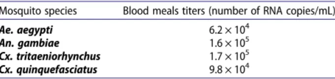
Table 2. Blood meals titres at the end of each mosquito feeding.
Mosquito species Blood meals titers (number of RNA copies/mL) Ae. aegypti 6.2 × 10⁴
An. gambiae 1.6 × 10⁵
Cx. tritaeniorhynchus 1.7 × 10⁵ Cx. quinquefasciatus 9.8 × 10⁴
Table 1.Virus strain used in this study.
Strain GenBank accession number Collection place Original host Passage number on cells Collection date ArD270551 MN622133 Barkedji (Senegal) Culex poicilipes (Mosquito pool) 2 2014
individually into separate tubes. Saliva was collected by inserting the proboscis of each mosquito into a capil-lary tube containing 1–2 μl of FBS for 30 min. After salivation, each mosquito body (whole body except legs and wings) and saliva sample was placed in a sep-arate tube. Detection of DKV in the mosquito body without infection of the legs, which contain haemo-lymph, is an indication of a non-disseminated infection limited to the midgut, whereas the presence of virus in both the mosquito body and legs indicates a dissemi-nated infection with virus in the haemocoel. The mos-quito bodies, legs/wings, and saliva were stored separately at−80°C until real-time RT–PCR could be performed. The infectiousness of positive saliva samples was tested with C6/36 cell cultures.
The samples were crushed and homogenized in 500μl of L-15 cell culture medium (GibcoBRL, Grand Island, NY, USA) containing 20% FBS and were then centrifuged for 10 min at 7500 rpm at 4°C to separate virus supernatant and debris. For real-time PCR, 100μl of supernatant was used for RNA extraction using the QIAamp Viral RNA Extraction Kit (QIAgen, Heiden, Germany), according to the manufacturer’s protocol. The RNA was amplified using ABI Prism 7000 SDS Real-Time apparatus (Applied BioSystems, Foster City, CA, USA) with the QuantiTect kit (QIAgen) and the pan Mesonivirus sys-tem as previously described [16]. The reaction mixture consisted of 5μl of extracted RNA, 10 μl of buffer (2 X QuantiTect Probe), 6.8μl of RNase free water, 1.25 μl of each primer, forward and reverse, 0.5μl of probe, and 0.2μl of enzymes to a total volume of 25 μl. The cycling conditions were RT step at 50.0°C for 10 min, at 95.0°C for 15 min, and 40 cycles of 15 s at 95.0°C and 1 min at 60°C.
Infectiousness of positive saliva samples
The infectiousness of positive saliva samples after amplification on a mosquito species was tested with C6/36 cell culture in 25 cm2flasks. The positive saliva samples were diluted in a 1:10 ratio in cell culture med-ium containing 10% FBS before filtration. Once cells reached a confluence of approximately 70%, the med-ium was discarded and 200μl of diluted positive saliva samples were inoculated into monolayer C6/36 cells as described above. Theflasks were gently agitated every 10 min during incubation to enhance viral dispersion. After 1 h, 5 ml of L-15 medium (5% FBS, 5% tryptose phosphate, 1% glutamine, 1% penicillin–streptomycin, and 0.05% amphotericin B) was added, and the infected cells were incubated (for 3 days) and then har-vested after CPE observation. The medium was then removed and centrifuged for 5 min at 10,000 rpm and 4°C. The supernatant was collected and stored at −80°C until analyses. The detection and quantification of virus was performed using the pan Mesonivirus
RT-qPCR assay. The presence of more viral particles in post-infection cultures than in saliva was taken to indi-cate infectiousness.
Binary logistic regression model
The effects of mosquito species and time after exposure (dpi) on each (i) infection, (ii) dissemination and (iii) transmission phenotype have been assessed using a binary logistic regression model. Possible interactions between independent factors were tested in the model, and likelihood ratio tests comparing models with and without the interaction term were used to estimate the significance of the interaction. The time post-exposure was treated as a continuous variable and Ae. aegypti the main arbovirus vector has been set up as reference compared to other species. The sig-nificance of risk factors was determined by calculating Odds ratio (OR) and 95% confidence interval (CI). N represents the number of mosquitoes tested.
Data analysis
Infection (number of positive bodies/total number of mosquitoes tested), dissemination (number of infected legs and wings/total number of infected bodies), and transmission (number of positive saliva/total number of infected legs and wings) rates were calculated for each species at each dpi. Rates were compared using Fisher’s exact test. Differences were considered statisti-cally significant at P < 0.05. Statistical tests were per-formed using R v. 2.15.1 (R Foundation for Statistical Computing, Vienna, Austria).
Results
A total of 223 Ae. aegypti, 131 An. gambiae, 88 Cx. tri-taeniorhynchus, and 38 Cx. quinquefasciatus were tested. Our results (Figures 1 and 2) showed that, except for An. gambiae, all species were highly suscep-tible to infection by DKV.
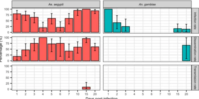
Infection rates of DKV in An. gambiae (Figure 1) decreased after thefirst dpi to 0% at 4 dpi. Recrudes-cence was observed at 15 and 20 dpi in infected An. gambiae with infection rates of 16% and 13.63%, respectively. Even if An. gambiae developed a dissemi-nated infection at 20 dpi, no transmission was observed.
Ae. aegypti was highly susceptible to infection with DKV strain. Infection rates again initially decreased after the first days, before a further increase at 6 dpi to reach 100% by 15 dpi. Dissemination was high and increased between thefirst to fourth dpi. Aedes aegypti were able to transmit DKV at 15 dpi (9.09%).
We limited our subsequent experiments to sample Cx. tritaeniorhynchus at 7, 10, and 15 dpi and Cx. quin-quefasciatus at 15 dpi because of the limited number of
engorged females obtained with these species not allowing testing samples at all dpi as previously.
Culex tritaeniorhynchus and Cx. quinquefasciatus were highly susceptible to infection and were able to transmit DKV. After 7 dpi, transmission rate in Cx. tri-taeniorhynchus reached 77.77%, 95.45% at 10 dpi and 52% at 15 dpi. For Cx. quinquefasciatus transmission rate was 28% at 15 dpi and comparable to those for Cx. tritaeniorhynchus (Fisher’s exact test: P = 0.1).
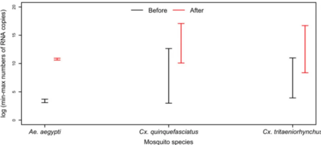
All positive saliva samples induced CPE in C6/36 cells. The presence of infectious viruses in saliva was further revealed by a higher virus RNA load after amplification of C6/36 cells (Figure 3).
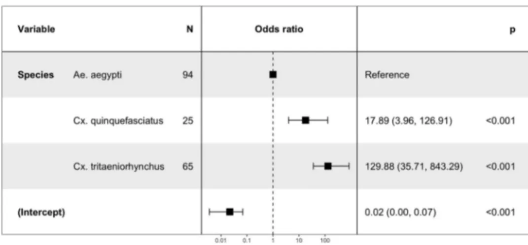
In binary logistic model, Cx. quinquefasciatus was removed from the analysis in infection and dissemina-tion; it was only followed on single time point (at 15 dpi) which does not allow assessing day exposure effect and species-days interaction. Each additional day exposure for Ae. Aegypti is associated with an increased chance of getting infected (OR = 1.14, P < 0.001).
Concerning the effect of mosquito species Cx. tritae-niorhynchus is more likely to become infected than Ae. aegypti (P < 0.001).
The exposure time (days) decreases the chance of infection for Cx. tritaeniorhynchus and An. gambiae
Figure 1.Infection, Dissemination and Transmission rates at 1, 2, 3… 20 dpi for Ae. aegypti and An. gambiae orally exposed to DKV. Errors bar represent the upper limits of the 95% confidence intervals.
Figure 2.Infection, Dissemination and Transmission rates at 7, 10 and 15 dpi for Cx. tritaeniorhynchus and at 15 dpi for Cx. quin-quefasciatus orally exposed to DKV. Errors bar represent the upper limits of the 95% confidence intervals.
(OR = 0.68, P < 0.001; OR = 0.89 (1.14*0.78), P < 0.001 respectively) (Figure 4).
In each additional day post-exposure, the chance of dissemination increases (OR = 1.06, P = 0.01) (Figure 5). DKV is more likely to be disseminated by Cx. tritae-niorhynchus (OR = 5.49, P < 0.001) than by Ae. aegypti and less likely to be disseminated by An. gambiae (OR = 0.06, P < 0.001) (Figure 5). Both Culex species were more likely to transmit DKV (Figure 6). The
interaction terms between species and days were not significant for dissemination and transmission (P > 0.1). The day’s effect was not significant for trans-mission (P = 0.15).
Discussion
DKV is a new mesonivirus species firstly reported in Senegal, West Africa, from several arthropods species,
Figure 3.The intervals (min-max) of numbers of RNA copies of DKV in saliva (black line) and post-infection C6/36 cells cultures (red line).
Figure 4.The risk factors for mosquito infection.
Figure 5.The risk factors for mosquito dissemination.
which include mosquitoes from the Culex, Aedes, Ano-pheles, and Mansonia genera [16]. Even if some studies have also described mesoniviruses infections in mos-quitoes with significant public health impact [1,2,6], there is very little knowledge on theirfitness in vectors. To our knowledge, this study is thefirst assessment of the possibility of transmission of viruses belonging to this group by mosquito saliva. A previous study only reported RNA detection/sequencing of other ISVs from salivary glands [19].
Of the anthropophilic mosquito species tested, the Culex mosquitoes were the most competent, followed by Ae. aegypti (Fisher’s exact test: P = 0.005 and Binary regression logistic: P < 0.001). An. gambiae populations were largely incompetent. Cx. tritaeniorhynchus and Cx. quinquefasciatus were the most susceptible to infection and were able to transmit DKV strain at all dpi tested. Both of these anthropophagic species are common and abundant in domestic environments where Cx. quinquefasciatus lays their eggs on artificial breeding sites (polluted water, septic tanks, etc.) [20] seldom colonized by Cx. tritaeniorhynchus which is often found in nearby ponds and ricefields [21]. The Cx. quinquefasciatus population used in this study has been proven competent for Rift Valley Fever virus (RVFV) [22]. Both Culex species are vectors of many other arboviruses, including West Nile, Japanese encephalitis viruses [23,24] and RVFV.
Our An. gambiae population was not competent for DKV strain. Mosquitoes appeared infected initially, but the virus disappeared by 4 dpi. Although the infection recrudesced at 15 dpi (to 16% infected) and dissemi-nated at 20 dpi, this is too late to be epidemiologically relevant given the short lifespan of the mosquitoes in nature [25]. Recrudescence may have been a result of a delayed immune response in some individuals or a long eclipse phase followed by virus replication [26]. An. gambiae is a poor vector of arboviruses, with the exception of O’nyong nyong fever virus [27,28].
Aedes aegypti mosquitoes were able to transmit DKV. Infection rates decreased over the first 3 days
after the infected bloodmeal was digested. Evidence of dissemination appeared, however, on the first dpi. The virus did not fully disseminate until 15 dpi with transmission rates of 9.09%. Ae. aegypti is an anthro-pophilic species but also biting a wide range of ver-tebrates [29,30], fully adapted to the human environment [31], abundant, worldwide distributed, active year-round because of its association with artifi-cial breeding sites. These vectorial capacity character-istics could make it – as it is for arboviruses like the dengue [32], yellow fever [33], zika [34] and chikungu-nya viruses [35] – an efficient vector of DKV in case that a second host would involve, while its trans-mission rate is low. Indeed it has been demonstrated with the yellow fever virus that an incompetent Ae. Aegypti population (transmission rate of 7%) can sup-port an epidemic transmission in the presence of high population density [36].
Our results showed that DKV viral particles remain infectious in anthropophilic mosquitoes’ saliva. One should note that DKV transmission to human or another vertebrate could occur during feeding, even if ISVs are characterized by their incapacity to repli-cate in vertebrate cells. The ISVs host range restriction could occur at different steps of the viral cycle, as Jun-glen et al. [37] showed a blocking at both attachment/ entry as well as at the assembly /release level in ver-tebrate cells. The restriction mechanisms are still not very well understood even if it was reported that ISVs are more sensitive to high-temperature effects than the arboviruses [38]. It was then observed for DKV in vitro replication, which was impacted by thermal conditions [16]. It is also pointed out that the innate immune system in vertebrate cells can strongly restrict ISVs replication as described by Tree et al. [39]. In this study, knockdown of some pat-tern recognition receptors led to the replication of the ISV Kamiti River virus in both Vero and A549 cells, suggesting to that several of these receptors are important in the detection and control of ISVs in ver-tebrate cells.
Figure 6.The risk factors for mosquito transmission.
Some studies showed evolutionary relationship between ISVs and arboviruses, suggesting that arbo-viruses could have been ISVs that later acquired the ability to expand their host-range to also include ver-tebrates [40]. Mesoniviruses share structural and gen-etic similarities to other vertebrates replicating Nidovirales [6]. Previous phylogenetic studies suggested that the coronaviruses and other viruses of the order may have evolved in arthropods [1,4]. The evolutionary process from ISVs to, at least, dual host viruses probably require a long period of time for adap-tation before the occurrence of an ancestral host switching.
This study is thefirst to evaluate the vector compe-tence of mosquitoes for DKV. Furthermore, using C6/ 36 cell culture, we demonstrated that the mosquitoes produced infectious virus particles in their saliva. This is an important point because, until now, vertical transmission was thought to be the main form of MESOV propagation [10,41–43]. This transmission modal had led previous studies on mesonivirus to use intrathoracic inoculation as infection route [44]. How-ever recent studies using the Negev and Eilat viruses showed that they successfully infected adult mosqui-toes fed with infectious blood [45,46].
Our work highlights the potential transmission of DKV to vertebrate through anthropophilic mosquito biting. Only the main arboviruses vectors were DKV competent while the malaria vector An. gambiae was not. The possibility of horizontal DKV transmission proved in this study highlights that DKV crosses all compartments within mosquito-like arboviruses. This shows potential interaction between DKV and arboviruses that can lead to different issues. For instance, it has been shown that dual viral infections in mosquito can alter viral infectivity [47] and ISVs are more and more considered as potential disease control agents in vector populations [40,48]. Virus restriction phenotypic could be more complex as Parry and Asgari [49] highlighted that restriction of Dengue virus replication in Ae.aegypti mosquitoes could be hindered by interactions between Aedes anphevirus (AeAV) a novel ISV and the endosymbio-tic bacterium Wolbachia pipientis. In contrast, Zhang et al. [50] pointed out that Cell fusing agent virus, another ISV, favour Dengue virus replication in Ae.aegypti cell lines. Based on these considerations, the issue of DKV interactions with arboviruses circu-lating in our subregion requires further studies. Ae. aegypti could be a good target for co-infection studies involving mesonivirus and arboviruses with real impact on public health due to the important role that the species is playing as a vector of major arbo-viruses of public health interest. Moreover, DKV could be a good model to study host switching from naturally infected mosquitoes to vertebrate organisms.
Acknowledgements
We thank Amadou Thiaw, Abdou K. Bodian for their tech-nical assistance duringfieldwork and mosquito rearing and Arame Ba during virus cultivation in C6/36 cells.
Disclosure statement
No potential conflict of interest was reported by the author(s).
Funding
This study was funded by the French National Research Agency (ANR) CEP&S 2011 under the references ANR-11-CEPL-0010 and ANR-11-JSV7-0006. Website: http://www. agence-nationale-recherche.fr/. The funders had no role in study design, data collection and analysis, decision to pub-lish, or preparation of the manuscript.
Author’s contributions
AG, MMD, AAS and MD conceived and designed the study. AG and EN collected the mosquito populations on field. AG, MMD, EN, MHDN and MF performed the laboratory experiments. AG, ID and MD analysed the data. CT performed the binary logistic regression model. AG, MMD and MD drafted the manuscript. AG, MMD, EN, MF, CT, GF, YB, DD, ID, OF, PH, AAS and MD critically revised, read and approved thefinal manuscript.
References
[1] Nga PT, del Carmen Parquet M, Lauber C, et al. Discovery of thefirst insect nidovirus, a missing evol-utionary link in the emergence of the largest RNA virus genomes. PLoS Pathog. 2011;7(9):e1002215. doi:10. 1371/journal.ppat.1002215
[2] Zirkel F, Kurth A, Quan P-L, et al. An insect nidovirus emerging from a primary tropical rainforest. MBio.
2011;2(3):e00077–11.
[3] Lauber C, Ziebuhr J, Junglen S, et al. Mesoniviridae: a proposed new family in the order Nidovirales formed by a single species of mosquito-borne viruses. Arch Virol.2012;157(8):1623–1628.
[4] Zirkel F, Roth H, Kurth A, et al. Identification and characterization of genetically divergent members of the newly established family Mesoniviridae. J Virol.
2013;87(11):6346–6358.
[5] Warrilow D, Watterson D, Hall RA, et al. A new species of mesonivirus from the Northern Territory, Australia. PLoS One.2014;9(3):e91103.
[6] Vasilakis N, Guzman H, Firth C, et al. Mesoniviruses are mosquito-specific viruses with extensive geo-graphic distribution and host range. Virol J.2014;11 (1):1.
[7] Moureau G, Ninove L, Izri A, et al. Flavivirus RNA in phlebotomine sandflies. Vector-Borne and Zoonotic Dis.2010;10(2):195–197.
[8] Calisher CH, Higgs S. The discovery of arthropod-specific viruses in hematophagous arthropods: an open door to understanding the mechanisms of
arbovirus and arthropod evolution? Annu Rev Entomol.2018;63:87–103.
[9] Werren JH, Baldo L, Clark ME. Wolbachia: master manipulators of invertebrate biology. Nat Rev Microbiol.2008;6(10):741–751.
[10] Bolling BG, Olea-Popelka FJ, Eisen L, et al. Transmission dynamics of an insect-specific flavivirus in a naturally infected Culex pipiens laboratory colony and effects of co-infection on vector competence for West Nile virus. Virology.2012;427(2):90–97.
[11] Kent RJ, Crabtree MB, Miller BR. Transmission of West Nile virus by Culex quinquefasciatus say infected with Culex Flavivirus Izabal. PLoS Negl Trop Dis.
2010;4(5):e671.
[12] Kenney JL, Solberg OD, Langevin SA, et al. Characterization of a novel insect-specific flavivirus from Brazil: potential for inhibition of infection of arthropod cells with medically important flaviviruses. J Gen Virol.2014;95(12):2796–2808.
[13] Burivong P, Pattanakitsakul S-N, Thongrungkiat S, et al. Markedly reduced severity of Dengue virus infec-tion in mosquito cell cultures persistently infected with Aedes albopictus densovirus (AalDNV). Virology.
2004;329(2):261–269.
[14] Mosimann ALP, Bordignon J, Mazzarotto GCA, et al. Genetic and biological characterization of a densovirus isolate that affects dengue virus infection. Memórias do Instituto Oswaldo Cruz.2011;106(3):285–292.
[15] Hobson-Peters J, Yam AWY, Lu JWF, et al. A new insect-specific flavivirus from northern Australia sup-presses replication of West Nile virus and Murray Valley encephalitis virus in co-infected mosquito cells. PLoS One.2013;8(2):e56534.
[16] Diagne MM, Gaye A, Ndione MHD, et al. Dianke virus: a new mesonivirus species isolated from mosqui-toes in Eastern Senegal. Virus Res.2019;197802.doi:10. 1016/j.virusres.2019.197802
[17] Rutledge L, Ward R, Gould D. Studies on the feeding response of mosquitoes to nutritive solutions in a new membrane feeder. Mosq News. 1964;24(4):407–
409.
[18] Diallo M, Ba Y, Faye O, et al. Vector competence of Aedes aegypti populations from Senegal for sylvatic and epidemic dengue 2 virus isolated in West Africa. Trans R Soc Trop Med Hyg.2008;102(5):493–498. [19] de Lara Pinto AZ, de Carvalho MS, de Melo FL, et al.
Novel viruses in salivary glands of mosquitoes from syl-vatic Cerrado, Midwestern Brazil. PLoS One. 2017;12 (11):e0187429.doi:10.1371/journal.pone.0187429
[20] Hougard JM, Mbentengam R, Lochouarn L, et al. Lutte contre Culex quinquefasciatus par Bacillus sphaericus: résultats d’une campagne pilote dans une grande agglomération urbaine d’Afrique équatoriale. Bull World Health Organ.1993;71(3-4):367.
[21] Roy M, Pramanik S, Chatterjee S, et al. Observations on the pupal productivity of Culex tritaeniorhynchus in rice fields of West Bengal, India: implications for vector management (Diptera: Culicidae). Fragm Entomol.2016;48(1):69–76.
[22] Ndiaye el H, Fall G, Gaye A, et al. Vector competence of Aedes vexans (Meigen). Culex poicilipes (Theobald) and Cx. quinquefasciatus say from Senegal for West and East African lineages of Rift Valley fever virus. Parasit Vectors. 2016;9(94). doi:10.1186/s13071-016-1383-y
[23] Rosen L. The natural history of Japanese encephalitis virus. Annual Reviews in Microbiology. 1986;40 (1):395–414.
[24] Kim N-H, Lee W-G, Shin E-H, et al. Prediction fore-cast for Culex tritaeniorhynchus populations in Korea. Osong Public Health Res Perspect. 2014;5 (3):131–137.
[25] Okech BA, Gouagna LC, Knols BG, et al. Influence of indoor microclimate and diet on survival of Anopheles gambiae ss (Diptera: Culicidae) in village house con-ditions in western Kenya. Int J Trop Insect Sci.
2004;24(3):207–212.
[26] Cheng G, Liu Y, Wang P, et al. Mosquito defense strat-egies against viral infection. Trends Parasitol.2016;32 (3):177–186.
[27] Williams M, Woodall J, Corbet PS, et al. O’nyong-nyong fever: An epidemic virus disease in East Africa VIII. Virus isolations from anopheles mosquitoes. Trans R Soc Trop Med Hyg.1965;59(3):300–306. [28] Vanlandingham DL, Tsetsarkin K, Klingler KA, et al.
Determinants of vector specificity of o’nyong nyong and chikungunya viruses in Anopheles and Aedes mosquitoes. Am J Trop Med Hyg.2006;74(4):663–669. [29] Scott TW, Chow E, Strickman D, et al. Blood-feeding patterns of Aedes aegypti (Diptera: Culicidae) collected in a rural Thai village. J Med Entomol.1993;30(5):922–
927.
[30] Jansen CC, Webb CE, Graham GC, et al. Blood sources of mosquitoes collected from urban and peri-urban environments in eastern Australia with species-specific molecular analysis of avian blood meals. Am J Trop Med Hyg.2009;81(5):849–857.
[31] Powell JR, Tabachnick WJ. History of domestication and spread of Aedes aegypti-a review. Memórias do Instituto Oswaldo Cruz.2013;108:11–17.
[32] Gaye A, Wang E, Vasilakis N, et al. Potential for sylva-tic and urban Aedes mosquitoes from Senegal to trans-mit the new emerging dengue serotypes 1, 3 and 4 in West Africa. PLoS Negl Trop Dis. 2019;13(2): e0007043.
[33] Jentes ES, Poumerol G, Gershman MD, et al. The revised global yellow fever risk map and recommen-dations for vaccination, 2010: consensus of the Informal WHO Working Group on Geographic Risk for Yellow Fever. Lancet Infect Dis. 2011;11(8):622– 632.
[34] Benelli G, Romano D. Mosquito vectors of Zika virus. Entomologia Generalis.2017;36(4):309–318.
[35] Leparc-Goffart I, Nougairede A, Cassadou S, et al. Chikungunya in the Americas. Lancet. 2014: 383 (9916):514.
[36] Miller B, Monath T, Tabachnick W, et al. Epidemic yellow fever caused by an incompetent mosquito vec-tor. Trop Med Parasitol.1989;40(4):396–399.
[37] Junglen S, Korries M, Grasse W, et al. Host range restriction of insect-specific Flaviviruses occurs at sev-eral levels of the viral life cycle. mSphere. 2017;2.
doi:10.1128/mSphere.00375-16
[38] Marklewitz M, Zirkel F, Kurth A, et al. Evolutionary and phenotypic analysis of live virus isolates suggests arthropod origin of a pathogenic RNA virus family. Proc Natl Acad Sci U S A.2015;112(24):7536–7541.
[39] Tree MO, McKellar DR, Kieft KJ, et al. Insect-specific flavivirus infection is restricted by innate immunity in the vertebrate host. Virology.2016;497:81–91.
[40] Öhlund P, Lundén H, Blomström A-L. Insect-specific virus evolution and potential effects on vector compe-tence. Virus Genes.2019;55(2):127–137.
[41] Wei W, Shao D, Huang X, et al. The pathogenicity of mosquito densovirus (C6/36DNV) and its interaction with dengue virus type II in Aedes albopictus. Am J Trop Med Hyg.2006;75(6):1118–1126.
[42] Lutomiah JJ, Mwandawiro C, Magambo J, et al. Infection and vertical transmission of Kamiti river virus in laboratory bred Aedes aegypti mosquitoes. Journal of Insect Science.2007;7(1):55.
[43] Saiyasombat R, Bolling BG, Brault AC, et al. Evidence of efficient transovarial transmission of Culex flavivirus by Culex pipiens (Diptera: Culicidae). J Med Entomol.
2011;48(5):1031–1038.
[44] Hall-Mendelin S, McLean BJ, Bielefeldt-Ohmann H, et al. The insect-specific Palm Creek virus modulates West Nile virus infection in and transmission by Australian mosquitoes. Parasit Vectors.2016;9(1):414. [45] Vasilakis N, Forrester NL, Palacios G, et al. Negevirus: a proposed new taxon of insect-specific viruses with
wide geographic distribution. J Virol. 2013;87 (5):2475–2488.
[46] Nasar F, Haddow AD, Tesh RB, et al. Eilat virus dis-plays a narrow mosquito vector range. Parasit Vectors.2014;7(1):595.
[47] Fujita R, Kato F, Kobayashi D, et al. Persistent viruses in mosquito cultured cell line suppress multiplication offlaviviruses. Heliyon.2018;4(8):e00736.
[48] Guzman H. Contreras-Gutierrez MA, da Rosa APT. et al. Characterization of three new insect-specific flavi-viruses: their relationship to the mosquito-borne flavi-virus pathogens. Am J Trop Med Hyg.2018;98(2):410–
419.
[49] Parry R, Asgari S. Aedes anphevirus: an insect-specific virus distributed worldwide in Aedes aegypti mosqui-toes that has complex interplays with Wolbachia and dengue virus infection in cells. J Virol. 2018;92(17): e00224–18.
[50] Zhang G, Asad S, Khromykh AA, et al. Cell fusing agent virus and dengue virus mutually interact in Aedes aegypti cell lines. Sci Rep.2017;7(1):1–8.