HAL Id: hal-02953925
https://hal.sorbonne-universite.fr/hal-02953925
Submitted on 30 Sep 2020HAL is a multi-disciplinary open access archive for the deposit and dissemination of sci-entific research documents, whether they are pub-lished or not. The documents may come from teaching and research institutions in France or abroad, or from public or private research centers.
L’archive ouverte pluridisciplinaire HAL, est destinée au dépôt et à la diffusion de documents scientifiques de niveau recherche, publiés ou non, émanant des établissements d’enseignement et de recherche français ou étrangers, des laboratoires publics ou privés.
Genetic forms of frontotemporal lobar degeneration:
Current diagnostic approach and new directions in
therapeutic strategies
Leila Sellami, Dario Saracino, Isabelle Le Ber
To cite this version:
Leila Sellami, Dario Saracino, Isabelle Le Ber. Genetic forms of frontotemporal lobar degeneration: Current diagnostic approach and new directions in therapeutic strategies. Revue Neurologique, Else-vier Masson, 2020, 176 (7-8), pp.571-581. �10.1016/j.neurol.2020.02.008�. �hal-02953925�
Review
Genetic forms of Frontotemporal lobar degeneration: current diagnostic approach and new directions in therapeutic strategies
Authors
Leila Sellami, MD, MSc1,2#, Dario Saracino, MD1,2#,Isabelle Le Ber, MD, PhD1,2,3*
Affiliations
1Sorbonne Université, Institut du Cerveau et de la Moelle épinière (ICM), Inserm U1127,
CNRS UMR 7225, AP-HP - Hôpital Pitié-Salpêtrière, Paris, France
2Centre de référence des démences rares ou précoces, IM2A, Département de Neurologie,
AP-HP - Hôpital Pitié-Salpêtrière, Paris, France
3Institut du Cerveau et de la Moelle épinière (ICM), FrontLab, Paris, France
#These authors equally contributed to the manuscript.
*Corresponding author:
Dr Isabelle Le Ber, Institut du cerveau et de la moelle épinière (ICM), AP-HP - Hôpital Pitié-Salpêtrière, 47-83 boulevard de l’Hôpital, CS21414 – 75646 Paris Cedex, France.
Email: isabelle.leber@upmc.fr
Disclosure of interests
I.L.B. is principal investigator for Alector in France and scientific consultant for Prevail Therapeutics. L.S. was supported by a grant from the Société Française de Neurologie - Revue Neurologique.
Abstract
Recent advances in the genetics of neurodegenerative diseases have substantially improved our knowledge about the genetic causes of Frontotemporal lobar degeneration (FTLD). Three major genes, namely progranulin (GRN), C9orf72 and MAPT, as well as several less common genes, are responsible of the majority of familial cases and of a significant proportion of sporadic forms, including FTLD with or without associated amyotrophic lateral sclerosis and some rarer clinical presentations. Plasma progranulin dosage and next-generation sequencing are currently available tools which allow the detection of a genetic cause in a more rapid and efficient way. This has important consequences for clinical practice and genetic counselling for patients and families. The ongoing investigations on some therapeutic candidates targeting different biological pathways involved in the most frequent genetic forms of FTLD, as well as a better understanding of the early pathophysiological modifications occurring during the presymptomatic phase of the disease could hopefully contribute to develop effective disease modifying therapies. The identification of a causal mutation in a family is of outmost importance indeed to propose to presymptomatic carriers the inclusion in clinical trials with the aim to prevent or delay the onset of disease.
Keywords: frontotemporal lobar degeneration; frontotemporal dementia; next-generation sequencing; progranulin (GRN); C9orf72; clinical trial.
1. Introduction
Frontotemporal lobar degeneration (FTLD) designates a spectrum of degenerative dementias with remarkable heterogeneity from a clinical, pathological and genetic point of view. FTLD is considered the second cause of adult-onset dementia after Alzheimer’s Disease (AD). Its estimated incidence is 1.6 new cases/100 000 subjects/year and its prevalence is as high as 10-15/100 000 subjects between 45 and 65 years [1,2].
The most frequent clinical phenotype is the behavioral variant of frontotemporal dementia (bvFTD), characterized by predominant frontal lobe dysfunction which manifests as behavioral changes, social cognition deficits and dysexecutive syndrome [3]. Predominant expressive and/or receptive language deficits at onset define the primary progressive aphasias (PPA) [4]. PPA is further distinguished into a nonfluent/agrammatic variant (nfvPPA,
previously known as progressive non-fluent aphasia, PNFA) and a semantic variant (svPPA, previously known as semantic dementia, SD). A third presentation, the logopenic variant (lvPPA) is more often associated with underlying AD pathology [5].
Other syndromes mainly characterized by atypical parkinsonism are included in the FTLD spectrum: the Richardson’s syndrome, which is predictive of underlying progressive supranuclear palsy (PSP) pathology (as well as other clinical syndromes associated with underlying PSP, such as progressive gait freezing) [6], and corticobasal syndrome (CBS), which may be due to underlying corticobasal degeneration (CBD) but also other pathological substrates (e.g. AD) [7]. Finally, amyotrophic lateral sclerosis (ALS) can be present in up to 15% of FTLD patients – mostly in those with bvFTD phenotype –or their relatives [8]. Similar to other neurodegenerative conditions, the pathological substrate of FTLD is the aggregation of insoluble proteins forming pathological inclusions within the neurons. A pathological classification of FTLD is based on the immunohistochemical identification of those proteins. The majority (50-60%) of FTLD cases are characterized by the accumulation
of the TAR DNA-binding protein of 43 kDa (TDP-43), and are designated as FTLD-TDP. Four different subtypes of FTLD-TDP (from A to D) have been established mainly depending on the relative abundance of TDP-43 deposits within the various cortical layers, on the shape of the stained neurites and on the presence of intranuclear inclusions [9]. The existence of a fifth subtype (FTLD-TDP type E) with distinctive pathological features and associated with a more severe clinical course has been recently proposed [10]. The accumulation of cleaved and hyperphosphorylated Tau protein characterizes FTLD-TAU, the second most common
pathological variant (30-40% of cases) [11]. In FTLD-FET/FUS (<10% of cases), other proteins including FUS, EWS and/or TAF15 are present. These proteins, like TDP-43, are located within the nuclei and play a role in RNA metabolism under physiological conditions [12]. In very rare cases, the accumulations are solely composed of ubiquitin and other proteasomal components (FTLD-UPS) (Figure 1).
Genetic factors play a crucial role in the FTLD-ALS continuum. A family history is present in 30-40% of patients, with evidence of autosomal dominant transmission in the majority of them. This has to be intended in the broadest sense, because, for instance, the presence of one or more cases of ALS in the pedigree of a patient affected by bvFTD should still raise the suspicion of a genetic etiology. An impressive amount of knowledge on FTLD genetics has developed during the past two decades. Twenty-two genes are currently associated with FTLD-ALS and a causative mutation is nowadays identifiable in more than 80% of familial cases and in 10-15% of sporadic forms approximately [8,13] (Figure 2). In this vast genetic landscape, three genes appear by far the most relevant ones due to their frequency in genetic FTLD: progranulin (GRN), chromosome 9 open reading frame 72 (C9orf72) and microtubule associated protein tau (MAPT) [14–18].
In this review, we will focus on the major FTLD genes, their associated phenotypes and disease mechanisms, and propose an updated diagnostic algorithm to include the less common
genes, in order to define a paradigm for the optimal use of genetic analyses. We will also give an overview on the ongoing clinical trials involving pathology-specific disease-modifying approaches in FTLD-related genetic mutations and highlight some promising therapeutic strategies that target the underlying pathology.
2. The major FTLD genes
2.1 Progranulin gene
GRN gene (formerly known as PGRN) mutations were identified in 2006 as responsible of
autosomal dominant FTLD [14,15]. They cause approximately 15-20% of familial FTLD and up to 5% of sporadic cases worldwide [19]. GRN encodes progranulin, a glycosylated
secretory protein. Progranulin is highly localized within the lysosomes, where it is cleaved into smaller peptides called granulins with complementary biological activities [20]. Its functions in the brain, though not completely elucidated, relate to neuronal proliferation and survival, axonal growth and neuroinflammation [21]. Progranulin is also expressed in peripheral tissues, mostly in epithelial cells and lymphocytes, and its pleiotropic functions include major roles in wound repair, tumorigenesis and metabolic homeostasis [22]. Heterozygous GRN mutations cause FTLD via haploinsufficiency, but the exact disease mechanism remains largely speculative. Currently, more than 70 pathogenic null mutations are known, mainly consisting in non-sense variations, exon deletions or small
insertions/deletions causing frameshift (www.molgen.ua.ac.be/FTDmutations/) [23]. Most lead to a truncated mRNA which is rapidly degraded, thus reducing the level of functional progranulin by about 50% [14,15]. Polymorphisms in TMEM106B gene, encoding another lysosomal protein, have been shown to modulate progranulin levels, likely altering the susceptibility to the disease [24]. Of note, GRN mutations when present at the homozygous
state lead to neuronal ceroid-lipofuscinosis type 11 (CLN11), a multi-systemic lysosomal storage disorder [25,26].
GRN-associated FTLD displays remarkable inter- and intra-familial variability of the age at
onset and the clinical phenotype. The age at onset ranges from the late fourth to the ninth decade, with a peak between 60 and 65 years [27,28]. The main clinical presentation is bvFTD, in which apathy commonly prevails over disinhibition among the behavioral manifestations. Besides, 15-20% of patients develop a language disorder at onset,
characterized by expressive deficits with reduced verbal output, resulting in nfvPPA, lvPPA or a mixed PPA variant [29,30]. Parkinsonism can be associated with these syndromes or can be found in the context of a CBS, another clinical presentation of GRN mutations [27]. Up to half of the patients show apraxia, dyscalculia and other parietal lobe dysfunctions, about 25% present visual hallucinations and 10-30% display an episodic memory deficit at the
neuropsychological assessment [27].
Neuroimaging studies reveal mostly asymmetric brain atrophy and/or hypometabolism
involving frontal, insular, temporal and parietal regions. Subcortical and periventricular white matter hyperintensities of likely degenerative nature are visible in T2-weighted MRI
sequences [31]. The neuropathological substrate is FTLD-TDP type A, with characteristic intranuclear “cat’s-eye” inclusions [9].
Due to its secretion, progranulin can be measured in the plasma, and a reduction of plasma progranulin levels is highly predictive of GRN mutations [32]. Progranulin dosage is
performed in symptomatic patients as part of the diagnostic work-up (see below), while it has no indications for their at-risk relatives.
One of the major advances in the genetics of the FTLD-ALS continuum has been the
identification, in 2011, of the GGGGCC hexanucleotide repeat expansion in the first intron of
C9orf72 gene in families concerned by FTLD, ALS or a combination of both [16,17]. This
mutation explains approximately 25% of familial FTLD and up to 80% of cases with the FTLD-ALS association [33]. Moreover, it is the first genetic cause of ALS, being responsible for 40% of familial and 6% of sporadic cases [34].
The protein encoded by C9orf72 is a guanine nucleotide exchange factor (GEF) interacting with various GTPases which is involved in several cellular functions including vesicular trafficking and phagosome formation [35]. Most individuals in control populations harbor less than 23 GGGGCC repeats, and most often only 2 to 8 repeats, in the first intron of the gene. An expansion above 30 is considered pathological, even if the majority of patients have many hundreds or thousands of repeats. Three different pathogenic mechanisms, not mutually exclusive, have been proposed: a) loss of physiological functions of the C9ORF72 protein; b) toxicity of mutant RNA that aggregates into nuclear foci; c) accumulation of dipeptide repeats proteins (DPR) generated by repeat-associated non-ATG (RAN) translation [36].
The mean age at onset in C9orf72 carriers is around 59 years, but it can vary, even in the same family, from 30 to more than 80 years [28]. The most common cognitive phenotype is bvFTD, characterized by remarkably slow progression of behavioral alterations and executive deficits in a subset of the mutation carriers. Rarer clinical presentations include PPA (nfvPPA or svPPA) or parkinsonian syndrome [33,37]. In ALS patients, the disease has no particular distinguishing features; bulbar onset and co-occurring cognitive deterioration are overall more frequent with respect to non-mutated cases. The appearance of ALS drastically shortens the disease course [33]. Interestingly, psychiatric symptoms and syndromes (such as auditory hallucinations, delusions, obsessive-compulsive disorder, bipolar disorder and schizophrenia) may be present in up to around 50% of C9orf72 patients [38]. They can coexist with the
neurological deficits or even appear years or decades before their onset [39]. In C9orf72 disease frontal and temporal atrophy is almost bilateral and symmetrical [40].
A mosaicism consisting of different sizes of the GGGGCC expansion amongst the different tissues, and among different central nervous system (CNS) regions, has been evidenced
C9orf72 carriers [41]. Additionally, the number of GGGGCC repeats in peripheral
lymphocytes appears to unpredictably vary over time in subjects with multiple blood samples, as well as through generations in parents-offspring pairs [42]. Consequently, no reliable correlations can be established between the size of the expansion in lymphocytes and the severity of the disease or its age of onset, differently from some other repeat expansion disorders [42]. The role of short expansions (e.g. a few dozen hexanucleotides) is even more controversial, as they have been found in symptomatic patients as well as in some old unaffected first-degree relatives [41]. Research on putative genetic modifiers, including but not limited to TMEM106B, is currently underway [24,43].
The underlying pathological substrate is FTLD-TDP type A or occasionally type B.
Widespread p62-positive inclusions, most notably in the cerebellum where TDP-43 pathology is absent, has been reported in C9orf72 patients, especially with predominant motor
phenotypes [44]. Additionally, intranuclear RNA foci and cytoplasmic DPR inclusions are present in the pyramidal cells of the hippocampus, the granular layer of the cerebellum and in several neocortical regions [45].
2.3 MAPT gene mutations
MAPT was the first gene identified in families affected by FTLD, in 1998, and the frequent
occurrence of parkinsonism together with cognitive and behavioral manifestations in those kindreds led to the descriptive term of “Frontotemporal dementia and parkinsonism linked to chromosome 17” (FTDP-17) [18]. This acronym should be avoided however, considering the
frequency of parkinsonism in other genetic forms of FTLD, and the absence of homogeneity in pathological subtypes of FTLD-TAU [46].
In France, the frequency of MAPT mutations is 5-10% in familial forms and about 3% in sporadic cases, but it can as be as high as 20% in some countries, as in northern Europe, due to founder effects [47].
The tau (tubulin-associated unit) protein encoded by MAPT exists in two different splice variants, 3R and 4R, and is involved in microtubule assembly, cytoskeleton stabilization and axonal transport. Currently more than 50 pathogenic mutations are known, mainly consisting of missense or non-sense base changes, which act by altering the physiological balance of tau isoforms, impairing its binding to axonal cytoskeleton and eventually leading to tau
hyperphosphorylation and accumulation [48,49].
The age at onset ranges between the third and the seventh decade, with a peak around 50 years, thus earlier than in the other principal genetic forms. The main clinical phenotype is bvFTD with prevailing disinhibition, at times associated with obsessive-compulsive
symptoms, episodic memory disturbances and semantic impairment [50]. SvPPA (without any behavioral troubles), as well as nfvPPA with prominent apraxia of speech, are rarer
presentations [50,51]. In addition to the atypical parkinsonism associated with bvFTD, other motor phenotypes include CBS and PSP [50].
Neuroimaging studies show predominant bilateral frontal and anterior temporal involvement [40]. The neuropathology associated with MAPT mutations is FTLD-TAU. Neuronal tau accumulations mainly consist of fibrillary tangles, straight filaments and Pick body-like inclusions. A glial pathology in form of tufted astrocytes, astrocytic plaques or
oligodendroglial coiled bodies (somewhat reminiscent of PSP or corticobasal degeneration) often coexists [46,49,50].
3. Other rare FTLD genes
3.1 Rare genes involved in FTLD-ALS association
TARDBP gene, coding the TDP-43 protein, is responsible of ~1% of FTLD and ~3% of ALS
cases, and is the most common genetic cause of FTLD-ALS after C9orf72. Almost all pathogenic mutations are located in exon 6 [52]. TANK-binding kinase 1 (TBK1) loss-of-function mutations are associated with late-onset familial ALS, with or without associated FTLD [53]. It is worth noticing that nfvPPA and svPPA are relatively frequent cognitive phenotypes for both TARDBP and TBK1 mutations [54,55]. Other genes occurring in <1% of familial FTLD-ALS cases each include TUB4A and CHMP2B [56,57]. Both have been associated with ALS, bvFTD and late-onset atypical parkinsonian syndromes. Notably, the pathology underlying CHMP2B mutations is the rare FTLD-UPS, with ubiquitin- and p62-positive inclusions, in absence of TDP-43 accumulation. Additionally, mutations in ANG,
OPTN, PFN1, UBQLN1 and MATR3 genes have been mainly reported in ALS families, with
or without associated cognitive impairment. FUS/TLS, EWSR1 and TAF15 genes, encoding for three RNA-binding proteins of the FET (Fused in sarcoma (FUS), Ewing’s sarcoma, TATA-binding protein associated factor (TAF) 15) family, are all associated with ALS (very rarely co-occurring with dementia), and are characterized by a distinctive underlying
pathological substrate (FTLD-FET/FUS) [58].
3.2 Rare genes involved in complex phenotypes
In some families, symptoms of FTLD or ALS are integrated in a more complex phenotypic association including other neurological, musculoskeletal or extra-neurological diseases. This emphasizes the importance of investigating associated features in the family during a
A variable association of FTLD, inclusion body myopathy (IBM), Paget’s disease of bone (PDB) and, rarely, ALS was initially described in some North American families and designated with the acronym IBMPFD (Inclusion-Body Myopathy with Paget’s disease of bone and Frontotemporal Dementia) or “multisystem proteinopathy” (MSP) [60]. VCP is the main gene involved in this complex phenotype. It is responsible of up to 3% of familial FTLD cases, with FTLD-TDP type D as pathological substrate [9]. Approximately 90% of VCP mutation carriers present IBM, 60% PDB and 30% bvFTD [59]. Rarer genes leading to MSP include SQSTM1, encoding the p62 protein, and two genes coding for heterogeneous
ribonucleoproteins, hnRNPA1 and hnRNPA2B1 [61,62].
Mutations in CSF1R (Colony stimulating factor receptor 1) gene cause a white matter disorder initially known as hereditary diffuse leukoencephalopathy with spheroids (HDLS) [63], and currently designated as adult-onset leukoencephalopathy with axonal spheroids and pigmented glia (ALSP), to encompass all its pathological features [64]. The onset of
symptoms occurs during the fourth decade (range 18-78 years). CSF1R mutations have been found in individuals showing bvFTD or CBS phenotypes [65,66] but the clinical presentations also variably include seizures, parkinsonism, cerebellar ataxia, spasticity and depression [65]. Extensive white matter T2-hyperintensities and thinning of corpus callosum are distinctive MRI findings [64,67]. A rapid clinical deterioration, the appearance of (mostly generalized) seizures and diffuse white matter involvement with bifrontal atrophy at MRI are important clues for this genetic form. Although rare, CSF1R mutations should be investigated in cases with autosomal dominant disease but also in ‘apparently sporadic’ cases, as the frequency of
de novo mutations can be as high as 40% [64].
CHCHD10 gene mutations lead to another complex syndrome associating FTLD-ALS with
cerebellar ataxia, mitochondrial myopathy and hearing impairment [56,68]. The mutations of
parkinsonism and Perry syndrome [69]. Finally, homozygous and compound heterozygous
TREM2 mutations cause polycystic lipomembranous osteodysplasia with sclerosing
leukoencephalopathy (PLOSL, also known as Nasu-Hakola syndrome), but have also been reported in association with an FTLD-like syndrome, without associated bone changes [70].
4. Genetic diagnosis in FTLD: new challenges and recommendations
The growing complexity of the genetic landscape of FTLD-ALS, with 22 genes involved thus far, represents an obvious difficulty in identifying the causal mutations. The analysis of all responsible genes with the standard sequencing techniques is time-consuming and expensive. The development of next generation sequencing (NGS) in the past decade has considerably eased the identification of a causal mutation in FTLD patients and it is nowadays possible to analyze most FTLD genes [71]. However, not all genes undergo mutations diagnosable by means of NGS. This is the case for C9orf72 gene, whose repeat expansions should be looked for separately with repeat primed-PCR or Southern blot, the gold standard methods for repeat expansion detection. Moreover, the interpretation of the huge load of data and the
considerable number of variants of uncertain significance (VUS) generated by NGS are new challenges for genetic diagnosis [72]. Significant diagnostic difficulties arise, for instance, in the presence of missense mutations in FTLD causative genes, such as GRN or TBK1, as they have a variable impact on protein synthesis and their pathogenic role is still unclear in the majority of cases [73]. Therefore, caution must be taken when interpreting uncertain results and a good expertise in the genotypes underlying FTLD phenotypes is needed. In this context, studies of familial segregation and additional evidence of pathogenicity (from in silico and/or
in vitro models) may be of paramount importance to validate the pathogenicity of genetic
variants. A higher level of complexity in the interpretation of NGS data and in the identification of the causal mutation comes from the identification of double mutations -
especially involving the rarest FTLD genes - in few FTLD patients [74]. Therefore, the extensive analysis of all FTLD genes might be warranted in selected cases, as the identification of a first mutation does not rule out the possibility of a digenic etiology. To maximize the efficacy of NGS approach, some recommendations for genetic diagnosis in FTLD-ALS patients and families should be proposed. In most genetic forms of FTLD, the core features display a remarkable intra- and interfamilial variability, hence the importance of investigating not only cognitive-behavioral symptoms, but also the existence of ALS, other neurological, musculoskeletal or extra-neurological (e.g. Paget’s disease of bone) diseases in the proband or in his family during a thorough clinical interview. The clinical phenotypes of the proband and his affected relatives should be carefully detailed, paying special attention to the heterogeneity in age at onset and clinical presentation.
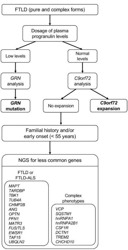
An algorithm for genetic diagnosis in FTLD-ALS, considering the role of the major genes and the additional contribution from NGS, is presented in Figure 3. Briefly, plasma progranulin dosage should be performed in all FTLD patients (with or without positive family history), and low levels should prompt immediate GRN gene analysis. The association of FTLD with ALS in the same individual or within the family is most often due to the C9orf72 repeat expansion. The research of C9orf72 repeat expansion should be carried out in all subjects with FTLD and FTLD-ALS (both non-familial and familial, considering the presence of ALS not only in the proband but in his family as well). In case of absence of GRN and C9orf72 mutations, a second-level approach for the genetic diagnosis is the implementation of an FTLD-ALS NGS. Atypical FTLD clinical presentations or unusual phenotypic associations should raise the suspicion of the involvement of different, rare genes, possibly prioritizing those more closely associated with the specific clinical context.
The considerable advances in the genetics of FTLD have paved the way to new therapeutic perspectives. Several novel therapies to delay the outbreak and progression of genetic FTD are emerging and some examples of molecules currently being tested are discussed below.
5.1 Therapeutic approaches for GRN gene mutations
The main pathogenic mechanism of GRN mutations arises from a loss of functional progranulin, and strategies to restore progranulin haploinsufficiency have emerged as promising avenues of research for GRN-related FTLD. A targeted intervention in GRN mutations might be the replacement of progranulin haploinsufficiency by upregulating expression of the nonmutant GRN allele. Histone deacetylase (HDAC) inhibitors were shown to promote progranulin expression through epigenetic regulation in human neuronal cells [75]. A phase II randomized, double-blind, placebo-controlled trial has tested HDAC
inhibitors (FRM-0334) (https://www.clinicaltrials.gov/, NCT02149160), for which results are still pending. Another avenue of research has focused on the Sortilin (SORT1)-progranulin axis. SORT1 is a clearance neuronal receptor via an endocytosis mechanism [76]. An open-label phase 2 study in human carriers is currently ongoing (https://www.clinicaltrials.gov/, NCT03987295). Other therapeutic approaches to rescue progranulin haploinsufficiency failed to demonstrate efficacy. A phase one 8-week clinical trial of Nimodipine, a blood-brain barrier-penetrant calcium channel blocker, did not show any significant effect on the
concentrations of progranulin in plasma and cerebrospinal fluid [77]. Similarly, a small phase 2 clinical trial of Amiodarone led to inconclusive results [78]. Other therapeutic approaches stemming from promising preclinical data in animal models, including oral small molecules, gene therapy and cell-based strategies are currently under development.
Since 2011, substantial advances have been achieved in our knowledge regarding C9orf72-mediated disease and its underlying pathogenesis. Toxic gain of function from C9orf72 repeat RNA and DPR proteins have been proposed as a crucial disease mechanism [36]. Hence, main drug discovery efforts aimed at reducing the gain of toxicity caused by the repeat-containing C9orf72 transcripts. Potential therapies based on this approach consist of antisense oligonucleotides (ASOs). ASOs are short DNA or RNA segments binding a complementary RNA sequence that are already used to modulate gene expression in several neurological diseases. A recent phase 2 clinical trial of ASO therapy targeting RNA-mediated toxicity in human has been conducted in Huntington disease (HTT Rx) another repeat expansions disease.
This trial showed its safety and tolerability, and a dose-dependent reduction in levels of the harmful mutant protein in the nervous system [79]. The most important breakthrough in the field of ASO therapy is undoubtedly the recent Food and Drug Administration approval for the Nusinersen, an antisense drug for the treatment of spinal muscular atrophy [80]. All these studies pave the way for the development of ASO therapies in neurodegenerative disorders and hold a great promise for the discovery of effective disease-modifying therapies for
C9orf72-associated disease. In vivo studies in C9orf72 mice showed a significant reduction of
RNA foci, DPR proteins and the development of behavioural deficits after a single dose of ASO therapy [81]. A phase 1 first-in-human study (BIIB078) assessing the safety and tolerability of ASO-mediated therapy administered intrathecally is currently underway in subjects with C9orf72-associated ALS (https://www.clinicaltrials.gov/, NCT03626012). Several other approaches including therapies based on small molecules and targeting DPR are also under development [13].
5.3 Therapeutic approaches for MAPT gene mutations
investigated, including protein kinases inhibitors for modulating tau phosphorylation, Microtubule stabilizers, tau aggregation inhibitors and anti-tau immunotherapy. Potentially relevant clinical trials for different tauopathies include a phase 3 trial of LMTX (TRx0237), a modified derivative of the tau aggregation inhibitor methylthionine chloride in subjects with bvFTD (https://www.clinicaltrials.gov/, NCT01626378) and a phase 2 trial of anti-tau immunotherapy, ABBV-8E12, a humanized recombinant anti-tau antibody, in patients with PSP (https://www.clinicaltrials.gov/, NCT03391765). These studies were completed in 2018, though their results have not been published yet. Gosuranemab (BIIB092), another tau-directed monoclonal antibody, demonstrated good tolerability in PSP patients in a phase 1b trial [82]. So far, these trials have mostly included PSP patients, but it is likely that some of these molecules will be tested in FTLD patients carrying MAPT mutations as well.
6. At the dawn of a new therapeutic era: the presymptomatic phase as a target window in genetic FTLD
The study of presymptomatic stage in mutation carriers is of outmost importance in light of the advent of promising preventive trials in genetic FTLD families. The presymptomatic phase indeed represents the optimal window for testing therapeutic molecules that target the FTLD-related neurobiological abnormalities at the earliest stage of the disease, before the occurrence of clinical symptoms. The identification of imaging and fluid biomarkers become crucial to monitor therapeutic trials at this stage, and research studies focusing on the
presymptomatic phase of genetic FTLD have become of increasing interest over the past few years [83,84]. Emerging data from several studies of well-characterised presymptomatic genetic cohorts, such as GENFI (Genetic Frontotemporal dementia Initiative) [84], LEFFTDS (Longitudinal Evaluation of Familial Frontotemporal Dementia Subjects) [85], Predict-PGRN [86] and PREV-DEMALS [87], suggested that the first pathological alterations associated
with FTLD gene mutations can be detected 15-10 years before onset of overt clinical symptoms. Accumulating results across these studies have identified early biomarkers in mutation carriers, encompassing neuropsychological, biological and multimodal
neuroimaging changes (i.e. structural and functional MRI and PET imaging) [84,87–91], but these markers still remain to be validated at the individual level. Once validated, the panel of early biomarkers identified at the presymptomatic stage could be used as efficacy endpoints in clinical trials. The significant advances that have been achieved in the understanding of the biomarkers cascade throughout the presymptomatic stage of genetic FTLD provide insight into the measurement of early disease progression and potential effects of disease-modifying therapies.
7. Conclusion
In the last few years, a major breakthrough has been achieved in the understanding of the genetics and molecular biology of FTLD. A remaining challenge is translating this substantial knowledge into therapeutic opportunities. Whilst much remains to be done in the field of drug discovery, it is noteworthy that we are now reaching a turning point as regards the
development of early biomarkers and preventive therapeutic approaches targeting the prodromal stage of genetic FTLD, before the onset of overt clinical symptoms in causative mutations carriers. The current investigations target different aspects of FTLD
pathophysiology and several appealing potential therapeutic candidates are currently being tested in the setting of clinical trials. These studies will hopefully expand the scope of potentially interesting disease modifying therapies in FTLD.
References
[1] Onyike CU, Diehl-Schmid J. The epidemiology of frontotemporal dementia. International Review of Psychiatry 2013;25:130–7.
https://doi.org/10.3109/09540261.2013.776523.
[2] Olney NT, Spina S, Miller BL. Frontotemporal Dementia. Neurologic Clinics 2017;35:339–74. https://doi.org/10.1016/j.ncl.2017.01.008.
[3] Rascovsky K, Hodges JR, Knopman D, Mendez MF, Kramer JH, Neuhaus J, et al. Sensitivity of revised diagnostic criteria for the behavioural variant of frontotemporal dementia. Brain 2011;134:2456–77. https://doi.org/10.1093/brain/awr179.
[4] Gorno-Tempini ML, Hillis AE, Weintraub S, Kertesz A, Mendez M, Cappa SF, et al. Classification of primary progressive aphasia and its variants. Neurology 2011;76:1006–14. https://doi.org/10.1212/WNL.0b013e31821103e6.
[5] Rogalski E, Sridhar J, Rader B, Martersteck A, Chen K, Cobia D, et al. Aphasic variant of Alzheimer disease: Clinical, anatomic, and genetic features. Neurology 2016;87:1337–43. https://doi.org/10.1212/WNL.0000000000003165.
[6] Höglinger GU, Respondek G, Stamelou M, Kurz C, Josephs KA, Lang AE, et al. Clinical diagnosis of progressive supranuclear palsy: The movement disorder society criteria: MDS Clinical Diagnostic Criteria for PSP. Movement Disorders 2017;32:853–64.
https://doi.org/10.1002/mds.26987.
[7] Armstrong MJ, Litvan I, Lang AE, Bak TH, Bhatia KP, Borroni B, et al. Criteria for the diagnosis of corticobasal degeneration. Neurology 2013;80:496–503.
https://doi.org/10.1212/WNL.0b013e31827f0fd1.
[8] Pottier C, Ravenscroft TA, Sanchez-Contreras M, Rademakers R. Genetics of FTLD: overview and what else we can expect from genetic studies. Journal of Neurochemistry 2016;138:32–53. https://doi.org/10.1111/jnc.13622.
[9] Mackenzie IRA, Neumann M, Baborie A, Sampathu DM, Du Plessis D, Jaros E, et al. A harmonized classification system for FTLD-TDP pathology. Acta Neuropathol
2011;122:111–3. https://doi.org/10.1007/s00401-011-0845-8.
[10] Lee EB, Porta S, Michael Baer G, Xu Y, Suh E, Kwong LK, et al. Expansion of the classification of FTLD-TDP: distinct pathology associated with rapidly progressive
frontotemporal degeneration. Acta Neuropathol 2017;134:65–78. https://doi.org/10.1007/s00401-017-1679-9.
[11] Irwin DJ, Cairns NJ, Grossman M, McMillan CT, Lee EB, Van Deerlin VM, et al. Frontotemporal lobar degeneration: defining phenotypic diversity through personalized medicine. Acta Neuropathol 2015;129:469–91. https://doi.org/10.1007/s00401-014-1380-1. [12] Neumann M, Bentmann E, Dormann D, Jawaid A, DeJesus-Hernandez M, Ansorge O, et al. FET proteins TAF15 and EWS are selective markers that distinguish FTLD with FUS pathology from amyotrophic lateral sclerosis with FUS mutations. Brain 2011;134:2595–609. https://doi.org/10.1093/brain/awr201.
[13] Desmarais P, Rohrer JD, Nguyen QD, Herrmann N, Stuss DT, Lang AE, et al. Therapeutic trial design for frontotemporal dementia and related disorders. J Neurol Neurosurg Psychiatry 2019;90:412–23. https://doi.org/10.1136/jnnp-2018-318603. [14] Cruts M, Gijselinck I, van der Zee J, Engelborghs S, Wils H, Pirici D, et al. Null mutations in progranulin cause ubiquitin-positive frontotemporal dementia linked to chromosome 17q21. Nature 2006;442:920–4. https://doi.org/10.1038/nature05017.
[15] Baker M, Mackenzie IR, Pickering-Brown SM, Gass J, Rademakers R, Lindholm C, et al. Mutations in progranulin cause tau-negative frontotemporal dementia linked to
chromosome 17. Nature 2006;442:916–9. https://doi.org/10.1038/nature05016.
et al. Expanded GGGGCC Hexanucleotide Repeat in Noncoding Region of C9ORF72 Causes Chromosome 9p-Linked FTD and ALS. Neuron 2011;72:245–56.
https://doi.org/10.1016/j.neuron.2011.09.011.
[17] Renton AE, Majounie E, Waite A, Simón-Sánchez J, Rollinson S, Gibbs JR, et al. A Hexanucleotide Repeat Expansion in C9ORF72 is the Cause of Chromosome 9p21-Linked ALS-FTD. Neuron 2011;72:257–68. https://doi.org/10.1016/j.neuron.2011.09.010.
[18] Hutton M, Lendon CL, Rizzu P, Baker M, Froelich S, Houlden H, et al. Association of missense and 5’-splice-site mutations in tau with the inherited dementia FTDP-17 1998;393:4. [19] Rademakers R, Neumann M, Mackenzie IR. Recent advances in the molecular basis of frontotemporal dementia. Nat Rev Neurol 2012;8:423–34.
https://doi.org/10.1038/nrneurol.2012.117.
[20] Paushter DH, Du H, Feng T, Hu F. The lysosomal function of progranulin, a guardian against neurodegeneration. Acta Neuropathol 2018;136:1–17. https://doi.org/10.1007/s00401-018-1861-8.
[21] Ahmed Z, Mackenzie IR, Hutton ML, Dickson DW. Progranulin in frontotemporal lobar degeneration and neuroinflammation. Journal of Neuroinflammation 2007;4:7. https://doi.org/10.1186/1742-2094-4-7.
[22] Cenik B, Sephton CF, Kutluk Cenik B, Herz J, Yu G. Progranulin: A Proteolytically Processed Protein at the Crossroads of Inflammation and Neurodegeneration. J Biol Chem 2012;287:32298–306. https://doi.org/10.1074/jbc.R112.399170.
[23] Cruts M, Theuns J, Van Broeckhoven C. Locus-specific mutation databases for neurodegenerative brain diseases. Human Mutation 2012;33:1340–4.
https://doi.org/10.1002/humu.22117.
[24] Lattante S, Le Ber I, Galimberti D, Serpente M, Rivaud-Péchoux S, Camuzat A, et al. Defining the association of TMEM106B variants among frontotemporal lobar degeneration patients with GRN mutations and C9orf72 repeat expansions. Neurobiology of Aging 2014;35:2658.e1-2658.e5. https://doi.org/10.1016/j.neurobiolaging.2014.06.023.
[25] Smith KR, Damiano J, Franceschetti S, Carpenter S, Canafoglia L, Morbin M, et al. Strikingly Different Clinicopathological Phenotypes Determined by Progranulin-Mutation Dosage. The American Journal of Human Genetics 2012;90:1102–7.
https://doi.org/10.1016/j.ajhg.2012.04.021.
[26] Huin V, Barbier M, Bottani A, Lobrinus JA, Clot F, Lamari F, et al. Homozygous GRN mutations: new phenotypes and new insights into pathological and molecular mechanisms. Brain 2020;143:303–19. https://doi.org/10.1093/brain/awz377.
[27] Le Ber I, Camuzat A, Hannequin D, Pasquier F, Guedj E, Rovelet-Lecrux A, et al. Phenotype variability in progranulin mutation carriers: a clinical, neuropsychological, imaging and genetic study. Brain 2008;131:732–46. https://doi.org/10.1093/brain/awn012. [28] Moore KM, Nicholas J, Grossman M, McMillan CT, Irwin DJ, Massimo L, et al. Age at symptom onset and death and disease duration in genetic frontotemporal dementia: an international retrospective cohort study. The Lancet Neurology 2019:S1474442219303941. https://doi.org/10.1016/S1474-4422(19)30394-1.
[29] Rohrer JD, Crutch SJ, Warrington EK, Warren JD. Progranulin-associated primary progressive aphasia: A distinct phenotype? Neuropsychologia 2010;48:288–97.
https://doi.org/10.1016/j.neuropsychologia.2009.09.017.
[30] Kim G, Ahmadian SS, Peterson M, Parton Z, Memon R, Weintraub S, et al.
Asymmetric pathology in primary progressive aphasia with progranulin mutations and TDP inclusions. Neurology 2016;86:627–36. https://doi.org/10.1212/WNL.0000000000002375. [31] Caroppo P, Le Ber I, Camuzat A, Clot F, Naccache L, Lamari F, et al. Extensive White Matter Involvement in Patients With Frontotemporal Lobar Degeneration: Think Progranulin. JAMA Neurol 2014;71:1562. https://doi.org/10.1001/jamaneurol.2014.1316.
[32] Ghidoni R, Stoppani E, Rossi G, Piccoli E, Albertini V, Paterlini A, et al. Optimal Plasma Progranulin Cutoff Value for Predicting Null Progranulin Mutations in
Neurodegenerative Diseases: A Multicenter Italian Study. Neurodegenerative Diseases 2012;9:121–7. https://doi.org/10.1159/000333132.
[33] Le Ber I, Guillot-Noel L, Hannequin D, Lacomblez L, Golfier V, Puel M, et al. C9ORF72 Repeat Expansions in the Frontotemporal Dementias Spectrum of Diseases: A Flow-chart for Genetic Testing. JAD 2013;34:485–99. https://doi.org/10.3233/JAD-121456. [34] Majounie E, Renton AE, Mok K, Dopper EG, Waite A, Rollinson S, et al. Frequency of the C9orf72 hexanucleotide repeat expansion in patients with amyotrophic lateral sclerosis and frontotemporal dementia: a cross-sectional study. The Lancet Neurology 2012;11:323– 30. https://doi.org/10.1016/S1474-4422(12)70043-1.
[35] Webster CP, Smith EF, Bauer CS, Moller A, Hautbergue GM, Ferraiuolo L, et al. The C9orf72 protein interacts with Rab1a and the ULK1 complex to regulate initiation of
autophagy. EMBO J 2016;35:1656–76. https://doi.org/10.15252/embj.201694401.
[36] Cruts M, Gijselinck I, Van Langenhove T, van der Zee J, Van Broeckhoven C. Current insights into the C9orf72 repeat expansion diseases of the FTLD/ALS spectrum. Trends in Neurosciences 2013;36:450–9. https://doi.org/10.1016/j.tins.2013.04.010.
[37] Snowden JS, Adams J, Harris J, Thompson JC, Rollinson S, Richardson A, et al. Distinct clinical and pathological phenotypes in frontotemporal dementia associated with MAPT, PGRN and C9orf72 mutations. Amyotrophic Lateral Sclerosis and Frontotemporal Degeneration 2015;16:497–505. https://doi.org/10.3109/21678421.2015.1074700.
[38] Snowden JS, Rollinson S, Thompson JC, Harris JM, Stopford CL, Richardson AMT, et al. Distinct clinical and pathological characteristics of frontotemporal dementia associated with C9ORF72 mutations. Brain 2012;135:693–708. https://doi.org/10.1093/brain/awr355. [39] Devenney EM, Ahmed RM, Halliday G, Piguet O, Kiernan MC, Hodges JR. Psychiatric disorders in C9orf72 kindreds: Study of 1,414 family members. Neurology 2018:1. https://doi.org/10.1212/WNL.0000000000006344.
[40] Whitwell JL, Weigand SD, Boeve BF, Senjem ML, Gunter JL, DeJesus-Hernandez M, et al. Neuroimaging signatures of frontotemporal dementia genetics: C9ORF72, tau,
progranulin and sporadics. Brain 2012;135:794–806. https://doi.org/10.1093/brain/aws001. [41] McGoldrick P, Zhang M, van Blitterswijk M, Sato C, Moreno D, Xiao S, et al. Unaffected mosaic C9orf72 case: RNA foci, dipeptide proteins, but upregulated C9orf72 expression. Neurology 2018;90:e323–31. https://doi.org/10.1212/WNL.0000000000004865. [42] Fournier C, Barbier M, Camuzat A, Anquetil V, Lattante S, Clot F, et al. Relations between C9orf72 expansion size in blood, age at onset, age at collection and transmission across generations in patients and presymptomatic carriers. Neurobiology of Aging 2019;74:234.e1-234.e8. https://doi.org/10.1016/j.neurobiolaging.2018.09.010.
[43] Barbier M, Camuzat A, Houot M, Clot F, Caroppo P, Fournier C, et al. Factors influencing the age at onset in familial frontotemporal lobar dementia: Important weight of genetics. Neurol Genet 2017;3:e203. https://doi.org/10.1212/NXG.0000000000000203. [44] Troakes C, Maekawa S, Wijesekera L, Rogelj B, Siklós L, Bell C, et al. An MND/ALS phenotype associated with C9orf72 repeat expansion: Abundant p62-positive, TDP-43-negative inclusions in cerebral cortex, hippocampus and cerebellum but without associated cognitive decline: p62 proteinopathy. Neuropathology 2012;32:505–14. https://doi.org/10.1111/j.1440-1789.2011.01286.x.
[45] Mackenzie IRA, Frick P, Neumann M. The neuropathology associated with repeat expansions in the C9ORF72 gene. Acta Neuropathol 2014;127:347–57.
https://doi.org/10.1007/s00401-013-1232-4.
[46] Forrest SL, Kril JJ, Stevens CH, Kwok JB, Hallupp M, Kim WS, et al. Retiring the term FTDP-17 as MAPT mutations are genetic forms of sporadic frontotemporal tauopathies.
Brain 2018;141:521–34. https://doi.org/10.1093/brain/awx328.
[47] Pickering-Brown S, Baker M, Bird T, Trojanowski J, Lee V, Morris H, et al. Evidence of a founder effect in families with frontotemporal dementia that harbor the tau +16 splice mutation. Am J Med Genet 2004;125B:79–82. https://doi.org/10.1002/ajmg.b.20083. [48] Pickering-Brown SM. The complex aetiology of frontotemporal lobar degeneration. Experimental Neurology 2007;206:1–10. https://doi.org/10.1016/j.expneurol.2007.03.017. [49] Ghetti B, Oblak AL, Boeve BF, Johnson KA, Dickerson BC, Goedert M. Invited review: Frontotemporal dementia caused by microtubule-associated protein tau gene (MAPT) mutations: a chameleon for neuropathology and neuroimaging. Neuropathol Appl Neurobiol 2015;41:24–46. https://doi.org/10.1111/nan.12213.
[50] van Swieten J, Spillantini MG. Hereditary Frontotemporal Dementia Caused by Tau Gene Mutations. Brain Pathology 2007;17:63–73.
https://doi.org/10.1111/j.1750-3639.2007.00052.x.
[51] Henz S, Ackl N, Knels C, Rominger A, Flatz W, Teipel S, et al. A Pair of Siblings with a rare R5H-Mutation in Exon 1 of the MAPT-Gene. Fortschr Neurol Psychiatr 2015;83:397–401. https://doi.org/10.1055/s-0035-1553236.
[52] Benajiba L, Le Ber I, Camuzat A, Lacoste M, Thomas-Anterion C, Couratier P, et al. TARDBP mutations in motoneuron disease with frontotemporal lobar degeneration. Ann Neurol 2009;65:470–3. https://doi.org/10.1002/ana.21612.
[53] Freischmidt A, Wieland T, Richter B, Ruf W, Schaeffer V, Müller K, et al. Haploinsufficiency of TBK1 causes familial ALS and fronto-temporal dementia. Nat Neurosci 2015;18:631–6. https://doi.org/10.1038/nn.4000.
[54] Caroppo P, Camuzat A, Guillot-Noel L, Thomas-Antérion C, Couratier P, Wong TH, et al. Defining the spectrum of frontotemporal dementias associated with TARDBP
mutations. Neurology Genetics 2016;2:e80. https://doi.org/10.1212/NXG.0000000000000080.
[55] Caroppo P, Camuzat A, De Septenville A, Couratier P, Lacomblez L, Auriacombe S, et al. Semantic and nonfluent aphasic variants, secondarily associated with amyotrophic lateral sclerosis, are predominant frontotemporal lobar degeneration phenotypes in TBK1 carriers. Alzheimer’s & Dementia: Diagnosis, Assessment & Disease Monitoring
2015;1:481–6. https://doi.org/10.1016/j.dadm.2015.10.002.
[56] Perrone F, Nguyen HP, Van Mossevelde S, Moisse M, Sieben A, Santens P, et al. Investigating the role of ALS genes CHCHD10 and TUBA4A in Belgian FTD-ALS spectrum patients. Neurobiology of Aging 2017;51:177.e9-177.e16.
https://doi.org/10.1016/j.neurobiolaging.2016.12.008.
[57] Skibinski G, Parkinson NJ, Brown JM, Chakrabarti L, Lloyd SL, Hummerich H, et al. Mutations in the endosomal ESCRTIII-complex subunit CHMP2B in frontotemporal
dementia. Nat Genet 2005;37:806–8. https://doi.org/10.1038/ng1609.
[58] Svetoni F, Frisone P, Paronetto MP. Role of FET proteins in neurodegenerative
disorders. RNA Biology 2016;13:1089–102. https://doi.org/10.1080/15476286.2016.1211225. [59] Al-Obeidi E, Al-Tahan S, Surampalli A, Goyal N, Wang AK, Hermann A, et al. Genotype-phenotype study in patients with valosin-containing protein mutations associated with multisystem proteinopathy. Clin Genet 2018;93:119–25.
https://doi.org/10.1111/cge.13095.
[60] Watts GDJ, Wymer J, Kovach MJ, Mehta SG, Mumm S, Darvish D, et al. Inclusion body myopathy associated with Paget disease of bone and frontotemporal dementia is caused by mutant valosin-containing protein. Nat Genet 2004;36:377–81.
https://doi.org/10.1038/ng1332.
[61] Le Ber I. SQSTM1 Mutations in French Patients With Frontotemporal Dementia or Frontotemporal Dementia With Amyotrophic Lateral Sclerosis. JAMA Neurol 2013.
https://doi.org/10.1001/jamaneurol.2013.3849.
[62] Kim HJ, Kim NC, Wang Y-D, Scarborough EA, Moore J, Diaz Z, et al. Mutations in prion-like domains in hnRNPA2B1 and hnRNPA1 cause multisystem proteinopathy and ALS. Nature 2013;495:467–73. https://doi.org/10.1038/nature11922.
[63] Rademakers R, Baker, Matt, Nicholson, Alexandra M, Rutherford, Nicola J, Finch, NiCole, Soto-Ortolaza, Alexandra, et al. Mutations in the colony stimulating factor 1 receptor (CSF1R) gene cause hereditary diffuse leukoencephalopathy with spheroids. Nature Genetics 2012;44:8. https://doi.org/10.1038/ng.1027.
[64] Konno T, Yoshida K, Mizuno T, Kawarai T, Tada M, Nozaki H, et al. Clinical and genetic characterization of adult-onset leukoencephalopathy with axonal spheroids and pigmented glia associated with CSF1R mutation. Eur J Neurol 2017;24:37–45.
https://doi.org/10.1111/ene.13125.
[65] Guerreiro R, Kara E, Le Ber I, Bras J, Rohrer JD, Taipa R, et al. Genetic Analysis of Inherited Leukodystrophies: Genotype-Phenotype Correlations in the CSF1R Gene. JAMA Neurol 2013;70:875. https://doi.org/10.1001/jamaneurol.2013.698.
[66] Kim E-J, Kim Y-E, Jang J-H, Cho E-H, Na DL, Seo SW, et al. Analysis of
frontotemporal dementia, amyotrophic lateral sclerosis, and other dementia-related genes in 107 Korean patients with frontotemporal dementia. Neurobiology of Aging 2018;72:186.e1-186.e7. https://doi.org/10.1016/j.neurobiolaging.2018.06.031.
[67] Codjia P, Ayrignac X, Mochel F, Mouzat K, Carra-Dalliere C, Castelnovo G, et al. Adult-Onset Leukoencephalopathy with Axonal Spheroids and Pigmented Glia: An MRI Study of 16 French Cases. AJNR Am J Neuroradiol 2018;39:1657–61.
https://doi.org/10.3174/ajnr.A5744.
[68] Chaussenot A, Le Ber I, Ait-El-Mkadem S, Camuzat A, de Septenville A, Bannwarth S, et al. Screening of CHCHD10 in a French cohort confirms the involvement of this gene in frontotemporal dementia with amyotrophic lateral sclerosis patients. Neurobiology of Aging 2014;35:2884.e1-2884.e4. https://doi.org/10.1016/j.neurobiolaging.2014.07.022.
[69] Wider C, Dachsel JC, Farrer MJ, Dickson DW, Tsuboi Y, Wszolek ZK. Elucidating the genetics and pathology of Perry syndrome. Journal of the Neurological Sciences
2010;289:149–54. https://doi.org/10.1016/j.jns.2009.08.044.
[70] Peplonska B, Berdynski M, Mandecka M, Barczak A, Kuzma-Kozakiewicz M, Barcikowska M, et al. TREM2 variants in neurodegenerative disorders in the Polish population. Homozygosity and compound heterozygosity in FTD patients. Amyotrophic Lateral Sclerosis and Frontotemporal Degeneration 2018;19:407–12.
https://doi.org/10.1080/21678421.2018.1451894.
[71] Goldman JS, Van Deerlin VM. Alzheimer’s Disease and Frontotemporal Dementia: The Current State of Genetics and Genetic Testing Since the Advent of Next-Generation Sequencing. Mol Diagn Ther 2018;22:505–13. https://doi.org/10.1007/s40291-018-0347-7. [72] Richards S, Aziz N, Bale S, Bick D, Das S, Gastier-Foster J, et al. Standards and guidelines for the interpretation of sequence variants: a joint consensus recommendation of the American College of Medical Genetics and Genomics and the Association for Molecular Pathology. Genet Med 2015;17:405–23. https://doi.org/10.1038/gim.2015.30.
[73] Saracino D, Sellami L, Clot F, Camuzat A, Lamari F, Rucheton B, et al. The missense p.Trp7Arg mutation in GRN gene leads to progranulin haploinsufficiency. Neurobiology of Aging 2020;85:154.e9-154.e11. https://doi.org/10.1016/j.neurobiolaging.2019.06.002. [74] Pottier C, Bieniek KF, Finch N, van de Vorst M, Baker M, Perkersen R, et al. Whole-genome sequencing reveals important role for TBK1 and OPTN mutations in frontotemporal lobar degeneration without motor neuron disease. Acta Neuropathol 2015;130:77–92.
https://doi.org/10.1007/s00401-015-1436-x.
Requirements of HDAC Inhibitors as Progranulin Enhancers for Treating Frontotemporal Dementia. Cell Chemical Biology 2017;24:892-906.e5.
https://doi.org/10.1016/j.chembiol.2017.06.010.
[76] Lee WC, Almeida S, Prudencio M, Caulfield TR, Zhang Y-J, Tay WM, et al. Targeted manipulation of the sortilin–progranulin axis rescues progranulin haploinsufficiency. Human Molecular Genetics 2014;23:1467–78. https://doi.org/10.1093/hmg/ddt534.
[77] Sha SJ, Miller ZA, Min S, Zhou Y, Brown J, Mitic LL, et al. An 8-week, open-label, dose-finding study of nimodipine for the treatment of progranulin insufficiency from GRN gene mutations. Alzheimer’s & Dementia: Translational Research & Clinical Interventions 2017;3:507–12. https://doi.org/10.1016/j.trci.2017.08.002.
[78] Alberici A, Archetti S, Pilotto A, Premi E, Cosseddu M, Bianchetti A, et al. Results from a pilot study on amiodarone administration in monogenic frontotemporal dementia with granulin mutation. Neurol Sci 2014;35:1215–9. https://doi.org/10.1007/s10072-014-1683-y. [79] Tabrizi SJ, Leavitt BR, Landwehrmeyer GB, Wild EJ, Saft C, Barker RA, et al. Targeting Huntingtin Expression in Patients with Huntington’s Disease. N Engl J Med 2019;380:2307–16. https://doi.org/10.1056/NEJMoa1900907.
[80] Finkel RS, Mercuri E, Darras BT, Connolly AM, Kuntz NL, Kirschner J, et al. Nusinersen versus Sham Control in Infantile-Onset Spinal Muscular Atrophy. N Engl J Med 2017;377:1723–32. https://doi.org/10.1056/NEJMoa1702752.
[81] Jiang J, Zhu Q, Gendron TF, Saberi S, McAlonis-Downes M, Seelman A, et al. Gain of Toxicity from ALS/FTD-Linked Repeat Expansions in C9ORF72 Is Alleviated by
Antisense Oligonucleotides Targeting GGGGCC-Containing RNAs. Neuron 2016;90:535–50. https://doi.org/10.1016/j.neuron.2016.04.006.
[82] Boxer AL, Qureshi I, Ahlijanian M, Grundman M, Golbe LI, Litvan I, et al. Safety of the tau-directed monoclonal antibody BIIB092 in progressive supranuclear palsy: a
randomised, placebo-controlled, multiple ascending dose phase 1b trial. The Lancet Neurology 2019;18:549–58. https://doi.org/10.1016/S1474-4422(19)30139-5.
[83] Bateman RJ, Xiong C, Benzinger TLS, Fagan AM, Goate A, Fox NC, et al. Clinical and Biomarker Changes in Dominantly Inherited Alzheimer’s Disease. N Engl J Med 2012;367:795–804. https://doi.org/10.1056/NEJMoa1202753.
[84] Rohrer JD, Nicholas JM, Cash DM, van Swieten J, Dopper E, Jiskoot L, et al. Presymptomatic cognitive and neuroanatomical changes in genetic frontotemporal dementia in the Genetic Frontotemporal dementia Initiative (GENFI) study: a cross-sectional analysis. The Lancet Neurology 2015;14:253–62. https://doi.org/10.1016/S1474-4422(14)70324-2. [85] Boeve B, Bove J, Brannelly P, Brushaber D, Coppola G, Dever R, et al. The
longitudinal evaluation of familial frontotemporal dementia subjects protocol: Framework and methodology. Alzheimer’s & Dementia 2019:S1552526019351131.
https://doi.org/10.1016/j.jalz.2019.06.4947.
[86] Caroppo P, Habert M-O, Durrleman S, Funkiewiez A, Perlbarg V, Hahn V, et al. Lateral Temporal Lobe: An Early Imaging Marker of the Presymptomatic GRN Disease? J Alzheimers Dis 2015;47:751–9. https://doi.org/10.3233/JAD-150270.
[87] Bertrand A, Wen J, Rinaldi D, Houot M, Sayah S, Camuzat A, et al. Early Cognitive, Structural, and Microstructural Changes in Presymptomatic C9orf72 Carriers Younger Than 40 Years. JAMA Neurology 2018;75:236. https://doi.org/10.1001/jamaneurol.2017.4266. [88] Jiskoot LC, Panman JL, van Asseldonk L, Franzen S, Meeter LHH, Donker Kaat L, et al. Longitudinal cognitive biomarkers predicting symptom onset in presymptomatic
frontotemporal dementia. J Neurol 2018;265:1381–92. https://doi.org/10.1007/s00415-018-8850-7.
[89] Jiskoot LC, Panman JL, Meeter LH, Dopper EGP, Donker Kaat L, Franzen S, et al. Longitudinal multimodal MRI as prognostic and diagnostic biomarker in presymptomatic
familial frontotemporal dementia. Brain 2019;142:193–208. https://doi.org/10.1093/brain/awy288.
[90] van der Ende EL, Meeter LH, Poos JM, Panman JL, Jiskoot LC, Dopper EGP, et al. Serum neurofilament light chain in genetic frontotemporal dementia: a longitudinal,
multicentre cohort study. The Lancet Neurology 2019;18:1103–11. https://doi.org/10.1016/S1474-4422(19)30354-0.
[91] Galimberti D, Fumagalli GG, Fenoglio C, Cioffi SMG, Arighi A, Serpente M, et al. Progranulin plasma levels predict the presence of GRN mutations in asymptomatic subjects and do not correlate with brain atrophy: results from the GENFI study. Neurobiology of Aging 2018;62:245.e9-245.e12. https://doi.org/10.1016/j.neurobiolaging.2017.10.016.
Figure 1. Pathological classification of frontotemporal lobar degeneration. TDP:
transactive response (TAR) DNA binding protein; 3R: 3-repeat Tau inclusions; 4R: 4-repeat Tau inclusions; FET: Fused in sarcoma (FUS), Ewing’s sarcoma, TATA-binding protein associated factor (TAF) 15; aFTLDU: atypical frontotemporal lobar degeneration with
ubiquitin-positive inclusions; NIFID: neuronal intermediate filament inclusion disease; BIBD: basophilic inclusion body disease; FTLD-UPS: frontotemporal lobar degeneration with
Figure 2. Landscape of the genes associated with the FTLD-ALS continuum. Major genes are represented as larger balloons. The list of ALS-associated genes is not exhaustive. Different pathological substrates are represented by different colors of the balloons’ lines (red: FTLD-TAU; blue: FTLD-TDP; green: FTLD-FET/FUS; brown: SOD1-positive inclusions; grey: other or unknown pathology). ALS: amyotrophic lateral sclerosis; FTLD: frontotemporal lobar degeneration; SOD1: superoxide dismutase 1.
Figure 3. Optimal diagnostic algorithm for genetic diagnosis in FTLD, including major and less common genes. ALS: amyotrophic lateral sclerosis; FTLD: frontotemporal lobar degeneration; NGS: next-generation sequencing.