HAL Id: hal-02346395
https://hal.archives-ouvertes.fr/hal-02346395
Submitted on 5 Nov 2019
HAL is a multi-disciplinary open access
archive for the deposit and dissemination of
sci-entific research documents, whether they are
pub-lished or not. The documents may come from
teaching and research institutions in France or
abroad, or from public or private research centers.
L’archive ouverte pluridisciplinaire HAL, est
destinée au dépôt et à la diffusion de documents
scientifiques de niveau recherche, publiés ou non,
émanant des établissements d’enseignement et de
recherche français ou étrangers, des laboratoires
publics ou privés.
Specific and Multiple Pathway Targeting
Julian Biau, E. Chautard, Pierre Verrelle, Marie Dutreix
To cite this version:
Julian Biau, E. Chautard, Pierre Verrelle, Marie Dutreix. Altering DNA Repair to Improve
Radia-tion Therapy: Specific and Multiple Pathway Targeting. Frontiers in Oncology, Frontiers, 2019, 9,
�10.3389/fonc.2019.01009�. �hal-02346395�
REVIEW published: 10 October 2019 doi: 10.3389/fonc.2019.01009
Edited by: Paul N. Span, Radboud University Nijmegen Medical Centre, Netherlands Reviewed by: Michael Wayne Epperly, University of Pittsburgh, United States Bevin P. Engelward, Massachusetts Institute of Technology, United States *Correspondence: Julian Biau [email protected]
Specialty section: This article was submitted to Radiation Oncology, a section of the journal Frontiers in Oncology Received: 15 July 2019 Accepted: 19 September 2019 Published: 10 October 2019 Citation: Biau J, Chautard E, Verrelle P and Dutreix M (2019) Altering DNA Repair to Improve Radiation Therapy: Specific and Multiple Pathway Targeting. Front. Oncol. 9:1009. doi: 10.3389/fonc.2019.01009
Altering DNA Repair to Improve
Radiation Therapy: Specific and
Multiple Pathway Targeting
Julian Biau
1,2,3,4,5,6*, Emmanuel Chautard
5,7, Pierre Verrelle
1,6,8,9and Marie Dutreix
1,2,3,41Institut Curie, PSL Research University, Centre de Recherche, Paris, France,2UMR3347, CNRS, Orsay, France,3U1021, INSERM, Orsay, France,4Université Paris Sud, Orsay, France,5Université Clermont Auvergne, INSERM, U1240 IMoST, Clermont Ferrand, France,6Radiotherapy Department, Université Clermont Auvergne, Centre Jean Perrin, Clermont-Ferrand, France,7Pathology Department, Université Clermont Auvergne, Centre Jean Perrin, Clermont-Ferrand, France,8U1196, INSERM, UMR9187, CNRS, Orsay, France,9Radiotherapy Department, Institut Curie Hospital, Paris, France
Radiation therapy (RT) is widely used in cancer care strategies. Its effectiveness relies
mainly on its ability to cause lethal damage to the DNA of cancer cells. However, some
cancers have shown to be particularly radioresistant partly because of efficient and
redundant DNA repair capacities. Therefore, RT efficacy might be enhanced by using
drugs that can disrupt cancer cells’ DNA repair machinery. Here we review the recent
advances in the development of novel inhibitors of DNA repair pathways in combination
with RT. A large number of these compounds are the subject of preclinical/clinical studies
and target key enzymes involved in one or more DNA repair pathways. A totally different
strategy consists of mimicking DNA double-strand breaks via small interfering DNA
(siDNA) to bait the whole DNA repair machinery, leading to its global inhibition.
Keywords: DNA damage, repair systems, radiotherapy, radioresistance, inhibition
INTRODUCTION
Radiation therapy (RT), in conjunction with surgery and systemic therapies (chemotherapy,
targeted therapies, immunotherapy. . . ), is a cornerstone of cancer care. About 50% of cancer
patients receive RT (
1
). The primary objective of RT is to increase the amount of radiation delivered
to the tumor to ensure local control and reduce the amount of radiation in adjacent healthy tissues.
Advanced developments such as image-guided RT (IGRT) or intensity-modulated RT (IMRT) have
led to the enhancement of this therapeutic ratio (
2
). Despite such improvements, many patients still
experience local recurrence of the disease after RT. Clinical factors such as tumor stage, frequently
associated with increased hypoxia, can explain some of the failures, but it is clear that biological
characteristics play a key part in successful treatment (
3
–
5
). RT-induced cell death is mostly due to
DNA damage, especially to double-strand breaks (DSBs) (
6
,
7
). Consequently, tumor cells with
highly efficient DNA repair are radioresistant (
8
), whereas deficiencies in pathways that repair
DSBs are particularly detrimental to the cells (
9
). Therefore, therapies that inhibit the DNA repair
machinery have the potential to enhance RT efficacy (
10
,
11
). Inhibiting DNA repair offers an
opportunity to target genetic differences between tumor and normal cells, as DNA repair is often
dysregulated in tumor cells (
10
,
12
–
14
). Tumor cells divide rapidly because of unregulated cell
cycle control. Thus, they have less time to repair DNA damage as compared to normal cells that
are not dividing or will stop dividing after activation of key checkpoints induced by RT (
15
,
16
).
Beside altered cell cycle control, cancer cells may also present defects in their DNA repair system,
inducing dependence on specific repair pathways and/or overexpression of alternative pathways
(
16
,
17
). Furthermore, cancer cells often develop under stress
conditions, thus raising the frequency of endogenous DNA
damage (
18
,
19
). This review will firstly focus on the distinct
categories of DNA lesions induced by RT and the DNA repair
pathways required for their repair. Subsequently, it will present
the approaches that are currently being developed to enhance RT
efficacy by modulating DNA repair.
RT-INDUCED DNA DAMAGE RESPONSE
DNA lesions induced by RT activate the DNA damage
response (DDR), which essentially involves post-translational
modifications of proteins to activate downstream signaling
pathways (
20
). DDR is based on an intricate network of
proteins that work together to manage DNA repair and cell
cycle coordination. DDR interrupts the cell cycle, thereby
inhibiting the spread of DNA damage to daughter cells and
facilitating repair. Cell division arrest is mainly controlled by the
checkpoint kinases CHK1 and CHK2, which are activated by the
phosphatidylinsositol-3-kinases (PI3K) of the DDR machinery
(
15
). DDR signaling is also essential for triggering apoptosis when
repair is unsuccessful, notably through modifications to the p53
protein (
20
).
RT induces a variety of DNA lesions. Approximately 10,000
damaged bases, 1,000 single strand breaks (SSBs) and 40 DSBs are
produced per gray per cell (
21
,
22
). Such lesions, if not corrected,
can lead to cell death by mitotic catastrophe and apoptosis. DSBs
are the most lethal to the cells despite their low proportion, as
one single unrepaired DSB can trigger cell death (
7
). DSBs are
produced directly and indirectly by RT. Indirect DSBs most often
occur during replication if the initial damage is unrepaired. As an
example, when a replication fork encounters an unrepaired SSB,
the fork is blocked and leads to the conversion of this SSB into a
DSB (
10
,
23
,
24
). The resulting DSB can directly trigger cell death
or activate DDR, which induces cell cycle arrest and promotes
DNA repair. This repair is usually error-free, allowing the cell to
survive without genetic consequences. It can also be error-prone,
leading either to cell death if the error is not viable or mutation
and chromosomic aberrations (
25
).
DNA REPAIR OF RT-INDUCED DAMAGE
Following RT, damaged bases induced by oxidative stress are
repaired by the base excision repair pathway (BER) (
26
–
31
).
In BER, damaged bases are excised by DNA glycosylases,
resulting in apurinic (AP) sites. Subsequently; these AP sites
are cleaved by apurinic endonuclease 1 (APE1) or an
AP-lyase activity, leading to SSBs. SSBs are repaired by the part
of the BER pathway called single strand break repair (SSBR)
(Figure 1A) (
12
,
32
). Either short-patch or long-patch SSBR
can then proceed, depending on several factors such as type
of lesion and cell cycle state. Single nucleotide insertion by
DNA polymerase (Pol) β and ligation by DNA ligase III are
described as short-patch SSBR, and interact with the protein
X-ray repair cross-complementing 1 (XRCC1). Long-patch SSBR
involves the removal of a larger DNA segment, which requires
several DNA replication factors such as proliferating cell nuclear
antigen (PCNA), Pol δ/ǫ, flap endonuclease 1 (FEN1), and DNA
ligase I. Concerning SSBs detection, poly-ADP-ribose polymerase
(PARP1 or PARP2) are required (
28
,
33
–
37
). The binding of
PARP to SSB activates its auto-PARylation, and leads to the
recruitment of BER/SSBR proteins. PARP-1 was also reported
as a regulator of DNA repair gene expression through the E2F1
pathway (
38
). Unrepaired SSB or a damaged base can block the
replication forks, resulting in fork collapse and DSB (
23
). The
great majority of oxidative damage induced by ionizing radiation
is corrected by BER. However, under hypoxic conditions, IR
causes the formation of cyclodeoxynucleosides that can be only
removed by nucleotide excision repair (NER). Several results
suggest that NER may be involved in the repair of oxidized DNA
damage. In addition, ionizing-radiation breast cancer risk has
been related to polymorphism in ERCC2 (one of the main NER
enzymes) (
39
).
Two
major
pathways
repair
DSBs:
homologous
recombination (HR) and non-homologous end joining (NHEJ)
(
40
). However, both mismatch repair (MMR) and NER pathways
have been reported to affect both HR- and NHEJ-mediated DSB
repair efficacy to a lesser extent (
41
). The formation of DSBs
triggers the activation of three key enzymes from the PIKK
family: ataxia telangiectasia mutated kinase (ATM), ATM-related
kinase (ATR), and DNA-dependent protein kinase (DNA-PK).
This leads to the phosphorylation of many proteins, signaling
damage and initiating DNA repair. One of the early steps is
the phosphorylation of histone H2AX (γ-H2AX), which signals
the presence of DSB to repair proteins where they aggregate
in ionizing radiation-induced foci (IRIF) (
42
). Besides their
crucial roles in DDR signaling, the kinases ATR and ATM are
also involved in maintaining replication fork stability (
14
) and
fork reversal in case of fork-stalling lesions, notably through
SMARCAL1 (
43
).
In mammalian cells, c-NHEJ (classical NHEJ, Figure 1B) is
the most efficient DSB repair mechanism. It acts by directly
ligating the broken DNA ends (
44
). c-NHEJ can occur during
the entire cell cycle. It is frequently accompanied by small
deletions at the repair break site and is considered to be the
main cause of DSB error-prone repair. The first step of NHEJ
is the binding of the heterodimer Ku70/Ku80 at the end of the
DSB (
45
), allowing the recruitment of catalytic subunit
DNA-PKcs forming the protein complex DNA-PK (
46
). DNA-PK,
bounded to DNA, is activated and phosphorylates numerous
proteins including H2AX (
47
), Artemis (
48
), X-ray repair
cross-complementing 4 (XRCC4) and ligase IV complex (
49
), and XLF
(XRCC4-like factor) (
50
) that are recruited on the site of the
DSB and participate in its repair. When c-NHEJ is impaired, an
alternative pathway called a-EJ (alternative EJ) or MHEJ (Micro
Homology End Joining) (Figure 1C) is activated (
51
). At the
initial breaking site, a deletion of 5–25 nucleotides is necessary to
reveal micro-homologies to realize a-EJ (
52
), while a maximum
of 4 deleted nucleotides is necessary for c-NHEJ (
44
). The
micro-homologies that are slightly longer in the case of a-EJ could
partly explain the higher number of large deletions and other
genomic rearrangements that occur (
53
,
54
). The a-EJ pathway
is differentiated from c-NHEJ by the fact that it is independent
Biau et al. DNA Repair Inhibition and Radiotherapy
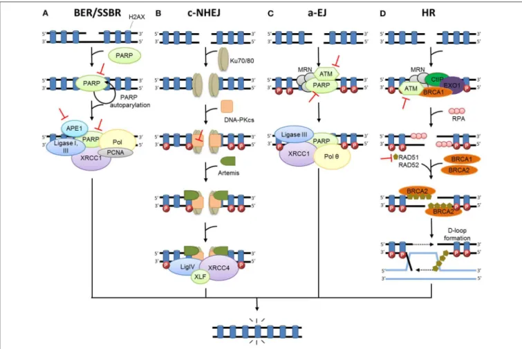
FIGURE 1 | DNA damage repair after radiation therapy. In irradiated cells, a number of DNA lesions are induced including single (SSB) and double-strand breaks (DSB). (A) SSBs are corrected by the part of base excision repair (BER) known as single-strand break repair (SSBR). The binding of PARP to SSB activates its auto-PARylation and leads to the recruitment of BER/SSBR proteins including AP endonucleases (APE1), XRCC1 (helper protein), PCNA, FEN, PNK, and DNA polymerases (Pol; damage processing) and DNA ligases. (B) In c-NHEJ, DSB is recognized by the Ku80-Ku70 heterodimer, which leads to DNA-dependent protein kinase catalytic subunit DNA-PKcs recruitment, gathering the DNA-PK complex and activating its kinase activity. This leads to the involvement of repair proteins (XRCC4, DNA ligase IV and others), which perform the processing and final junction reaction. (C) When c-NHEJ is impaired, an alternative pathway called a-EJ (alternative EJ) takes place and involves mainly PARP1, XRCC1, ligase III, MRN complex and the DNA polymerase θ. (D) In HR, after ATM activation, the DSB site is bounded by the MRE11-RAD50-NBS1 complex (MRN). The consequence is the phosphorylation of a set of targets including H2AX (γ-H2AX), localized at the site of DSB. HR uses the sister chromatid as a model to repair DSB. First, the resection of the DNA at DSB results in a 3′single-strand DNA, which is then coated by
proteins of replication A (RPA). Subsequently, proteins of the RAD family are recruited and mediate the invasion of the homologous strand of the sister chromatid, leading to the formation of Holliday junctions. DNA polymerases can then synthetize across the missing regions. The Holliday junctions are finally resolved by cleavage and followed by ligation of adjacent ends. Represents inhibitors of DNA repair in preclinical or clinical development.
of Ku proteins (
52
). a-EJ involves mainly PARP1, XRCC1, ligase
III (LIGIII), and the MRE11/RAD50/NBS1 (MRN) complex (
55
,
56
). DNA polymerase theta (Pol θ or PolQ) is specifically involved
in nucleotide incorporation in the a-EJ mechanism through the
TMEJ (theta-mediated end joining) pathway (
57
).
HR is an alternative pathway for repairing DSBs that uses
the sister chromatid as a model, restricting this mechanism to
the S and G2 cell cycle phases (Figure 1D) (
40
). HR is the
most conservative and least error-prone repair mechanism. It
necessitates the presence of BRCA proteins, defects of which
increase susceptibility to breast or ovarian cancer. The DSB
site is bounded by several factors such as the MRN complex,
EXO1 (exonuclease 1), DNA2-BLM (Bloom syndrome), BRCA1
and CTIP (CtBP-interacting protein) that contribute to DNA
resection and formation of a 3
′single-strand DNA (
58
–
60
),
which is then coated by proteins of replication A (RPA). After
the RPA protein’s displacement by RAD51, BRCA2 together
with the localizer of BRCA2 (PALB2), RAD54, and BARD1
(BRCA1-associated RING domain protein 1) mediates the
nucleoprotein filament invasion of the homologous strand of the
sister chromatid and creates the “D-loop” (
61
). DNA polymerases
can then synthetize across the missing regions. The resulting
Holliday junctions are finally resolved by cleavage and followed
by ligation of adjacent ends (
62
). However, HR can sometimes be
error-prone, especially if template switching occurs, e.g., in repeat
sequences (
63
).
The choice between the two major mechanisms for DSB repair
(NHEJ and HR pathways) seems to be linked to several factors
such as cell-cycle phase, chromatin context, or availability of key
actors such as the Ku complex, 53BP1 or RAD51 (
64
,
65
).
CURRENT STRATEGIES INVOLVED IN DDR
INHIBITION IN COMBINATION WITH RT
Targeting Key Enzymes Involved in a
Specific DNA Repair Pathway
Inhibiting BER/SSBR
BER and SSBR pathways repair damaged bases and SSBs.
Inhibiting BER/SSBR may lead to unrepaired damages that are
converted to DSBs when encountering a replication fork (
23
).
Therefore, in cells already defective for HR, such as BRCA
−/−breast or ovarian cancer tumors, the inhibition of BER by PARP
inhibitors leads to unrepaired DSBs and cell death (
14
). This
effect, called synthetic lethality, has been extensively described
(
17
,
66
,
67
) and studied in numerous clinical trials (
68
–
70
). Since
the majority of RT-induced damages are repaired by BER/SSBR,
inhibition of this pathway should highly sensitize cells to RT even
in HR-proficient cells (
29
). The preclinical evaluation of PARP
inhibitors has shown enhanced RT efficacy both in vitro and in
vivo (
71
–
73
). Several PARP inhibitors have already been tested
in or entered into numerous clinical trials in association with RT
for brain metastases, ovarian cancer, breast cancer, rectal cancer,
or glioblastoma, among others (Table 1). However, early data
did not demonstrate convincing and coherent proof of synergy,
although neither did they demonstrate unexpected toxic effects
(
74
,
75
). Another strategy for the inhibition of BER/SSBR is the
development of APE1 inhibitors. APE1 is crucial for BER/SSBR
and is commonly overexpressed in cancer cells (
80
,
81
), giving
to this strategy some tumor specificity. APE1 inhibitors have
shown efficacy in combination with RT in preclinical studies (
82
).
Lucanthone, an APE1 inhibitor, combined with temozolomide,
has recently been tested in a phase 2 clinical trial in glioblastoma
(Table 1). The results are not yet published.
Inhibiting NHEJ
DNA-PK, a key enzyme in NHEJ, is a member of the PI3K
family that performs a central role in many cellular functions
(
83
). Selective DNA-PK inhibitors have led to radiosensitization
in preclinical studies (
84
–
86
). Three phase 1 trials are currently
testing the safety and tolerability of a DNA-PK inhibitor (M3814)
in combination with palliative RT +/- immunotherapy in
advanced solid tumors (NCT02516813 and NCT03724890) and
curative-intent radiotherapy in locally advanced rectal cancer
(NCT03770689) (Table 1). Such strategies, which are not based
on a selective effect on the tumor, are considered promising by
some (
14
) though they have been criticized by others (
87
). Early
reports of the clinical combination of M3814 and palliative RT
showed enhanced normal tissue reactions including dysphagia,
prolonged mucosal inflammation/stomatitis, and skin injury (
87
,
88
). Inhibition of Ku subunits could also result in reduced
DNA-PK activity and NHEJ. This is consistent with the existing
data reporting that shRNA depletion of Ku70 or Ku80 showed
cytotoxicity and radiosensitization in pancreatic cancer cells (
89
,
90
). CC-115, a dual inhibitor of DNA-PK and mammalian target
of rapamycin (mTOR), is being tested; preliminary anti-tumor
activity has been reported, although whether these responses
are attributable to activity against DNA-PK or mTOR is unclear
(
14
,
91
). A phase 1 trial testing CC-115 in combination with RT
and temozolomide in the treatment of glioblastoma is ongoing
(NCT02977780). NHEJ can also be indirectly inhibited via the
EGFR pathway (see below).
Inhibiting HR
Cancer cells are known to be more proliferative than normal
cells (
92
). Inhibitors of replication-associated processes such
as HR exploit this specificity to enhance the therapeutic ratio.
Nevertheless, there are few specific inhibitors of HR. It has been
shown that RAD51 expression and functional HR can be reduced
using imatinib during experimental RT, leading to increased
radiosensitization (
13
,
93
). Indirect inhibition of HR can also be
obtained via cell cycle checkpoint targeting (see below).
Targeting Key Enzymes Involved in
Multiple Repair Pathways
DNA damage detection and signaling is the first step common
to all DNA repair pathways. Acting on this step will
alter several pathways. Therefore, several approaches have
been tested to disable part or all of the DNA damage
recognition/signaling steps.
Inhibiting ATM
ATM is one of the key enzymes in DNA damage signaling
of DSBs for HR but also NHEJ (
94
). Defective cells in ATM
are extremely radiosensitive, independent of their p53 status
(
95
). ATM inhibitors have shown radiosensitization in preclinical
studies (
96
–
98
). A single 15Gy RT dose suppressed tumor
growth in a preclinical model when ATM was deleted in cancer
cells vs. when deleted in endothelial cells (
99
), underlining the
interest in testing ATM inhibitors in combination with highly
conformal RT. Like DNA-PK, ATM is part of the PI3K family
and has many cellular functions. A phase 1 study is currently
testing an ATM inhibitor (AZD1390) in combination with RT
in brain tumors including glioblastoma and brain metastases
(NCT03423628). Indirectly, inhibition of the TGFβ-signaling or
mitogen-activated protein kinase (MAPK) pathway can lead to
reduced ATM activation and increased tumor cell radiosensitivity
through reduced DSB repair (
100
–
102
).
Inhibiting ATR
ATR is a critical kinase that is activated in reaction to replication
stress and blocked replication forks. ATR is one of the key
enzymes in DNA damage signaling of DSBs (
103
). Cancer
cells, which exhibit relatively elevated levels of replication
stress, are more susceptible to dependence on ATR signaling
for survival (
104
). An ATR inhibitor (AZD6738) has given
encouraging preclinical results (
67
,
105
) and is currently in
phase I trials as monotherapy or in combination with olaparib,
RT (NCT02223923), carboplatin and immunotherapy agents.
Another ATR inhibitor (M6620) is being tested in three phase
1 trials with radiotherapy in esophageal cancer (NCT03641547),
Biau et al. DNA Repair Inhibition and Radiotherapy
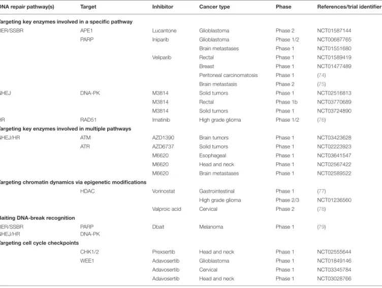
TABLE 1 | Examples of clinical trials of inhibitors of the DNA damage response in combination with radiation therapy.
DNA repair pathway(s) Target Inhibitor Cancer type Phase References/trial identifier Targeting key enzymes involved in a specific pathway
BER/SSBR APE1 Lucantone Glioblastoma Phase 2 NCT01587144
PARP Iniparib Glioblastoma Phase 1/2 NCT00687765
Brain metastases Phase 1 NCT01551680
Veliparib Rectal Phase 1 NCT01589419
Breast Phase 1 NCT01477489
Peritoneal carcinomatosis Phase 1 (74) Brain metastasis Phase 2 (75)
NHEJ DNA-PK M3814 Solid tumors Phase 1 NCT02516813
M3814 Rectal Phase 1b NCT03770689
M3814 Solid tumors Phase 1 NCT03724890
HR RAD51 Imatinib High grade glioma Phase 1/2 (76)
Targeting key enzymes involved in multiple pathways
NHEJ/HR ATM AZD1390 Brain tumors Phase 1 NCT03423628
ATR AZD6737 Solid tumors Phase 1 NCT02223923
M6620 Esophageal Phase 1 NCT03641547
M6620 Head and neck Phase 1 NCT02567422 M6620 Brain metastases Phase 1 NCT02589522 Targeting chromatin dynamics via epigenetic modifications
HDAC Vorinostat Gastrointestinal Phase 1 (77) High grade glioma Phase 2/3 NCT01236560 Valproic acid Cervical Phase 2 (78) Baiting DNA-break recognition
BER/SSBR NHEJ/HR
PARP DNA-PK
Dbait Melanoma Phase 1 (79)
Targeting cell cycle checkpoints
CHK1/2 Prexsertib Head and neck Phase 1 NCT02555644
WEE1 Adavosertib Glioblastoma Phase 1 NCT01849146
Adavosertib Cervical Phase 1 NCT03345784 Adavosertib Head and neck Phase 1 NCT03028766
locally advanced head and neck squamous cell carcinoma
(NCT02567422) and brain metastases (NCT02589522).
Inhibiting MRN Complex
Mirin is an inhibitor of MRE11 endonuclease and thus of HR
function. However, MRE11 is also upstream of NHEJ, and so
mirin inhibits NHEJ and its effects are not specific to HR (
16
,
106
,
107
). It might be of particular interest in combination with RT.
Baiting DNA Breaks Signaling
This approach developed recently is represented by the molecules
called Dbait/AsiDNA
TM. Dbait/AsiDNA
TMconsist of
double-strand oligonucleotides that mimic DNA double-strand breaks and
consequently bind and trap the signaling and repair proteins
DNA-PK (
24
,
108
,
109
) and PARP (
110
), leading to inhibition
of both SSB and DSB repair. In preclinical studies, the
proof of concept that a RT-Dbait association could be used
in treating melanoma (
24
) and high-grade glioma (
111
) has
been established. A first-in-man phase 1 trial was conducted
combining Dbait/AsiDNA
TMin combination with palliative RT
in in-transit metastases of melanoma (
79
) (Table 1). In this
trial, no dose-limiting toxicity was reported and the maximum
tolerated dose was not met.
Targeting Chromatin Dynamics via
Epigenetic Modifications
Epigenetics is an emerging field in cancer biology (
112
). It
focuses on functionally relevant genome modifications that do
not modify the nucleotide sequence. Such modifications include
DNA methylation or histone modifications that may regulate
gene expression but do not alter the associated DNA sequence.
These modifications could also affect DNA repair ability. The
loss of ARID1A, a piece of the SWI/SNF chromatin remodeling
complex, was recently reported to induce a selective vulnerability
to combined RT and PARP inhibitor therapy (
113
).
Inhibiting Histone Deacetylases (HDACs)
HDAC inhibitors are epigenetic therapeutics. They have the
capacity to lower RT-induced damage repair both at the level of
damage signaling, via inhibition of the ATM or MRN complex,
and by directly affecting proteins involved in NHEJ and HR
(
112
,
114
–
117
). Several clinical trials have been carried out for
various cancer types (
77
,
78
) (Table 1).
Inhibition of Kinases Involved in
DDR-Related Survival Pathways
Sorafenib, a multi-kinase inhibitor, is currently utilized in the
clinic for the treatment of hepatocellular and renal cancers. It
inhibits MAPK signaling together with additional intracellular
Ser/Thr kinases, leading to both NHEJ and HR inhibition.
Sorafenib has shown a radiosensitization effect in preclinical
studies (
118
). Dasatinib is another multi-kinase inhibitor
inhibiting ABL and SRC tyrosine kinases. In preclinical studies,
it has shown a radiosensitization effect partly due to blocking of
DNA repair pathways involved in DSB repair (
119
). Sorafenib
and dasatinib are clinically evaluated in association with RT.
Because of their large spectrum of targets, most of these inhibitors
may show high toxicity, which prevents them from being used at
the dosage required to be efficient in combination with RT in the
management of aggressive tumors that overexpress some of their
targets (
120
,
121
).
After RT, EGFR has been found to translocate into the
nucleus and modulate DNA repair (especially NHEJ) through
association with DNA-PKcs (
122
,
123
). A current in-clinic
approach is using the monoclonal antibody cetuximab to inhibit
this nuclear translocation of EGFR. Cetuximab combined with
RT has improved patients’ overall survival in a phase III trial in
head and neck cancer (
124
). Furthermore, EGFR signaling may
be interrupted by small-molecule tyrosine kinase inhibitors such
as erlotinib or gefitinib, especially in the case of specific EGFR
mutation; these are currently being tested in combination with
RT (
125
,
126
).
Targeting Cell Cycle Checkpoints
Checkpoint dysfunction represents a common molecular defect
acquired during tumorigenesis (
15
,
127
), underlying the
importance of its regulation in cancer development. Interfering
with cell cycle checkpoint signaling is an alternative approach
to modulating DNA repair activity and potentially improving
the therapeutic ratio. The induction of DNA lesions by RT in
normal cells stops their progression in the cell cycle, thereby
avoiding the accumulation of other lesions and their damaging
effects (
20
). This cell cycle arrest is subtly correlated with DNA
repair to fine-tune cell cycle restart with the disappearance of
damage. In cancer cells with an altered G1 checkpoint, cell cycle
progression goes on relentlessly and, as a result, the removal
of the G2 block increases unrepaired damage and its transfer
to the daughter cells. This finally causes the loss of essential
genetic material and cell death, a process that strengthens
checkpoint inhibition strategies. Combination of RT with a dual
CHK1 and CHK2 inhibitors (AZD7762 and prexsertib) showed
a radiosensitization effect with an increase in mitotic catastrophe
in different cancer cell lines and xenografts (
128
–
130
). A phase
1b trial was completed that combined prexsertib with RT and
cisplatin or cetuximab in locally advanced head and neck cancer
(NCT02555644), the results of which are not yet published.
However, in addition to checkpoint activation, CHK1 is also
involved in HR (
131
,
132
) and it is uncertain if this is only
a result of checkpoint inhibition or if it is partly due to HR
inhibition (
133
).
Another target is the WEE1 kinase, which has been shown to
be a major regulator of the G2-M checkpoint (
134
). This tyrosine
kinase inhibits the entrance in mitosis by adding an inhibitory
phosphorylation to Cdc2 (the human homolog of tyrosine kinase
1[Cdk1]) to tyrosine 15 (Y15). As a consequence, the Cdc2/cyclin
B complex becomes inactivated, which stops the cells in
G2-M and allows DNA repair. Preclinical studies have shown the
potential use of WEE 1 inhibitors as radiosentizers (
135
,
136
).
Several ongoing clinical trials are testing WEE1 inhibitors with
RT. In addition, several phase 1 trials are currently testing
the WEE1 inhibitor adavosertib (AZD1775) in combination
with RT and temozolomide in the treatment of glioblastoma
(NCT01849146), with RT and cisplatin in cervical, vaginal or
uterine cancer (NCT03345784), or in combination with RT and
cisplatin in advanced head and neck cancer (NCT03028766
and NCT02585973).
Combining DNA Repair Targeting,
Immunotherapy, and Radiotherapy
DNA repair proteins preserve the integrity of the genome;
therefore, DNA repair targeting may enhance the tumor
mutational burden, which may lead to the production of
neoantigens and increased activity of anti-cancer T cells.
Some clinical trials have been set to investigate the use
of immune checkpoint inhibitors, notably by combining
Dravulumab (anti-PD-L1) with PARP (NCT02484404), ATR
(NCT02264678) or WEE1 inhibition (NCT02617277). To date,
the interplay between radiation and the immune system is far
from being completely deciphered, but several interesting facts
have been reported. The cytotoxic action of radiotherapy on
tumor cells provides T lymphocytes with tumor neoantigens
and releases pro-inflammatory cytokines, thus promoting
the immune response (
137
). The cell death inducing this
type of immune response is called immunogenic cell death.
Combining immunotherapy with radiotherapy (several recent
trials: NCT02707588, NCT02952586, NCT02999087) could
increase the ability to cause immunogenic cell death by removing
locks that block the immune system (
138
). The non-overlapping
toxicities of DNA repair targeting and immune checkpoint
inhibitors render the use of combinations of these agents with
radiotherapy appealing (
14
,
139
).
CONCLUSION
Targeted therapies are beginning to demonstrate activity across a
number of tumor types. The most promising approach toward
improving the efficiency of a treatment and gaining a reliable
response is to develop therapy combinations that decrease the
chance of resistance and to treat resistance when it emerges.
There has been a considerable renewed emphasis on new targeted
treatments such as radiosensitizers that do not cause overlapping
dose-limiting toxicities. Selection of appropriate targeted agents
represents a challenge. As predicted, during preclinical and
clinical trials, particular attention was paid to proteins involved
Biau et al. DNA Repair Inhibition and Radiotherapy
in DNA repair pathways. Various strategies have been explored,
ranging from specific protein targeting to global inhibition, and
many DNA repair inhibitors have been developed. Up until now,
only a few of them have reached the clinical stage, while even
fewer have been tested in combination with RT. The several
clinical trials currently underway will tell whether these new
compounds can be tolerable and efficient.
RT-induced lesions can be corrected by various DNA repair
pathways. The intricacy of crosstalk between DNA repair
pathways suggests that biomarker assays to determine the
status of multiple DNA repair pathways could provide essential
information on the sensitivity and resistance of cancer cells to
repair inhibitors. Understanding these DNA repair pathways and
identifying effective stratification biomarkers from the various
DNA repair pathways that are specifically altered in some tumors
would be required to characterize patients’ responses to specific
DNA repair inhibitors.
AUTHOR CONTRIBUTIONS
JB and EC analyzed the literature and wrote the manuscript. PV
and MD gave important intellectual input and carefully revised
the manuscript. All authors approved the final manuscript
for submission.
REFERENCES
1. Delaney G, Jacob S, Featherstone C, Barton M. The role of
radiotherapy in cancer treatment: estimating optimal utilization
from a review of evidence-based clinical guidelines. Cancer. (2005) 104:1129–37. doi: 10.1002/cncr.21324
2. Elshaikh M, Ljungman M, Ten Haken R, Lichter AS.
Advances in radiation oncology. Annu Rev Med. (2006)
57:19–31. doi: 10.1146/annurev.med.57.121304.131431
3. Fertil B, Malaise EP. Intrinsic radiosensitivity of human cell lines is correlated with radioresponsiveness of human tumors: analysis of 101 published survival curves. Int J Radiat Oncol Biol Phys. (1985) 11:1699– 707. doi: 10.1016/0360-3016(85)90223-8
4. Taghian A, Ramsay J, Allalunis-Turner J, Budach W, Gioioso D, Pardo F, et al. Intrinsic radiation sensitivity may not be the major determinant of the poor clinical outcome of glioblastoma multiforme. Int J Radiat Oncol Biol Phys. (1993) 25:243–9. doi: 10.1016/0360-3016(93)90345-V
5. Gerweck LE, Vijayappa S, Kurimasa A, Ogawa K, Chen DJ. Tumor cell radiosensitivity is a major determinant of tumor response to radiation. Cancer Res. (2006) 66:8352–5. doi: 10.1158/0008-5472.CAN-06-0533
6. Khanna KK, Jackson SP. DNA double-strand breaks: signaling, repair and the cancer connection. Nat Genet. (2001) 27:247–54. doi: 10.1038/ 85798
7. Radford IR. The level of induced DNA double-strand breakage correlates with cell killing after X-irradiation. Int J Radiat Biol Relat Stud Phys Chem Med. (1985) 48:45–54. doi: 10.1080/09553008514551051
8. Jeggo P, Lavin MF. Cellular radiosensitivity: how much better
do we understand it? Int J Radiat Biol. (2009) 85:1061–
81. doi: 10.3109/09553000903261263
9. O’Driscoll M, Jeggo PA. The role of double-strand break repair - insights from human genetics. Nat Rev Genet. (2006) 7:45–54. doi: 10.1038/nrg1746 10. Helleday T, Petermann E, Lundin C, Hodgson B, Sharma RA. DNA repair
pathways as targets for cancer therapy. Nat Rev Cancer. (2008) 8:193– 204. doi: 10.1038/nrc2342
11. Stover EH, Konstantinopoulos PA, Matulonis UA, Swisher EM. Biomarkers of response and resistance to DNA repair targeted therapies. Clin Cancer Res. (2016) 22:5651–60. doi: 10.1158/1078-0432.CCR-16-0247
12. Begg AC, Stewart FA, Vens C. Strategies to improve radiotherapy with targeted drugs. Nat Rev Cancer. (2011) 11:239–53. doi: 10.1038/nrc3007 13. Thoms J, Bristow RG. DNA repair targeting and radiotherapy: a
focus on the therapeutic ratio. Semin Radiat Oncol. (2010) 20:217– 22. doi: 10.1016/j.semradonc.2010.06.003
14. Pilié PG, Tang C, Mills GB, Yap TA. State-of-the-art strategies for targeting the DNA damage response in cancer. Nat Rev Clin Oncol. (2019) 16:81– 104. doi: 10.1038/s41571-018-0114-z
15. Kastan MB, Bartek J. Cell-cycle checkpoints and cancer. Nature. (2004) 432:316–23. doi: 10.1038/nature03097
16. Curtin NJ. DNA repair dysregulation from cancer driver to therapeutic target. Nat Rev Cancer. (2012) 12:801–17. doi: 10.1038/nrc3399
17. Shaheen M, Allen C, Nickoloff JA, Hromas R. Synthetic lethality: exploiting the addiction of cancer to DNA repair. Blood. (2011) 117:6074– 82. doi: 10.1182/blood-2011-01-313734
18. Kryston TB, Georgiev AB, Pissis P, Georgakilas AG. Role of oxidative stress and DNA damage in human carcinogenesis. Mutat Res. (2011) 711:193– 201. doi: 10.1016/j.mrfmmm.2010.12.016
19. Nowsheen S, Wukovich RL, Aziz K, Kalogerinis PT, Richardson CC, Panayiotidis MI, et al. Accumulation of oxidatively induced clustered DNA lesions in human tumor tissues. Mutat Res. (2009) 674:131– 6. doi: 10.1016/j.mrgentox.2008.09.010
20. Jackson SP, Bartek J. The DNA-damage response in human biology and disease. Nature. (2009) 461:1071–8. doi: 10.1038/nature08467
21. Ward JF. DNA damage and repair. Basic Life Sci. (1991) 58:403–15; discussion 415–21. doi: 10.1007/978-1-4684-7627-9_15
22. Burkart W, Jung T, Frasch G. Damage pattern as a function of radiation quality and other factors. C R Acad Sci III, Sci Vie. (1999) 322:89– 101. doi: 10.1016/S0764-4469(99)80029-8
23. Kuzminov A. Single-strand interruptions in replicating chromosomes cause double-strand breaks. Proc Natl Acad Sci USA. (2001) 98:8241– 6. doi: 10.1073/pnas.131009198
24. Biau J, Devun F, Jdey W, Kotula E, Quanz M, Chautard E, et al. A preclinical study combining the DNA repair inhibitor Dbait with radiotherapy for the treatment of melanoma. Neoplasia. (2014) 16:835– 44. doi: 10.1016/j.neo.2014.08.008
25. Jalal S, Earley JN, Turchi JJ. DNA repair: from genome maintenance to biomarker and therapeutic target. Clin Cancer Res. (2011) 17:6973– 84. doi: 10.1158/1078-0432.CCR-11-0761
26. Krokan HE, Bjørås M. Base excision repair. Cold Spring Harb Perspect Biol. (2013) 5:a012583. doi: 10.1101/cshperspect.a012583
27. van Loon B, Markkanen E, Hübscher U. Oxygen as a friend and enemy: how to combat the mutational potential of 8-oxo-guanine. DNA Repair. (2010) 9:604–16. doi: 10.1016/j.dnarep.2010.03.004
28. Visnes T, Grube M, Hanna BMF, Benitez-Buelga C, Cázares-Körner A, Helleday T. Targeting BER enzymes in cancer therapy. DNA Repair. (2018) 71:118–26. doi: 10.1016/j.dnarep.2018.08.015
29. Vens C, Begg AC. Targeting base excision repair as a sensitization
strategy in radiotherapy. Semin Radiat Oncol. (2010) 20:241–
9. doi: 10.1016/j.semradonc.2010.05.005
30. Hyun J-W, Cheon G-J, Kim H-S, Lee Y-S, Choi E-Y, Yoon B-H, et al. Radiation sensitivity depends on OGG1 activity status in human leukemia cell lines. Free Radic Biol Med. (2002) 32:212– 20. doi: 10.1016/S0891-5849(01)00793-6
31. Yang N, Galick H, Wallace SS. Attempted base excision repair of ionizing radiation damage in human lymphoblastoid cells produces lethal and mutagenic double strand breaks. DNA Repair. (2004) 3:1323– 34. doi: 10.1016/j.dnarep.2004.04.014
32. Fortini P, Dogliotti E. Base damage and single-strand break
repair: mechanisms and functional significance of short- and
long-patch repair subpathways. DNA Repair. (2007) 6:398–
33. Carter RJ, Parsons JL. Base excision repair, a pathway regulated
by posttranslational modifications. Mol Cell Biol. (2016)
36:1426–37. doi: 10.1128/MCB.00030-16
34. Wallace SS. Base excision repair: a critical player in many games. DNA Repair. (2014) 19:14–26. doi: 10.1016/j.dnarep.2014.03.030
35. Limpose KL, Corbett AH, Doetsch PW. BERing the burden of damage: pathway crosstalk and posttranslational modification of base excision repair proteins regulate DNA damage management. DNA Repair. (2017) 56:51– 64. doi: 10.1016/j.dnarep.2017.06.007
36. Jacobs AL, Schär P. DNA glycosylases: in DNA repair and beyond. Chromosoma. (2012) 121:1–20. doi: 10.1007/s00412-011-0347-4
37. Dizdaroglu M. Oxidatively induced DNA damage and its
repair in cancer. Mutat Res Rev Mutat Res. (2015) 763:212–
45. doi: 10.1016/j.mrrev.2014.11.002
38. Schiewer MJ, Mandigo AC, Gordon N, Huang F, Gaur S, de Leeuw R, et al. PARP-1 regulates DNA repair factor availability. EMBO Mol Med. (2018) 10:e8816. doi: 10.15252/emmm.201708816
39. Rajaraman P, Bhatti P, Doody MM, Simon SL, Weinstock RM, Linet MS, et al. Nucleotide excision repair polymorphisms may modify ionizing radiation-related breast cancer risk in US radiologic technologists. Int J Cancer. (2008) 123:2713–6. doi: 10.1002/ijc.23779
40. Wyman C, Kanaar R. DNA double-strand break repair:
all’s well that ends well. Annu Rev Genet. (2006) 40:363–
83. doi: 10.1146/annurev.genet.40.110405.090451
41. Zhang Y, Rohde LH, Wu H. Involvement of nucleotide excision and mismatch repair mechanisms in double strand break repair. Curr Genomics. (2009) 10:250–8. doi: 10.2174/138920209788488544
42. Belyaev IY. Radiation-induced DNA repair foci: spatio-temporal aspects of formation, application for assessment of radiosensitivity and biological dosimetry. Mutat Res. (2010) 704:132–41. doi: 10.1016/j.mrrev.2010.01.011 43. Cortez D. Replication-coupled DNA repair. Mol Cell. (2019) 74:866–
76. doi: 10.1016/j.molcel.2019.04.027
44. Pannunzio NR, Watanabe G, Lieber MR. Nonhomologous DNA end-joining for repair of DNA double-strand breaks. J Biol Chem. (2018) 293:10512– 23. doi: 10.1074/jbc.TM117.000374
45. Britton S, Coates J, Jackson SP. A new method for high-resolution imaging of Ku foci to decipher mechanisms of DNA double-strand break repair. J Cell Biol. (2013) 202:579–95. doi: 10.1083/jcb.201303073
46. Gottlieb TM, Jackson SP. The DNA-dependent protein kinase: requirement for DNA ends and association with Ku antigen. Cell. (1993) 72:131– 42. doi: 10.1016/0092-8674(93)90057-W
47. Park E-J, Chan DW, Park J-H, Oettinger MA, Kwon J. DNA-PK is activated by nucleosomes and phosphorylates H2AX within the nucleosomes in an acetylation-dependent manner. Nucleic Acids Res. (2003) 31:6819– 27. doi: 10.1093/nar/gkg921
48. Ma Y, Pannicke U, Schwarz K, Lieber MR. Hairpin opening and overhang processing by an Artemis/DNA-dependent protein kinase complex in nonhomologous end joining and V(D)J recombination. Cell. (2002) 108:781–94. doi: 10.1016/S0092-8674(02)00671-2
49. Nick McElhinny SA, Snowden CM, McCarville J, Ramsden DA. Ku recruits the XRCC4-ligase IV complex to DNA ends. Mol Cell Biol. (2000) 20:2996– 3003. doi: 10.1128/MCB.20.9.2996-3003.2000
50. Ahnesorg P, Smith P, Jackson SP. XLF interacts with the XRCC4-DNA ligase IV complex to promote DNA nonhomologous end-joining. Cell. (2006) 124:301–13. doi: 10.1016/j.cell.2005.12.031
51. Sallmyr A, Tomkinson AE. Repair of DNA double-strand breaks by mammalian alternative end-joining pathways. J Biol Chem. (2018) 293:10536–46. doi: 10.1074/jbc.TM117.000375
52. McVey M, Lee SE. MMEJ repair of double-strand breaks (director’s cut): deleted sequences and alternative endings. Trends Genet. (2008) 24:529– 38. doi: 10.1016/j.tig.2008.08.007
53. Boboila C, Jankovic M, Yan CT, Wang JH, Wesemann DR, Zhang T, et al. Alternative end-joining catalyzes robust IgH locus deletions and translocations in the combined absence of ligase 4 and Ku70. Proc Natl Acad Sci USA. (2010) 107:3034–9. doi: 10.1073/pnas.0915067107
54. Yan CT, Boboila C, Souza EK, Franco S, Hickernell TR, Murphy M, et al. IgH class switching and translocations use a robust non-classical end-joining pathway. Nature. (2007) 449:478–82. doi: 10.1038/nature06020
55. Haince J-F, McDonald D, Rodrigue A, Déry U, Masson J-Y, Hendzel MJ, Poirier GG. PARP1-dependent kinetics of recruitment of MRE11 and NBS1 proteins to multiple DNA damage sites. J Biol Chem. (2008) 283:1197– 208. doi: 10.1074/jbc.M706734200
56. El-Khamisy SF, Masutani M, Suzuki H, Caldecott KW. A requirement for PARP-1 for the assembly or stability of XRCC1 nuclear foci at sites of oxidative DNA damage. Nucleic Acids Res. (2003) 31:5526– 33. doi: 10.1093/nar/gkg761
57. Kent T, Chandramouly G, McDevitt SM, Ozdemir AY, Pomerantz RT. Mechanism of microhomology-mediated end-joining promoted by human DNA polymerase θ. Nat Struct Mol Biol. (2015) 22:230– 7. doi: 10.1038/nsmb.2961
58. Lavin MF. The Mre11 complex and ATM: a two-way functional interaction in recognising and signaling DNA double strand breaks. DNA Repair. (2004) 3:1515–20. doi: 10.1016/j.dnarep.2004.07.001
59. Sartori AA, Lukas C, Coates J, Mistrik M, Fu S, Bartek J, et al. Human CtIP promotes DNA end resection. Nature. (2007) 450:509– 14. doi: 10.1038/nature06337
60. Prakash R, Zhang Y, Feng W, Jasin M. Homologous recombination
and human health: the roles of BRCA1, BRCA2, and
associated proteins. Cold Spring Harb Perspect Biol. (2015)
7:a016600. doi: 10.1101/cshperspect.a016600
61. Heyer W-D, Li X, Rolfsmeier M, Zhang X-P. Rad54: the Swiss Army knife of homologous recombination? Nucleic Acids Res. (2006) 34:4115– 25. doi: 10.1093/nar/gkl481
62. Solinger JA, Heyer WD. Rad54 protein stimulates the postsynaptic phase of Rad51 protein-mediated DNA strand exchange. Proc Natl Acad Sci USA. (2001) 98:8447–53. doi: 10.1073/pnas.121009898
63. Rodgers K, McVey M. Error-prone repair of DNA double-strand breaks. J Cell Physiol. (2016) 231:15–24. doi: 10.1002/jcp.25053
64. Scully R, Panday A, Elango R, Willis NA. DNA double-strand break repair-pathway choice in somatic mammalian cells. Nat Rev Mol Cell Biol. (2019). doi: 10.1038/s41580-019-0152-0. [Epub ahead of print].
65. Fattah F, Lee EH, Weisensel N, Wang Y, Lichter N, Hendrickson EA. Ku regulates the non-homologous end joining pathway choice of DNA double-strand break repair in human somatic cells. PLoS Genet. (2010) 6:e1000855. doi: 10.1371/journal.pgen.1000855
66. Chan DA, Giaccia AJ. Harnessing synthetic lethal interactions
in anticancer drug discovery. Nat Rev Drug Discov. (2011)
10:351–64. doi: 10.1038/nrd3374
67. Brown JS, O’Carrigan B, Jackson SP, Yap TA. Targeting DNA repair in cancer: beyond PARP inhibitors. Cancer Discov. (2017) 7:20–37. doi: 10.1158/2159-8290.CD-16-0860
68. Gelmon KA, Tischkowitz M, Mackay H, Swenerton K, Robidoux A, Tonkin K, et al. Olaparib in patients with recurrent high-grade serous or poorly differentiated ovarian carcinoma or triple-negative breast cancer: a phase 2, multicentre, open-label, non-randomised study. Lancet Oncol. (2011) 12:852–61. doi: 10.1016/S1470-2045(11)70214-5
69. Kaye SB, Lubinski J, Matulonis U, Ang JE, Gourley C, Karlan BY, et al. Phase II, open-label, randomized, multicenter study comparing the efficacy and safety of olaparib, a poly (ADP-ribose) polymerase inhibitor, and pegylated liposomal doxorubicin in patients with BRCA1 or BRCA2 mutations and recurrent ovarian cancer. J Clin Oncol. (2012) 30:372– 9. doi: 10.1200/JCO.2011.36.9215
70. Ledermann J, Harter P, Gourley C, Friedlander M, Vergote I, Rustin G, et al. Olaparib maintenance therapy in platinum-sensitive relapsed ovarian cancer. N Engl J Med. (2012) 366:1382–92. doi: 10.1056/NEJMoa11 05535
71. Calabrese CR, Almassy R, Barton S, Batey MA, Calvert AH, Canan-Koch S, et al. Anticancer chemosensitization and radiosensitization by the novel poly(ADP-ribose) polymerase-1 inhibitor AG14361. J Natl Cancer Inst. (2004) 96:56–67. doi: 10.1093/jnci/djh005
72. Dungey FA, Löser DA, Chalmers AJ. Replication-dependent
radiosensitization of human glioma cells by inhibition of poly(ADP-Ribose) polymerase: mechanisms and therapeutic potential. Int J Radiat Oncol Biol Phys. (2008) 72:1188–97. doi: 10.1016/j.ijrobp.2008.07.031 73. Donawho CK, Luo Y, Luo Y, Penning TD, Bauch JL, Bouska JJ, et al.
Biau et al. DNA Repair Inhibition and Radiotherapy
DNA-damaging agents in preclinical tumor models. Clin Cancer Res. (2007) 13:2728–37. doi: 10.1158/1078-0432.CCR-06-3039
74. Reiss KA, Herman JM, Armstrong D, Zahurak M, Fyles A, Brade A, et al. A final report of a phase I study of veliparib (ABT-888) in combination with low-dose fractionated whole abdominal radiation therapy (LDFWAR) in patients with advanced solid malignancies and peritoneal carcinomatosis with a dose escalation in ovarian and fallopian tube cancers. Gynecol Oncol. (2017) 144:486–90. doi: 10.1016/j.ygyno.2017.01.016
75. Chabot P, Hsia T-C, Ryu J-S, Gorbunova V, Belda-Iniesta C, Ball D, et al. Veliparib in combination with whole-brain radiation therapy for patients with brain metastases from non-small cell lung cancer: results of a randomized, global, placebo-controlled study. J Neurooncol. (2017) 131:105–15. doi: 10.1007/s11060-016-2275-x
76. Pollack IF, Jakacki RI, Blaney SM, Hancock ML, Kieran MW, Phillips P, et al. Phase I trial of imatinib in children with newly diagnosed brainstem and recurrent malignant gliomas: a pediatric brain tumor consortium report. Neuro-oncology. (2007) 9:145–60. doi: 10.1215/15228517-2006-031 77. Ree AH, Dueland S, Folkvord S, Hole KH, Seierstad T, Johansen M,
et al. Vorinostat, a histone deacetylase inhibitor, combined with pelvic palliative radiotherapy for gastrointestinal carcinoma: the Pelvic Radiation and Vorinostat (PRAVO) phase 1 study. Lancet Oncol. (2010) 11:459– 64. doi: 10.1016/S1470-2045(10)70058-9
78. Candelaria M, Cetina L, Pérez-Cárdenas E, de la Cruz-Hernández E, González-Fierro A, Trejo-Becerril C, et al. Epigenetic therapy and cisplatin chemoradiation in FIGO stage IIIB cervical cancer. Eur J Gynaecol Oncol. (2010) 31:386–91.
79. Le Tourneau C, Dreno B, Kirova Y, Grob JJ, Jouary T, Dutriaux C, et al. First-in-human phase I study of the DNA-repair inhibitor DT01 in combination with radiotherapy in patients with skin metastases from melanoma. Br J Cancer. (2016) 114:1199–205. doi: 10.1038/bjc.2016.120
80. Tell G, Fantini D, Quadrifoglio F. Understanding different
functions of mammalian AP endonuclease (APE1) as a
promising tool for cancer treatment. Cell Mol Life Sci. (2010) 67:3589–608. doi: 10.1007/s00018-010-0486-4
81. Naidu MD, Mason JM, Pica RV, Fung H, Peña LA. Radiation resistance in glioma cells determined by DNA damage repair activity of Ape1/Ref-1. J Radiat Res. (2010) 51:393–404. doi: 10.1269/jrr.09077
82. Xiang D-B, Chen Z-T, Wang D, Li M-X, Xie J-Y, Zhang Y-S, et al. Chimeric adenoviral vector Ad5/F35-mediated APE1 siRNA enhances sensitivity of human colorectal cancer cells to radiotherapy in vitro and in vivo. Cancer Gene Ther. (2008) 15:625–35. doi: 10.1038/cgt.2008.30
83. Mohiuddin IS, Kang MH. DNA-PK as an emerging therapeutic target in cancer. Front Oncol. (2019) 9:635. doi: 10.3389/fonc.2019.00635
84. Ismail IH, Mårtensson S, Moshinsky D, Rice A, Tang C, Howlett A, McMahon G, Hammarsten O. SU11752 inhibits the DNA-dependent protein kinase and DNA double-strand break repair resulting in ionizing radiation sensitization. Oncogene. (2004) 23:873–82. doi: 10.1038/sj.onc.1207303 85. Shinohara ET, Geng L, Tan J, Chen H, Shir Y, Edwards E, et al.
DNA-dependent protein kinase is a molecular target for the development of noncytotoxic radiation-sensitizing drugs. Cancer Res. (2005) 65:4987– 992. doi: 10.1158/0008-5472.CAN-04-4250
86. Davidson D, Amrein L, Panasci L, Aloyz R. Small molecules, inhibitors of DNA-PK, targeting DNA repair, and beyond. Front Pharmacol. (2013) 4:5. doi: 10.3389/fphar.2013.00005
87. Brown JM. beware of clinical trials of DNA repair inhibitors. Int J Radiat Oncol Biol Phys. (2019) 103:1182–3. doi: 10.1016/j.ijrobp.2018.11.063 88. Mau-Sorensen M, van Bussel M, Kuipers M, Nielsen DL, Verheul
HM, Aftimos P, et al. 1845PSafety, clinical activity and pharmacological biomarker evaluation of the DNA-dependent protein kinase (DNA-PK) inhibitor M3814: results from two phase I trials. Ann Oncol. (2018) 29. doi: 10.1093/annonc/mdy303.015
89. Li Y-H, Wang X, Pan Y, Lee D-H, Chowdhury D, Kimmelman AC. Inhibition of non-homologous end joining repair impairs pancreatic cancer growth and enhances radiation response. PLoS ONE. (2012) 7:e39588. doi: 10.1371/journal.pone.0039588
90. Gavande NS, VanderVere-Carozza PS, Hinshaw HD, Jalal SI,
Sears CR, Pawelczak KS, et al. DNA repair targeted therapy:
The past or future of cancer treatment? Pharmacol Ther. (2016) 160:65–83. doi: 10.1016/j.pharmthera.2016.02.003
91. Munster PN, Mahipal A, Nemunaitis JJ, Mita MM, Paz-Ares LG, Massard C, et al. Phase I trial of a dual TOR kinase and DNA-PK inhibitor (CC-115) in advanced solid and hematologic cancers. JCO. (2016) 34:2505. doi: 10.1200/JCO.2016.34.15_suppl.2505
92. Hanahan D, Weinberg RA. The hallmarks of cancer. Cell. (2000) 100:57– 70. doi: 10.1016/S0092-8674(00)81683-9
93. Qiao B, Kerr M, Groselj B, Teo MTW, Knowles MA, Bristow RG, et al. Imatinib radiosensitizes bladder cancer by targeting
homologous recombination. Cancer Res. (2013) 73:1611–
20. doi: 10.1158/0008-5472.CAN-12-1170
94. Shrivastav M, De Haro LP, Nickoloff JA. Regulation of DNA
double-strand break repair pathway choice. Cell Res. (2008)
18:134–47. doi: 10.1038/cr.2007.111
95. Westphal CH, Hoyes KP, Canman CE, Huang X, Kastan MB, Hendry JH, et al. Loss of atm radiosensitizes multiple p53 null tissues. Cancer Res. (1998) 58:5637–9.
96. Rainey MD, Charlton ME, Stanton RV, Kastan MB. Transient inhibition of ATM kinase is sufficient to enhance cellular sensitivity to ionizing radiation. Cancer Res. (2008) 68:7466–74. doi: 10.1158/0008-5472.CAN-08-0763 97. Hickson I, Zhao Y, Richardson CJ, Green SJ, Martin NMB, Orr AI, et al.
Identification and characterization of a novel and specific inhibitor of the ataxia-telangiectasia mutated kinase ATM. Cancer Res. (2004) 64:9152– 9. doi: 10.1158/0008-5472.CAN-04-2727
98. Golding SE, Rosenberg E, Adams BR, Wignarajah S, Beckta JM, O’Connor MJ, et al. Dynamic inhibition of ATM kinase provides a strategy for glioblastoma multiforme radiosensitization and growth control. Cell Cycle. (2012) 11:1167–73. doi: 10.4161/cc.11.6.19576
99. Torok JA, Oh P, Castle KD, Reinsvold M, Ma Y, Luo L, et al. Deletion of Atm in tumor but not endothelial cells improves radiation response in a primary mouse model of lung adenocarcinoma. Cancer Res. (2019) 79:773–82. doi: 10.1158/0008-5472.CAN-17-3103
100. Andarawewa KL, Paupert J, Pal A, Barcellos-Hoff MH. New rationales for using TGFbeta inhibitors in radiotherapy. Int J Radiat Biol. (2007) 83:803– 11. doi: 10.1080/09553000701711063
101. Kirshner M, Rathavs M, Nizan A, Essers J, Kanaar R, Shiloh Y, et al. Analysis of the relationships between ATM and the Rad54 paralogs involved in homologous recombination repair. DNA Repair. (2009) 8:253– 61. doi: 10.1016/j.dnarep.2008.11.005
102. Golding SE, Rosenberg E, Neill S, Dent P, Povirk LF, Valerie K. Extracellular signal-related kinase positively regulates ataxia telangiectasia mutated, homologous recombination repair, and the DNA damage response. Cancer Res. (2007) 67:1046–53. doi: 10.1158/0008-5472.CAN-06-2371
103. Minchom A, Aversa C, Lopez J. Dancing with the DNA damage response: next-generation anti-cancer therapeutic strategies. Ther Adv Med Oncol. (2018) 10:1758835918786658. doi: 10.1177/1758835918786658
104. Rundle S, Bradbury A, Drew Y, Curtin NJ. Targeting the ATR-CHK1 axis in cancer therapy. Cancers. (2017) 9:E41. doi: 10.3390/cancers9050041 105. Vendetti FP, Lau A, Schamus S, Conrads TP, O’Connor MJ, Bakkenist
CJ. The orally active and bioavailable ATR kinase inhibitor AZD6738 potentiates the anti-tumor effects of cisplatin to resolve ATM-deficient non-small cell lung cancer in vivo. Oncotarget. (2015) 6:44289–305. doi: 10.18632/oncotarget.6247
106. Dupré A, Boyer-Chatenet L, Sattler RM, Modi AP, Lee J-H, Nicolette ML, et al. A forward chemical genetic screen reveals an inhibitor of the Mre11-Rad50-Nbs1 complex. Nat Chem Biol. (2008) 4:119– 25. doi: 10.1038/nchembio.63
107. Kuroda S, Urata Y, Fujiwara T. Ataxia-telangiectasia mutated and the Mre11-Rad50-NBS1 complex: promising targets for radiosensitization. Acta Med Okayama. (2012) 66:83–92. doi: 10.18926/AMO/48258
108. Quanz M, Berthault N, Roulin C, Roy M, Herbette A, Agrario C, et al. Small-molecule drugs mimicking DNA damage: a new strategy for sensitizing tumors to radiotherapy. Clin Cancer Res. (2009) 15:1308– 16. doi: 10.1158/1078-0432.CCR-08-2108
109. Quanz M, Chassoux D, Berthault N, Agrario C, Sun J-S, Dutreix M. Hyperactivation of DNA-PK by double-strand break mimicking
molecules disorganizes DNA damage response. PLoS ONE. (2009) 4:e6298. doi: 10.1371/journal.pone.0006298
110. Croset A, Cordelières FP, Berthault N, Buhler C, Sun J-S, Quanz M, et al. Inhibition of DNA damage repair by artificial activation of PARP with siDNA. Nucleic Acids Res. (2013) 41:7344–55. doi: 10.1093/nar/gkt522 111. Biau J, Chautard E, Berthault N, de Koning L, Court F, Pereira B,
et al. Combining the DNA repair inhibitor Dbait with radiotherapy
for the treatment of high grade glioma: efficacy and protein
biomarkers of resistance in preclinical models. Front Oncol. (2019) 9:549. doi: 10.3389/fonc.2019.00549
112. Groselj B, Sharma NL, Hamdy FC, Kerr M, Kiltie AE. Histone deacetylase inhibitors as radiosensitisers: effects on DNA damage signalling and repair. Br J Cancer. (2013) 108:748–54. doi: 10.1038/bjc.2013.21
113. Park Y, Chui MH, Suryo Rahmanto Y, Yu Z-C, Shamanna RA, Bellani MA, et al. Loss of ARID1A in tumor cells renders selective vulnerability to combined ionizing radiation and PARP inhibitor therapy. Clin Cancer Res. (2019) 25:5584–94. doi: 10.1158/1078-0432.CCR-18-4222
114. Blattmann C, Oertel S, Ehemann V, Thiemann M, Huber PE, Bischof M, et al. Enhancement of radiation response in osteosarcoma and rhabdomyosarcoma cell lines by histone deacetylase inhibition. Int J Radiat Oncol Biol Phys. (2010) 78:237–45. doi: 10.1016/j.ijrobp.2010.03.010 115. Lee J-H, Choy ML, Ngo L, Foster SS, Marks PA. Histone deacetylase inhibitor
induces DNA damage, which normal but not transformed cells can repair. Proc Natl Acad Sci USA. (2010) 107:14639–44. doi: 10.1073/pnas.1008522107 116. Konsoula Z, Cao H, Velena A, Jung M. Adamantanyl-histone deacetylase inhibitor H6CAHA exhibits favorable pharmacokinetics and augments prostate cancer radiation sensitivity. Int J Radiat Oncol Biol Phys. (2011) 79:1541–8. doi: 10.1016/j.ijrobp.2010.11.057
117. Munshi A, Tanaka T, Hobbs ML, Tucker SL, Richon VM, Meyn
RE. Vorinostat, a histone deacetylase inhibitor, enhances the
response of human tumor cells to ionizing radiation through
prolongation of gamma-H2AX foci. Mol Cancer Ther. (2006)
5:1967–74. doi: 10.1158/1535-7163.MCT-06-0022
118. Yu W, Gu K, Yu Z, Yuan D, He M, Ma N, et al. Sorafenib potentiates irradiation effect in hepatocellular carcinoma in vitro and in vivo. Cancer Lett. (2013) 329:109–17. doi: 10.1016/j.canlet.2012.10.024
119. Raju U, Riesterer O, Wang Z-Q, Molkentine DP, Molkentine JM, Johnson FM, et al. Dasatinib, a multi-kinase inhibitor increased radiation sensitivity by interfering with nuclear localization of epidermal growth factor receptor and by blocking DNA repair pathways. Radiother Oncol. (2012) 105:241– 9. doi: 10.1016/j.radonc.2012.08.010
120. Khurshid H, Dipetrillo T, Ng T, Mantripragada K, Birnbaum A, Berz D, et al. A phase I study of dasatinib with concurrent chemoradiation
for stage III non-small cell lung cancer. Front Oncol. (2012)
2:56. doi: 10.3389/fonc.2012.00056
121. Kroeze SGC, Fritz C, Hoyer M, Lo SS, Ricardi U, Sahgal A, et al. Toxicity of concurrent stereotactic radiotherapy and targeted therapy or immunotherapy: a systematic review. Cancer Treatment Rev. (2017) 53:25– 37. doi: 10.1016/j.ctrv.2016.11.013
122. Liccardi G, Hartley JA, Hochhauser D. EGFR nuclear translocation modulates DNA repair following cisplatin and ionizing radiation treatment. Cancer Res. (2011) 71:1103–14. doi: 10.1158/0008-5472.CAN-10-2384 123. Ang KK, Berkey BA, Tu X, Zhang H-Z, Katz R, Hammond EH, et al. Impact
of epidermal growth factor receptor expression on survival and pattern of relapse in patients with advanced head and neck carcinoma. Cancer Res. (2002) 62:7350–6.
124. Bonner JA, Harari PM, Giralt J, Azarnia N, Shin DM, Cohen RB, et al. Radiotherapy plus cetuximab for squamous-cell carcinoma of the head and neck. N Engl J Med. (2006) 354:567–78. doi: 10.1056/NEJMoa 053422
125. Martins RG, Parvathaneni U, Bauman JE, Sharma AK, Raez LE, Papagikos MA, et al. Cisplatin and radiotherapy with or without erlotinib in locally advanced squamous cell carcinoma of the head
and neck: a randomized phase II trial. J Clin Oncol. (2013) 31:1415– 21. doi: 10.1200/JCO.2012.46.3299
126. Sato Y, Ebara T, Sunaga N, Takahashi T, Nakano T. Interaction of radiation and gefitinib on a human lung cancer cell line with mutant EGFR gene in vitro. Anticancer Res. (2012) 32:4877–81.
127. Aziz K, Nowsheen S, Pantelias G, Iliakis G, Gorgoulis VG, Georgakilas AG. Targeting DNA damage and repair: embracing the pharmacological era for successful cancer therapy. Pharmacol Ther. (2012) 133:334– 50. doi: 10.1016/j.pharmthera.2011.11.010
128. Mitchell JB, Choudhuri R, Fabre K, Sowers AL, Citrin D, Zabludoff SD, et al. In vitro and in vivo radiation sensitization of human tumor cells by a novel checkpoint kinase inhibitor, AZD7762. Clin Cancer Res. (2010) 16:2076–84. doi: 10.1158/1078-0432.CCR-09-3277
129. Zabludoff SD, Deng C, Grondine MR, Sheehy AM, Ashwell S, Caleb BL, et al. AZD7762, a novel checkpoint kinase inhibitor, drives checkpoint abrogation and potentiates DNA-targeted therapies. Mol Cancer Ther. (2008) 7:2955–66. doi: 10.1158/1535-7163.MCT-08-0492
130. Zeng L, Beggs RR, Cooper TS, Weaver AN, Yang ES. Combining Chk1/2 inhibition with cetuximab and radiation enhances in vitro and in vivo cytotoxicity in head and neck squamous cell carcinoma. Mol Cancer Ther. (2017) 16:591–600. doi: 10.1158/1535-7163.MCT-16-0352
131. Goldstein M, Kastan MB. The DNA damage response: implications for tumor responses to radiation and chemotherapy. Annu Rev Med. (2015) 66:129–43. doi: 10.1146/annurev-med-081313-121208
132. Ciccia A, Elledge SJ. The DNA damage response: making it safe to play with knives. Mol Cell. (2010) 40:179–204. doi: 10.1016/j.molcel.2010.09.019 133. Morgan MA, Parsels LA, Zhao L, Parsels JD, Davis MA, Hassan
MC, et al. Mechanism of radiosensitization by the Chk1/2 inhibitor AZD7762 involves abrogation of the G2 checkpoint and inhibition of homologous recombinational DNA repair. Cancer Res. (2010) 70:4972– 81. doi: 10.1158/0008-5472.CAN-09-3573
134. Mueller S, Haas-Kogan DA. WEE1 kinase as a target for cancer therapy. JCO. (2015) 12:3159–64. doi: 10.1200/JCO.2015.62.2290
135. Bridges KA, Hirai H, Buser CA, Brooks C, Liu H, et al. MK-1775, a novel Wee1 kinase inhibitor, radiosensitizes p53-defective human tumor cells. Clin Cancer Res. (2011) 17:5638–48. doi: 10.1158/1078-0432.CCR-11-0650 136. Caretti V, Hiddingh L, Lagerweij T, Schellen P, Koken PW, Hulleman
E, et al. WEE1 kinase inhibition enhances the radiation response of diffuse intrinsic pontine gliomas. Mol Cancer Ther. (2013) 12:141– 50. doi: 10.1158/1535-7163.MCT-12-0735
137. Ko EC, Formenti SC. Radiation therapy to enhance tumor immunotherapy: a novel application for an established modality. Int J Radiat Biol. (2019) 95:936–9. doi: 10.1080/09553002.2019.1623429
138. Golden EB, Apetoh L. Radiotherapy and immunogenic cell death. Semin Radiat Oncol. (2015) 25:11–7. doi: 10.1016/j.semradonc.2014.07.005 139. Ahmad SS, Crittenden MR, Tran PT, Kluetz PG, Blumenthal GM, Bulbeck
H, et al. Clinical development of novel drug-radiotherapy combinations. Clin Cancer Res. (2019) 25:1455–61. doi: 10.1158/1078-0432.CCR-18-2466
Conflict of Interest:MD is the cofounder of DNA Therapeutics/Onxeo, which is involved in DT01 development.
The remaining authors declare that the research was conducted in the absence of any commercial or financial relationships that could be construed as a potential conflict of interest.
Copyright © 2019 Biau, Chautard, Verrelle and Dutreix. This is an open-access article distributed under the terms of the Creative Commons Attribution License (CC BY). The use, distribution or reproduction in other forums is permitted, provided the original author(s) and the copyright owner(s) are credited and that the original publication in this journal is cited, in accordance with accepted academic practice. No use, distribution or reproduction is permitted which does not comply with these terms.