HAL Id: hal-01396592
https://hal.sorbonne-universite.fr/hal-01396592
Submitted on 14 Nov 2016HAL is a multi-disciplinary open access archive for the deposit and dissemination of sci-entific research documents, whether they are pub-lished or not. The documents may come from teaching and research institutions in France or abroad, or from public or private research centers.
L’archive ouverte pluridisciplinaire HAL, est destinée au dépôt et à la diffusion de documents scientifiques de niveau recherche, publiés ou non, émanant des établissements d’enseignement et de recherche français ou étrangers, des laboratoires publics ou privés.
Update on infections with human herpesviruses 6A, 6B,
and 7
H. Agut, P. Bonnafous, A. Gautheret-Dejean
To cite this version:
H. Agut, P. Bonnafous, A. Gautheret-Dejean. Update on infections with human herpesviruses 6A, 6B, and 7. Médecine et Maladies Infectieuses, Elsevier Masson, 2016, �10.1016/j.medmal.2016.09.004�. �hal-01396592�
Mise au point sur les herpesvirus humains 6A, 6B et 7 Update on infections with human herpesviruses 6A, 6B, and 7
H. Agut1, 2, P. Bonnafous2, A. Gautheret-Dejean1, 2, 3
1Assistance Publique –Hôpitaux de Paris, Hôpitaux Universitaires La Pitié Salpêtrière-Charles Foix,
Service de Virologie ;
2Sorbonne Universités, UPMC Univ Paris 06, INSERM, CIMI-Paris UMR 1135, Équipe 1 PVI ; 3Université Paris-Descartes, Faculté de Pharmacie, Paris, France.
Corresponding author:
Henri Agut
Service de Virologie CERVI
Hôpitaux Universitaires La Pitié Salpêtrière-Charles Foix 83 Boulevard de l'Hôpital
75651 Paris Cedex 13
Telephone: 06 76 07 99 23
Email: henri.agut@gmail.com
Co-authors:
Pascale Bonnafous: pascale.bonnafous@aphp.fr Agnès Gautheret-Dejean: agnes.gautheret@aphp.fr
Mots clés : HHV-7, exanthème subit, infections opportunistes, intégration chromosomique, HHV-6 Keywords: HHV-7, exanthema subitum, opportunistic infections, chromosomal integration, HHV-6
Résumé
Les herpesvirus humains 6A, 6B et 7 (HHV-6A, HHV-6B, HHV-7) sont génétiquement proches du cytomégalovirus. Ils appartiennent au genre des Roseolovirus et à la sous-famille des Betaherpesvirinae. Ils infectent les lymphocytes T, mais aussi les monocytes/macrophages, certaines cellules épithéliales et les cellules du système nerveux central. Ces virus sont ubiquistes et provoquent une infection chronique à vie, le plus souvent asymptomatique, chez la quasi-totalité de la population générale adulte. Le HHV-6B est responsable de l'exanthème subit, maladie bénigne du très jeune enfant. Les HHV-6A et HHV-6B sont responsables d'infections opportunistes chez les patients immunodéprimés : encéphalites, hépatites, insuffisances médullaires, colites et pneumopathies. Leur rôle étiologique dans des maladies chroniques comme sclérose en plaques, myocardiopathies ou thyroïdites est encore controversé. La pathogénicité du HHV-7 est moins bien connue et paraît beaucoup plus restreinte. L'intégration chromosomique des HHV-6A et HHV-6B est transmissible de façon héréditaire et concerne environ 1 % de la population générale. Elle pose la question des éventuelles maladies associées et peut être un facteur de confusion pour le diagnostic des infections actives par ces deux virus. Le diagnostic des infections à HHV-6A, HHV-6B et HHV-7 se fonde plus sur la PCR, qui permet la détection et la quantification du génome viral, que sur la sérologie, indiquée essentiellement en cas de primo-infection. Le ganciclovir, le foscarnet et le cidofovir inhibent la réplication de ces virus, ce qui permet le traitement des infections graves. Les indications ne sont cependant pas encore bien codifiées.
Abstract
Human herpesviruses 6A, 6B, and 7 (HHV-6A, HHV-6B, HHV-7) are genetically related to cytomegalovirus. They belong to the Roseolovirus genus and to the Betaherpesvirinae subfamily. They infect T cells, monocytes-macrophages, epithelial cells, and central nervous system cells. These viruses are ubiquitous and are responsible for lifelong chronic infections, most often asymptomatic, in the vast majority of the general adult population. HHV-6B is responsible for exanthema subitum, which is a benign disease of infants. HHV-6A and HHV-6B also cause opportunistic infections in immunocompromised patients: encephalitis, hepatitis, bone marrow suppression, colitis, and pneumonitis. Their etiological role in chronic diseases such as multiple sclerosis, cardiomyopathy, and thyroiditis is still controversial. The pathogenicity of HHV-7 is less clear and seems to be much more restricted. Chromosomal integration of HHV-6A and HHV-6B is transmissible from parents to offspring and observed in about 1% of the general population. This integration raises the question of potential associated diseases and can be a confounding factor for the diagnosis of active infections by both viruses. The diagnosis of HHV-6A, HHV-6B, and HHV-7 infections is rather based on gene amplification (PCR), which allows for the detection and quantification of the viral genome, than on serology which is mainly indicated in case of primary infection. Ganciclovir, foscarnet, and cidofovir inhibit the replication of HHV-6A, HHV-6B, and HHV-7. Severe infections may thus be treated but these therapeutic indications are still poorly defined.
Introduction
Human herpesvirus 6A (HHV-6A) was isolated by accident in 1986 from T cell blood cultures performed to look for new retroviruses [1]. Human herpesvirus 6B (HHV-6B) was identified in 1988 under similar circumstances [2, 3]. It is however just recently, in 2014, that converging results of phenotypic and genetic studies led to the classification of HHV-6A and HHV-6B as two distinct species among viruses defined as HHV-6 [4]. HHV-7 is genetically related to HHV-6 and was identified in 1990 [5]. These three viruses are very common in the general population worldwide. Just like other human herpesviruses, they persist indefinitely in the infected organ and may lead to reactivations, which are usually asymptomatic or associated with more or less severe clinical syndromes. The variety of clinical presentations, the high prevalence of the infection, and its chronicity make it difficult to precisely determine the pathogenic role of the three viruses. This issue is associated with at least one major medical challenge: diagnosing and treating severe presentations of active HHV-6A and HHV-6B infections, which correspond to primary infections or reactivations and are susceptible to antiviral drugs that proved effective against cytomegalovirus (CMV), a virus genetically related to HHV-6A, HHV-6B, and HHV-7 [6].
Characteristics of the viruses
HHV-6A, HHV-6B, and HHV-7 have an envelope of 160 to 200 nanometers of diameter, and their genome is a double-stranded linear DNA. The genomes of the viruses are respectively 159, 165, and 145 kilo base pair long [7]. These genomes are quite homologous and present with a similar organization despite their different size; all three viruses belong to the Roseolovirus genus. They harbor similar repeated sequences at each of their extremities, with patterns similar to those of telomeric repeated sequences of human chromosomes. The three viruses are also genetically homologous to CMV as all four of them belong to the Betaherpesvirinae subfamily. Viral particles (also known as virions) consist, from the outside to the inside, of the envelope carrying viral glycoproteins, the tegument, and the nucleocapsid containing the genomic DNA. The lipid nature of the envelop confers poor resistance to chemical or physical inactivating agents. HHV-6 species can be distinguished from one another thanks to their different characteristics in terms of culture, genome sequence, and antigenicity [4]. These differences are lacking among HHV-7 strains, even though genetic variations have been observed [8].
HHV-6A and HHV-6B infect many cells in vivo: T cells, especially CD4+ T cells, but also CD8+ T cells, monocytes-macrophages, hematopoietic cells of the bone marrow, epithelial cells of the kidney and salivary glands, endothelial cells, microglial cells, oligodendrocytes, and astrocytes. The CD46 molecule is a cellular receptor for 6A, while the CD134 molecule is a cellular receptor for HHV-6B [9, 10]. HHV-7 has a selective tropism for CD4+ T cells, and CD4 is a receptor for that virus. However, in vivo, HHV-7 is observed in the skin, salivary glands, and many other organs [11].
Isolation and repeated cultures of both HHV-6 species are performed at the laboratory with peripheral blood mononuclear cells of healthy donors, while adaptation to lymphocytic, glial, oligodendrocytic, and megakaryocytic cell lines or to human embryonic fibroblasts is not a common characteristic of all isolates [12, 13]. Isolating HHV-7 is best performed in purified blood CD4+ T cell cultures. HHV-7 culture is, however, difficult as it does not usually trigger any cytopathic effect and adaptation to lymphocytic cell lines varies. The viral stocks obtained always have a low infectious titer. There is currently no known natural animal model of productive infection with these viruses, even though transgenic mice expressing human CD46 have experimentally been infected with HHV-6A via intracerebral injections [14].
Pathophysiology of the infection
Following penetration of the virus into the target cells, the genomic DNA migrates to the cell nucleus where key steps of viral replication take place. Viral genes go through three stages to be transcribed and expressed: immediate early genes, early genes, and late genes. The nucleocapsid is ultimately formed and binds to the proteins of the tegument in the nucleus cell before acquiring the envelope in cytoplasmic vacuoles derived from the Golgi apparatus. These vacuoles then release virions into the cellular membrane by exocytosis [12]. This replication cycle corresponds to the lytic (or productive) cycle that leads to the death of host cells and to virus production that will disseminate through the organism at the time of primary infection or during reactivations. Following primary infection, both species of HHV-6 and HHV-7 latently persist in various sites and cells of the organism, especially in monocytes-macrophages [15]. Various hypotheses have been put forward to explain this latency mechanism; all of these mechanisms might be present among various cell : presence of the sole viral genome in its episomal form with a potentially limited expression of some genes, replication cycle blocked at the intermediate stage or replication cycle completed but controlled and
compatible with the prolonged survival of the host cell. From this latent state, a productive cycle may be reactivated and may lead to a new production of infectious viruses in the blood and in other body compartments such as saliva. However, the selective expression of some viral genes may be enough to induce modifications of cell functions, even in the absence of a complete replication cycle as demonstrated in an experimental study [13].
Chromosomal integration of HHV-6A and HHV-6B DNA is a unique phenomenon among human herpesviruses. It is observed in approximately 1% of the general population [16]. It corresponds to the covalent binding of the viral genome to the cell DNA in the telomeric region of a given chromosome. This binding is present in all cells of the organism and the viral genome may be vertically transmitted by germ cells or horizontally transmitted via tissue or organ transplants. Viral reactivation from an integrated genomic form has already been demonstrated [17].
HHV-6 primary infection triggers a specific immune response that can be identified through its humoral and cellular components [7, 18, 19]. Serum antibodies react to various viral proteins, and some of these proteins seem to be dominant antigens in serological tests. Cellular immune response may be detected with the proliferation of CD4+ and CD8+ T cells following exposure to viral antigens. However, the number of HHV-6-specific T cells circulating in the blood is low and a major cross-reactivity between HHV-6A and HHV-6B epitopes may be observed. These viruses are able to modulate the inflammatory response and the specific immune response by i) stimulating the synthesis of proinflammatory cytokines, ii) reducing the expression of HLA class I antigens at the surface of infected cells, iii) producing analogues of chemokines and chemokine receptors. These phenomena are believed to help the virus actively replicate by escaping from the immune response [13]. Despite this immune response observed as early as the primary infection, reinfection by exogenous viral strains from the same viral species is possible and these strains coexist with homologous viral strains that are already latently present. This could trigger genetic recombination events [20].
Epidemiology
The ubiquitous viruses HHV-6A, HHV-6B, and HHV-7 are widely observed in the general population as Humans are the sole natural hosts [12]. They are primarily transmitted through saliva, very early in life. Breastfeeding does not seem to play any role in the transmission of HHV-6A and HHV-6B, but
the presence of HHV-7 in breast milk has been demonstrated and suggests a potential route of transmission [21]. The in utero transmission of HHV-6 as a traditional virus is observed in approximately 1% of pregnancies. This figure is similar to that observed with CMV, but it is believed to be often associated with the presence of viral DNA integrated to the mother’s chromosomes even though this mechanism is still not clearly understood [22]. The transmission of HHV-6 through organ transplants has also been reported while its transmission via labile blood products has never been observed [23].
HHV-6B primary infection usually occurs in early childhood, between 6 months and 3 years of age. HHV-7 and HHV-6A primary infections occur later on. Thus, anti-HHV-6 antibodies reappear as early as 6 months of age when the mother’s antibodies disappear, while HHV-7 seroconversion occurs at a later stage. There is currently no specific serological test for both HHV-6 species; the exact mean age and frequency of HHV-6A primary infection thus remain unknown. The prevalence of HHV-6B and HHV-7 infections is very high, above 90% in the general adult population. The prevalence of HHV-6A infection is probably very high as well, but it has yet to be precisely measured.
Associated diseases Primary infection
Many HHV-6A, HHV-6B, and HHV-7 primary infections are asymptomatic. HHV-6B primary infection is often associated with exanthema subitum (also known as roseola infantum or sixth disease), a benign acute disease of infants [3, 24] (Table I). This disease is usually observed in infants aged between 6 months and 3 years. It usually consists of two consecutive phases: fever for 3 to 5 days, often associated with seizures, and then a rubella-like rash on the neck and chest observed when temperature drops and lasting for 1 or 2 days. HHV-6B primary infection may be associated with less specific symptoms and, in some cases, more severe ones with isolated fever, infectious respiratory or digestive syndromes, meningoencephalitis, liver impairment, viral-like syndrome with mononuclear cells in blood, and macrophage activation syndrome [25-28]. Some encephalitis cases associated with HHV-6B primary infection have a particularly poor outcome and may lead to the patient’s death or to significant neurological sequelae [29, 30]. In this context, the role of HHV-6B in the onset of severe pediatric cases of temporal lobe epilepsy is questioned. Symptomatic HHV-6A primary infections are not as well documented as those caused by 6B [31, 32]. 6A or
HHV-6B primary infection observed in fetuses during pregnancy leads to a congenital infection that never seems to be associated with clear clinical signs at birth, unlike CMV. However, an impact on the child’s future neuropsychological development has been described [33].
Clear HHV-7 primary infections are poorly documented, except for a few potential cases of exanthema subitum [34]. Viral-like syndrome with mononuclear cells in blood, seizures, or other neurological impairments have also been associated with HHV-7 in studies lacking statistical power, while the association with pityriasis rosea is still being questioned [35, 36].
Reactivations and chronic infection
HHV-6A and HHV-6B are responsible for opportunistic infections that mainly correspond to reactivations of these viruses from their latent state during immunodeficiency (Table I). Symptoms and diseases observed include fever with rash and cytopenia, pneumonitis, hepatitis, and encephalitis in organ transplant recipients, as well as colitis and retinitis in HIV-infected patients [37-39]. The most common presentations are limbic encephalitis, neurocognitive impairment, and delayed engraftment in patients who underwent an hematopoietic stem-cell transplant [40, 41]. HHV-6A and HHV-6B reactivations less often lead to severe diseases in solid organ transplant recipients [42]. Besides, CMV is often concomitantly detected with HHV-6A or HHV-6B and raises the question of each virus sole responsibility in the onset of these diseases. HHV-6 reactivation may be associated with acute transplant rejection but it is still difficult to know if the viral reactivation is the cause or consequence of the rejection.
The role of HHV-6A as a cofactor of the human immunodeficiency virus (HIV) in the progression towards AIDS is mainly supported by in vitro experimental findings: joint infection of CD4+ T cells by both viruses, induction of CD4 receptor expression by HHV-6 in CD8+ T cells and natural killer cells (NK cells) that would thus become susceptible to HIV, and HIV gene transactivation by HHV-6 [43]. However, even though HHV-6A and HHV-6B are frequently detected and responsible for opportunistic infections in HIV-infected patients, their aggravating role in the progression towards AIDS has never been formally proven [44].
Drug-induced hypersensitivity syndrome, also known as drug rash with eosinophilia and systemic symptoms (DRESS), is often associated with HHV-6B reactivations, to such an extent that this association is used as a diagnostic criterion for DRESS by Japanese dermatologists [45]. The disease
usually occurs after intake of antibiotics or antiepileptic drugs. It may be characterized by fever, skin rash, adenopathy, cytolytic hepatitis, blood hypereosinophilia, and a significant deterioration of the patient’s general status. It has been demonstrated that some of these drugs stimulate the in vitro replication of HHV-6B, that could thus be the cause of the disease [46].
The association between both species of HHV-6 and lymphoid tissue tumors has been suggested based on their experimental carcinogenic power on mouse cells and more recently because of their chromosomal integration capacity. In humans, the viral genome has been isolated from tumor tissues of patients presenting with lymphoma and Hodgkin’s disease, sometimes as an integrated form to the cellular genome [47, 48]. However, findings from a study conducted on healthy control individuals showed that HHV-6A and HHV-6B are also frequently detected in lymphoid tissues in the absence of tumor [49].
The role of HHV-6A and HHV-6B in the occurrence of autoimmune diseases has been discussed in patients presenting with multiple sclerosis and, more recently, with Hashimoto's thyroiditis [50, 51]. These viruses are detected in the central nervous system of patients presenting with multiple sclerosis, especially in demyelinated plaques. Serological data also demonstrates a higher antiviral immune response in individuals presenting with multiple sclerosis, but the topic is still the object of much debate. The same goes for the potential etiological role of these viruses in some cardiomyopathies or chronic arteriopathies [52, 53]. Although this finding still needs to be confirmed, the chromosomal integration of HHV-6A and HHV-6B could be a risk factor for the development of angina pectoris [54].
The role of HHV-7 in opportunistic infections and some tumors has been mentioned, but available data is still weak. Paradoxically, detecting HHV-7 infection seems to be correlated with better prognosis for HIV infection. One reason might just be due to the presence of a sufficient number of CD4+ T cells [55].
Diagnosis Direct diagnosis
Direct diagnosis aims at detecting the viruses responsible for the infections, at characterizing them – and especially at differentiating HHV-6A from HHV-6B -, at defining the indication for antiviral treatment, and at monitoring treatment efficacy. Direct diagnosis relies on the direct detection of
viruses and viral components (Table II). This type of diagnosis targets the viral particles that are released in biological fluids and infected cells of these fluids and tissues. Infected cells are crucial in case of HHV-6A, HHV-6B, and HHV-7 infections which, in vivo, are mostly intracellular viruses [56]. Biological specimens used for diagnostic tests are whole blood (sampled with an anticoagulant) rather than plasma, saliva, other bodily fluids such as cerebrospinal fluid (CSF) or bronchoalveolar lavage specimen, and tissue biopsies. Biological specimens must be adapted to the observed clinical syndromes and to the relevant diagnostic questions.
Virus isolation from cultures of primary lymphocytic cells is the historical reference method [1, 5, 7]. However, culture techniques lack sensitivity, are time/staff/reagent-consuming, and are associated with risks related to the production of infectious viruses. They are now mainly used for research purposes.
Viral antigens of HHV-6A, HHV-6B, and HHV-7 may be directly detected in infected cells or tissues using the immunofluorescence or immunohistochemistry technique thanks to monoclonal antibodies [57]. This technique is also mainly used for biomedical research purposes as it has a relatively low sensitivity and is limited by the low number of functional antibodies available.
Detecting genomic DNA by gene amplification (PCR) is currently the reference technique; it is accessible, specific, and very sensitive. Quantitative PCR methods, notably real-time PCR, constituted a significant advance as they reduced the risk of contamination by amplified DNA and provided a relatively precise value of the viral load in a given body organ or compartment [58, 59]. Detecting and quantifying viral messenger (or transcript) RNA using RT-PCR is an interesting additional strategy to better characterize the active or latent nature of the infection, but this technique needs to be improved [60]. Sequencing viral nucleic acids helps in characterizing them. Among the latest technical innovations of molecular assays, droplet digital PCR helps in quantifying viral DNA without having to use a range of benchmarks. Next-generation sequencing also helps in analyzing the various viral subpopulations within a single specimen [61, 62]. All molecular techniques used for the direct diagnosis of HHV-6A, HHV-6B, and HHV-7 infections need to be meticulously standardized using international benchmarks.
The indirect diagnosis relies on the detection of specific serum antibodies, immunoglobulins G (IgG) and M (IgM), most often using the immunofluorescence technique [63]. Western blot techniques and serum neutralization tests have been described in the diagnosis of HHV-6 infection; they are however mainly used for research purposes. Several factors restrict the use of viral serology, including cross-reactions between HHV-6A, HHV-6B, HHV-7, and CMV antibodies; the absence of a specific serological test able to distinguish HHV-6A infections from HHV-6B infections; the highly frequent HIV-infected status of the general adult population; the absence of correlation between the absolute value or variations of the antibody titer and the presence of an active viral infection; and the alteration of the humoral immune response in some immunocompromised patients. Serological assays are indicated in only two cases: primary infection detected through seroconversion and through the identification of specific IgM, and seroprevalence studies where seropositivity is indicative of a chronic latent infection.
Diagnostic strategy and result interpretation
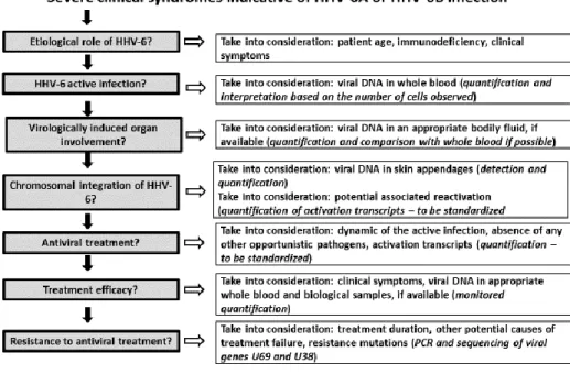
The diagnosis of HHV-6A, HHV-6B, and HHV-7 infections is usually established based on severe symptoms indicative of primary infection or reactivation. In these situations, priority must be given to the direct diagnostic method [63] (Figure1). Indirect diagnosis remains interesting because of its retrospective diagnosis of a primary infection. Otherwise, the identification of a seropositive status is associated with a poor diagnostic impact.
An active infection may be distinguished from a latent one by measuring the DNAemia of HHV-6A and HHV-6B; the threshold in-between those two states is roughly set around 1,000 copies of viral genome per milliliter of whole blood [6]. Successive measures of DNAemia allows for a better assessment of the reactivation curve. As HHV-6A, HHV-6B, and HHV-7 are essentially intracellular viruses, it is quite useful to express the DNAemia in terms of the number of cells present in the circulating blood to avoid underestimating viral replication in case of severe leukopenia [64]. Chromosomal integration of HHV-6A or HHV-6B must be considered in case of a very high and stable DNAemia, reaching or above a million copies of genomic DNA per milliliter of whole blood [13, 16]. This chromosomal integration must be confirmed by the presence of HHV-6 DNA in skin appendage specimens (hair follicles, nails), which are devoid of such DNA in the absence of integration, or by the parallel quantification of viral and cellular DNA by digital PCR - a method that still needs to be
improved. Viral reactivations have, however, been described in patients presenting with chromosomal integration, and are currently very tricky to identify given the lack of standardized techniques for quantifying viral transcripts [17].
Detecting HHV-6A or HHV-6B DNA in the CSF is highly indicative of an active infection of the central nervous system, especially of encephalopathy. This diagnosis must be considered even if the results of the concomitant DNAemia analysis are not significantly high as reactivation limited to the central nervous system may occur. In case of DNA detection in the CSF, chromosomal integration must be ruled out as it can yet again lead to a false negative result [65]. As for other body compartments susceptible to be the target of HHV-6 active infection, comparing the DNA viral load in tissues and whole blood helps in confirming the presence of in situ viral replication.
The diagnostic strategy and result interpretation would be similar for HHV-7 infections even though: the virus pathogenicity is far from being demonstrated; diagnostic strategies are less well developed and used than the ones for HHV-6; and chromosomal integration has so far never been described in this infection.
Antiviral treatment
Available antiviral treatment
In vitro, the replication of HHV-6A, HHV-6B, and HHV-7 is inhibited by foscarnet, ganciclovir, and
cidofovir. All these drugs are inhibitors of the viral polymerase DNA encoded by the U38 gene, and are active at concentrations close to those effective against CMV [13]. In contrast, aciclovir and its prodrug (valaciclovir) have no significant activity at the concentrations obtained in treated patients. Ganciclovir and cidofovir need to have gone through three or two phosphorylation steps, respectively, inside the cells to trigger their inhibiting effect, while foscarnet acts directly without any modification required. Phosphorylations are performed by cellular kinases, except for the first step of ganciclovir phosphorylation process which is catalyzed by a viral phosphotransferase (or protein kinase) encoded by the U69 gene. This mechanism of action is in line with the localization of the mutations responsible for HHV-6 resistance to antiviral drugs: within the U69 gene for the resistance to ganciclovir and the U38 gene for the resistance to the three molecules [66, 67]. Note that HHV-6 resistance can appear during the prolonged treatment of CMV infection [66].
Indications and treatment follow-up
There is no official indication for initiating antiviral treatments targeting HHV-6A and HHV-6B as their effects have only been reported in a few isolated clinical case patients.
However, these treatments may reasonably be proposed as a monotherapy or in combination to patients presenting with severe clinical presentations when one of these viruses is likely to be involved, all the more in immunocompromised patients (Figure 1). It is, however, too early to suggest a preventive treatment before diagnosing HHV-6 active infection, or a preemptive treatment for a diagnosed active infection with no associated clinical symptoms. All HHV-6A or HHV-6B active infections do not necessarily progress into severe diseases; some are spontaneously controlled thanks to the immune response. Thus, healthcare professionals seem to agree in saying that limbic encephalitis observed in hematopoietic stem-cell transplant recipients is an indication for curative treatment [13, 68].
The clinical and virological efficacy of all antiviral treatments initiated must be monitored, including measuring the viral load using quantitative PCR. The clinical impact of the emergence of resistant viruses is for now quite mild, but resistance must be considered in case of therapeutic failure and must lead healthcare professionals to look for associated mutations using viral genome sequencing techniques.
We stress the need for developing and conducting therapeutic studies with a well-designed method to validate treatments for HHV-6A, HHV-6B, and HHV-7 infections.
Acknowledgement
The authors would like to thank the Pierre et Marie Curie University, the INSERM, the HHV-6
Foundation, and the Association for Viral Infection Research (ARIV) for supporting the research
works conducted on HHV-6 and HHV-7.
Conflicts of interest
References
1. Salahuddin SZ, Ablashi DV, Markham PD, Josephs SF, Sturzenegger S, Kaplan M, et al. Isolation of a new virus, HBLV, in patients with lymphoproliferative disorders. Science 1986;234:596-601. 2. Lopez C, Pellett P, Stewart J, Goldsmith C, Sanderlin K, Black J, et al. Characteristics of human
herpesvirus-6. J Infect Dis 1988;157:1271-3.
3. Yamanishi K, Okuno T, Shiraki K, Takahashi M, Kondo T, Asano Y, et al. Identification of human herpesvirus-6 as a causal agent for exanthem subitum. Lancet 1988;1:1065-7.
4. Ablashi D, Agut H, Alvarez-Lafuente R, Clark DA, Dewhurst S, DiLuca D, et al. Classification of HHV-6A and HHV-6B as distinct viruses. Arch Virol 2014;159:863-70.
5. Frenkel N, Schirmer EC, Wyatt LS, Katsafanas G, Roffman E, Danovich RM, et al. Isolation of a new herpesvirus from human CD4+ T cells. Proc Natl Acad Sci U S A 1990;87:748-52.
6. Agut H. Deciphering the clinical impact of acute human herpesvirus 6 (HHV-6) infections. J Clin
Virol 2011;52:164-71.
7. Yamanishi K, Mori Y, Pellett PE. Human Herpesviruses 6 and 7. In: Knipe DM, Howley PM, Cohen JI, Griffin BE, Lamb RA, Martin MA, et al., editors. Fields Virology. Sixth Edition ed. Philadelphia: Wolters Kluwer / Lippincott Williams & Wilkins; 2013. p. 2058-79.
8. Franti M, Aubin JT, Gautheret-Dejean A, Malet I, Cahour A, Huraux JM, et al. Preferential associations of alleles of three distinct genes argue for the existence of two prototype variants of human herpesvirus 7. J Virol 1999;73:9655-8.
9. Santoro F, Kennedy PE, Locatelli G, Malnati MS, Berger EA, Lusso P. CD46 is a cellular receptor for human herpesvirus 6. Cell 1999;99:817-27.
10. Tang H, Serada S, Kawabata A, Ota M, Hayashi E, Naka T, et al. CD134 is a cellular receptor specific for human herpesvirus-6B entry. Proc Natl Acad Sci U S A 2013;110:9096-9.
11. Black JB, Pellett PE. Human herpesvirus 7. Rev Med Virol 1999;9:245-62.
12. De Bolle L, Naesens L, De Clercq E. Update on human herpesvirus 6 biology, clinical features, and therapy. Clin Microbiol Rev 2005;18:217-45.
13. Agut H, Bonnafous P, Gautheret-Dejean A. Laboratory and clinical aspects of human herpesvirus 6 infections. Clin Microbiol Rev 2015;28:313-35.
14. Reynaud JM, Jegou JF, Welsch JC, Horvat B. Human herpesvirus 6A infection in CD46 transgenic mice: viral persistence in the brain and increased production of proinflammatory chemokines via Toll-like receptor 9. J Virol 2014;88:5421-36.
15. Kondo K, Kondo T, Okuno T, Takahashi M, Yamanishi K. Latent human herpesvirus 6 infection of human monocytes/macrophages. J Gen Virol 1991;72 ( Pt 6):1401-8.
16. Pellett PE, Ablashi DV, Ambros PF, Agut H, Caserta MT, Descamps V, et al. Chromosomally integrated human herpesvirus 6: questions and answers. Rev Med Virol 2012;22:144-55.
17. Endo A, Watanabe K, Ohye T, Suzuki K, Matsubara T, Shimizu N, et al. Molecular and virological evidence of viral activation from chromosomally integrated human herpesvirus 6A in a patient with X-linked severe combined immunodeficiency. Clin Infect Dis 2014;59:545-8.
18. Becerra A, Gibson L, Stern LJ, Calvo-Calle JM. Immune response to HHV-6 and implications for immunotherapy. Curr Opin Virol 2014;9C:154-61.
19. Gerdemann U, Keukens L, Keirnan JM, Katari UL, Nguyen CT, de Pagter AP, et al. Immunotherapeutic strategies to prevent and treat human herpesvirus 6 reactivation after allogeneic stem cell transplantation. Blood 2013;121:207-18.
20. Achour A, Malet I, Le Gal F, Dehee A, Gautheret-Dejean A, Bonnafous P, et al. Variability of gB and gH genes of human herpesvirus-6 among clinical specimens. J Med Virol 2008;80:1211-21. 21. Fujisaki H, Tanaka-Taya K, Tanabe H, Hara T, Miyoshi H, Okada S, et al. Detection of human
herpesvirus 7 (7) DNA in breast milk by polymerase chain reaction and prevalence of HHV-7 antibody in breast-fed and bottle-fed children. J Med Virol 1998;56:2HHV-75-9.
22. Hall CB, Caserta MT, Schnabel K, Shelley LM, Marino AS, Carnahan JA, et al. Chromosomal integration of human herpesvirus 6 is the major mode of congenital human herpesvirus 6 infection. Pediatrics 2008;122:513-20.
23. Ward KN, Gray JJ, Efstathiou S. Brief report: primary human herpesvirus 6 infection in a patient following liver transplantation from a seropositive donor. J Med Virol 1989;28:69-72.
24. Zerr DM, Meier AS, Selke SS, Frenkel LM, Huang ML, Wald A, et al. A population-based study of primary human herpesvirus 6 infection. N Engl J Med 2005;352:768-76.
25. Asano Y, Yoshikawa T, Suga S, Yazaki T, Kondo K, Yamanishi K. Fatal fulminant hepatitis in an infant with human herpesvirus-6 infection. Lancet 1990;335:862-3.
26. Asano Y, Yoshikawa T, Kajita Y, Ogura R, Suga S, Yazaki T, et al. Fatal encephalitis/encephalopathy in primary human herpesvirus-6 infection. Arch Dis Child 1992;67:1484-5.
27. Hall CB, Long CE, Schnabel KC, Caserta MT, McIntyre KM, Costanzo MA, et al. Human herpesvirus-6 infection in children. A prospective study of complications and reactivation. N
Engl J Med 1994;331:432-8.
28. Crawford JR, Kadom N, Santi MR, Mariani B, Lavenstein BL. Human herpesvirus 6 rhombencephalitis in immunocompetent children. J Child Neurol 2007;22:1260-8.
29. Yoshikawa T, Ohashi M, Miyake F, Fujita A, Usui C, Sugata K, et al. Exanthem subitum-associated encephalitis: nationwide survey in Japan. Pediatr Neurol 2009;41:353-8.
30. Tesini BL, Epstein LG, Caserta MT. Clinical impact of primary infection with roseoloviruses. Curr
Opin Virol 2014;9C:91-6.
31. Bates M, Monze M, Bima H, Kapambwe M, Clark D, Kasolo FC, et al. Predominant human herpesvirus 6 variant A infant infections in an HIV-1 endemic region of Sub-Saharan Africa. J
Med Virol 2009;81:779-89.
32. Tembo J, Kabwe M, Chilukutu L, Chilufya M, Mwaanza N, Chabala C, et al. Prevalence and Risk Factors for Betaherpesvirus DNAemia in Children >3 Weeks and <2 Years of Age Admitted to a Large Referral Hospital in Sub-Saharan Africa. Clin Infect Dis 2015;60:423-31.
33. Caserta MT, Hall CB, Canfield RL, Davidson P, Lofthus G, Schnabel K, et al. Early Developmental Outcomes of Children With Congenital HHV-6 Infection. Pediatrics 2014;134:1111-8.
34. Tanaka K, Kondo T, Torigoe S, Okada S, Mukai T, Yamanishi K. Human herpesvirus 7: another causal agent for roseola (exanthem subitum). J Pediatr 1994;125:1-5.
35. Ward KN, Andrews NJ, Verity CM, Miller E, Ross EM. Human herpesviruses-6 and -7 each cause significant neurological morbidity in Britain and Ireland. Arch Dis Child 2005;90:619-23.
36. Rebora A, Drago F, Broccolo F. Pityriasis rosea and herpesviruses: facts and controversies. Clin
Dermatol 2010;28:497-501.
37. Clark DA, Griffiths PD. Human herpesvirus 6: relevance of infection in the immunocompromised host. Br J Haematol 2003;120:384-95.
38. Boutolleau D, Fernandez C, Andre E, Imbert-Marcille BM, Milpied N, Agut H, et al. Human herpesvirus (HHV)-6 and HHV-7: two closely related viruses with different infection profiles in stem cell transplantation recipients. J Infect Dis 2003;187:179-86.
39. Gautheret-Dejean A, Agut H, Nicolas JC, Beaugerie L. Roseolovirus DNA in the colonic mucosa of HIV-seropositive patients with diarrhea. Clin Infect Dis 2003;36:1348-9.
40. Ogata M. Human herpesvirus 6 in hematological malignancies. J Clin Exp Hematop 2009;49:57-67.
41. Zerr DM, Fann JR, Breiger D, Boeckh M, Adler AL, Xie H, et al. HHV-6 reactivation and its effect on delirium and cognitive functioning in hematopoietic cell transplantation recipients. Blood 2011;117:5243-9.
42. Razonable RR. Human herpesviruses 6, 7 and 8 in solid organ transplant recipients. Am J
Transplant 2013;13 Suppl 3:67-77; quiz -8.
43. Lusso P, Gallo RC. Human herpesvirus 6 in AIDS. Immunol Today 1995;16:67-71.
44. Spira TJ, Bozeman LH, Sanderlin KC, Warfield DT, Feorino PM, Holman RC, et al. Lack of correlation between human herpesvirus-6 infection and the course of human immunodeficiency virus infection. J Infect Dis 1990;161:567-70.
45. Yoshikawa K. HHV-6B and HHV-7 in exanthema subitum and related skin diseases. In: Flamand L, Lautenschlager I, Krueger G, Ablashi D, editors. Human herpesviruses HHV-6A, HHV-6B & HHV-7. Third Edition ed. Amsterdam: Elsevier; 2014. p. 153-66.
46. Mardivirin L, Valeyrie-Allanore L, Branlant-Redon E, Beneton N, Jidar K, Barbaud A, et al. Amoxicillin-induced flare in patients with DRESS (Drug Reaction with Eosinophilia and Systemic Symptoms): report of seven cases and demonstration of a direct effect of amoxicillin on Human Herpesvirus 6 replication in vitro. Eur J Dermatol 2010;20:68-73.
47. Torelli G, Barozzi P, Marasca R, Cocconcelli P, Merelli E, Ceccherini-Nelli L, et al. Targeted integration of human herpesvirus 6 in the p arm of chromosome 17 of human peripheral blood mononuclear cells in vivo. J Med Virol 1995;46:178-88.
48. Lacroix A, Jaccard A, Rouzioux C, Piguet C, Petit B, Bordessoule D, et al. HHV-6 and EBV DNA quantitation in lymph nodes of 86 patients with Hodgkin's lymphoma. J Med Virol 2007;79:1349-56.
49. Fillet AM, Raphael M, Visse B, Audouin J, Poirel L, Agut H. Controlled study of human herpesvirus 6 detection in acquired immunodeficiency syndrome-associated non-Hodgkin's lymphoma. The French Study Group for HIV-Associated Tumors. J Med Virol 1995;45:106-12. 50. Leibovitch EC, Jacobson S. Evidence linking HHV-6 with multiple sclerosis: an update. Curr Opin
Virol 2014;9C:127-33.
51. Caselli E, Zatelli MC, Rizzo R, Benedetti S, Martorelli D, Trasforini G, et al. Virologic and immunologic evidence supporting an association between HHV-6 and Hashimoto's thyroiditis.
PLoS Pathog 2012;8:e1002951.
52. Leveque N, Boulagnon C, Brasselet C, Lesaffre F, Boutolleau D, Metz D, et al. A fatal case of Human Herpesvirus 6 chronic myocarditis in an immunocompetent adult. J Clin Virol 2011;52:142-5.
53. Takatsuka H, Wakae T, Mori A, Okada M, Fujimori Y, Takemoto Y, et al. Endothelial damage caused by cytomegalovirus and human herpesvirus-6. Bone Marrow Transplant 2003;31:475-9. 54. Gravel A, Dubuc I, Morissette G, Sedlak RH, Jerome KR, Flamand L. Inherited chromosomally
integrated human herpesvirus 6 as a predisposing risk factor for the development of angina pectoris. Proc Natl Acad Sci U S A 2015;112:8058-63.
55. Boutolleau D, Bonduelle O, Sabard A, Devers L, Agut H, Gautheret-Dejean A. Detection of human herpesvirus 7 DNA in peripheral blood reflects mainly CD4+ cell count in patients infected with HIV. J Med Virol 2005;76:223-8.
56. Achour A, Boutolleau D, Slim A, Agut H, Gautheret-Dejean A. Human herpesvirus-6 (HHV-6) DNA in plasma reflects the presence of infected blood cells rather than circulating viral particles. J
Clin Virol 2007;38:280-5.
57. Halme L, Arola J, Hockerstedt K, Lautenschlager I. Human herpesvirus 6 infection of the gastroduodenal mucosa. Clin Infect Dis 2008;46:434-9.
58. Flamand L, Gravel A, Boutolleau D, Alvarez-Lafuente R, Jacobson S, Malnati MS, et al. Multicenter comparison of PCR assays for detection of human herpesvirus 6 DNA in serum. J Clin
Microbiol 2008;46:2700-6.
59. Fernandez C, Boutolleau D, Manichanh C, Mangeney N, Agut H, Gautheret-Dejean A. Quantitation of HHV-7 genome by real-time polymerase chain reaction assay using MGB probe technology. J Virol Methods 2002;106:11-6.
60. Ihira M, Enomoto Y, Kawamura Y, Nakai H, Sugata K, Asano Y, et al. Development of quantitative RT-PCR assays for detection of three classes of HHV-6B gene transcripts. J Med Virol 2012;84:1388-95.
61. Leibovitch EC, Brunetto GS, Caruso B, Fenton K, Ohayon J, Reich DS, et al. Coinfection of human herpesviruses 6A (HHV-6A) and HHV-6B as demonstrated by novel digital droplet PCR assay.
PLoS ONE 2014;9:e92328.
62. Sedlak RH, Cook L, Huang ML, Magaret A, Zerr DM, Boeckh M, et al. Identification of chromosomally integrated human herpesvirus 6 by droplet digital PCR. Clin Chem 2014;60:765-72.
63. Gautheret-Dejean A, Agut H. Practical Diagnostic Procedures for HHV-6A, HHV-6B, and HHV-7. In: Flamand L, Lautenschlager I, Krueger G, Ablashi D, editors. Human herpesviruses HHV-6A, HHV-6B & HHV-7. Third Edition ed. Amsterdam: Elsevier; 2014. p. 9-34.
64. Gautheret-Dejean A, Henquell C, Mousnier F, Boutolleau D, Bonnafous P, Dhedin N, et al. Different expression of human herpesvirus-6 (HHV-6) load in whole blood may have a significant impact on the diagnosis of active infection. J Clin Virol 2009;46:33-6.
65. Ward KN, Leong HN, Thiruchelvam AD, Atkinson CE, Clark DA. Human herpesvirus 6 DNA levels in cerebrospinal fluid due to primary infection differ from those due to chromosomal viral integration and have implications for diagnosis of encephalitis. J Clin Microbiol 2007;45:1298-304.
66. Manichanh C, Olivier-Aubron C, Lagarde JP, Aubin JT, Bossi P, Gautheret-Dejean A, et al. Selection of the same mutation in the U69 protein kinase gene of human herpesvirus-6 after prolonged exposure to ganciclovir in vitro and in vivo. J Gen Virol 2001;82:2767-76.
67. Bonnafous P, Naesens L, Petrella S, Gautheret-Dejean A, Boutolleau D, Sougakoff W, et al. Different mutations in the HHV-6 DNA polymerase gene accounting for resistance to foscarnet.
Antivir Ther 2007;12:877-88.
68. Razonable RR. Infections due to human herpesvirus 6 in solid organ transplant recipients. Curr
Tableau I. Maladies associées aux infections à HHV-6A et HHV-6B Table I. Diseases associated with HHV-6A and HHV-6B infections
Status of viral infection Proven association Suggested association to be confirmed
Congenital infection - Impairment of neuropsychological development in
infants Postnatal primary infection Exanthema subitum (roseola infantum or sixth disease) for
HHV-6B Fever Seizures
Mild respiratory and digestive symptoms Thrombocytopenia
Viral-like syndrome with mononuclear cells in blood Encephalitis
Hepatitis, colitis
Exanthema subitum for HHV-6A Macrophage activation syndrome Temporal lobe epilepsy
Reactivation Fever
Skin rash
Thrombocytopenia, leukopenia, anemia Myelosuppression
Encephalitis, neurocognitive deficit Hepatitis, colitis, gastroenteritis Retinitis
Pneumonitis
Drug-induced hypersensitivity syndrome (DRESS)
Temporal lobe epilepsy Transplant rejection
Graft-versus-host disease (GVHD) Thrombotic microangiopathy
Chronic infection - Multiple sclerosis
Hashimoto's thyroiditis
Myocarditis, chronic cardiomyopathy
Rapid progression towards AIDS in HIV-infected patients for HHV-6A
Chromosomal integrationa - Increased risk of angina pectoris
Increased risk of HHV-6 congenital infection
a
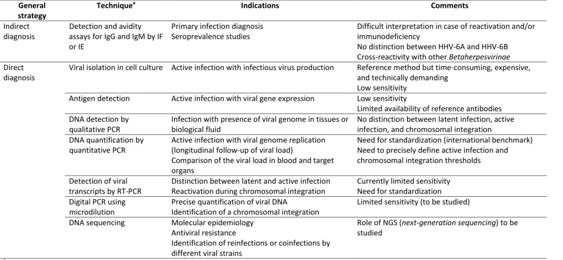
Tableau II. Approches diagnostiques des infections à HHV-6A, HHV-6B et HHV-7 Table II. Diagnostic strategies for HHV-6A, HHV-6B, and HHV-7 infections
General strategy
Techniquea Indications Comments
Indirect diagnosis
Detection and avidity assays for IgG and IgM by IF or IE
Primary infection diagnosis Seroprevalence studies
Difficult interpretation in case of reactivation and/or immunodeficiency
No distinction between HHV-6A and HHV-6B Cross-reactivity with other Betaherpesvirinae Direct
diagnosis
Viral isolation in cell culture Active infection with infectious virus production Reference method but time-consuming, expensive, and technically demanding
Low sensitivity Antigen detection Active infection with viral gene expression Low sensitivity
Limited availability of reference antibodies DNA detection by
qualitative PCR
Infection with presence of viral genome in tissues or biological fluid
No distinction between latent infection, active infection, and chromosomal integration DNA quantification by
quantitative PCR
Active infection with viral genome replication (longitudinal follow-up of viral load)
Comparison of the viral load in blood and target organs
Need for standardization (international benchmark) Need to precisely define active infection and chromosomal integration thresholds
Detection of viral transcripts by RT-PCR
Distinction between latent and active infection Reactivation during chromosomal integration
Currently limited sensitivity Need for standardization Digital PCR using
microdilution
Precise quantification of viral DNA
Identification of a chromosomal integration
Limited sensitivity (to be studied) DNA sequencing Molecular epidemiology
Antiviral resistance
Identification of reinfections or coinfections by different viral strains
Role of NGS (next-generation sequencing) to be studied
a
IF: immunofluorescence; IE: immunoenzymology; PCR: polymerase chain reaction (gene amplification); RT-PCR: reverse transcriptase –polymerase chain reaction (gene amplification following reverse transcription)
Figure 1. Schéma décisionnel pour le diagnostic et le traitement d’une infection sévère à HHV-6A ou HHV-6B. Figure 1. Decision tree for diagnosing and treating severe HHV-6A or HHV-6B infection.
Plusieurs des examens virologiques présentés sur ce schéma nécessitent d’être standardisés et/ou approuvés pour l’utilisation diagnostique proposée. Le terme HHV-6 se réfère collectivement aux deux espèces virales HHV-6A et HHV-6B.
Several of the virological examinations mentioned in this diagram need to be standardized and/or approved for the suggested diagnostic use. The term “HHV-6” refers to both viral species (HHV-6A and HHV-6B).