Metal reduction by spores of Desulfotomaculum
reducens
Pilar Junier,1Manon Frutschi,1
Nicholas S. Wigginton,1Eleanor J. Schofield,2 John R. Bargar2and Rizlan Bernier-Latmani1* 1Environmental Microbiology Laboratory, Ecole Polytechnique Fédérale de Lausanne (EPFL), Lausanne, CH 1015, Switzerland.
2Stanford Synchrotron Radiation Laboratory, Menlo Park, CA 94025, USA.
Summary
The bioremediation of uranium-contaminated sites is designed to stimulate the activity of microorganisms able to catalyze the reduction of soluble U(VI) to the less soluble mineral UO2. U(VI) reduction does not necessarily support growth in previously studied bac-teria, but it typically involves viable vegetative cells and the presence of an appropriate electron donor. We characterized U(VI) reduction by the sulfate-reducing bacterium Desulfotomaculum reducens strain MI-1 grown fermentatively on pyruvate and observed that spores were capable of U(VI) reduction. Hydrogen gas – a product of pyruvate fermentation – rather than pyruvate, served as the electron donor. The presence of spent growth medium was required for the process, suggesting that an unknown factor produced by the cells was necessary for reduction. Ultrafiltration of the spent medium followed by U(VI) reduction assays revealed that the factor’s molecular size was below 3 kDa. Pre-reduced spent medium displayed short-term U(VI) reduction activity, suggesting that the missing factor may be an electron shuttle, but neither anthraquinone-2,6-disulfonic acid nor ribofla-vin rescued spore activity in fresh medium. Spores of D. reducens also reduced Fe(III)-citrate under experi-mental conditions similar to those for U(VI) reduction. This is the first report of a bacterium able to reduce metals while in a sporulated state and underscores the novel nature of the mechanism of metal reduction by strain MI-1.
Introduction
As a result of nuclear fuel and weapon production, U(VI) contamination is a severe problem in many US Depart-ment of Energy sites and similar locales worldwide (Riley et al., 1992; Abdelouas et al., 1999; Meinrath et al., 2003). The microbial reduction of U(VI) to the less soluble and less bio-available U(IV), which precipitates as the mineral UO2, is touted as a potential solution for the immobiliza-tion of uranium in the subsurface at contaminated sites. The strategy considered for the direct enzymatic reduc-tion of U(VI) involves the addireduc-tion of an appropriate elec-tron donor that would stimulate the indigenous microbial community and promote the use of U(VI) as a terminal electron acceptor (Sanford et al., 2007; Yabusaki et al., 2007; N’Guessan et al., 2008).
Species from four genera capable of U(VI) reduction have been studied most extensively: Shewanella (Bencheikh-Latmani et al., 2005), Geobacter (Shelobolina et al., 2007), Desulfovibrio (Payne et al., 2004) and Anaer-omyxobacter (Wu et al., 2006). The former two are genera of dissimilatory metal-reducing bacteria exhibiting a remarkable respiratory versatility and able to couple U(VI) reduction to growth (Lovley et al., 1991) whereas the third is a genus of sulfate-reducing bacteria some species of which couple U(VI) reduction to growth (Pietzsch and Babel, 2003). The final genus, Anaeromyxobacter, encom-passes anaerobic myxobacteria and one of its species was reported to couple growth to U(VI) reduction (Sanford et al., 2007). In all four genera, U(VI) reduction also takes place readily under non-growth conditions, but metabolically active cells are needed to catalyse the process (Bencheikh-Latmani et al., 2005; Wall and Krumholz, 2006; Marshall et al., 2009).
In this study, we characterize U(VI) reduction by the bacterium Desulfotomaculum reducens strain MI-1, a sulfate-reducing bacterium isolated from marine sedi-ments contaminated with Cr(VI) (Tebo and Obraztsova, 1998). U(VI) reduction by this organism was deemed interesting and unusual for several reasons. First, strain MI-1 was reported to grow on U(VI) as a sole terminal electron acceptor with butyrate as an electron donor (Tebo and Obraztsova, 1998). Second, several field sites contaminated with U(VI) have been shown to harbour Desulfotomaculum and/or its close relative Desulfos-porosinus either prior to (Chang et al., 2001) or during a *For correspon-dence. E-mail rizlan.bernier-latmani@epfl.ch; Tel.
(+41) 21 693 5001; Fax (+41) 21 693 6205.
bioremediation effort (Anderson et al., 2003; Nevin et al., 2003; Suzuki et al., 2003; Chandler et al., 2006), suggest-ing that these microorganisms may play a role in the environmental U(VI) reduction process. Finally, strain MI-1 differs phylogenetically from other studied U(VI)-reducing organisms because it is a spore-forming Firmic-utes, whereas Geobacter, Shewanella, Desulfovibrio and Anaeromyxobacter are all Proteobacteria.
The distinct phylogeny of strain MI-1 raises the possi-bility that its mechanism of U(VI) reduction is significantly different from those previously described. Therefore, the goal of the study was to characterize the mechanism of U(VI) reduction by strain MI-1. We found that this microorganism exhibited a novel mechanism of U(VI) and Fe(III) reduction that involved its spore form and an unidentified factor excreted by cells.
Results
U(VI) reduction by strain MI-1
The ability to ferment pyruvate is a common trait in the genus Desulfotomaculum (Widdel, 1992). Desulfoto-maculum reducens is no exception as it grew with pyuvate as the sole substrate and produced acetate, H2and CO2 (Fig. 1 and Fig. S1). The presence of U(VI) during fermen-tative growth on pyruvate had no effect on the temporal change in protein concentration (Fig. 1) or on the trans-formation of pyruvate to its products (Fig. S1). In addition, little U(VI) reduction took place during active growth because only 8.2mM U(VI) was reduced in the first 48 h of growth (Fig. 1), whereas 50mM U(VI) was reduced
between 48 and 180 h, after pyruvate was depleted and growth ceased (Fig. 1). A total of 85mM U(VI) was reduced after 324 h. Considering that the maximum amount of biomass produced was 18mg ml-1, we calcu-lated that a maximum of 4.7mmoles of U(VI) were reduced per mg protein over that time frame.
Interestingly, U(VI) reduction proceeded (48–180 h) during a steady decline in the total protein concentration, which occurred independently of the presence of U(VI) (Fig. 1). Microscopic observation of the cultures revealed that 14% of the D. reducens cells had sporulated after 108 h. Desulfotomaculum spp. are known to sporulate in the absence of an appropriate electron donor and in the presence of acetate (Widdel, 1992). Hereafter, the reader may assume that cultures devoid of pyruvate (typically 48 h old or older) will contain some fraction of spores, unless otherwise indicated. Thus, we attribute the decrease in protein concentration, assayed in centrifuged culture pellets, to sporulation and to the release of soluble protein from disintegrating vegetative cells. The observed temporal relationship suggests that U(VI) reduction could be linked to the formation of spores.
U(VI) reduction by spores of D. reducens
To evaluate the possibility of spore involvement in U(VI) reduction more rigorously, we considered the reduction of U(VI) in the absence of pyruvate by: (i) spores alone (variously pasteurized, unpasteurized or glutaraldehyde-fixed), (ii) vegetative cells alone (growing or pasteurized) and (iii) an abiotic control containing neither vegetative cells nor spores. The results (Table 1) show that U(VI) reduction proceeded when spores (pasteurized or unpas-teurized) were present along with both H2 and spent medium. Spent fermentation medium (Table 1) was the filtrate from a stationary-phase (> 48 h) culture that had converted all of the originally added pyruvate to H2, acetate and CO2.
The extent of U(VI) reduction by spores depended on the chemical state of the spores. Glutaraldehyde-fixed spores, which were unable to germinate (data not shown) and are expected to be more extensively modified chemi-cally than pasteurized spores, did not reduce U(VI) (Table 1). In contrast, pasteurized spores were viable (able to germinate) and reduced U(VI), as did untreated spores (Table 1). As pasteurized vegetative cells did not reduce U(VI) (Table 1), the observed activity of pasteur-ized spores cannot be explained by residual vegetative cell debris that might have remained in the pasteurized spore preparations despite repeated washes to remove them. We therefore conclude that strain MI-1 spores are directly involved in U(VI) reduction.
Vegetative cells grown fermentatively in the presence of U(VI) exhibited some U(VI) reduction, although 100-fold
0 5 10 15 20 25 0 20 40 60 80 100 120 140 160 180 200 Time (h) 0 20 40 60 80 100 U(VI) concentration (µM) Prot ein con c entrat ion (µg ml –1 ) U(VI) protein with U protein no U
Fig. 1. Pyruvate fermentation by MI-1 in the presence (filled
symbols) and absence (open symbols) of U(VI). Results of growth (measured by protein concentration) and U(VI) concentration are displayed. The corresponding pyruvate and acetate data are in Fig. S1. Triplicates were run for all conditions and the error bars represent the standard deviation within the set.
less than that with spores alone (Table 1). Yields were 10-8nmol reduced U(VI) per cell.h versus 10-6nmol reduced U(VI) per spore.h.
As described previously, U(VI) reduction commences after the depletion of pyruvate from the medium. In the absence of pyruvate, H2, one of the products of fermen-tation, is presumed to act as the electron donor for U(VI) reduction. A requirement for the presence of H2was dem-onstrated by the lack of U(VI) reduction in the N2-purged aliquot of spent fermentative growth medium, which was examined both unsupplemented and supplemented with 20 mM H2(Table 1). Amendment with H2rescues reduc-tion activity, suggesting that it is the electron donor for spore-mediated U(VI) reduction. In addition, the lack of U(VI) reduction in the spent abiotic control (Table 1) con-firms that U(VI) reduction is a biological process requiring the presence of spores, rather than a chemical process driven by H2.
We tested the dependence of the rate of U(VI) reduction on the concentration of spores (Fig. 2) and found a propor-tional relationship – twice the concentration of spores yields approximately twice the rate – which further confirms the role of spores in U(VI) reduction and suggests an enzymatic process. A comparison of the rate of reduction of U(VI) by D. reducens spores and Shewanella oneidensis MR-1 cells yields surprisingly similar rates: 10 nmoles h-1 per 107spores (this study) and 9.5 nmoles h-1per 107cells (Liu et al., 2002).
We observed the formation of a dark precipitate in all samples after the reduction of U(VI) by spores of strain MI-1. In order to characterize the localization of uranium relative to the spores, a spore preparation incubated in the presence of U(VI), spent growth medium and H2was
imaged by transmission electron microscopy (TEM). U was the only electron-dense material in the sample and appeared to be located mainly in association with the spore coat (Fig. 3).
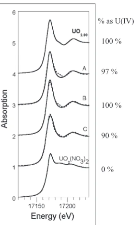
We confirmed that the disappearance of U(VI) from solution did indeed stem from U(VI) reduction to U(IV) by characterizing the solid-phase product of U(VI) reduction by X-ray absorption spectroscopy. The X-ray absorption near-edge structure (XANES) spectra of samples contain-ing pyruvate-grown D. reducens cultures after 300 h of Table 1. U(VI) reduction by spores and vegetative cells under different conditions in the presence of pyruvate-free spent media amended with or
free of H2, compared with fresh medium amended with acetate and H2.
% U(VI) reduced
Spent fermentative
mediuma Fresh WLP medium
N2-purged spent fermentative mediuma
20 mM H2added 20 mM H2added 20 mM H2added No H2
Pasteurized sporesb 73.5⫾ 2.4 3.1⫾ 6.4 ND ND
Unpasteurized sporesb 83.9⫾ 1.0 2.8⫾ 4.0 52.3⫾ 1.9 4.9⫾ 0.4
Glutaraldehyde-fixed sporesb 0.9⫾ 1.3 ND ND ND
Pasteurized vegetative cellsc 1.9⫾ 5.6 1.9⫾ 5.6 ND ND
Live vegetative cellsd 11.2⫾ 4.5 ND ND ND
Abiotic (no spores or cells) 2.1⫾ 5.9 2.5⫾ 5.9 4.6⫾ 0.4 ND
a. Spent fermentation medium was the filtrate from a stationary-phase culture in which all detectable pyruvate had been removed (fermented)
during growth.
b. The spore concentration was 1.6¥ 106spores ml-1and the vegetative cell concentration 2.3¥ 107cells ml-1. Pasteurized and unpasteurized spores were viable (able to germinate) and were expected to have less chemical damage than glutaraldehyde-fixed spores, which were unable to produce viable vegetative cells under germination conditions.
c. Pasteurized vegetative cell preparations contain cell debris comparable to, but in higher concentrations than, the vegetative cell debris found
in partially purified, pasteurized spore preparations.
d. Cells grown fermentatively in the presence of U(VI). H2was present as a result of pyruvate fermentation. ND, not determined. 0 10 20 30 40 50 60 70 80 90 0 50 100 150 Time (h) Redu ced U concentrat ion (µ M) 1.1 x 10⁶spores ml–1 2.2 x 10⁶spores ml–1
Fig. 2. Concentration of reduced U over time after incubation in
spent growth medium, H2and different concentration of spores of
exposure to U(VI) (i.e., in which some fraction of the cells had sporulated), as well as pasteurized and unpasteur-ized spore preparation exposed to U(VI) for 72 h were best fit as a linear combination of U(IV) and U(VI) stan-dards with U(IV) representing 97%, 100% and 90% of the total U for the three samples respectively (Fig. 4). Dependence of U(VI) reduction on spent
growth medium
U(VI) reduction by strain MI-1 spores occurred only in spent growth medium but not in fresh pyruvate-free fer-mentation medium (Table 1). Spores of strain MI-1 did not reduce U(VI) even when fresh medium was amended with the products of pyruvate fermentation (acetate and H2, data not shown) or with H2alone (Table 1). The require-ment for spent growth medium suggests that germinating and/or growing cells release a stimulatory factor that is involved in the U(VI) reduction process.
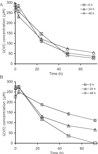
Because strain MI-1 cultures are typically inoculated from a spore preparation, it is conceivable that the factor could be excreted during the spore germination process or could be excreted by vegetative cells during growth. To evaluate both possibilities, the ability of spent growth medium to support U(VI) reduction by spores was tested for cultures in which germination had or had not occurred (Fig. 5). Spent growth media from two types of cultures were assayed: cultures inoculated from a cell suspension (Fig. 5A) and cultures inoculated from a spore suspension (Fig. 5B). In the former, the absence of spores precluded germination and provided a condition in which only cell growth could account for the presence of the factor. In the case of the culture inoculated from a spore suspension, both germination and subsequent cell growth took place. Spent growth medium collected at different times from the cell-inoculated culture supported U(VI) reduction by spores at the same rate for all time points. This is evident
when the spent medium collected from the same culture at different times is amended with spores and H2and the rate of U(VI) reduction is similar regardless of the culture age (Fig. 5A). This suggests that the factor is produced by growing vegetative cells (no germination occurs under these conditions), that its concentration is sufficient to support U(VI) reduction in the cell-inoculated culture for up to 48 h (last time point) and that it is stable enough to remain in the medium for that amount of time.
In contrast, in the spore-inoculated culture, the longer the incubation time of the culture, the slower the rate of U(VI) reduction supported by the spent medium from that culture. The rate of U(VI) reduction decreases as the age of the culture increases (Fig. 5B). Thus, as the spore-inoculated culture ages, the stimulatory factor appears to be consumed.
Comparative microscopic observation of the two cul-tures (cell- and spore-inoculated) over time indicated that sporulation occurred before 24 h in the culture inoculated from spores (data not shown). In contrast, in the cell-inoculated culture, no sporulation was detected even at 48 h. This result suggests that the factor may be consumed during sporulation, leading to a decrease of the reduction capacity of the corresponding spent growth medium. Fig. 3. Transmission electron micrograph of a cell pellet embedded
in resin and thin-sectioned showing uranium associated with the coat of a D. reducens spore.
% as U(IV) 100 % 97 % 100 % 90 % 0 %
Fig. 4. U LIII-edge XANES spectra collected at 77 K. Sample A is a fermentation culture exposed to U(VI) for 300 h. Samples B and C are pasteurized and unpasteurized spores (respectively) exposed to spent fermentation medium for 72 h. Model compounds for U(IV) [UO2.00] and U(VI) [UO2(NO3)2] are included for comparison. The solid lines represent data and the dotted lines the fit. The fit results are reported along the graph.
Additionally, the data in Fig. 5B indicate that the factor is secreted early during growth (6 h), prior to biomass reach-ing concentrations high enough to be detected by protein or OD600 measurements. At 6 h, in the spore-inoculated culture, there is a sufficient concentration of factor to support U(VI) reduction at the usual rate despite the fact that no biomass is detectable by OD600(data not shown). This suggests that very low cell concentrations excrete a sufficient concentration of the factor to sustain U(VI) reduction and that the process likely relies on catalytic amounts of the factor rather than equimolar concentra-tions with U(VI). We therefore propose that the factor is secreted during cell growth (although we cannot exclude the possibility that spore germination also releases the factor), is consumed during sporulation and is required in small concentrations.
The identity of the stimulatory factor is unknown and currently under investigation. To narrow down the molecu-lar size of the factor, spent fermentative medium was filtered through a 3 kDa nominal molecular weight cut-off centrifugal filter and the filtrate and retentate tested for their ability to support U(VI) reduction by MI-1 spores. The results show that the active compound was < 3 kDa in size because the stimulatory activity was recovered in the flow-through supplemented with spores and H2, but not when the retained fraction (retentate) was added to fresh medium supplemented with spores and H2(Fig. 6).
Dipicolinic acid (DPA or pyridine-2,6-dicarboxylic acid) is a compound secreted by germinating spores (Scott and Ellar, 1978) and consumed by sporulating cells and, as such, was deemed a possible candidate factor. However, it was found not to affect U(VI) reduction when added to fresh medium either in a reduced state (after treatment with Pd and H2) or in an anaerobic state (after flushing
A
B
0 50 100 150 200 250 300 0 20 40 60 Time (h) 6 h 24 h 48 h 0 50 100 150 200 250 300 0 20 40 60 Time (h) 6 h 24 h 48 h U(VI) co n c en tration (µM) U(VI) co n c en tration (µM)Fig. 5. Reduction of U(VI) by spores of D. reducens strain MI-1
over time using spent fermentation medium from cultures inoculated from a cell suspension (A) or cultures inoculated from spores (B). H2is provided in all cases.
Fig. 6. Reduction of U(VI) by spores of
D. reducens strain MI-1 in fresh medium supplemented with retentate of spent medium ultrafiltered using 3 kDa molecular weight cut-off membranes and compared with the flow-throw. Initial time in grey and t= 72 h in white. 0 20 40 60 80 100
abiotic (+) spores (+) spores (+) retentate
abiotic (+) spores
Fresh medium Filtrate
% initial
U(VI
)
with N2) (data not shown). No variation of pH was detected after filtration or addition of DPA.
We sought evidence of the involvement of an electron shuttle in spore-mediated uranium reduction by the incu-bation of U(VI) with pre-reduced spent medium. The ability of the factor to be reduced and transfer electrons to U(VI) was tested by reducing a 3 kDa ultrafiltered spent medium by exposure to Pd and H2. Subsequently, the reduced, ultrafiltered spent medium was incubated with U(VI) and H2in the presence and absence of spores. The pre-reduced ultrafiltered spent medium showed U(VI) reduction in the absence of spores that was initially com-parable to that in the presence of spores and non-reduced spent medium (Fig. S2). However, the reduction was not sustained beyond 50 h, which is consistent with the deple-tion of the reduced factor. This result suggests that a factor present in the spent medium is likely to be an electron shuttle.
Anthraquinone-2,6-disulfonic acid (AQDS) is a humic acid analogue and a known electron shuttle for metal reduction (Hernandez and Newman, 2001), including the reduction of U(VI) (Finneran et al., 2002). AQDS and the reduced form of the compound – anthrahydroquinone-2,6-disulfonic acid (AH2DS) – were tested in order to evaluate the possibility that AQDS could serve as an electron shuttle for U(VI) reduction by spores. AH2DS was found to reduce U(VI) in fresh medium in the absence of spores but only to a limited extent (~20%) despite being present at a concentration equal to that of U(VI) (Fig. S3). AQDS added to spores suspended in fresh medium con-taining H2 did not restore any U(VI) reduction activity (Fig. S3). We attribute the lack of U(VI) reduction to the inability of AQDS to serve as an electron shuttle for the spore and U(VI) system.
Riboflavin, a compound shown to be involved in metal reduction in Shewanella (von Canstein et al., 2008; Marsili et al., 2008) was also tested but did not rescue U(VI) reduction activity when added to fresh medium (contain-ing spores and H2) in either an oxidized or reduced form (Fig. S3). This result suggests that riboflavin is not an electron shuttle for U(VI) reduction by spores.
Fe(III)-citrate reduction by MI-1 spores
Desulfotomaculum reducens MI-1 has been reported to reduce metals such as Fe(III), Mn(IV) and Cr(VI) (Tebo and Obraztsova, 1998). To test whether MI-1 spores are able to reduce iron, the reduction of chelated iron [Fe(III)-citrate] was assayed under the same experimental conditions used for U(VI) reduction. Immediately after the addition of Fe(III)-citrate to spent growth medium, a change in colour was observed suggesting partial reduc-tion of Fe(III) in the absence of spores. Moreover, in the presence of spores, further Fe(III) reduction was
pre-sumed due to a change in medium colour (yellow-orange to clear) after a 48 h incubation and the formation of a dark precipitate. The production of Fe(II) was confirmed quantitatively by recovering the totality of the iron concen-tration (added to the system as Fe(III)) as measured solid phase Fe(II) by a 0.1 M HCl extraction (data not shown). In addition, we carried out the same experiment with ultra-filtered spent medium pre-reduced with H2and Pd. The results (Fig. 7C) show that: (i) the addition of Fe(III) to pre-reduced spent medium in the absence of spores results in the production of~2 mM Fe(II) and (i) the pres-ence of spores is required for the complete reduction of Fe(III). Concomitantly with the measured production of Fe(II), we observed a change in medium colour with the formation of a dark precipitate (Fig. 7B) and TEM images of whole-mount samples revealed Fe precipitates associ-ated with spores (Fig. 7A). These findings combined support the hypothesis that an electron shuttle is present in the spent medium and involved in metal reduction and that spores are needed for sustained Fe(III) reduction.
Discussion
In this study, we found that D. reducens cells incubated with pyruvate and U(VI) grew fermentatively and did not reduce uranium (Fig. 1). This finding would seem to be in contradiction with the previously reported growth of strain MI-1 coupled to butyrate oxidation and U(VI) reduction (Tebo and Obraztsova, 1998). However, this is not the case because we confirmed that, in contrast to limited uranium reduction under the conditions of this study, D. reducens vegetative cells carry out substantial reduc-tion of U(VI) with butyrate as an electron donor (data not shown). However, because the focus of this work is on spore-driven reduction, we did not systematically investi-gate the reasons for these differences in metabolism between electron donors.
Desulfotomaculum reducens shows an unusual strategy for U(VI) reduction distinct from that identified for the four most studied genera: Shewanella, Geobacter, Des-ulfovibrio and Anaeromyxobacter. In the latter cases, the reduction of U(VI) is catalyzed by live vegetative cells that use c-type cytochromes to transfer electrons from an elec-tron donor to U(VI) (for review see Wall and Krumholz, 2006). In contrast, D. reducens strain MI-1 is capable of reducing U(VI) when in its sporulated state, provided that H2(an electron donor) and an unknown factor (or several factors) found in spent growth medium are present. A similar process is described for Fe(III) reduction.
This is the first report of metal reduction by bacterial spores. The only previously known redox interaction between spores and metals is the catalysis of Mn(II) oxi-dation by Bacillus spores (Francis and Tebo, 2002). The enzyme responsible for Mn oxidation is localized in the
exosporium, the outer coating of Bacillus sp. SG-1 spores and is part of a protein family, the multicopper oxidases (Francis et al., 2002). Thus, Mn(II) serves as an electron donor for the reduction of molecular oxygen, a process catalysed by a spore-associated enzyme.
MI-1 spores could catalyze the reduction of U(VI) with H2 as an electron donor by a similar mechanism. However, in the case of D. reducens, the process requires a factor present in spent medium from fermentative cul-tures. The identity of this factor is currently unknown. Nonetheless, some information about its properties can be gleaned from the experiments conducted to date. The factor is small: it has a molecular weight of < 3 kDa (Fig. 6), which makes it unlikely to be a protein. In addi-tion, it is secreted by growing cells and consumed by sporulating cells (Fig. 5). Furthermore, results from the pretreatment of spent medium with a reducing agent suggest that the factor is able to directly reduce U(VI). We observe significant U(VI) reduction in the presence of a chemically reduced spent medium and in the absence of spores (Fig. S2). This behaviour is consistent with the factor being an electron shuttle: a small but measurable initial decrease in U(VI) concentration corresponding to the reduction of U(VI) by the reduced factor followed by a constant U(VI) concentration after its depletion. Thus, the stimulatory factor present in D. reducens spent medium could be an electron shuttle that is reduced by a spore surface protein receiving electrons from H2 and that, in turn, reduces U(VI). AQDS, a humic acid analogue and electron shuttle for metal reduction did not restore U(VI)
reduction by spores in fresh medium supplemented with H2(Fig. S3), suggesting that it does not act as an electron shuttle in this system.
Previous studies have shown that cells can produce electron shuttles to transfer electrons to extracellular electron acceptors. For instance, S. oneidensis strain MR-1 is thought to excrete a small (150–300 Da) quinone-containing molecule that transfers electrons to Fe(III) minerals (Newman and Kolter, 2000; Hernandez and Newman, 2001), thereby circumventing the need for direct contact between the bacterium and the solid phase terminal electron acceptor. More recent studies have shown that flavin mononucleotide (450 Da) and riboflavin are excreted by Shewanella strains and act as electron shuttles for Fe(III) oxide as well as soluble Fe(III) reduc-tion (von Canstein et al., 2008; Marsili et al., 2008).
In addition, electron transport molecules are not always involved in shuttling electrons from cells to insoluble elec-tron acceptors; they can also reduce extracellular soluble electron acceptors directly. For example, pyridine-2,6-thiocarboxylic acid (PDTC) is a small (< 500 Da) redox-active compound secreted by the denitrifying bacterium Pseudomonas stutzeri strain KC. PDTC is able to reduce carbon tetrachloride when complexed with metals (e.g. Cu2+) (Lee et al., 1999). It can also complex and reduce soluble selenite, tellurite and chromate (Zawadzka et al., 2006; 2007) and precipitate elemental Se and Te as well as Cr(III).
The redox potential of all of the above [AQDS/AH2DS E0
w= -0.184 V (Clark, 1972); flavin(ox)/flavin(red) Fig. 7. Fe(III) reduction by D. reducens
spores.
A. Transmission electron micrograph showing Fe precipitates around spores of D. reducens. B. Evidence of an Fe precipitate formed after 24 h of incubation (picture taken after 48 h of incubation).
C. Iron(III)-citrate reduction by spores of D. reducens strain MI-1 in spent medium that underwent ultrafiltration (UF) through a 3 kDa cut-off and that was subjected to a 20 min incubation with H2and Pd. Fe(II) measured is 0.1 M HCl-extractable Fe(II) from the Fe precipitate formed during Fe(III) reduction.
0 1 2 3 4 5 6 0 10 20 30 40 50 Time (h) Spores Abiotic control 1 µm
A
B
C
Fe(II) co n c en tration (mM)E0
w= -0.185 to -0.220 V (Clark, 1972); Cu : PDTC complex E0
w= -0.512 V (Cortese et al., 2002)) are suffi-ciently reduced to donate electrons to U(VI). We suggest that, based on its redox potential, a flavin may be a good candidate for an electron shuttle. Indeed, reduced ribofla-vin was found to reduce U(VI) experimentally (Fig. S3), but re-oxidation of U(IV) was observed subsequently. No effect of riboflavin was observed in a fresh medium amended with spores, suggesting that spore proteins may be unable to catalyze the reduction of the oxidized form of riboflavin.
Based on the observations collected to date, we suggest that the stimulatory factor likely acts as an elec-tron shuttle between spores and U(VI) but that it is not AQDS or riboflavin.
In addition to D. reducens, a few Gram-positive spore-forming bacteria have been identified as able to reduce U(VI). Among those, non-SRB bacteria, such as the fermentative bacterium Clostridium acetobutylicum (Gao and Francis, 2008) and the thermophile Carboxydother-mus ferrireducens (Khijniak et al., 2005), have been iden-tified. While, to date, the ability of spores to reduce U(VI) is confined to D. reducens, this mechanism may not be exclusive to this organism. Field studies of U(VI)-reducing microbial communities increasingly suggest the relevance of Gram-positive bacteria (N’Guessan et al., 2008; Madden et al., 2009). If spores are also involved in the reduction of U(VI) by other Firmicutes, it may become apparent that U(VI) reduction by spores is more wide-spread than considered and may have a significant impact on uranium biogeochemistry.
In summary, this work shows that D. reducens is an organism that exhibits a novel mechanism of metal reduc-tion involving spores. This result is significant both bio-chemically – the spore protein involved is unlikely to resemble known metal reductases – and environmentally – because strategies for bioremediation may be altered if this pathway is important in the subsurface. The involve-ment of a presumed electron shuttle in the reduction may also provide opportunity for the study of a hitherto unknown excreted molecule in a Gram-positive bacte-rium. Thus, understanding the mechanism of metal reduc-tion by spores and identifying the missing factor in spent medium are topics that warrant continued scrutiny.
Experimental procedures Cultivation conditions
Desulfotomaculum reducens strain MI-1 was kindly provided
by Anna Obraztsova. Standard anaerobic conditions were used throughout the study (Balch et al., 1979). Widdel low phosphate (WLP) medium, modified from Widdel and Bak (1992), was used for growth experiments. In addition to vita-mins and trace minerals, the constituents of WLP were as
follows (per litre, all supplied by Acros): NH4Cl, 0.25 g;
CaCl2·2H2O, 0.1 g; MgCl2·6H2O, 0.5 g; NaCl, 5 g; KCl,
0.5 g; and KH2PO4, 0.03 g adjusted to pH 7.2. The medium
was dispensed in 50 ml volumes into 100 ml glass serum bottles and autoclaved. The following solutions were added from sterile anaerobic stocks (final concentration): yeast
extract (Difco), 0.05%; NaHCO3 (Acros), 30 mM;
1,4-piperazinediethane sulfonic acid disodium salt monohydrate (PIPES, Applichem), 20 mM; pyruvic acid (Acros), 10 mM.
The final pH of the medium was 7.2⫾ 0.2. For fermentative
growth no electron acceptor was added to the medium. For
reduction assays, 100mM U(VI) (as uranyl acetate, Fluka)
was added from an anaerobic stock. A sporulated culture (2 ml) was used as an inoculum for 50 ml of medium. Two controls were included in the experiments: one did not contain cells and the other lacked electron donor. All experi-ments were carried out in biological triplicates. Cultures were incubated at 37°C and sampled using disposable syringes in the anaerobic chamber. The presence or absence of spores was determined by optical microscopy.
Spore preparation and assays
Pasteurized spore suspensions were obtained by centrifug-ing a 60 h pyruvate-grown culture at 8000 g for 15 min and resuspending it in fresh anaerobic medium containing 20 mM acetate and 1 mM sulfate to induce sporulation. The spores were centrifuged (as above), washed three times and resus-pended in aerobic MilliQ (18 MW) water. They were pasteur-ized at 80°C for 20 min and washed five times with anaerobic water. Unpasteurized spore preparations were obtained in the same way but omitting the pasteurization step. The final pasteurized or unpasteurized spore preparations were diluted 100-fold in anaerobic MilliQ water and yielded a spore
suspension containing~1.6 ¥ 108spores ml-1as determined
by direct counting with a Petroff-Hauser counting chamber. Controls with pasteurized cells were prepared using the same procedure but cells from a 48 h fermentation culture (the absence of spores was verified with an optical
micro-scope) were pasteurized directly. Glutaraldehyde-fixed
spores were prepared by exposing the unpasteurized spore suspension to 2.5% glutaraldehyde (Sigma) in anaerobic WLP, incubating overnight anaerobically and washing exten-sively with anaerobic MilliQ before use. The viability of the spores and cells was tested before the U(VI) reduction assays by placing them in fresh medium containing 10 mM pyruvate and 5 mM sodium sulfate (Acros). Microscopic observations and production of sulfide were used to test growth.
U(VI) and Fe(III) reduction assays in spent medium Spent medium for U(VI) reduction assays was obtained from a pyruvate fermentation culture after the depletion of pyru-vate. Cells and spores were removed by centrifugation. The supernatant was filter-sterilized in the anaerobic chamber
(5% H2) and transferred anaerobically to a new sterile
con-tainer. Aliquots of 5 ml of spent medium were added to sterile Hungate tubes with an atmosphere of 100%
con-centration of 100mM anaerobic U(VI) or 5 mM chelated-iron [as Fe(III)-citrate, Sigma]. Pasteurized or unpasteurized spores or pasteurized cells were added to the assays. A no
H2 spent medium control was obtained by purging the
medium with N2for over 1 h. The experiments were carried
out in serum bottles outside the glove box to prevent H2
contamination.
U(VI) reduction assays in the presence of DPA, AQDS, riboflavin and/or after pre-reduction with Pd
Spent medium from a fermentation culture was prepared as described above. Fresh medium was supplemented with
100mM of pyridine-2,6-dicarboxylic acid (dipicolinic acid or
DPA, Sigma) from an anaerobic stock prepared in MQ water, flushed with nitrogen and sterilized by autoclaving. In addi-tion, AQDS (Sigma) or riboflavin (Sigma) also prepared in
anaerobic stocks were added to separate cultures (100mM
each) either untreated or after treatment with Pd and H2to
pre-reduce the compounds. The treatment consisted of incu-bation with 10% w/v of palladium pellets (0.5% w/w on 3.2 mm alumina pellets, Sigma) in the presence of hydrogen
(100% H2in the 25 ml headspace) for 20 min. Palladium was
removed by filtration and the filtrate used to amend spent medium.
Spent medium was passed through a 3 kDa molecular weight cut-off filter (Millipore) and exposed to palladium and hydrogen as described above. The 3 kDa filter was pre-treated prior to use with anaerobic MQ water to remove impurities as suggested in the user manual guidelines.
Pal-ladium was removed by filtration through a 0.2mm filter.
For all experiments, 5 ml of the medium (fresh, spent or amended with a compound) was transferred to fresh Hungate
tubes supplemented with hydrogen (100% H2 in the 10 ml
headspace) and amended with a final concentration of
100mM anaerobic U(VI). Unpasteurized spores were added
when indicated. Abiotic control from spent medium, fresh medium and fresh medium supplemented with DPA/AQDS/ riboflavin were also prepared.
Analytical methods
Protein from a centrifugally pelleted culture was extracted by incubation at 95°C for 10 min in 0.1% Triton X-100, 0.1% SDS, 10 mM EDTA and 1 mM Tris-HCl. After a 100-fold dilu-tion, the protein concentration was determined using the Bradford assay (Bio-Rad, Hercules, CA). Uranium was analy-sed by kinetic phosphorescence analysis (KPA-11; Chem-check Instruments, Richland, WA) after anaerobic filtration
(Millipore Millex-GV PVDF 0.2mm). HCl-extractable Fe(II)
was measured with ferrozine as previously described (Lovley and Phillips, 1987). Total iron in the samples was measured using a Perkin-Elmer 2000 Inductively Coupled Plasma
Emission Spectrometer in 1% HNO3. Organic acids were
measured using an ion chromatograph (DX-3000, Dionex, Sunnyvale, CA) with an IonPac AS11-HC column. Elution was carried out using a gradient of 0.5–30 mM KOH. Hydro-gen was measured using a GC-TCD (Hewlett Packard HP6890-TCD) with a carboxen capillary column and nitrogen as a carrier gas.
XANES
The samples were collected anaerobically and shipped to the Stanford Synchrotron Radiation Laboratory in Menlo Park, CA in an anaerobic serum bottle. The wet sample pellet was loaded into an aluminum sample holder in an anaerobic
chamber (95% N2, 5% H2). Fluorescence U L-III edge XANES
spectra were collected at 77 K at beamline 11-2 using a Si(220) double crystal monochromator, detuned to attenuate harmonic content in the beam. Beam size was set to 1 mm
vertical¥ 3 mm horizontal. Data were background-subtracted
and normalized using the software packageSIXPACK(Webb,
2005). The standards used were uranyl nitrate [UO2(NO3)2]
for U(VI) and a pure, crystalline, stoichiometric, chemically
produced UO2.00(Conradson et al., 2004) for U(IV). Linear
combination fits inSIXPACKusing the model compounds given
provided the amount of U(VI) and U(IV) present respectively
(Table 1) and carried an error of⫾10%. Sensitivity to
oxida-tion state is based on the energy posioxida-tion of the X-ray absorp-tion edge [relatively blue shifted for U(VI)] and by the presence or absence of the strong multiple-scattering peak at
c. 17185 eV, characteristic of the uranyl trans-dioxo cation.
The details of data correction and fitting are given in Support-ing information.
Electron microscopy
The spore suspension incubated with U(VI) was collected and pelleted by centrifugation at 8000 g for 5 min at 10 000 r.p.m. The sample was fixed overnight with 2.5% glutaraldehyde (Sigma). Fixed cells were dehydrated by sequential washes with 25%, 50%, 75%, 90% and 100% ethanol. Washes with 25–75% ethanol were performed twice while the washes with 90–100% ethanol were carried out three times each. The final wash was carried out with dry ethanol from a sealed bottle. The cells were resuspended in
N2-purged LR-White resin (Electron Microscopy Sciences).
After a 15 min incubation, the resin was replaced by fresh resin. Up to this point, all the steps were carried out inside the glove box. The polymerization was carried out at 60°C over-night. The sample was thin-sectioned and the sections mounted onto a Cu grid with a formvar film. For the whole-mount sample, a spore suspension incubated with Fe(III)-citrate for 96 h was collected by centrifugation at 7000 g for 3 min and washed twice with anaerobic MQ to remove
excess salt. The sample was resuspended in 20ml of
anaero-bic MQ and mounted onto a Cu grid with a formvar film in the anaerobic chamber. For imaging, the grids were transferred directly into a transmission electron microscope (FEI CM-10) and images were collected using a side-mounted Morada Soft Imaging System camera.
Acknowledgements
This work was funded by the Swiss National Science Founda-tion Division III through project 33100A0-112337 and through the US Department of Energy Grant Number DE-FG02-06ER64227. Support for this project was also provided by BER-ERSD Project Number SCW0041. Portions of this research were carried out at the Stanford Synchrotron
Radia-tion Laboratory, a naRadia-tional user facility operated by Stanford University on behalf of the US Department of Energy, Office of Basic Energy Sciences. We thank Brad Tebo and Anna Obraztsova for providing the D. reducens isolate, Felippe de Alencastro and the ISTE Central Environmental Laboratory for their great analytical support, Martin Schroth at ETHZ for access to the IC and Subrahmanyam Challapalli at EPFL for
help in measuring H2. We are especially grateful to Dorothy
Parker for her thorough and constructive comments on the manuscript. We also thank three anonymous reviewers whose feedback helped improve the manuscript.
References
Abdelouas, A., Lutze, W., and Nuttall, H.E. (1999) Uranium contamination in the subsurface: characterization and remediation. Rev Miner Geochem 38: 433–473.
Anderson, R.T., Vrionis, H.A., Ortiz-Bernad, I., Resch, C.T., Long, P.E., Dayvault, R., et al. (2003) Stimulating the in situ activity of Geobacter species to remove uranium from the groundwater of a uranium-contaminated aquifer. Appl
Environ Microbiol 69: 5884–5891.
Balch, W.E., Fox, G.E., Magrum, L.J., Woese, C.R., and Wolfe, R.S. (1979) Methanogens – re-evaluation of a unique biological group. Microbiol Rev 43: 260–296. Bencheikh-Latmani, R., Williams, S.M., Haucke, L., Criddle,
C.S., Wu, L., Zhou, J., and Tebo, B.M. (2005) Global tran-scriptional profiling of Shewanella oneidensis MR-1 during Cr(VI) and U(VI) reduction. Appl Environ Microbiol 71: 7453–7460.
von Canstein, H., Ogawa, J., Shimizu, S., and Lloyd, J.R. (2008) Secretion of flavins by Shewanella species and their role in extracellular electron transfer. Appl Environ
Micro-biol 74: 615–623.
Chandler, D.P., Jarrell, A.E., Roden, E.R., Golova, J., Chernov, B., Schipma, M.J., et al. (2006) Suspension array
analysis of 16S rRNA from Fe- and SO42–-reducing bacteria
in uranium-contaminated sediments undergoing bioreme-diation. Appl Environ Microbiol 72: 4672–4687.
Chang, Y., Peacock, A.D., Long, P., Stephen, J.R., McKinley, J.P., Macnaughton, S.J., et al. (2001) Diversity and char-acterization of sulfate-reducing bacteria in groundwater at a uranium mill tailings site. Appl Environ Microbiol 67: 3149–3160.
Clark, W.M. (1972) Oxidation-Reduction Potentials of
Organic Systems. Huntington, NY, USA: Robert E. Krieger
Publishing Company.
Conradson, S.D., Manara, D., Wastin, F., Clark, D.L., Lander, G.H., Morales, L.A., et al. (2004) Local structure and
charge distribution of the UO2-U4O9 system. Inorg Chem
43: 6922–6935.
Cortese, M.S., Paszczynski, A., Lewis, T.A., Sebat, J.L., Borek, V., and Crawford, R.L. (2002) Metal chelating prop-erties of pyridine-2,6-bis (thiocarboxylic acid) produced by
Pseudomonas spp. & the biological activities of the formed
complexes. Biometals 15: 103–120.
Finneran, K.T., Anderson, R.T., Nevin, K.P., and Lovley, D.R. (2002) Potential for bioremediation of uranium-contaminated aquifers with microbial U(VI) reduction. Soil
Sediment Contam 11: 339–357.
Francis, C.A., and Tebo, B.M. (2002) Enzymatic manga-nese(II) oxidation by metabolically dormant spores of diverse Bacillus species. Appl Environ Microbiol 68: 874– 880.
Francis, C.A., Casciotti, K.L., and Tebo, B.M. (2002) Local-ization of Mn(II)-oxidizing activity and the putative multi-copper oxidase, MnxG, to the exosporium of the marine
Bacillus sp strain SG-1. Arch Microbiol 178: 450–456.
Gao, W.M., and Francis, A.J. (2008) Reduction of urani-um(VI) to uranium(IV) by Clostridia. Appl Environ Microbiol
74: 4580–4584.
Hernandez, M.E., and Newman, D.K. (2001) Extracellular electron transfer. Cell Mol Life Sci 58: 1562–1571. Khijniak, T.V., Slobodkin, A.I., Coker, V., Renshaw, J.C.,
Livens, F.R., Bonch-Osmolovskaya, E.A., et al. (2005) Reduction of uranium(VI) phosphate during growth of the thermophilic bacterium Thermoterrabacterium
ferrire-ducens. Appl Environ Microbiol 71: 6423–6426.
Lee, C.H., Lewis, T.A., Paszczynski, A., and Crawford, R.L. (1999) Identification of an extracellular catalyst of carbon tetrachloride dehalogenation from Pseudomonas stutzeri strain KC as pyridine-2,6-bis (thiocarboxylate). Biochem
Biophys Res Commun 261: 562–566.
Liu, C.X., Gorby, Y.A., Zachara, J.M., Fredrickson, J.K., and Brown, C.F. (2002) Reduction kinetics of Fe(III), Co(III), U(VI), Cr(VI) and Tc(VII) in cultures of dissimilatory metal-reducing bacteria. Biotechnol Bioeng 80: 637– 649.
Lovley, D.R., and Phillips, E.J.P. (1987) Rapid assay for microbially reducible ferric iron in aquatic sediments. Appl
Environ Microbiol 53: 1536–1540.
Lovley, D.R., Phillips, E.J.P., Gorby, Y.A., and Landa, E.R. (1991) Microbial reduction of uranium. Nature 350: 413– 416.
Madden, A.S., Palumbo, A.V., Ravel, B., Vishnivetskaya, T.A., Phelps, T.J., Schadt, C.W., and Brandt, C.C. (2009) Donor-dependent extent of uranium reduction for bioreme-diation of contaminated sediment microcosms. J Environ
Qual 38: 53–60.
Marshall, M.J., Dohnalkova, A.C., Kennedy, D.W., Plymale, A.E., Thomas, S.H., Loffler, F.E., et al. (2009) Electron donor-dependent radionuclide reduction and nanoparticle formation by Anaeromyxobacter dehalogenans strain 2CP-C. Environ Microbiol 11: 534–543.
Marsili, E., Baron, D.B., Shikhare, I.D., Coursolle, D., Gralnick, J.A., and Bond, D.R. (2008) Shewanella secretes flavins that mediate extracellular electron transfer. Proc
Natl Acad Sci USA 105: 3968–3973.
Meinrath, A., Schneider, P., and Meinrath, G. (2003) Uranium ores and depleted uranium in the environment, with a ref-erence to uranium in the biosphere from Erzgebirge/ Sachsen, Germany. J Environ Radioact 64: 175–193. Nevin, K.P., Finneran, K.T., and Lovley, D.R. (2003)
Micro-organisms associated with uranium bioremediation in a high-salinity subsurface sediment. Appl Environ Microbiol
69: 3672–3675.
Newman, D.K., and Kolter, R. (2000) A role for excreted quinones in extracellular electron transfer. Nature 405: 94–97.
N’Guessan, A.L., Vrionis, H.A., Resch, C.T., Long, P.E., and Lovley, D.R. (2008) Sustained removal of uranium from
contaminated groundwater following stimulation of dissimi-latory metal reduction. Environ Sci Technol 42: 2999–3004. Payne, R.B., Casalot, L., Rivere, T., Terry, J.H., Larsen, L., Giles, B.J., and Wall, J.D. (2004) Interaction between uranium and the cytochrome c (3) of Desulfovibrio
desulfu-ricans strain G20. Arch Microbiol 181: 398–406.
Pietzsch, K., and Babel, W. (2003) A sulfate-reducing bacte-rium that can detoxify U(VI) and obtain energy via nitrate reduction. J Basic Microbiol 43: 348–361.
Riley, R.G., Zachara, J.M., and Wobber, F.J. (1992)
Chemi-cal Contaminants on DOE Lands and Selection of Con-taminant Mixtures for Subsurface Science Research.
Washington, DC, USA: US DOE.
Sanford, R.A., Wu, Q., Sung, Y., Thomas, S.H., Amos, B.K., Prince, E.K., and Loffler, F.E. (2007) Hexavalent uranium supports growth of Anaeromyxobacter dehalogenans and
Geobacter spp. with lower than predicted biomass yields. Environ Microbiol 9: 2885–2893.
Scott, I.R., and Ellar, D.J. (1978) Study of calcium dipicolinate release during bacterial spore germination by using a new, sensitive assay for dipicolinate. J Bacteriol 135: 133–137. Shelobolina, E.S., Coppi, M.V., Korenevsky, A.A., DiDonato, L.N., Sullivan, S.A., Konishi, H., et al. (2007) Importance of c-type cytochromes for U(VI) reduction by Geobacter
sul-furreducens. BMC Microbiol 7: 16.
Suzuki, Y., Kelly, S.D., Kemner, K.M., and Banfield, J.F. (2003) Microbial populations stimulated for hexavalent uranium reduction in uranium mine sediment. Appl Environ
Microbiol 69: 1337–1346.
Tebo, B.M., and Obraztsova, A.Y. (1998) Sulfate-reducing bacterium grows with Cr(VI), U(VI), Mn(IV), and Fe(III) as electron acceptors. FEMS Microbiol Lett 162: 193– 198.
Wall, J.D., and Krumholz, L.R. (2006) Uranium reduction.
Ann Rev Microbiol 60: 149–166.
Webb, S.M. (2005) SIXPACK: a graphical user interface for XAS analysis using IFEFFIT. Phys Scr T115: 1011–1014. Widdel, F. (1992) The genus Desulfotomaculum. In The
Prokaryotes. Ballows, A., Truper, H.G., Dworkin, M.,
Harder, W., and Schleifer, K.-H (eds). Berlin, Germany: Springer, pp. 1792–1799.
Widdel, F., and Bak, F. (1992) Gram-negative mesophilic sulfate-reducing bacteria. In The Prokaryotes. Ballows, A.,
Truper, H.G., Dworkin, M., Harder, W., and Schleifer, K.-H (eds). Berlin, Germany: Springer, pp. 3352–3378. Wu, Q., Sanford, R.A., and Loffler, F.E. (2006) Uranium(VI)
reduction by Anaeromyxobacter dehalogenans strain 2CP-C. Appl Environ Microbiol 72: 3608–3614.
Yabusaki, S.B., Fang, Y., Long, P.E., Resch, C.T., Peacock, A.D., Komlos, J., et al. (2007) Uranium removal from groundwater via in situ biostimulation: field-scale modeling of transport and biological processes. J Contam Hydrol 93: 216–235.
Zawadzka, A.M., Crawford, R.L., and Paszczynski, A.J. (2006) Pyridine-2,6-bis (thiocarboxylic acid) produced by
Pseudomonas stutzeri KC reduces and precipitates
sele-nium and tellurium oxyanions. Appl Environ Microbiol 72: 3119–3129.
Zawadzka, A.M., Crawford, R.L., and Paszczynski, A.J. (2007) Pyridine-2,6-bis (thiocarboxylic acid) produced by
Pseudomonas stutzeri KC reduces chromium(VI) and
pre-cipitates mercury, cadmium, lead and arsenic. Biometals
20: 145–158.
Supporting information
Additional Supporting Information may be found in the online version of this article:
Fig. S1. Pyruvate fermentation by MI-1 with and without
U(VI). These data correspond to the same experiment shown
in Fig. 1.
Fig. S2. U(VI) reduction in the presence and absence of
spores and spent medium that underwent ultrafiltration (UF)
through a 3 kDa cut-off and that was subjected to 20 min H2
and Pd incubation. Total concentration of U(VI) reduced for the treatments incubated in the presence of spores.
Fig. S3. Percentage of reduced U(VI) over time after
incu-bation with spores of D. reducens strain MI-1 in fresh medium supplemented with AQDS, AHDS or riboflavin (100 mM each)
and H2. Spent medium was used as positive control.