HAL Id: tel-02050234
https://tel.archives-ouvertes.fr/tel-02050234
Submitted on 27 Feb 2019
HAL is a multi-disciplinary open access
archive for the deposit and dissemination of sci-entific research documents, whether they are pub-lished or not. The documents may come from teaching and research institutions in France or abroad, or from public or private research centers.
L’archive ouverte pluridisciplinaire HAL, est destinée au dépôt et à la diffusion de documents scientifiques de niveau recherche, publiés ou non, émanant des établissements d’enseignement et de recherche français ou étrangers, des laboratoires publics ou privés.
Structural studies on inhibition mechanisms,
oligomerization and DNA binding of the transcription
regulator Fur : from in silico simulations to in vitro
biological assays
Serge Nader
To cite this version:
Serge Nader. Structural studies on inhibition mechanisms, oligomerization and DNA binding of the transcription regulator Fur : from in silico simulations to in vitro biological assays. Quantitative Meth-ods [q-bio.QM]. Université Grenoble Alpes, 2018. English. �NNT : 2018GREAV037�. �tel-02050234�
THÈSE
Pour obtenir le grade de
DOCTEUR DE LA COMMUNAUTE UNIVERSITE
GRENOBLE ALPES
Spécialité : Biologie Structurale et Nanobiologie
Arrêté ministériel : 25 mai 2016
Présentée par
« Serge NADER »
Thèse dirigée par Serge CROUZY, Directeur de Recherche, CEA de Grenoble, et coencadrée par Julien PERARD, Ingénieur de Recherche, Institut de Biosciences et Biotechnologies de Grenoble Préparée au sein du Laboratoire de Chimie et Biologie des
Métaux dans l'École Doctorale Chimie et Sciences du Vivant
Études structurales des mécanismes
d’inhibition, d’oligomérisation et de
liaison à l’ADN du régulateur de
transcription Fur : des simulations in
silico aux tests biologiques in vitro
Thèse soutenue publiquement le « 23 novembre 2018 », devant le jury composé de :
Mme Isabelle SCHALK Rapporteur
Directrice de Recherche, CNRS, Ecole Supérieur de Biotechnologie de Strasbourg
M. Thomas SIMONSON Rapporteur
Professeur, Ecole Polytechnique
Mme Joanna TIMMINS Examinateur
Chargée de Recherche, CNRS, Institut de Biologie Structurale – Grenoble
M. Renaud DUMAS Président du jury
Directeur de Recherche, CRNS, Institut de Biosciences et Biotechnologies de Grenoble
M. Serge CROUZY Directeur de thèse
Directeur de Recherche, CEA, Institut de Biosciences et Biotechnologies de Grenoble
M. Julien PERARD Invité
Ingénieur de Recherche, CNRS, Institut de Biosciences et Biotechnologies de Grenoble
Mme Isabelle MICHAUD-SORET Invité
THESIS
To obtain the degree of
DOCTOR OF PHILOSOPHY AT LA COMMUNAUTE
UNIVERSITE GRENOBLE ALPES
Specialty : Structural Biology and Nanobiology
Ministerial decree : 25th of May 2016
Presented by
« Serge NADER »
Thesis supervised by Serge CROUZY, Research director, CEA de Grenoble, and Julien PERARD, Research Engineer, Biosciences and Biotechnologies Institute of Grenoble
Prepared within the Chemistry and Biology of Metals Laboratory in the Chemistry and Life Sciences Doctoral School
Structural studies on inhibition
mechanisms, oligomerization and DNA
binding of the transcription regulator
Fur : from in silico simulations to in
vitro biological assays
Thesis publicly defended on the « 23rd of November 2018 »,
in front of the jury composed by :
Mrs. Isabelle SCHALK External Examiner
Senior Researcher, CNRS, Ecole Supérieur de Biotechnologie de Strasbourg
Mr. Thomas SIMONSON External Examiner
Professor, Ecole Polytechnique
Mrs. Joanna TIMMINS Internal Examiner
Researcher, CNRS, Institut de Biologie Structurale – Grenoble
Mr. Renaud DUMAS Jury president
Senior Researcher, CNRS, Biosciences and Biotechnologies Institute of Grenoble
Mr. Serge CROUZY Thesis Supervisor
Senior Researcher, CEA, Biosciences and Biotechnologies Institute of Grenoble
Mr. Julien PERARD Invited guest
Research Engineer, CNRS, Biosciences and Biotechnologies Institute of Grenoble
Mrs. Isabelle MICHAUD-SORET Invited guest
v
"Nothing in Biology Makes Sense
Except in the Light of Evolution"
Preface
This manuscript is submitted for the degree of Doctor of Philosophy at the University of Grenoble Alpes. The research described herein was conducted under the supervi-sion of Dr. Serge Crouzy and Dr. Julien Pérard in the Chemistry and Biology of Metals Laboratory, at the French Alternative Energies and Atomic Energy Commission (CEA), between October 2015 and November 2018. Financial support was granted by the "Region Auvergne-Rhone Alpes" academic research community: ARC 1 "Santé".
To better introduce, and emphasise the significance of the results obtained from my studies, the introduction will start by describing antimicrobial resistance, highlighting its mechanisms and emergence in nature, in addition to world wide efforts put into surveil-lance and drug development. The following introduction chapter shows how in the search for new therapeutic targets, one can consider interfering with iron regulation in bacteria. This second chapter, presents how iron became a key element in living organisms and at the same time an exploitable weakness in our battle against infection. The introduction ends with details on the protein studied in this work, the Ferric Uptake regulator, by describing how it works, its structure and my team’s previously obtained data.
This work presents original results, except when references are made to previous re-search. Part of this work has been presented in the following publication:
Julien Pérard, Serge Nader et al. “Structural and functional studies of the metalloreg-ulator Fur identify a promoter-binding mechanism and its role in Francisella tularensis virulence”. In: Communications Biology 1.1 (July 2018), p. 93. issn: 2399-3642. url: https://doi.org/10.1038/s42003-018-0095-6
Contents
Preface vii
List of acronyms xv
Fur proteins abbreviations xvi
1 Antimicrobial resistance 1
1.1 Mechanisms of antibiotic resistance . . . 2
1.1.1 Decreased membrane permeability and use of efflux pumps . . . 3
1.1.2 Resistance mutations . . . 3
1.1.3 Drug inactivation . . . 4
1.1.4 Coupling of resistance mechanisms . . . 4
1.2 Antibiotic resistance is ancient . . . 5
1.3 Alternative roles of antibiotic resistance in nature . . . 7
1.4 Alternative roles of antibiotics in nature . . . 9
1.5 Antibiotic pollution and resistance in the biosphere . . . 9
1.6 Problems facing new drug development . . . 12
1.7 Worldwide surveillance and control centers . . . 13
1.8 Species under high surveillance . . . 14
1.9 Sustaining the therapeutic life of current antibiotics . . . 17
1.10 Chapter conclusion . . . 18
2 Iron: a key element in living organisms 21 2.1 Evolution was chemically constrained . . . 22
2.1.1 Iron and early life . . . 22
2.1.2 The impact of early ocean chemistry . . . 23 ix
2.2 Emergence of metal binding proteins . . . 25
2.3 Metal transport in bacteria. . . 26
2.4 The need for metal sensors . . . 28
2.5 Iron pools in organisms . . . 29
2.6 The battle for iron . . . 30
2.7 Chapter conclusion . . . 33
3 The Ferric Uptake Regulator 35 3.1 Fur discovery . . . 36
3.1.1 Fur-like proteins. . . 36
3.2 Fur is a global regulator . . . 38
3.2.1 Auto-regulation of fur . . . 39
3.2.2 Negative regulation . . . 39
3.2.3 Direct positive regulation. . . 40
3.2.4 Indirect positive regulation . . . 40
3.3 The Fur box . . . 42
3.4 Structural description of Fur proteins . . . 42
3.5 Metal binding in Fur proteins . . . 43
3.6 The case of Fur from E. coli . . . 45
3.7 Developing Fur inhibitors. . . 46
3.7.1 Fur and virulence . . . 46
3.7.2 In vitro screening for Fur inhibitors . . . 47
3.7.3 Effects of inhibiting Fur in vivo . . . 51
3.7.4 Coupled in silico & in vitro approach . . . 51
3.8 Chapter conclusion . . . 54
Thesis objectives 56 4 Studying Fur inhibitors 57 4.1 Docking of cyclic peptides on Fur from E. coli . . . 58
4.2 Inhibitory peptides : pL1 and pL2 . . . 59
4.4 X-ray absorption spectroscopy studies on EcFur . . . 62
4.5 Structure of Fur from P. aeruginosa . . . 66
4.5.1 Conventional metal sites of Mn-PaFurΔS3 and Zn-PaFurΔS3. . . . 68
4.5.2 PaFurΔS3 tetramers and unconventional metal sites . . . 69
4.5.3 Crystallization trials with molecule B . . . 70
4.5.4 Co-crystallization trials with pL1 and pL2 . . . 73
4.6 Structure of Fur from E.coli . . . 73
4.6.1 EcFurΔS3 crystallization trials . . . 74
4.6.2 EcFur-140 crystallization . . . 75
4.6.3 Metal site S1 in the EcFur-140 structure . . . 81
4.6.4 Unconventional S2 site in the EcFur-140 structure . . . 82
4.6.5 Classical accessory S3 site in the EcFur-140 structure . . . 84
4.7 Biochemical characterization of EcFur-140 . . . 84
4.7.1 Activity tests . . . 84
4.7.2 Electrophoretic mobility shift assay (EMSA) . . . 85
4.8 Biophysical characterisation of EcFur-140 . . . 86
4.8.1 SEC-MALLS . . . 86
4.8.2 Small-angle X-ray scattering experiments . . . 88
4.9 Docking of pF2 on the structure of EcFur-140 . . . 92
4.10 Chapter conclusion . . . 96
5 Properties of Fur oligomeric states 103 5.1 Phylogenetic analysis of Fur proteins . . . 104
5.2 Studying Fur from Francisella tularensis . . . 105
5.2.1 Construction of the models. . . 106
5.2.2 Computing free energy profiles . . . 107
5.2.3 Computation of average interaction energy profiles. . . 111
5.2.4 Major interacting residues in the FtFur tetramer complex . . . 113
5.3 The wild type tetramer from P. aeruginosa . . . 117
5.4 The ΔS3 mutant from P. aeruginosa . . . 119
5.5 A ”false” tetramer from V. cholerae . . . 121
5.6 Summary of PMFs for Fur tetramer models . . . 123 xi
5.7 FtFur + Fur box . . . 124
5.8 Fur from M. gryphiswaldense + Fur box . . . 128
5.9 Fur from M. gryphiswaldense + feoAB1 . . . 131
5.10 Comparing MgFur binding to different DNA sequences . . . 134
5.11 EcZur with DNA . . . 137
5.12 EcZur + chain C + DNA . . . 139
5.12.1 Interaction of chain A and B with the DBD of chain C . . . 139
5.12.2 Interaction of chain A and B with DNA . . . 140
5.13 Summary of PMFs for Fur / DNA models . . . 145
5.14 Chapter conclusion . . . 147
6 Experimental materials and methods 190 6.1 Biophysical characterisation . . . 190
6.1.1 SEC-MALLS . . . 190
6.1.2 Small-angle X-ray scattering . . . 192
6.1.3 X-ray absorption spectroscopy . . . 194
6.2 Crystallization assays . . . 197
6.2.1 Manual crystallization assays . . . 197
6.2.2 Automated crystallization assays: HTXLab . . . 198
6.2.3 Crystallization of PaFurΔS3 . . . 198
6.2.4 Crystallization of PaFurΔS3 with inhibitors . . . 199
6.2.5 Crystallization of EcFurΔS3 . . . 201
6.3 Cloning, expression and purification of Fur proteins . . . 201
6.3.1 EcFur-WT, EcFurΔS2 and EcFurΔS3 . . . 201
6.3.2 EcFur-140 . . . 202
6.4 Nuclease protection assay . . . 202
6.5 Electrophoretic mobility shift assay . . . 203
6.6 Inductively coupled plasma atomic emission spectroscopy . . . 203
6.6.1 DTNB assay. . . 203
7 Theoretical Methods 205 7.1 Theory . . . 205
7.1.2 Molecular mechanics force fields and parameters . . . 208
7.1.3 Minimization algorithms . . . 212
7.1.4 Non-bonded interactions . . . 213
7.1.5 Periodic boundary conditions . . . 214
7.1.6 Statistical mechanics . . . 215
7.1.7 Implicit solvation . . . 219
7.1.8 Free energy calculations and potential mean force . . . 221
7.2 GROMACS software and the GROMOS force field. . . 224
7.2.1 Non-bonded interactions . . . 224
7.2.2 Potential of mean force calculation . . . 227
7.2.3 Computation of average interaction energy profiles. . . 229
7.3 CHARMM . . . 230
7.3.1 CHARMM data structures . . . 230
7.3.2 EEF1 energy function . . . 231
7.4 Docking with Autodock . . . 233
7.5 Phylogeny: tree construction and software . . . 233
7.6 Building structural models . . . 234
7.6.1 FtFur complexes . . . 235
7.6.2 PaFur complexes . . . 235
7.6.3 VcFur tetramer . . . 236
7.6.4 MgFur complexes . . . 236
7.6.5 EcZur + DNA. . . 238
7.6.6 EcZur + chain C + DNA . . . 238
7.6.7 Solvation and equilibration . . . 238
8 General conclusion and perspectives 241
Bibliography 261
Thesis summary in French 283
Acknowledgments 291
Abstract 293
List of acronyms
AMR Antimicrobial Resistance
BIF Banded Iron Formation
CHARMM Chemistry at HARvard Macromolecular Mechanics
COM Center Of Mass
DNA Deoxyribonucleic Acid
DMSO Dimethyl Sulfoxide
DTxR Difteria Toxin Receptor
EDTA Ethylenediaminetetraacetic Acid
EMBL European Molecular Biology Laboratory
EMSA Electrophoretic Mobility Shift Assay
ESRF European Synchrotron Radiation Facility
EXAFS Extended X-Ray Absorption Fine Structure
Fur Ferric Uptake Regulator
GOE Great Oxygenation Event
GROMACS GROningen MAchine for Chemical Simulations
HTG Horizontal Gene Transfer
IACT Integrated Average Correlation Times
ICP-AES Inductively Coupled Plasma Atomic Emission Spectroscopy
IC50 The half maximal inhibitory concentration
LECA Last Eukaryotic common Ancestor
LUCA Last Universal Common Ancestor
MALLS Multiangle Laser Light Scattering
MD Molecular Dynamics
MDR Multidrug-Resistant
MRSA Meticillin-Resistant S. aureus
NOE Nuclear Overhauser Effect
PDB Protein Data Bank
PEG Polyethylene Glycol
PMF Potential of Mean Force
QS Quorum Sensing
RI Refractive Index
RNA Ribonucleic Acid
SCOP Structural Classification Of Proteins
SEC Size-Exclusion Chromatography
SAXS Small-Angle X-ray Scattering
TCEP tris(2-carboxyethyl)phosphine
WT Wild Type
WHO World Health Organisation
XANES X-ray Absorption Near Edge Structure
XAS X-ray Absorption Spectroscopy
Zur Zinc Uptake Regulator
Fur proteins abbreviations
CjFur Fur from C. jejuni
EcFur Fur from E. coli
FtFur Fur from F. tularensis
HpFur Fur from H. pylori
MgFur Fur from M. gryphiswaldense
PaFur Fur from P. aeruginosa
VcFur Fur from V. cholerae
Chapter 1
Antimicrobial resistance
The discovery of antibiotics has dramatically changed human and veterinary medicine, preventing and curing infections and saving millions of lives. A bacterium eventually becomes resistant to an-timicrobial treatment through the natural process of adaptative evolution. However, the misuse of antimicrobial agents greatly ac-celerates the rate at which resistance emerges. Nowadays, microbial resistance to antibiotics is considered to be a major public health threat as currently available antimicrobial agents lose their effec-tiveness, and very few new drugs are being developed, many types of infection are becoming life-threatening again especially in poor and overpopulated countries (The Regional Office for Europe of the World Health Organization, 2017; O’Neill, 2016). Even in de-veloped nations, the excessive financial cost of such public health episodes reaches billions of euros. The World Health Organisation (WHO) predicts that by 2050 every 3 seconds one death will be linked to antimicrobial resistance (O’Neill, 2016). It is interesting to note that this is faster than the average person blinking rate of once every 5 seconds.
CHAPTER 1. ANTIMICROBIAL RESISTANCE
Antimicrobial drugs are medicines that are effective against infections caused by bac-teria, viruses, fungi and parasites. Antimicrobial resistance (AMR) arises when the micro-organism survives exposure to a medicine that would under normal conditions kill it or stop its growth. This allows the surviving strains to spread and grow due to a lack of competition, leading to the emergence of ”superbugs”. According to the Central Asian and Eastern European Surveillance of Antimicrobial Resistance last annual report “the world is heading towards a post-antibiotic era in which common infections could once again kill” (The Regional Office for Europe of the World Health Organization, 2017). Resistance to antimicrobial molecules is a natural process observed with a lot of interest since the discovery of antibiotics. Resistant strains to streptomycin were isolated about one year after the discovery of the antibiotic (Price et al., 1947). Even if genes conferring resis-tance to antibiotics are ancient, AMR has become a problem in recent times mainly due to the misuse of actual drugs, increasing the development and spread of resistance. This is making us face a growing enemy with a largely depleted armoury, risking to lose the ground we gained in the last century.
Previously, resistant strains were associated with hospitals and controlled laboratory settings, but in the last decades the number of resistant infections in communities is increasing (O’Neill, 2016). Another example of increasing resistance comes from fluoro-quinolone resistance mechanisms. After their introduction in 1987, it was thought that a resistance to this type of gyrase inhibitors required two independent mutations and was therefore unlikely. However, mutants of the genes involved were later characterised and the fluoroquinolone resistance have increasingly been encountered since (Davies et al., 2010). Whenever resistance, or any other biological process, is biochemically possible it will occur if its evolutionary trajectory and selective pressure are provided.
1.1
Mechanisms of antibiotic resistance
Bacteria have evolved sophisticated mechanisms of drug resistance in order to sur-vive. These resistance qualities are acquired from a pool of resistance genes from other bacterial species including antibiotic-producing organisms. The gene sequences involved in resistance were integrated by recombination, typically via integrons (Davies, 1994), that are DNA sequences structured in what is known as cassettes on which integrase enables genetic material to be integrated into the DNA. Resistance to antibiotics can be achieved through multiple biochemical pathways. In what will follow, a list of the major
CHAPTER 1. ANTIMICROBIAL RESISTANCE
Figure 1.1: Mechanisms of antibiotic resistance in Gram-negative bacteria (Allen et al.,2010)
1.1.1
Decreased membrane permeability and use of efflux pumps
Decreased membrane permeability is frequent in gram-negative bacteria where molecules such as tetracyclines and some fluoroquinolones are affected by changes in permeability since they use porins to cross the outer membrane, Figure 1.1: a. Porin-mediated an-tibiotic resistance can be achieved through a change in the expression levels and types of porins, sometimes the impairment of porin function is observed. For example, in antibiotic resistant P. aeruginosa, mutations of the oprD gene, used for basic amino acids acquisi-tion, leads to decreased uptake of antibiotics. Another way of resisting antibiotics is the production of complex machineries capable of extruding the toxic compound, Figure 1.1: b. These efflux pumps can act on a wide range of antimicrobial classes and are divided into families that differ in structural conformation, energy source, extruded substrates and bacterial host. For example, Tet efflux pumps extrude tetracyclines using proton ex-change and are considered a classic example of efflux-mediated resistance (Munita et al., 2016).
1.1.2
Resistance mutations
Bacteria have evolved tactics such as protection and modifications of the target site to achieve resistance, Figure 1.1: c. An example of target protection is the tetracycline resistance determinants Tet(M) and Tet(O) acting as elongation factors they interact with 3
CHAPTER 1. ANTIMICROBIAL RESISTANCE
the ribosome and remove tetracycline from its binding site. In addition, this interaction alters the ribosomal conformation preventing rebinding of the antibiotic. Modification of target sites is the most common mechanism of antibiotic resistance. It can be achieved by point mutations, enzymatic alterations of the binding site and replacement of the original target. Point mutations occur in rifampin resistance where mutations of RNA polymerase decrease affinity of the drug for its target. Enzymatic alterations of the binding site has been characterized in macrolide resistance where methylation of the ribosome occurs through an enzyme encoded by the erm genes. Replacement or bypass of the target sites is encountered in bacteria capable of evolving new macro-molecules accomplishing similar functions as the original target but not inhibited by the antimicrobial molecule. Methicillin resistance in S. aureus (MRSA) uses this mechanism after acquisition of mecA genes encoding a penicillin binding protein PBP2a that has low affinity for all β-lactams (Munita et al.,2016), allowing transpeptidase activity in the presence of these molecules.
1.1.3
Drug inactivation
The most successful bacterial strategy is to produce enzymes that inactivate the drug. This can be done by adding specific chemical groups to the antibiotic or by destroying the molecule itself, which makes the antibiotic unable to interact with its target, Figure 1.1: d. Chemical alteration can be achieved by acetylation, phosphorylation, or adenylation creating a steric hindrance that decreases the avidity of the drug for its target. An example is the modification of chloramphenicol by acetytransferases known as CATs, cat genes have been described in both Gram-positive and Gram-negative bacteria. In the case of destruction of the antibiotic molecule, β-lactam resistance is a good example since it relies on β-lactamases that destroy the β-lactam ring.
1.1.4
Coupling of resistance mechanisms
In several bacterial species, resistance occurs through a combination of mechanisms. Fluoroquinolone resistance can occur using different biochemical pathways : over-expression of efflux pumps, protection of topoisomerases binding sites by the Qnr protein, and mu-tations in genes encoding DNA gyrase and topoisomerase IV modifiying the target sites. In other cases, evolution promotes some mechanisms of resistance over others. The main mechanism of resistance to β-lactams in Gram-negative bacteria is the production of β-lactamases, while resistance to these compounds in Gram-positive bacteria is accom-plished by modification of their target : the penicillin-binding proteins (Munita et al., 2016).
Nevertheless, familiar mechanisms of antibiotic resistance, like the ones cited above, do not seem to be responsible for the protection of bacteria in biofilms. Several hypothesis
CHAPTER 1. ANTIMICROBIAL RESISTANCE The second is the altered chemical microenvironment within the biofilm such as anaerobic niches, local accumulation of acids and variable osmotic pressure that can antagonise the action of antibiotics or affect bacterial permeability. The third hypothesis is the pres-ence of highly protected subpopulations in spore-like states providing a powerful defpres-ence against antibiotics (Stewart et al.,2001).
1.2
Antibiotic resistance is ancient
Trying to understand the origins of genes conferring resistance to antibiotics, scientists debate on the presence of such genes in the pre-antibiotic era and the selection pressure placed on them as a result of human activity. For example, oxa genes encode β-lactamases and confer resistance to β-lactam antibiotics, these genes are found both on chromosomes and on plasmids. Phylogeny of plasmid-borne oxa genes show that they have existed since the Cambrian Explosion, for over 500 million years, and were mobilized from chromosomes to plasmids a least two times independently at 116 and 42 million years ago. Plasmid-encoded beta-lactamase are suggested to have been originally penicillin-binding proteins involved in the synthesis of peptidoglycans (Martinez, 2009).
This observation contradicts the common impression that mobilization of resistance genes is a strict result of modern use of antibiotics (Barlow et al., 2002; Allen et al., 2010). Additional research on other β-lactamase groups such as the serine β-lactamase and metallo-β-lactamase groups from remote areas showed that both are very ancient to the point that homology between them is lost. Using structure based phylogeny, it was shown that they both originated on bacterial chromosomes more than two billion years ago (Davies,1994). Moreover, genetic elements encoding resistance to β-lactam and tetracycline have been isolated from 30000 year old permafrost sediments (D’Costa et al., 2011).
Another origin of current resistance genes comes from housekeeping genes such as the sugar kinases and acyltransferases that may have evolved to modify aminoglycoside antibiotics (Davies, 1994). A phylogenetic analysis of antibiotic resistance genes encod-ing ribosomal protection proteins (RPP) that confer resistance to tetracyclines, shows a branching and diversification of clusters before the modern antibiotic era. The same ob-servation was obtained for the erm gene family encoding enzymes that protect ribosomes from macrolide antibiotics, the quionolone resistance genes (qnr) and of the vanHAX cluster confering high-level vancomycin resistance, proving their existence way before the use of modern antibiotics (Aminov et al., 2007; Davies et al., 2010).
A study of the Murray collection comprising several bacterial strains, mainely En-terobacteriaceae, collected in the pre-antibiotic era between 1917 and 1954 showed that numerous old plasmids belong to the same group as today’s resistance plasmids indicating that the lineage of the latter is from plasmid resident in enterobacteria before synthetic 5
CHAPTER 1. ANTIMICROBIAL RESISTANCE
antibiotics were used (Datta et al.,1983). More recent work showed that genetic determi-nants of mobilisable AMR are indeed present in the collection (Baker et al., 2015). Gene encoding for antibiotic resistance are often associated with the mobilome, the ensemble of mobile genetic elements, and can be transferred between distantly related species. Trans-posable elements with almost all combinations of antibiotic resistance genes have been identified (Davies, 1994).
Figure 1.2: Horizontal gene transfer between organisms (Holmes et al.,2016).
The most common mechanism of horizontal gene transfer (HTG) is plasmid-mediated transmission and is shown in Figure 1.2. The prevalent mechanism of genetic exchange being conjugation, when genetic material is transferred through direct contact between two bacteria. Transduction is the process by which DNA is transferred from one bacterium to another by a virus. Bacteriophages carrying antibiotic resistance genes are rarely identified in hospital isolates of resistant strains but are common in the case of S. aureus. And transformation that involves taking up DNA molecules form the external environment to be incorporated into the genome of the recipient cell. Other processes may exist, for example cell to cell fusion such as those found in biofilms (Davies et al., 2010).
In several cases, the presence of antibiotic producing species in the same environment as other bacteria provokes the transfer of their own resistance genes. In addition some an-tibiotics have been shown to promote plasmid transfer between different bacterial species and by doing so being labelled as bacterial pheromones (Davies, 1994) playing major roles in bacterial evolution and speciation (Mazodier et al., 1991).Given the appropriate
CHAPTER 1. ANTIMICROBIAL RESISTANCE gene flow in nature is structured by ecology (Wellington et al.,2013).
HTG played a key part in evolution, but what happened during the evolution of bac-teria over billions of years cannot be compared to the phenomenon of antibiotic resistance that took place over the last century. What we are witnessing in our lifetime is an evo-lutionary process intensified by anthropogenic influence rather than the slower course of natural evolution (Davies et al.,2010).
1.3
Alternative roles of antibiotic resistance in nature
Antibiotic producing strains carry genes encoding resistance to the antibiotics that they produce. In addition to providing self protection, resistance genes, found in the same gene cluster as the antibiotic biosynthesis pathway, could be involved in the regulation of antibiotic production (Allen et al.,2010).
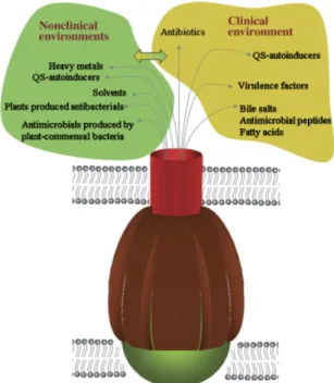
Toxic compounds can be found in non-clinical settings, some have biotic origins such as antibiotics and antimicrobial agents produced by plants and fungi, others have abiotic origins such as heavy metals derived from the earth crust and it’s erosion. The presence of offensive compounds or environments might select for antibiotic resistance mechanisms. Bacteria cultured form the marine air-water interface were shown to be more resistant to antibiotics than bacteria cultured directly from water. The same was observed for environments with radiation or pollution. So some genes conferring antibiotic resistance are likely to have other primary roles.
Figure 1.3: Role of multidrug resistant (MDR) pumps in nonclinical and clinical environments (Martinez et al.,2009). QS : Quorum Sensing.
Genes encoding multidrug efflux pumps are present in chromosomes across the three domains of life and are generally conserved, showing that they are not exclusive to an-tibiotic producing species and suggesting that their major roles are not resisting the antibi-otics used in therapy. The potential roles pro-posed are relevant to the behaviour of bacte-ria in their natural ecosystems. It has been demonstrated that efflux pumps are necessary for detoxification of intracellular metabolites, bacterial virulence, cell homeostasis and in-tercellular signal trafficking (Martinez et al., 2009). Certain classes of efflux pumps are known to evacuate various toxins and heavy metals, offering a general mechanism of resis-tance not only to antibiotics Allen et al., 2010. Figure 1.3 summarizes these mechanisms in 7
CHAPTER 1. ANTIMICROBIAL RESISTANCE
the case of an RND pump from Gram-negative bacteria.
The first step of bacterial infection is colonization. This can be inhibited by host-produced compounds such as bile salts, long-chained fatty acids and antimicrobial pep-tides. To avoid these compounds and colonize a tissue bacteria use MDR efflux pumps. For example, the AcrAB system in E. coli confers resistance to bile salts in vitro. In the case of Salmonella typhimurium and Francisella tularensis inactivation of the AcrAB sys-tem impairs colonization, showing that MDR efflux pumps are required for full virulence in certain species (Martinez et al.,2009).
In quorum sensing bacteria produce small molecules, sometimes called autoinducers, to regulate its population. Quorum sensing includes biosynthesis of antimicrobial peptides, polysaccharide synthesis and virulence factor such as elastases and proteases. An inter-esting aspect of quorum sensing is how the autoinducers accumulate in the environment as the population density increases. Some diffuse freely but others require transporters and this is where multidrug resistant efflux pumps play an important role. In P. aerug-inosa a mutation of the MexAB-OprM efflux pump affects release of N-3-oxododecanoyl homoserine lactone (3-Oxo-C12-HSL) decreasing the production of quorum sensing con-trolled virulence factors, the same was observed for other efflux pumps mutants. Loss of multidrug resistant efflux pumps in Vibrio cholerae decreased the expression of the virulence gene regulator tcpP, possibly by affecting cell-to-cell signalling (Martinez et al., 2009). In some carbapenems producing saprophytic bacteria, living on decaying organic matter, the genes implicated in the synthesis of antibiotics are shown to have a role in the quorum-sensing apparatus and formation of biofilms (Holmes et al.,2016).
These efflux pumps are important for some species that thrive in extreme environ-ments. Cupriavidus metallidurans grows in biotopes rich in heavy metals, volcanic biotopes for example, and uses like all micro-organisms trace amounts of heavy metals as cofactors of several proteins. Therefore, to maintain a finely tuned metal homeostasis, bacteria uti-lize efflux pumps to regulate their intracellular heavy metal concentrations. In addition, organic solvents present in non-anthropogenic petroleum in soil and water, as a product of biosphere activity, can be toxic compounds to bacteria, their tolerance is mediated by efflux pumps. This mechanism is also used in species that are not specialized in growth on high concentrations of heavy metals, since genes coding for efflux pumps are shared with great degree of conservation (Martinez et al., 2009).
Some bacterial species can grow on antibiotics as sole carbon and nitrogen sources. Pseudomonas fluorescens grows on streptomycin, Burkholderia cepacia grows on peni-cillin, Streptomyces venezuelae grows on chloramphenicol. Without their resistance genes these organisms would not be able to survive. Some of these isolates exhibit resistotypes not seen before, emphasising the necessity of their study for a better prediction of the
CHAPTER 1. ANTIMICROBIAL RESISTANCE
1.4
Alternative roles of antibiotics in nature
Microbial populations are capable of producing a wide spectrum of small bioactive molecules, this diverse mix of molecules is termed the parvome (Davies, 2013). Usually used for quorum-sensing reactions and affecting pathogenesis, these molecules can activate biochemical pathways in target organisms at low concentrations. Interestingly at high concentrations, some molecules of the parvome have antibiotic activity Barlow et al., 2002; Allen et al., 2010. The different effects of the same molecules can be interpreted as signalling responses that adapt the bacterial metabolism in mixed microbial communities. In addition, antibiotics have an important role in some cases of symbiosis. For example, some ant species rely on fungus growing as food source. They carry on their cuticle an antibiotic producing actinomycete, Pseudonocardia sp., which is used to biologically control their fungal garden. In other cases of parasitism, bacteria evolved to recognise antibiotics secreted by plants as signalling molecules to induce adaptation to the plant defence system allowing colonisation of the rhizosphere (Allen et al.,2010).
1.5
Antibiotic pollution and resistance in the biosphere
In contrast to intrinsic resistance, developed by bacterial population over the past millions of years, acquired antibiotic resistance is a recent event in the evolution of human pathogens with the main selective force being the human use of antibiotics (Martinez, 2009). The major role played by human activities in the generation of environmental reservoirs of antibiotic resistance cannot be disputed. Starting from the 1940s, increasing amounts of antibiotics have been produced and widely disseminated in the environment, thus providing constant selection pressure for resistant strains in the biosphere. For example, genomic and microbial studies of waste-water treatment plants have pointed out that they are now rich reservoirs for resistant genes and organisms (Davies et al., 2010). Antibiotics released into the sewage system after use in humans will be degraded, associated with sewage sludge or released into rivers. Sludge-associated drugs will pollute agricultural systems with the use of sludge as fertiliser. Some antibiotics are not easily biodegradable and some persist in soils for long periods of time. Fluoroquinolones can persist in the environment for month or even years (Wellington et al.,2013).
To take into account the extended reach of this antibiotic pollution, recent studies have shown the presence of antibiotic resistance genes in the gut flora of people who live in isolated areas and not exposed to modern civilization antibiotic therapy. Researchers were able to demonstrate that the acquired resistance genes were of the same type as those found in antibiotic-exposed settings. These data suggest that the resistance in such remote communities is likely to be the consequence of clonal expansion and horizontal transfer of genetic elements harbouring resistance genes from antibiotic-exposed settings 9
CHAPTER 1. ANTIMICROBIAL RESISTANCE
and not the result of an independent in situ selection (Pallecchi et al., 2007).
But how can antibiotic resistant bacteria travel great distances ? Physical forces like wind and watershed can spread antibiotic resistance genes. Even bacteria from environ-ments that are thought to be stationary such as soil, can be moved, one example is the intercontinental transport of bacteria on desert dust. Proximity to human activities can also influence antibiotic resistance profiles in wild animals. Bacterial isolates from cap-tured mice in rural England are resistant to β-lactam antibiotics. Likewise, isolates from wild animals in Mexico and Australia have a hight frequency of antibiotic resistance. In Africa, apes that are in contact with humans harbour more antibiotic resistant strains than those that inhabit more remote areas of the continent (Allen et al., 2010).
Moreover, a key reservoir for long distance dissemination of antibiotic resistance is wild migratory birds with their long distance travel and the wide variety of environments they inhabit. Their impact can be seen on Arctic birds that are showing pattern of antibiotic resistance similar to clinical isolates, this can be explained by the fact that many migratory birds breed in the Arctic and migrate to other continents. The environmental reservoirs of resistance are poorly understood demonstrating the need for more investigations on this subject (Allen et al., 2010).
Antibiotics are needed in agriculture and aquaculture to maintain animal health and welfare as well as food security. This wide scale use of antibiotics encourage the devel-opment of resistance. Studies suggest that 75-90% of used antibiotics are excreted from animals un-metabolised and enter water courses and sewage systems. The same problem is observed in human waste from hospitals and in the environment of active pharmaceutical ingredients manufacturers (O’Neill, 2016).
More antibiotics are used in food production than human medicine with an estimation of 400 mg of antibiotics per kg of meat produced in some European countries. In the United Kingdom alone, around 400 tonnes of antibiotics are used each year for food producing animals. In the European Union the use of antibiotics to promote growth in livestock was banned in 2006. It is interesting to note that such practices are still present worldwide like in the United States of America for example. The use of copper, or other metals, as bactericide in fields or its natural occurrence in certain areas raises concerns since metals can co-select for resistance to antibiotics and heavy metals. Alarmingly, evidence of β-lactamase genes transmission from livestock to humans have been reported. Educating farmers to reduce antibiotic use in addition to legislative enforcement can help improve the current situation (Wellington et al., 2013; Holmes et al.,2016).
Large scale manufacturing of antibiotics is also problematic in countries where regula-tions tend to be vaguely defined. In India and China this problem arises as studies show extremely high antibiotic concentrations, few milligrams per litre instead of the accepted nanograms per litre, in river water downstream of pharmaceutical industries (Wellington
CHAPTER 1. ANTIMICROBIAL RESISTANCE
Figure 1.4: Ecological landscapes of antibiotic resistance (Martinez,2009). R: resistance determinants present in antibiotic producers; r : metabolic or signalling determinant that can be used for resistance at high antibiotic load.
social issues can drive antibiotic resistance through prescription of unnecessary antibiotics to retain patients loyalty. Moreover, the increased internet access opened the way to even more unregulated purchasing of antibiotics, automedication and unregulated disposal in the environment (Wellington et al.,2013).
Most works studying the impact of human activity on the biosphere, such as the over use of antibiotics, are founded on the study of higher organisms. However, the majority of life on earth is microbial and the consequences of environmental changes on the microbiosphere are largely ignored. What should be taken into account is the existence of three different landscapes important in the evolution of resistance of human pathogens (Martinez, 2009), Figure 1.4.
The first level, all white panel, is the natural biosphere where there is a large number of genes capable of conferring antibiotic resistance to human pathogens through HGT (R genes). other genes, (r genes) have also been selected for detoxification, communication or other functions different from antibiotic resistance, unless they are expressed at high concentrations. The second level, grey square in Figure1.4, consists of habitats where con-tact of human associated bacteria is frequent with the environmental microbiota. These places, like waste waters, are considered to be hotspots for antibiotic resistance genes acquisition by pathogenic bacteria. Furthermore, since waste waters usually contains an-tibiotics the selection for resistant strains increases. The third level, dark grey square in Figure 1.4, is the clinical environment or even the patient where selection will favour al-ready acquired resistance genes and mutation-driven diversification genes whose function is only resistance (Martinez,2009).
Humans can be exposed to antibiotic resistance genes or antibiotic resistant bacteria through several ways : crops exposed to contaminated sludge or manure, livestock treated with veterinary drugs and containing resistant flora, fish exposed to drugs soluble in water, contaminated water sources and groundwater used for drinking, contaminated coastal waters used for bathing or shellfish production (Wellington et al., 2013).
CHAPTER 1. ANTIMICROBIAL RESISTANCE
1.6
Problems facing new drug development
It is clear that investment in the development of new antimicrobial drugs and strategies is urgently needed. Lack of such investments shows the fears that resistance will eventually develop, limiting returns on investments due to restrictions in use of the no longer efficient newly developed drugs. This makes development of new antibiotics seem less attractive than business investment in medicines for chronic diseases, pushing major pharmaceutical companies to stop research in this area. And instead, investing in scientifically challenging but commercially lucrative disease area like cancer (World Health Organization, 2015a). Moreover sales of patented antibiotics make up only 10%, around 4 billion USD, of a relatively large total market of antibiotics worth 40 billion USD of sales a year. Adding to that the unattractive commercial return on R&D investment until widespread resistance has emerged against previous generations of drugs. So it is not surprising that firms are not investing in antibiotics despite the very high medical needs, Figure 1.5.
Figure 1.5: Distribution of investments in pharmaceutical R&D (O’Neill,2016).
The same scheme is true in the allocation of public research funds by governments, the United States of America National Institutes of Health gave 1.2% of its grants to fund AMR related research compared to 18.6% to cancer research between 2009 and 2014 (O’Neill, 2016). What is needed is new processes that facilitate investments in research and development of new antibiotics and ensure a governed public health framework for the use of new products to conserve their effectiveness and longevity (World Health Or-ganization, 2015a; O’Neill,2016).
CHAPTER 1. ANTIMICROBIAL RESISTANCE
1.7
Worldwide surveillance and control centers
Worldwide, several control centers are on the lookout for every new case of infection by a resistant micro-organism. All these centres share the same objective of preventing any possible high scale spread of the antibiotic resistance infections. The main surveillance networks are the following :
• The European Antimicrobial Resistance Surveillance Network (EARS-Net) coordi-nated by the European Centre for Disease Prevention and Control. The Central Asian and Eastern European Surveillance of Antimicrobial Resistance (CAESAR) network, an initiative that aims to support all countries of the European Region that are not part of the EARS-Net.
• The Global Antimicrobial Resistance Surveillance System (GLASS) promotes a worldwide understanding and engagement to support the global effort to control AMR by collecting an unprecedented set of information related to AMR at global level through a network of surveillance sites linked to national reference laboratories and national coordination centers. This World Health Organisation (WHO) sup-ported system aims to analyse and share data in order to optimize decision-making and drive local and regional action.
• The Interagency Coordination Group on Antimicrobial Resistance (IACG). The United Nations Secretary-General has established IACG to improve coordination between international organizations and to ensure effective global action.
All these organisations share the same goals to improve awareness and understanding of AMR, strengthen surveillance and research, reduce the incidence of infection, optimize the use of antimicrobial medicines, develop the economically sustainable investment in new medicines (The Regional Office for Europe of the World Health Organization, 2016; World Health Organization,2015a; World Health Organization, 2015b).
In addition to all international networks, joint public-private partnerships like the Global Antibiotic Research and Development Partnership (GARDP) are encouraging re-search and development in the aim of delivering new treatments. Annual events are also used to raise awareness like the World Antibiotic Awareness Week an WHO initiative held every November since 2015 to increase awareness of global antibiotic resistance and to encourage correct practices among the general public.
CHAPTER 1. ANTIMICROBIAL RESISTANCE
1.8
Species under high surveillance
With the alarming rate at which antibiotic resistance infections are rising, especially in the case of community acquired resistant infections, some bacterial species draw attention more than others. This is the case for example of E. coli and S. aureus infections with one in four infections being resistant to classical, or first-line, antibiotic treatment (The Regional Office for Europe of the World Health Organization, 2017). To face this, the WHO placed the species listed below under GLASS surveillance (World Health Organi-zation,2015b). Figure1.6 summarizes the different classes of antibiotics and their modes of actions.
Acinetobacter spp.
Acinetobacter spp., especially species belonging to the A. baumannii group, are intrin-sically resistant to many antimicrobial agents due to their ability to exclude various molecules from penetrating their outer membrane. Acquiring a multidrugresistant strain is usually caused by prolonged mechanical ventilation and intensive care unit hospitalisa-tion. Colistin is the only effective antibiotic left but emerging resistance is being detected.
Escherichia coli
Despite the fact that it is part of the normal flora in the intestine in humans and animals, E. coli is the cause of most community and hospital-acquired urinary tract and blood-stream infections. in the last few years, E. coli became the leading cause of foodborne infections worldwide. Resistance in E. coli develops through mutations and acquisition of mobile genetic elements. Usually carbapenems, members of the beta lactam class of antibiotics, are the only available treatment for severe infections, although carbapenem re-sistance mediated by carbapenemases is emerging. Alternatively, colistin use is increasing with rare cases of resistance first described in China.
Klebsiella pneumoniae
The majority of human infections caused by K. pneumoniae are health-care associated. Resistance in K. pneumoniae develops through mutations and acquisition of mobile ge-netic elements. With high proportions of cephalosporin resistance the treatment of severe K. pneumoniae rely on carbapenems. However, K. pneumoniae is today the main cause of carbapenem-resistant bacterial infections worldwide rendering almost all available treat-ment options ineffective. Which leads to the use of last resort drugs like tigecycline, a
CHAPTER 1. ANTIMICROBIAL RESISTANCE
D
IF
FE
RE
N
T C
LA
SS
ES O
F A
N
TI
BI
O
TI
CS - A
N O
VE
RV
IE
W
BY NC ND© COMPOUND INTEREST 2014 - WWW.COMPOUNDCHEM.COM
| Twitter: @compoundchem | Facebook: www.facebook.com/compoundchem
Shared under a Creative Commons Attribution-NonCommercial-NoDerivatives licence.
C 19 30 19 40 19 70 19 80 ß-L AC TA MS N H O R O S O OH AM IN OG LY CO SID ES ch lo ra mp hen ico l gl yc op ep tid es su lfon am ide s te tr ac ycl in es ma cro lid es lip op ep tid es EXAMPLES Penicillins (shown) such as
amoxicillin and flucloxacillin;
Cephalosporins
such as cefalexin.
MODE OF ACTION
Inhibit bacteria cell wall biosynthesis.
S O O N H2 N H H
All contain a beta-lactam ring
MOST WIDELY USED ANTIBIOTICS
IN THE NHS EXA M PL ES Pr on to si l, su lfa ni la mi de (sh ow n) , sul fa di az in e, s ul fis ox az ol e. M OD E OF A C TI ON D o n ot k ill b ac te ri a b ut p re ve nt t he ir gr ow th a nd m ul tip lic at io n. C au se al ler gi c r ea ct ion s i n s om e p at ien ts .
All contain the sulfonamide group
FI R ST C O M M ER CI A L A N TI B IO TI CS W ER E SU LF O N A M IDE S EXAMPLES
Streptomycin (shown), neomycin,
kanamycin, paromomycin.
MODE OF ACTION
Inhibit the synthesis of proteins by bacteria, leading to cell death.
All contain aminosugar substructures
FAMILY OF OVER 20 ANTIBIOTICS
O O O O HO HO OH OH N N HO HO HN HO H2 N NH 2 NH 2 NH 2 O EX A M PL ES Te tr ac yc line (s how n) , d ox yc ycl in e, lim ec yc lin e, o xy te tr ac yc lin e. M OD E OF A C TI ON In hib it s yn th esi s of p rot eins b y ba ct er ia , pr ev en tin g g row th .
All contain 4 adjacent cyclic
hydrocarbon rings B EC OM ING LE SS P OP UL A R D UE T O DE VEL O PM EN T O F R ES IS TA N CE OH O OH O O NH 2 OH N OH OH H H
COMMONLY USED IN LOW INCOME
COUNTRIES
MODE OF ACTION
Inhibits synthesis of proteins,
preventing growth.
No longer a first line drug in any developed nation (except for conjunctivitis) due to increased
resistance and worries about safety.
Distinct individual compound
OH N H Cl Cl O N + O -O OH EX A M PL ES Er yt hr om yci n ( sho w n) , cla ri th ro m yc in , a zi th ro m yc in . M OD E OF A C TI ON In hi bi t p ro te in s yn th es is b y b ac te ri a, oc ca si on al ly l ea di ng t o c el l d ea th .
All contain a 14-, 15-, or 16-membered
macrolide ring SE CO N D M O ST P RE SC RI B ED A NT IB IO TI CS IN T H E N HS O O OH HO CH 3 OH C2 H5 O O O O OH OCH 3 O HO N CH 3
COMMON ‘DRUGS OF LAST RESORT’ Consist of carbohydrate linked to a
peptide formed of amino acids
EXAMPLES
Vancomycin (shown), teicoplanin.
MODE OF ACTION
Inhibit bacteria cell wall biosynthesis.
NH O O ClH N O O N H O H N O O N H N H O HN H2 N O OH OH HO O HO HO Cl O O O O OH NH 2 OH HO HO OH an sa my cin s EXAMPLES
Geldanamycin (shown), rifamycin,
naphthomycin.
MODE OF ACTION
Inhibit the synthesis of RNA by bacteria, leading to cell death.
All contain an aromatic ring bridged by
an aliphatic chain.
CAN ALSO DEMONSTRATE
ANTIVIRAL ACTIVITY NH 2 O O HN O O O H3 CO OCH 3 H3 CO OH 19 60 qu inol on es EX A M PL ES Ci pr ofl ox aci n (s ho w n) , l ev ofl ox aci n, tr ova flo xa ci n. M OD E OF A C TI ON In te rf er e w ith b ac te ri a D N A rep lic at ion a nd tr an scr ipt ion .
All contain fused aromatic rings with a
carboxylic acid group attached
RE SI ST AN CE E VO LVE S R AP ID LY HN N N F O O OH st rep to gr am in s EXAMPLES
Pristinamycin IIA (shown),
Pristinamycin IA. MODE OF ACTION
Inhibit the synthesis of proteins by bacteria, leading to cell death. Combination of two structurally differing compounds, from groups denoted A & B
TWO GROUPS OF ANTIBIOTICS THAT
ACT SYNERGISTICALLY N O NH O N O O O O OH EX A M PL ES Da pt om yc in (s ho w n) , s ur fa ct in . M OD E OF A C TI ON D is ru pt m ul tip le c el l m em br an e fu nc tio ns , l ea di ng t o c el l d ea th .
All contain a lipid bonded to a peptide
IN ST A N CE S O F R ES IS TA N CE R A RE O OH O H N O N H NH C9 H19 O H N NH 2 O N H O O NH HN HN NH HN N H NH NH HN O O O O O O O O O O OH OH O O HO O OH H2 N O NH 2 Key: COMMONLY ACT AS BACTERIOSTATIC AGENTS
, RESTRICTING GROWTH & REPRODUCTION
COMMONLY ACT AS
BACTERICIDAL AGENTS
, CAUSING BACTERIAL CELL DEATH
19 50 EX A M PL ES Li ne zol id (s ho w n) , p os iz ol id , te di zo lid , c yclos er ine . M OD E OF A C TI ON In hib it s yn th esi s of p rot eins b y ba ct er ia , pr ev en tin g g row th .
All contain 2-oxazolidone somewhere
in their structure PO TE NT A NT IB IO TI CS C O M M O N LY U SE D A S ‘ D RU G S O F L A ST R ES O RT ’ O H N O N O N O F ox az ol idi non es D IS CO VE RY N H
Figure 1.6: Timeline showing the discovery of major classes of antibiotics and their modes of action (Different classes of antibiotics - An overview).
CHAPTER 1. ANTIMICROBIAL RESISTANCE Neisseria gonorrhoeae
N. gonorrhoeae is the causal agent of gonorrhoea, the acute sexually transmitted infection of the reproductive tract in humans. Actually the last remaining option for treatment are third-generation cephalosporins. With few new treatment options in the drug development pipeline and several countries reporting treatment failures the WHO fears that gonorrhoea will emerge as a silent epidemic in the foreseeable future.
Salmonella spp.
Salmonella spp. can be found in the intestines of food-producing animals and in hu-man or animal faeces. Infections are usually acquired by consumption of contaminated food which can lead to foodborne outbreaks with severe cases of enteric fever caused by Salmonella typhi. First detected in 1989, multidrug-resistant strains of Salmonella spp. spread worldwide. Fluoroquinolones are suggested for the treatment of enteric fever caused by species resistant to first-line antibiotics. Reduced susceptibility to oral drugs and reports of treatment failures are of concern.
Shigella spp.
Shigella spp. are the major cause of dysentery and diarrhoea throughout the world. They are usually transmitted by ingestion of contaminated food or water and through human contact in poor sanitation conditions. After resistance to first-line antibiotics like cotrimoxazole and ciprofloxacin, Shigella spp. strains are now resistant to second-line antimicrobial drugs, such as the third-generation cephalosporin ceftriaxone and the macrolide azithromycin.
Staphylococcus aureus
S. aureus can be part of the normal human microbiota but remains one of the most important human pathogens, causing a variety of infections, most notably skin, soft tis-sue, bone and bloodstream infections. S. aureus developed resistance to penicillin, a few years after it’s introduction in the 1940s, by the production of a beta-lactamase. There-fore beta-lactamase inhibitors were developed to be administered with the antibacterial drugs. However, strains of S. aureus have become resistant to these penicillinase-stable antibacterial drugs and termed meticillin-resistant S. aureus (MRSA). Second-line drugs used to treat MRSA infections are expensive and treatments of last resort using glycopep-tides such as vancomycin and teicoplanin require special care to avoid adverse side-effects.
CHAPTER 1. ANTIMICROBIAL RESISTANCE Streptococcus pneumoniae
S. pneumoniae is the leading cause of community acquired pneumonia worldwide causing also high mortality invasive disease such as meningitis. Resistance to antibacterial drugs occurs by acquiring mutations in the genes coding for the penicillin-binding proteins. In addition, S. pneumoniae has a variety of virulence factors that facilitate the host’s infec-tion. Resistance has been linked to worse clinical outcomes in patients with pneumococcal meningitis, most likely leaving survivors with permanent residual symptoms.
Based on the rising drug resistance in pathogens under surveillance, estimations show that unless action is taken, death toll from AMR will reach 10 million each year by 2050. This is a staggering one person every three seconds (O’Neill, 2016). Which explain why the listed species are on the Priority Pathogens List of antibiotic-resistant bacteria as critical priorities for research and development of new and effective antibiotic treatments (World Health Organization,2015b).
1.9
Sustaining the therapeutic life of current antibiotics
Several approaches have been proposed to to prolong the useful therapeutic life of present antibiotics. Drug cycling can be used to maintain heterogeneity of antibiotic agents and to avoid acute selective pressure. It involves the periodic replacement of first-line antibiotics with alternative structural classes. The weakness in this drug cycling, is that it does not provide a long-term solution since resistant strains will be reselected when related antibiotics are used at each cycle. In addition, in large hospitals it may be difficult to decontaminate infected intensive care units while at the same time cycling between different antibiotics.
A related tactic is a combination of compounds that have different modes of action in the hope that resistance will not have time to emerge since carefully studied combinations will quickly eliminate the strain. This combinatorial approach has been applied with success in the treatment of infections in cancer and HIV patients (Holmes et al.,2016).
Non-antibiotic approaches exist for the treatment of bacterial infection and involve the stimulation and recruitment of the innate immune system and the human gut micro-biome. Another proposition, that is the work base of this manuscript, is the inhibition of bacterial virulence to stop the infection process without the need for antibiotics. The main advantage of this strategy is that selection for resistance might not occur because the growth of the infecting organism would not be impaired. Nevertheless, it is noteworthy to mention that in an ideal world, the effective use of vaccines against all infectious diseases would reduce drastically the use of antibiotics that will be limited to surgical procedures under strict controls (Davies et al.,2010).
In addition to innovative techniques, effort should be made on improvement of di-17
CHAPTER 1. ANTIMICROBIAL RESISTANCE
agnosis and prescription practices, perusing studies on withdrawn or underused antimi-crobial and encouraging de-novo drug discovery. Areas of particular interest for de-novo drug discovery include molecules capable of attenuating bacterial virulence and disrupting biofilm formation, bacteriophage therapy, eco-biological approaches (Nood et al., 2013) and enhancement of host immune responses. In addition allowing guaranteed access to medicines and ensuring the use of adequate serum drug concentrations in low-income countries through the prosecution of falsified, substandard and generally low quality drugs (Holmes et al., 2016).
Moving forward and reducing demand for antibiotics requires a series of key strate-gic objectives. Starting with a massive global public awareness campaign, followed by improvement in hygiene standards and the prevention of infection spread. Besides unnec-essary use of antimicrobials in agriculture and their dissemination into the environment should be reduced. Improvements in global surveillance of drug resistance and antimi-crobial consumption are to be undertaken. Rapid diagnostics to cut unnecessary use of antibiotics need to be promoted as well as the use and development of vaccines and alternatives. Improvements in the numbers, pay and recognition of people working on infectious diseases are to be made. Establishing a global innovation fund for early-stage and non-commercial research as well as better incentives to promote investments in new drugs and their development are to be carried out. All this global coalition for action is to be supported by the G20 and the UN (O’Neill, 2016).
1.10
Chapter conclusion
The most commonly prescribed drugs in human medicine are antibiotics but up to 50% of the prescriptions are considered unnecessary (Holmes et al., 2016). The period after World War II saw a ”golden era” of antibiotic discovery with new products reaching the market from 1940s to the 1970s. Since then the rate of discovery has dramatically fallen (O’Neill,2016). In addition, drug resistant bacteria are omnipresent in the biosphere, but their consequences are aggravated in situations such as civil unrest, famine and natural disasters (Davies et al., 2010).
For the pharmaceutical industry, medicines that are no longer effective lose their value. Hence the need for industry leaders to support a responsible use of medicines in order to prolong their effectiveness. Through research and development the industry can slow down the emergence of resistance. New concepts are needed to promote innovation and cooperation among policy makers, academia and the pharmaceutical industry, to establish new globally available technologies to prevent and treat resistant infections (World Health Organization, 2015a).
CHAPTER 1. ANTIMICROBIAL RESISTANCE over, the ability to rapidly characterize emerging resistance and elucidate the underlying mechanisms should be the main objective. This is crucial for keeping surveillance and diagnostic tool up to date. In addition, effort should be made to endorse the use and development of existing vaccines that can prevent infectious diseases whose treatment would require antimicrobial drugs. In fact, vaccination reduces the prevalence of viral infections which are often inappropriately treated with antibiotics, resulting in secondary infections that require antibiotic treatment (World Health Organization, 2015a).
With these warnings and recommendations in mind, the development of new strategies to fight pathogens should be a priority. The new ideal therapeutic targets should exert weak evolutionary pressure, disarm or weaken the pathogen and be unique to the micro-organism.
One way to do so is by interfering with the iron regulation and its homeostasis within bacteria. The next chapter shows how iron became important for life and at the same time, an exploitable weakness in our battle against infection.
Chapter 2
Iron: a key element in living organisms
The evolutionary path that early life took was not a product ofchance, it was constrained by the changing thermodynamic equi-librium of the environment which forced a one way progression (Williams et al.,2003). The majority of organisms require the first row transition metals that include manganese, iron, cobalt, nickel, copper and zinc. The ability of these metals to cycle between oxi-dation states contribute to their catalytic importance and toxicity (Palmer et al., 2016). The regulation of such important elements resides in several sensing mechanisms able to regulate the import and export of metals. With such processes in place, a competition between organisms gave rise to what is known as the battle for metals between hosts and pathogens. We can make use of this bat-tle and its insights to explore new opportunities for anti-virulence therapeutic potential.
CHAPTER 2. IRON: A KEY ELEMENT IN LIVING ORGANISMS
The availability of elements and their oxidation states were extremely strong con-straints placed on primitive cells and their energy systems. Chemical equilibrium de-pended on the solubility, redox potentials and stability of inorganic complexes (Williams et al., 2003). Transition metals played an extremely important role in the evolution of the biosphere. Their bioavailability varied over geological times, driven by the emergence of oxygenic photosynthesis that gave rise to the Great Oxygenation Event (GOE) around 2.45 billion years ago Boyd et al., 2014. Many biochemical pathways require metalloen-zymes, proteins containing ions of inorganic elements such as transition metals, forming around one third of all proteins (Dupont et al., 2010).
Many of the essential elements for early life, needed in their reduced state in the cytoplasm, became oxidized by the environment and had to be scavenged then reduced. This is the case of iron (Williams et al., 2003).
2.1
Evolution was chemically constrained
Iron is an abundant element in the universe due to its nuclear stability and its freezing of nuclear kinetics of element formation in evolving stars. Once iron is reached, fusion is halted and the core temperature drops leading to stellar death. On Earth, iron is close in abundance to oxygen though most of it is in the central metallic core of our planet (Williams, 2012). Nevertheless, it is the fourth most abundant element in the earth’s crust, following oxygen, silicon and aluminium. At first glance it would seem unlikely that iron can limit biochemical processes, however the bioavailability of iron is dependant of its redox state and has forged the evolution of our biosphere (Falkowski et al.,1998).
2.1.1
Iron and early life
Hydrothermal vents are still today, as they were in the Archean ocean, a major source of dissolved elements in oceans. They are continuously providing high concentrations of precursors compounds with biological importance by circulating fluids through tem-perature gradients and various reaction surfaces. Less than 500 million years after the formation of the ocean, they provided multiple pathways for abiotic synthesis of chemical compounds later used as building blocks for early life (Baross et al.,1985). The predom-inant source of Fe2+ in deep Archean ocean basins was hydrothermal water circulating through basalt ridges (Boyd et al., 2014).
Fe2+ often forms complexes with sulfide near hydrothermal discharges giving rise to an iron-sulphur mineral phase, in particular pyrrhotite shown to catalyse the reduction of N2 and CO2, and the production of H2 under high temperature and pressure (Boyd et al.,