HAL Id: hal-03031395
https://hal.archives-ouvertes.fr/hal-03031395
Submitted on 30 Nov 2020
HAL is a multi-disciplinary open access
archive for the deposit and dissemination of sci-entific research documents, whether they are pub-lished or not. The documents may come from teaching and research institutions in France or abroad, or from public or private research centers.
L’archive ouverte pluridisciplinaire HAL, est destinée au dépôt et à la diffusion de documents scientifiques de niveau recherche, publiés ou non, émanant des établissements d’enseignement et de recherche français ou étrangers, des laboratoires publics ou privés.
Nanoparticle detection by solid state nanopore
integrated into a reusable microfluidic device
Izadora Mayumi Fujinami Tanimoto, Benjamin Cressiot, Jean Roman,
Nathalie Jarroux, Gilles Patriarche, Bruno Le Pioufle, Juan Pelta, Laurent
Bacri
To cite this version:
Izadora Mayumi Fujinami Tanimoto, Benjamin Cressiot, Jean Roman, Nathalie Jarroux, Gilles Pa-triarche, et al.. Nanoparticle detection by solid state nanopore integrated into a reusable microfluidic device. MicroTAS, Oct 2020, on-line, France. �hal-03031395�
NANOPARTICLE DETECTION BY SOLID-STATE NANOPORE INTEGRATED
INTO A REUSABLE MICROFLUIDIC DEVICE
Izadora Mayumi Fujinami Tanimoto
1,2, Benjamin Cressiot
3, Jean Roman
2, Nathalie Jarroux
1,
Gilles Patriarche
4, Bruno Le Pioufle
2, Juan Pelta
1,3and Laurent Bacri
11
Université Paris‐Saclay, Univ Evry, LAMBE, France,
2
ENS Paris-Saclay, Université Paris-Saclay, Institut d’Alembert, LUMIN, France,
3CY Cergy Paris Université, LAMBE, France and
4
C2N, Université Paris-Saclay, CNRS, France.
ABSTRACT
Solid-state nanopores can be used as electrical biosensors at the single molecule resolution. Such high resolution sensors provide a promising approach to address societal challenges in health and biotechnology. Nevertheless, the analytes detection is still limited by non-specific interactions between these molecules and the membrane supporting the nanopore. Therefore, we performed a polymer functionalization of the nanopore surface that allows to passivate the membrane and to control the pore size. Moreover, we integrated the solid-state nanopore in a reusable microfluidic device, thereby reducing the sample volume needed to perform the detection. The combination of these two approaches is applied to the sensing of single thyroglobulin protein.
KEYWORDS: Solid-state nanopore, microfluidics, protein detection, polymer functionalization INTRODUCTION
Solid-state nanopores have been used to characterize the biomolecular interactions in a controlled environment.1
One can tune their diameter to better fit the analyte size and they present better stability than the biological ones.2,3
Nanopore sensing is performed from an electric detection. Whenever the analyte transiently resides inside the nanopore there is an ionic current drop. However, the interactions between nanoparticles and the nanopore are not well established. A high surface energy of the membrane,2 missing events due to high translocation speed and the
pore-to-pore variability limit the experiments reproducibility. Another challenge is that in classical microfluidic devices the nanopore chip needs to be irreversibly sealed into it, whereas the analytes chamber are not hermetically sealed leading to potential cross contamination during the solution exchange process. Hence, this work will focus on improving the membrane chemical and physical surface properties, by functionalizing its surface to avoid non-specific interactions and to control the nanopore size, and on the integration of solid-state nanopores into a microfluidic device (Fig. 1a-b). The efficiency of such set-up was confirmed by the detection of single thyroglobulin transport.
EXPERIMENTAL
Si3N4 membranes are pierced with a TEM. Polyethylene glycol (PEG) 7 kDa chains are grafted over the
nanopore surface.3 This nanopore is inserted in between two identical subunits that formed the microfluidic device.3
Part of the surfaces of those subunits are not covalently bonded, in order to achieve the reversible packaging (Fig. 1a).3 The polydimethylsiloxane (PDMS) microfluidic circuit device is casted on a 3D-printed polyethylene
terephthalate (PET) mold on a silicon wafer. The ionic current is measured using Ag/AgCl electrodes.
RESULTS AND DISCUSSION
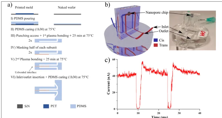
The fabrication of PDMS based devices needs a master mold, usually obtained by photolithography techniques, which are time consuming and require a clean room. Here, we show that a 3D-printer can make molds in laboratory and usable for fast conception of microfluidic devices (Fig. 1a). This approach allows us to work with a hermetic system, avoiding possible contamination and fluid evaporation (Fig. 1b) and to reduce the analyte volume to 100 µL, which is important for biologic fluids. Besides, this device facilitates the changes in fluids. The final set-up confirms the capacity to detect protein at single molecule level, from variation of ionic current trace after addition of thyroglobulin (Ø = 18 nm) (Fig. 1c). The current blockades show the presence of thyroglobulin. The high signal to noise ratio - 98% - shows a high confinement controlled by the PEG chain grafting .
Figure 1: a) Microfluidic device protocol. b) 3D sketch of microfluidic device with the nanopore inserted on it (left) and a picture of the device with the nanopore chip (right). c) Current trace measured after the addition of thyroglobulin by one
nanopore (Ø = 28 nm, length 20 nm) grafted by PEG 7 kDa chain. ΔV = 1 V, 50 mM LiCl, 0.1 M Tris, pH 7.4.
CONCLUSION
Here, we stated the feasibility of the nanopore integration in reusable microfluidic device without permanent sealing between the nanopore and the device, which makes possible changes of small amount of electrolyte solutions and on nanopore manipulation. We demonstrate the control on PEG chain conformation along the nanopore surface. The system allows the detection of thyroglobulin transport through the nanopores with a high signal-to-noise ratio.
ACKNOWLEDGEMENTS
This work is supported by DIM Respore, DEFI Instrumentation aux limites 2016/2017, ANR Epsilomics.
REFERENCES
[1] B.Malekian, R.L. Schoch, T. Robson, G.F.D. del Castillo, K. Xiong, G. Emilsson, L.E. Kapinos, R.Y.H. Lim,
A. Dahlin, “Detecting Selective Protein Binding Inside Plasmonic Nanopores: Toward a Mimic of the Nuclear Pore Complex,” Front. Chem., 6, 637-644, 2018.
[2]D.J.Niedzwiecki, J. Grazul, L. Movileanu, “Single-Molecule Observation of Protein Adsorption onto an Inorganic Surface,” J. Am. Chem. Soc., 132, 10816-10822, 2010.
[3] J.Roman, O. Français, N. Jarroux, G. Patriarche, J. Pelta, L. Bacri, B. Le Pioufle, “Solid-State Nanopore Easy
Chip Integration in a Cheap and Reusable Microfluidic Device for Ion Transport and Polymer Conformation Sensing,” ACS Sens., 3, 2129-2137, 2018.
CONTACT