HAL Id: hal-03168070
https://hal.archives-ouvertes.fr/hal-03168070v2
Submitted on 2 Apr 2021HAL is a multi-disciplinary open access
archive for the deposit and dissemination of sci-entific research documents, whether they are pub-lished or not. The documents may come from teaching and research institutions in France or abroad, or from public or private research centers.
L’archive ouverte pluridisciplinaire HAL, est destinée au dépôt et à la diffusion de documents scientifiques de niveau recherche, publiés ou non, émanant des établissements d’enseignement et de recherche français ou étrangers, des laboratoires publics ou privés.
Bilateral control of interceptive saccades: evidence from
the ipsipulsion of vertical saccades after caudal fastigial
inactivation
Clara Bourrelly, Julie Quinet, Laurent Goffart
To cite this version:
Clara Bourrelly, Julie Quinet, Laurent Goffart. Bilateral control of interceptive saccades: evidence from the ipsipulsion of vertical saccades after caudal fastigial inactivation. Journal of Neurophysiology, American Physiological Society, In press. �hal-03168070v2�
Bilateral control of interceptive saccades: evidence from
1the ipsipulsion of vertical saccades after caudal fastigial
2inactivation
34
Clara Bourrelly*, Julie Quinet and Laurent Goffart*
5
Aix Marseille Université, Centre National de la Recherche Scientifique,
6
Institut de Neurosciences de la Timone, Marseille, France.
7 8
Laurent Goffart : https://orcid.org/0000-0001-8767-1867
9
Julie Quinet : https://orcid.org/0000-0003-3043-3424
10
Title for running head: Neural control of interceptive saccades
11
12
*: C. Bourrelly and L. Goffart contributed equally to this work.
13
Address for correspondence:
14
Laurent Goffart, PhD,
15
E-mail: laurent.goffart@univ-amu.fr
16
Institut de Neurosciences de la Timone, Campus de Sante, 27 Bd Jean Moulin, 13385 Marseille
17
cedex 5, France.
18 19
Abstract (219 words <= 250 words)
21
The caudal fastigial nuclei (cFN) are the output nuclei by which the medio-posterior cerebellum
22
influences the production of saccades toward a visual target. On the basis of the organization
23
of their efferences to the premotor burst neurons and the bilateral control of saccades, the
24
hypothesis was proposed that the same unbalanced activity accounts for the dysmetria of all
25
saccades during cFN unilateral inactivation, regardless of whether the saccade is horizontal,
26
oblique, or vertical. We further tested this hypothesis by studying, in two head-restrained
27
macaques, the effects of unilaterally inactivating the caudal fastigial nucleus on saccades
28
toward a target moving vertically with a constant, increasing or decreasing speed. After local
29
muscimol injection, vertical saccades were deviated horizontally toward the injected side with
30
a magnitude that increased with saccade size. The ipsipulsion indeed depended upon the tested
31
target speed, but not its instantaneous value because it did not increase (decrease) when the
32
target accelerated (decelerated). By subtracting the effect on contralesional horizontal saccades
33
from the effect on ipsilesional ones, we found that the net bilateral effect on horizontal saccades
34
was strongly correlated with the effect on vertical saccades. We explain how this correlation
35
corroborates the bilateral hypothesis and provide arguments against the suggestion that the
36
instantaneous saccade velocity would somehow be “encoded” by the discharge of Purkinje cells
37
in the oculomotor vermis.
38
NEW & NOTEWORTHY (73 <= 75 words)
40
Besides causing dysmetric horizontal saccades, unilateral inactivation of caudal fastigial
41
nucleus causes an ipsipulsion of vertical saccades. This study is the first to quantitatively
42
describe this ipsipulsion during saccades toward a moving target. By subtracting the effects on
43
contralesional (hypometric) and ipsilesional (hypermetric) horizontal saccades, we find that this
44
net bilateral effect is strongly correlated with the ipsipulsion of vertical saccades, corroborating
45
the suggestion that a common disorder affects all saccades.
46
Introduction
48
Saccades are rapid changes in eye orientation whose time course is characterized by an
49
interval during which the rotation speed increases to a maximum (acceleration) followed by an
50
interval during which it returns to zero (deceleration). Starting with the eyes centered in the
51
orbits, the changes in the contraction of lateral and medial recti muscles rotate the eyes
52
horizontally whereas combined changes in the contraction of superior and inferior recti
53
muscles, and the superior and inferior oblique muscles rotate them vertically (Robinson 1975).
54
During vertical saccades, the tension in muscles involved in horizontal rotations exhibit a
55
transient small reduction at the level of medial recti (Miller and Robins 1992). A burst of spikes
56
from motoneurons causes the phasic contraction of agonist muscles while the relaxation of
57
antagonist muscles is enabled by a pause of motoneurons which innervate them (Fuchs and
58
Luschei 1970; Robinson 1970; Schiller 1970). Premotor commands for horizontal and vertical
59
eye rotations originate in distinct regions of the reticular formation: burst neurons in the
60
pontomedullary reticular formation drive horizontal saccades whereas those controlling vertical
61
saccades are located in the rostral midbrain tegmentum (Fuchs et al. 1985; Hepp et al., 1989;
62
Horn 2006; Moschovakis et al. 1996).
63
Robinson and Fuchs (2001) proposed that these populations of premotor neurons and
64
the time course of saccades are under the control of neurons in the medio-posterior cerebellum:
65
burst neurons in the contralateral caudal fastigial nucleus (cFN) would support the acceleration
66
of saccades while those in the ipsilateral cFN would help their deceleration. This biphasic
67
pattern was suggested by the delay between the peaks of cFN discharge during contralateral
68
and ipsilateral saccades (Fuchs et al. 1993; Ohtsuka and Noda 1991; Kleine et al. 2003).
69
Moreover, after unilateral cFN inactivation, contralesional saccades are hypometric and exhibit
70
lower peak velocity whereas ipsilesional saccades are hypermetric with a longer deceleration
(Buzunov et al. 2013; Goffart et al. 2004). Thus, it was suggested that a shift of activity from
72
the contralateral to the ipsilateral cFN would guide the trajectory of saccades (Optican 2005).
73
Numerous observations lead to questioning this isomorphism which was assumed
74
between the brainstem activity and the kinematics of saccades. Firstly, Davis-Lopez de
75
Carrizosa et al. (2011) showed that a model factoring muscle tension and its first derivative
76
accounted for the firing rate of motoneurons better than a model factoring eye position and its
77
first and second derivatives (velocity and acceleration). Secondly, the resemblance between the
78
firing rate of premotor burst neurons and saccade instantaneous velocity (see for example Fig.
79
5B of Cromer and Waitzman (2007)) disappears when the spikes are not convolved with a
80
Gaussian kernel (see Figs. 2 and 3 in Hu et al. (2007); Fig. 3C in Sparks & Hu (2006) and Fig.
81
4 in van Gisbergen et al. (1981)). Thirdly, the inspection of cFN bursts reveals that the transition
82
between the activity of the left and right cFN is not as sharp as the separation between the
83
acceleration and deceleration phases. In fact, the bursts in the two nuclei largely overlap around
84
the time of saccade peak velocity (Kleine et al. 2003). Moreover, some neurons burst after
85
saccade onset during large contralateral head unrestrained gaze shifts, casting doubt on the
86
cFN’s role in accelerating saccades (Fuchs et al. 2010). Involvement in only decelerating
87
ipsilateral saccades is also called into question by the changes in the acceleration of saccades
88
after cFN inactivation (Buzunov et al. 2013; Quinet and Goffart 2007). Finally, a horizontal
89
deviation (ipsipulsion) affects the full time course of vertical saccades, including both
90
acceleration and deceleration, following unilateral cFN inactivation (Goffart et al. 2004;
91
Iwamoto and Yoshida 2002; Quinet and Goffart 2007).
92
The hypothesis that cFN activity helps to accelerate contralateral horizontal saccades
93
and to decelerate ipsilateral ones does not explain why vertical saccades exhibit this ipsipulsion.
94
Robinson et al. (1993) proposed a different explanation: “during saccades to vertical targets,
95
there is activity in the horizontal burst generator on one side that is not balanced by activity on
the other. Such unbalanced activity might arise because in the normal animal, caudal fastigial
97
neurons on each side burst at about the same time during vertical saccades”. Emphasizing the
98
organization of cFN efferences to premotor burst neurons (Sparks and Barton 1993) and the
99
bilateral control of saccades (van Gisbergen et al. 1981), Goffart et al. (2004) went further by
100
proposing that the unbalanced activity accounts for the dysmetria of all saccades during cFN
101
unilateral inactivation, regardless of whether the saccade is horizontal, oblique, or vertical.
102
According to this bilateral hypothesis, both cFN influence the premotor process, from saccade
103
onset to saccade end. Regardless of its direction, the saccade is influenced by the discharge
104
from both cFN. If the causes are the same, the ipsipulsion of vertical saccades should be
105
correlated with the dysmetria of horizontal (ipsilesional and contralesional) saccades; this
106
inference is not deducible from the scheme proposed by Robinson and Fuchs (2001). In the
107
present study, we characterize the ipsipulsion of vertical saccades, and confirm this prediction
108
using saccades toward a moving target as a probe. We investigated such saccades because they
109
offer the methodological advantage of enabling to vary both target eccentricity and the saccade
110
amplitude more easily than saccades toward static targets. Goffart et al. (2004) indeed found
111
that the magnitude of the ipsipulsion increased when target eccentricity (see also Iwamoto and
112
Yoshida 2002) or saccade duration increased. Which of these features (target eccentricity or
113
saccade duration) determines the size of the horizontal error could not be determined from their
114
data. Our results do not support a link between the ipsipulsion and saccade duration because
115
across all our testing conditions, its magnitude was less frequently dependent upon the duration
116
than upon the amplitude of vertical saccades.
Materials and Methods
118
The materials and methods used in this work are identical to those described in Bourrelly
119
et al. (2018a, b) and comply with the ARRIVE (Animal Research: Reporting of In Vivo
120
Experiments) guidelines. We will remind them only briefly.
121
Subjects and surgical procedures
122
Two adult male rhesus monkeys (Macaca mulatta, 11-12 kg) participated in this study.
123
Their eye movements were recorded with the electromagnetic induction technique. Training to
124
the eye movement tasks started after full recovery (> 1 month after surgery). A craniotomy was
125
made to permit the electrophysiological localization of saccade-related neurons in both fastigial
126
nuclei. The surgical procedures and experiments were performed in accordance with the
127
guidelines from the French Ministry of Agriculture and the European Community, after
128
approval from the Regional Ethics Committee (authorization # A13/01/13). Care and
129
maintenance of animals were made under the auspices of a veterinarian and the assistance of
130
animal facilities staff.
131
Eye movement recording and visual stimulation
132
During the experimental sessions, the monkeys were seated in a chair with their head
133
restrained, facing a LCD video monitor (Samsung SyncMaster, P227f; 1,280 × 1,024 pixels,
134
100-Hz refresh rate, 39 × 29 cm) located at a viewing distance of 38 cm. The visual target was
135
a Gaussian blurred white disk of 0.4° diameter displayed over a gray background. Eye
136
movements were measured with a phase-angle detection system (CNC Engineering, 3-ft. coil
137
frame) and voltage signals encoding the horizontal and vertical positions of one eye were
138
sampled at 500 Hz. The triggering of stimuli, the on-line recording of the oculomotor
139
performance and the data acquisition were controlled by a PC using the Beethoven software
package (Ryklin Software). Eye position signals were calibrated by having the animal fixate
141
stationary targets presented at ±16° along the horizontal or vertical meridians.
142
Oculomotor tests
143
The experiments were performed more than one year after the monkeys were tested and
144
trained to make saccades toward moving targets (Quinet and Goffart 2015; Bourrelly et al.
145
2014, 2016). Each experiment consisted of several pre-injection (control) sessions and one
post-146
injection session. During the control sessions, the eye movements were recorded with a series
147
of trials performed over 3-4 successive days before the injection whereas the post-injection
148
session consisted of recordings made during trials collected approximately 30-50 minutes after
149
the muscimol was injected in the cFN (after removing the cannula from the brain and recording
150
saccades toward static targets). Each trial started with a brief tone followed by the appearance
151
of a target at the center of the visual display (straight ahead). After the monkey directed its gaze
152
toward its location within a surrounding invisible window (12° centered on the target) and for
153
a variable interval (ranging from 750 to 1500 ms by increments of 250 ms), the central target
154
moved centrifugally along a cardinal (horizontal and vertical meridians) or oblique axis with a
155
constant speed (10°/s during 1,200 ms, 20°/s during 600 ms or 40°/s during 300 ms), or with an
156
increasing (from 0 to 40°/s during 600 ms) or decreasing speed (from 40 to 0°/s during 600 ms).
157
The different types of target speed were tested in separate blocks of trials. Within each block,
158
the direction of target motion (cardinal or oblique) was pseudo-randomly selected (one out of
159
eight possible directions) in order to prevent the generation of anticipatory eye movements. The
160
monkeys’ task was to track the target until the location where it disappeared at the end of the
161
trial. The fixation constraints for controlling the accuracy of eye movements were relaxed with
162
respect to requirements for obtaining their reward. Indeed, the monkeys had to direct their gaze
163
within a large invisible window around the moving target (12° centered on the moving target).
164
Otherwise, the trial was aborted and a new trial started. Our recordings show that these relaxed
constraints do not incite monkeys to perform inaccurate saccades (see Figs. 3, 5, 7 and 9 in
166
Bourrelly et al. 2018; see also Fleuriet and Goffart 2012; Quinet and Goffart 2015). Each
167
session (lasting 1 to 2 hours) was composed of different blocks of trials whose number
168
depended upon the monkey’s motivation. Water-deprived in their home cage, the monkeys
169
obtained the water they need during daily sessions. A session ended when they stopped fixating
170
the central target for more than ten successive trials.
171
Muscimol injection
172
A thin cannula (beveled tip) was connected to a Hamilton syringe with polyethylene
173
tubing. We filled the inner volume with a saline solution of muscimol (2 μg/μl). The cannula
174
was lowered through the same guide tube used during the electrophysiological sessions and
175
aimed at the fastigial region where neurons emitted bursts during saccades. Small volumes (0.2–
176
1.1 μl) of muscimol were injected by small pulses over a time interval of approximately 15
177
minutes. The injected volume was checked by the displacement of a meniscus of air in the
178
tubing (1 mm corresponding to 0.1 μl). Before starting the recording of eye movements toward
179
a moving target, saccades toward static targets were recorded for approximately 15-30 minutes
180
in order to complete a companion study (Goffart et al. 2005, 2017b; Quinet and Goffart 2016).
181
During the muscimol sessions, the blocks that tested the tracking of accelerating or decelerating
182
targets were always the last ones.
183
Data analysis
184
The results presented in this paper concern the data obtained before and after ten
185
unilateral muscimol injections performed in the cFN of two monkeys (6 injections in monkey
186
A, 4 in monkey Bi). For each experiment, the performance after muscimol injection was
187
compared with that recorded during the 3-4 preceding control daily sessions. No noticeable
188
difference was observed between the responses collected during the pre-injection sessions, so
movements were pooled together to include some day-to-day variability in our control dataset.
190
For each monkey, we injected muscimol into the left cFN in half of the experiments and in the
191
right cFN in the others. All data were digitized on-line and analyzed off-line using a
custom-192
made software program that detected the onset and offset of the horizontal and vertical
193
components of saccades on the basis of a velocity threshold (30°/s). The results of this automatic
194
detection were checked by inspecting each trial individually, and were adjusted manually when
195
necessary. The present study describes the effect of unilateral cFN inactivation on the
196
interceptive saccades made in response to the target when it moved along the vertical meridian.
197
The effects of fastigial inactivation on movements toward the horizontally moving target were
198
thoroughly described elsewhere (Bourrelly et al. 2018a, b). The interceptive saccade
199
corresponds to the first saccade made from a static target toward the target after the onset of its
200
motion. To make sure that the interceptive saccade was not triggered prematurely, those with
201
latency < 80 ms were excluded from analysis (0.3% of the total number of trials). The Statistica
202
software (Statsoft) was used for statistical analyses and figure illustrations. Statistical
203
significance threshold was set to P-value < 0.05. Because the size of deficits could differ
204
between different injections, the mean values of the pre- and post-injection movements were
205
compared with the non-parametric Wilcoxon test (for paired comparison). Thus, our main
206
conclusions are based on the systematic effects that were revealed by this overall comparison
207
across different experiments. The correlations between different behavioral measurements were
208
evaluated with the (parametric) Bravais-Pearson test whereas the correlation between a
209
behavioral parameters and the target speed was evaluated with the non-parametric Spearman
210
test.
Results
212
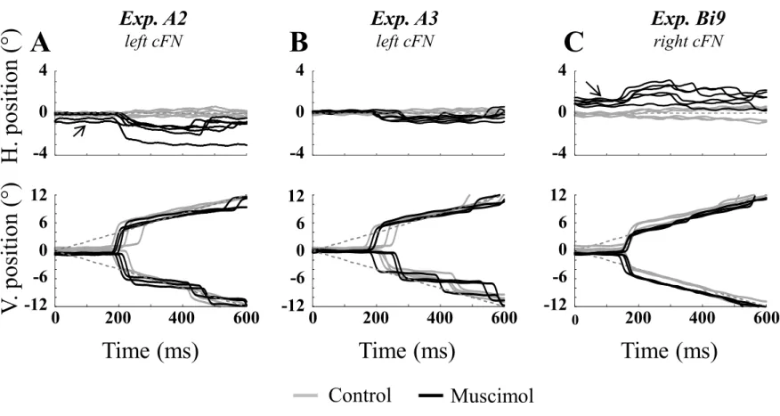
Figure 1 approximately here
213
Figure 1 illustrates with a few selected trials how a unilateral injection of muscimol in the cFN
214
affects the eye movements made in response to a centrifugal target moving at a constant speed
215
(20°/s) along the vertical meridian. The time course of the horizontal (top row) and vertical
216
(bottom row) components is shown for movements produced before (gray) and after muscimol
217
injection (black) in the left (A and B: experiments A2 and A3 in monkey A) or right cFN (C:
218
experiment Bi9 in monkey Bi). As described elsewhere (Guerrasio et al. 2010), inactivation of
219
cFN caused a fixation offset toward the injected side. Most starting eye positions were shifted
220
to the right after muscimol injection in the right cFN (see black arrow in C) and to the left after
221
muscimol injection in the left cFN (black arrow in A). The ipsilesional offset was not observed
222
on every trial. We show examples (recorded during the experiments A2 and A3) in which the
223
saccades were initiated from positions near the vertical meridian (horizontal position values
224
close to zero in the upper graphs) but below the horizontal meridian (negative values of vertical
225
position in the lower graphs). In addition to altering gaze direction during fixation, unilateral
226
cFN inactivation also perturbed vertical saccades. Their trajectory was deviated horizontally
227
toward the injected side, regardless of whether the target moved upward or downward. In the
228
following paragraphs, after describing the velocity of their vertical component, we describe the
229
ipsipulsion that alters the trajectory of vertical saccades.
230
Figure 2 approximately here
231
As for horizontal saccades (see Fig. 2 in Bourrelly et al. 2018a), muscimol injection in
232
the cFN barely affected the vertical velocity of saccades. Figure 2 illustrates the vertical
233
amplitude-peak velocity relationship for four different experiments (two per monkey; A and B:
234
experiments A2 and A3 in monkey A; C and D: Bi7 and Bi8 in monkey Bi). The peak velocity
values are plotted for all upward and downward saccades (positive and negative amplitude
236
values, respectively) made in response to the different target speeds. When we consider
237
saccades of comparable vertical amplitude, we can see that the peak velocity is similar between
238
the pre and post-injection movements.
239
Figure 3 approximately here
240
Figure 3 describes the landing position and time of interceptive saccades made toward
241
a target moving with a constant speed (20°/s) along the vertical meridian (black: upward
242
motion, gray: downward). For three different experiments (A3 and Bi7: left cFN inactivation;
243
Bi9: right cFN inactivation), the vertical (top row) and horizontal (bottom row) landing
244
positions are plotted as a function of their times relative to target motion onset. During the
245
control sessions (open squares), the vertical landing position increased as the landing time
246
increased, and the saccades were directed toward locations close to the physical target
247
(represented by the dashed lines). After muscimol injection (filled squares), the vertical landing
248
positions still increased as the landing time increased, regardless of the motion direction. The
249
overlap between pre- and post-injection data points indicates that unilateral cFN inactivation
250
did not noticeably affect the vertical component of interceptive saccades. Similar findings were
251
found with respect to saccades toward a static target (Goffart et al. 2004). By contrast, the
252
horizontal landing positions changed after muscimol injection. While they were scattered along
253
the vertical meridian (position values close to zero) during the control sessions, their
254
distribution was shifted toward the injected side after muscimol injection (to the left in
255
experiments A3 and Bi7, to the right in experiment Bi9). This effect is rather small in
256
experiment A3 (B: mean ± standard deviation values changing from -0.1 ± 0.3° to -0.5 ± 0.4°
257
after muscimol injection), but the change of horizontal position was much larger in experiments
258
Bi7 (D: from 0.0 ± 0.3° to -3.5 ± 1.6°), with Bi9 displaying intermediate values (F: from 0.3 ±
259
0.4° to 2.0 ± 0.7°). Regardless of whether the target moved upward (black symbols) or
downward (gray symbols), the horizontal landing position was always shifted toward the
261
injected side. The size of the ipsipulsion was, in some cases, relatively constant (as in B) or
262
increased with the landing time (as in D). It is worth noting that the hypermetria of ipsilesional
263
horizontal saccades was relatively small during experiment A3 whereas it was larger during
264
experiments Bi7 and Bi9 (see Figures 7 and 10 in Bourrelly et al. 2018a). In summary, an
265
ipsilesional deviation was observed in the trajectory of vertical saccades during cFN
266
inactivation. The magnitude of the ipsipulsion varied between experiments, but as we will show,
267
it is correlated to the magnitude of the dysmetria that impairs the horizontal saccades.
268
To quantify the accuracy of vertical saccades, we calculated for each component the
269
ratio of landing position to landing time. This parameter (called position/time landing)
270
characterizes both spatial and temporal aspects of saccades made toward a moving target
271
(Bourrelly et al. 2018a). When we consider saccades toward a target moving along the vertical
272
meridian, the values of vertical position/time landing are expected to be close to the target speed
273
if gaze lands next to the target location. A ratio larger than the target speed indicates an
274
overshoot (too large amplitude) whereas a smaller value means an undershoot (too small
275
amplitude). Concerning the horizontal component, if gaze lands close to the target, its value is
276
expected to be close to zero because the target does not move horizontally. We will use the
277
convention that a positive horizontal ratio indicates an ipsilesional deviation whereas a negative
278
value is the signature of a contralesional deviation.
279
Figure 4 approximately here
280
Figure 4 shows the average vertical position/time landing relationships for each monkey
281
and each experiment in which the target moved at constant speed. During the control sessions,
282
the values were close to the target speed (11.2 ± 1.3, 21.9 ± 2.0 and 38.9 ± 4.6 °/s for the 10, 20
283
and 40 °/s targets, respectively). After muscimol injection, these ratios were larger (12.3 ±
3.8°/s, 24.3 ± 4.0°/s and 44.2 ± 6.3°/s for the 10, 20 and 40 °/s targets, respectively) (Fig. 4A).
285
This increase was small but statistically significant (Wilcoxon test, all P values < 0.05). A
286
stronger affect was observed in the horizontal component of vertical saccades. Figure 4B shows
287
the average horizontal position/time landings for each tested constant speed, in each experiment
288
and both monkeys. After muscimol injection, the horizontal position/time landings increased
289
from 0.0 ± 0.7°/s (range: -1.1 – 1.6°/s) to 5.4 ± 3.6°/s (range: 0.8 – 12.1°/s) when the target
290
speed was 10°/s, from 0.0 ± 1.1°/s (range: -2.6 – 2.9 °/s) to 8.0 ± 4.9°/s (range: 1.8 – 18.6°/s)
291
with the 20°/s target and from -0.2 ± 1.6°/s (range: -2.5 – 2.9°/s) to 12.0 ± 7.6°/s (range: 3.1 –
292
24.2°/s) with the 40°/s target. The ranges of mean values and the examination of data points in
293
Fig 4B indicate more variability after muscimol injection than before (see the scattering toward
294
more positive values; note also the different scales for the x and y axes). During the control
295
sessions, the average horizontal position/time landings were close to zero (grand averages =
296
0.0, 0.0 and -0.2°/s for the 10, 20 and 40°/s target, respectively; all monkeys considered). After
297
muscimol injection, the values were larger and also increased with target speed (5.4, 8.0 and
298
12.0°/s for the 10, 20 and 40°/s target, respectively). Figure 4C compares the average horizontal
299
position/time landing between saccades toward the 20°/s target and saccades to the 10°/s (gray
300
symbols) and 40°/s (black symbols) targets. The values with the 10°/s target were smaller than
301
the values with the 20°/s target (gray symbols are situated below the diagonal line of equality)
302
whereas the values with the 40°/s target were larger than those obtained with the 20°/s target
303
(almost all black symbols above the diagonal line). Thus, within the same experiment, the size
304
of the ipsipulsion was larger during saccades to the fastest target than during saccades to the
305
slowest ones. Moreover, the injections which led to the larger horizontal errors with the 20°/s
306
target were those caused the bigger changes with the other speeds. Statistically significant
307
correlations were indeed found between the average horizontal position/time landing values
308
calculated with the 20°/s and 10°/s targets (Bravais-Pearson correlation coefficient R = 0.94, P
< 0.05) as well as between the mean values obtained with the 20°/s and 40°/s targets (R(x,y) =
310
0.93, P < 0.05).
311
Figure 5 approximately here
312
In our previous report, we documented the dysmetria of horizontal interceptive saccades
313
after cFN inactivation: ipsilesional saccades are hypermetric whereas contralesional saccades
314
are hypometric (Bourrelly et al. 2018a). Like the dysmetria that affects horizontal saccades
315
toward a static target (Goffart et al. 2004, in preparation), the sizes of the ipsilesional and
316
contralesional dysmetria are unrelated. However, it is known that the cFN projects to the region
317
in the contralateral medullary reticular formation (medRF) where premotor inhibitory burst
318
neurons (IBNs) are located (Noda et al. 1990) and that these neurons, in addition to firing a
319
burst during horizontal saccades, also fire during vertical saccades (Scudder et al. 1988; van
320
Gisbergen et al. 1981). Because of this involvement of IBNs in horizontal and vertical saccades,
321
it is quite reasonable to consider that a common dysfunction causes the ipsipulsion of vertical
322
saccades and the dysmetria of horizontal saccades (Goffart et al. 2004). Therefore, we tested
323
the relation between these two disorders and found a significant correlation between the average
324
horizontal position/time landings of ipsilesional horizontal saccades and vertical saccades
325
toward the 10°/s (R = 0.77, P < 0.05), 20°/s (R = 0.62, P < 0.05) and 40°/s (R = 0.81, P < 0.05)
326
targets (Fig. 5A). When the correlation was instead tested between the horizontal position/time
327
landings of contralesional horizontal saccades and vertical saccades, the coefficients were
328
smaller and negative. Furthermore, the correlation did not reach statistical significance when
329
the target moved at 10°/s (R = -0.41, P > 0.05) and 40°/s (R = -0.13, P > 0.05). A significant
330
correlation was found only with the 20°/s target (R = -0.47, P<0.05) (Fig. 5B). Then, for each
331
experiment, we subtracted the average contralesional position/time landing from the
332
ipsilesional position/time landing, the resulting difference (hereafter called net bilateral effect
333
on horizontal saccades) characterizing the dysmetria of saccades made toward a target moving
horizontally (leftward or rightward). In this case, we found significant correlations between the
335
horizontal position/time landings of vertical saccades and the net bilateral effect on horizontal
336
saccades (Fig. 5C) that were stronger than those obtained with ipsilesional horizontal saccades
337
(10°/s target: R = 0.91 versus 0.77; 20°/s R = 0.81 versus 0.62; 40°/s: R = 0.90 versus 0.81).
338
In summary, after muscimol injection in the cFN, all vertical saccades made toward a
339
target moving at a constant speed along the vertical meridian were deviated horizontally toward
340
the injected side. Their vertical components were slightly affected but not as dramatically as
341
the horizontal component. The size of the ipsipulsion differed between experiments and the
342
average horizontal position/time landing increased with the target speed. Moreover, the
343
ipsipulsion is correlated with the dysmetria that impaired the horizontal (ipsilesional and
344
contralesional) saccades.
345
Figure 6 approximately here
346
In the figure 4C, we showed that the ipsipulsion of vertical saccades depends upon the
347
target speed. To further explore this dependence, we tested saccades toward a target that moved
348
with continuous acceleration or deceleration. If the ipsipulsion depends on target speed, the
349
horizontal landing position should increase with later landing times when the target accelerates
350
but decrease or stagnate when it decelerates. Figure 6 describes the horizontal landing position
351
and time of saccades to a target moving with an increasing (top row) and decreasing speed
352
(bottom row) for three experiments (A3: left column; Bi8: middle and Bi9: right). During the
353
control sessions (open squares), the saccades landed on locations situated along the vertical
354
meridian (open symbols are close to the line of zero horizontal position) whereas after muscimol
355
injection (filled squares), they landed on locations which were shifted toward the injected side
356
(see negative position values in experiments A3 and Bi8 and positive values in experiment Bi9).
357
For these three experiments, the ipsipulsion of vertical saccades did not depend on the
instantaneous speed of the target. Statistically significant correlations were found in only two
359
experiments: A6 (positive correlation R = 0.54, injection in the right cFN) and A2 (negative
360
correlation R = -0.35, left cFN). For the other experiments, no correlation was found between
361
the horizontal landing position and the landing time when the target accelerated (R values
362
ranged from -0.25 to 0.19, P-values > 0.05). Concerning the saccades toward a decelerating
363
target, no statistically significant correlation was found between the horizontal landing position
364
and the landing time (R values ranged from -0.42 to 0.34, P-values > 0.05). In summary, we
365
found no evidence suggesting that the ipsipulsion depends on the instantaneous target speed:
366
the horizontal landing position neither increased with later landing times when the target
367
accelerated nor decreased when it decelerated.
368
Figure 7 approximately here
369
Fig. 7 documents the mean values of vertical (A) and horizontal (B) position/time
370
landing of saccades toward the accelerating (gray symbols) and decelerating (black symbols)
371
targets, before and after muscimol injection, as a summary of our results. The vertical
372
position/time landing of saccades was changed after muscimol injection, more with the
373
decelerating target than with the accelerating one (Fig. 7A). With the accelerating target, the
374
pre- and post-injection mean values of vertical position/time landing were not significantly
375
different (Wilcoxon test, P = 0.47) whereas with the decelerating target, the post-injection
376
values were significantly different from pre-injection values (16% average increase, P < 0.05).
377
Regarding the horizontal position/time landing (Fig. 7B), it increased from 0.1 ± 0.7°/s (range:
378
-0.8 – 1.9°/s) to 5.0 ± 2.9°/s (range: 0.8 – 9.5°/s) after muscimol injection with the accelerating
379
target and from 0.0 ± 1.8°/s (range: -3.6 – 3.4°/s) to 11.6 ± 5.9°/s (range: 2.6 – 20.1°/s) with the
380
decelerating target. In both groups of saccades, the changes were statistically significant (P <
381
0.05). Like saccades toward a target moving with a constant speed, the mean values of
382
horizontal landing position were also variable between the different muscimol injections (see
range values reported above and the scatter of post-lesion data points in Fig. 7B). Finally, the
384
injections which led to the larger horizontal position/time landings with the 20°/s moving target
385
were those which led to the bigger changes with the accelerating and decelerating targets (Fig.
386
7C). Statistically significant correlations were indeed found between the horizontal
387
position/time landings of saccades toward the 20°/s and those directed toward the accelerating
388
(Bravais-Pearson correlation coefficient R(x,y) = 0.86, P < 0.05) or decelerating target (R(x,y)
389
= 0.90 P < 0.05). In summary, all vertical saccades toward a target moving with a changing
390
speed were shifted horizontally after muscimol injection in the cFN. Like saccades toward a
391
target moving at a constant speed, the amplitude of their vertical component was slightly
392
increased after muscimol inactivation. Their horizontal component was biased toward the
393
injected side with a magnitude which differed between experiments. The ipsipulsion was larger
394
for saccades aimed toward a fast target than toward a slow target. It was also smaller when the
395
target accelerated than when it decelerated.
396
As for saccades to a target moving horizontally, the larger deficit with fast targets can
397
result from the fact that at identical landing times, a fast target is more eccentric than a slow
398
one. The dependency of the ipsipulsion upon the target speed can result from the fact that a
399
larger amplitude was required to intercept the target when it moves fast. Studies of vertical
400
saccades toward a static target do indeed show that after cFN inactivation, the ipsipulsion
401
increases with more eccentric targets (Goffart et al. 2004; Iwamoto and Yoshida 2002; Quinet
402
and Goffart 2007). However, increasing target eccentricity enhances both the amplitude and
403
duration of saccades for larger saccades that last longer. It is not known which of these
404
parameters affect more the magnitude of ipsipulsion. Therefore, we tested the correlation
405
between the horizontal landing position and saccade duration for each post-injection session.
406
The parameters of the equations fitting the relation between the horizontal landing position and
407
the saccade duration after muscimol injection are documented for all the experiments (upward
and downward movements pooled together) in table 1 (constant speeds) and table 2 (changing
409
speeds). The correlation was statistically significant in 13 out of 44 test sessions (corresponding
410
to 30% of cases) with coefficients of correlation ranging from 0.37 to 0.72. When we instead
411
considered the relation between the horizontal landing position and vertical saccade amplitude,
412
statistically significant correlations were found in 30 out of 44 sessions (corresponding to 68%
413
of the post-injection sessions), with coefficients of correlation ranging from 0.38 to 0.81. The
414
parameters of the equations fitting the relation between the horizontal landing position and the
415
vertical saccade amplitude after muscimol injection are documented for all the experiments
416
(upward and downward movements pooled together) in table 1 (target moving at a constant
417
speed) and table 2 (accelerating and decelerating targets). In several instances, the correlation
418
between the saccade duration and the horizontal landing position (R(Dur,HorP)) did not reach
419
statistical significance whereas the correlation between the saccade amplitude and the
420
horizontal landing position (R(Amp,HorP)) was statistically significant (see Table 1:
421
experiments A3, A5, A6 and Bi9 for the 10°/s target; A2, A3, A6, Bi9 and Bi10 for the 20°/s
422
target; in A3, A5 and Bi7 for the 40°/s target). Likewise, when we consider the saccades toward
423
an accelerating target (leftmost columns in Table 2), R(Dur,HorP) was not statistically
424
significant although R(Amp,HorP) was during the experiments A2 and A6. Finally, when the
425
target was decelerating (rightmost columns in Table 2), R(Dur,HorP) was not statistically
426
significant but R(Amp,HorP) was significant during the experiments A3, A6, Bi8 and Bi10.
427
Considering all 44 sessions, a significant correlation R(Dur,HorP) without significant
428
correlation R(Amp,HorP) was observed in one single experiment (Bi8, accelerating target).
429
Altogether, these results strongly suggest that the ipsipulsion increased mostly because
430
saccades had larger amplitudes than because they lasted longer.
431
Figure 8 approximately here
To illustrate this correlation between the vertical amplitude of saccades and the
433
ipsipulsion, Fig. 8 plots the horizontal landing position as a function of vertical saccade
434
amplitude before and after muscimol injection. Both upward and downward saccades were
435
included (the amplitude of downward saccades was multiplied by -1). The relations are shown
436
for four injections made in the left cFN of monkey A (experiments A2 and A3, top row) and Bi
437
(experiments Bi7 and Bi8, bottom) for saccades to targets moving with a constant speed (light
438
gray, dark gray and black symbols correspond to the 10, 20 and 40°/s target speeds,
439
respectively). Negative values of horizontal eye position indicate that the saccades were
440
deviated toward the left (inactivated side). Thus, we can see that while the target speed increases
441
from 10 to 40°/s (symbols with different shades of grey), the horizontal landing position and
442
the vertical saccade amplitude increased. During experiment A2 (Fig. 8A), as the target speed
443
increased, the horizontal landing position changed from -0.3 ± 0.3° (10°/s) to -0.7 ± 0.5° (20°/s)
444
and -1.4 ± 0.8° (40°/s), while the vertical amplitude increased from 2.8 ± 0.6° (10°/s) to 5.1 ±
445
1.1° (20°/s) and 10.2 ± 1.9° (40°/s). Likewise, during experiment A3 (Fig. 8B), the horizontal
446
landing position increased from -0.3 ± 0.3° to -0.5 ± 0.4° and -1.3 ± 0.6° while the vertical
447
amplitude increased from 2.7 ± 0.6° to 5.1 ± 1.1° and 10.5 ± 1.1° (values for the 10, 20 and
448
40°/s target speed, respectively). The same relationship was found in monkey Bi. During
449
experiment Bi7 (Fig. 8C), the horizontal landing position increased from -2.4 ± 0.7° to -3.4 ±
450
1.6° and -4.3 ± 1.1° and the vertical amplitude from 2.2 ± 0.7° to 5.1 ± 1.2° and 9.0 ± 1.3°.
451
Finally, during experiment Bi8 (Fig. 8D), the horizontal landing position increased from -2.0 ±
452
0.7° (10°/s) to -2.8 ± 0.8° (20°/s) to -3.7 ± 1.0° (40°/s), while the vertical amplitude increased
453
from 2.2 ± 0.6° to 5.0 ± 1.6°and 9.1 ± 1.1°. A relation was observed between the horizontal
454
landing position and the vertical amplitude of saccades. Each inset graph plotted in the right
455
part of the panels of Figure 8 shows an overlap of data points corresponding to saccades to a
456
target moving at constant speed (gray symbols) and to an accelerating or decelerating target
(black symbols). In summary, after cFN inactivation, the landing position of vertical saccades
458
was deviated toward the injected side by an amount that varied between experiments and that
459
was larger when the target moved quickly than when it moved slowly. The dependency of the
460
ipsipulsion upon the target speed indicates that the saccade oculomotor system has access to
461
different signals when the target moves at different speeds. However, the use of accelerating
462
and decelerating target enabled us to exclude a dependency upon the instantaneous speed of the
463
target. Further analyses revealed that the ipsipulsion was more frequently correlated with the
464
vertical amplitude of saccades rather than with their duration.
Discussion
466
This study is the first to describe the consequences of unilateral pharmacological
467
inactivation of cFN on the generation of vertical saccades toward a moving target. As for the
468
saccades toward a static target, interceptive saccades exhibit a horizontal deviation of their
469
trajectory toward the lesioned side (ipsipulsion; Figs. 3, 4B and 7B) whereas the vertical
470
component is barely affected (Figs. 2, 3, 4A and 7A). The magnitude of the ipsipulsion is larger
471
for fast targets than for slowly moving targets (Fig. 4C) but the use of accelerating and
472
decelerating targets indicates that this relation is due less to the instantaneous target speed (Fig.
473
6) than to the saccade size. Indeed, like saccades toward a static target, the ipsipulsion increased
474
with the vertical amplitude of saccades (Fig. 8). Statistical analyses made across all sessions
475
indicate that the correlations between the ipsipulsion and the vertical amplitude were more
476
frequent than the correlations between the ipsipulsion and the duration. Moreover, in several
477
sessions, the correlation between the ipsipulsion and the duration of saccades did not reach
478
statistical significance whereas the correlation with the vertical amplitude was significant
479
(Tables 1 and 2). Below, we explain the neurophysiological mechanisms leading to the
480
ipsipulsion of vertical saccades and further develop facts which call for an alternative approach
481
to investigate the neurophysiology of visually-guided eye movements and their cerebellar
482
control.
483
Figure 9 approximately here
484
How does bilateral fastigial activity influence the trajectory of saccades?
485
Numerous works show that the activity of cFN neurons plays a crucial role in the
486
cerebellar control of saccade generation. Two major reasons led us to investigate their influence
487
on the trajectory of saccades toward a target moving along the vertical meridian: 1) vertical
488
saccades are deviated horizontally after unilateral cFN inactivation and 2) the same etiology
489
can account for this ipsipulsion and the dysmetria of horizontal saccades. In the framework of
negative feedback control in which an internal estimate of current eye position (or
491
displacement) is compared to a desired eye position (or displacement) command (Robinson
492
1975; Sparks 1989; Becker 1995; Pola 2002), a horizontal bias could impair vertical saccades
493
in a cumulative manner as long as motor error is not zeroed and omnipause neurons do not
494
resume their activity. Our results do not support this link between the horizontal targeting error
495
and the duration of vertical saccades because the magnitude of the ipsipulsion was less
496
frequently dependent upon the duration than the amplitude, and because in many cases, the
497
correlation between the ipsipulsion and the duration did not reach statistical significance
498
whereas the correlation between the ipsipulsion and the amplitude was statistically significant.
499
An independence between the dysmetria and duration of gaze shifts was already noticed in the
500
head unrestrained cat after cFN inactivation (see Figs. 5 and 6 in Goffart et al. 1998). Finally,
501
it is worth reminding that neither Kaneko (1996) nor Soetedjo et al. (2000) reported changes in
502
the accuracy of saccades when the nucleus raphe interpositus is lesioned or inactivated. The
503
dysmetria of saccades after cFN inactivation is therefore not caused (but permitted) by the pause
504
of OPNs (under the influence of the vertical burst generator). We propose that the ipsipulsion
505
is driven by action potentials that abducens neurons emit under the influence of unbalanced
506
input from excitatory and inhibitory burst neurons.
507
The known physiology of the saccade-related circuitry indeed explains the ipsipulsion
508
of vertical saccades: if the activity of neurons in the left and right abducens nuclei is not
509
balanced during vertical saccades, there will be horizontal deviation of the eyes. The elevated
510
speed and the large magnitude of the ipsipulsion during head-unrestrained gaze shifts (Quinet
511
and Goffart 2007) indicate that this imbalance is not caused by a reduced inhibition that IBNs
512
in the contralateral medullary reticular formation exert upon neurons in the abducens nucleus
513
ipsilateral to the inactivated cFN (synapses a and b in Fig. 9). An excitatory drive is required
514
and we hypothesize that it is conveyed by neurons in the opposite (unaffected) cFN (synapse
f). Transmitted by crossed projections (Noda et al. 1990), their spikes excite premotor burst
516
neurons in the pontomedullary reticular formation ipsilateral to the inactivated cFN, which in
517
turn activate motor and internuclear neurons in the ipsilateral abducens nucleus (synapses g and
518
h). This crossed excitatory influence upon premotor burst neurons is supported by the fact that
519
fastigial electrical stimulation evokes contralateral saccades (Cogdell et al. 1977; Noda et al.
520
1988; Quinet and Goffart 2015). It is also consistent with the contrapulsion of vertical saccades
521
when the oculomotor vermis is asymmetrically lesioned (Takagi et al. 1998) or inactivated with
522
muscimol (Nitta et al. 2007). This crossed fastigioreticular projection does not concern the
523
reticulospinal neurons that Takahashi et al. (2014) identified in the cat. Indeed, if it also exists
524
in the macaque, this fastigio-reticulospinal connection is likely involved in the postural tone of
525
the head because the head barely moves when the cFN is electrically stimulated (Quinet and
526
Goffart 2009). Also, during cFN inactivation in both the cat and monkey, the head exhibits an
527
ipsilesional deviation (Goffart and Pélisson 1998; Quinet and Goffart 2005). Furthermore, the
528
increase of ipsipulsion with saccade amplitude indicates that abducens neurons and excitatory
529
burst neurons (EBNs) fire more as the vertical amplitude increases. Although EBNs do not
530
normally fire during vertical saccades (Strassman et al. 1986a; Sparks et al. 2002) and unilateral
531
inactivation of their territory yields no ipsipulsion (Barton et al. 2003), it is possible that after
532
cFN inactivation, ipsilateral MNs, INs and EBNs abnormally fire during vertical saccades
533
because they are released from the inhibition exerted by burst neurons (IBNs) in the
534
contralateral reticular formation (synapses a, b and d; Cullen and Guitton 1997; Scudder et al.
535
1988; Strassman et al. 1986b). Those IBNs are silenced because of the suppression of their
536
fastigial drive (synapse i) by the muscimol injection, because also of the inhibition exerted by
537
IBNs located in the opposite side (synapse j) and recruited by neurons in the unaffected cFN
538
(synapse e). Scudder et al. (1988) indeed showed that IBNs fire more spikes as the size of
539
vertical saccades increases (see also van Gisbergen et al. 1981). Thus, released from the
inhibitory influence of contralateral IBNs (synapses a and b), MNs and INs emit spikes that
541
deviate vertical saccades with a magnitude that increases with saccade size. The burst would be
542
triggered by excitatory input from EBNs or from other neurons innervating the abducens
543
nucleus (Langer et al. 1986; Ugolini et al. 2006).
544
According to the bilateral hypothesis, the activity of saccade-related fastigial neurons
545
influences the horizontal component of all saccades (vertical and/or horizontal). Ipsilesional
546
saccades overshoot horizontally their target because following cFN inactivation, the agonist
547
motor neurons receive a drive from excitatory burst neurons (synapses g and h in Fig. 9) that
548
exceeds the pre-lesional drive, as it is no longer attenuated by spikes emitted by IBNs in the
549
contralateral side (synapses a, b and d). Indeed, these IBNs not only lack their fastigial
550
excitatory input (synapse i) but they are also inhibited by IBNs driven by neurons in the
551
unaffected cFN (synapse e). Likewise, contralesional saccades prematurely stop for two
552
reasons: because the agonist motor neurons lack the drive they usually receive from EBNs (as
553
a result of suppressing their fastigial input; synapse k) and because the opposite (unaffected)
554
cFN excites IBNs which in turn inhibit the agonist motor neurons (synapses l and m). Thus,
555
regardless of saccade direction, the same bilateral mechanism (i.e., an altered balance of
556
excitatory and inhibitory input to the motor neurons) explains the effects on the horizontal
557
component of all saccades. This conclusion is further supported when the results from the
558
present study are synthesized with those reported in Bourrelly et al. (2018a). Indeed, the net
559
bilateral effect of inactivation on horizontal saccades (calculated by subtracting the average
560
contralesional position/time landing ratio from the ipsilesional ratio) is strongly correlated with
561
the horizontal position/time landing of vertical saccades.
562 563
Recasting the neurophysiology of saccades
In the preceding section as well as in our previous reports (Bourrelly et al. 2018a,b;
565
Goffart et al. 2004; Guerrasio et al. 2010; Quinet & Goffart 2007), the oculomotor disorders
566
observed after cFN inactivation were explained without assuming perturbations of some
567
internal (and putative) estimate of the eye kinematics, as one might expect for example, if their
568
input from Purkinje cells in the oculomotor vermis were to encode the instantaneous velocity
569
of saccades (Herzfeld et al. 2015). In this section, we want to further develop additional
570
concerns which support our alternative viewpoint and call for recasting the neurophysiology of
571
eye movements (Goffart et al. 2018; Goffart 2019).
572
First of all, it is important to realize that if the recording of eye muscle tension had been
573
used more frequently (Davis-Lopez de Carrizosa et al., 2011; Gamlin and Miller; 2012;
574
Lennerstrand et al. 1993; Miller and Robins 1992) than the recording the orientation of the eyes
575
during neurophysiological experiments, different processes and models might have been
576
proposed. Then, it is also important to consider the fact that the time course of saccades is not
577
exclusively determined by the discharge of premotor and motor neurons. It involves changes in
578
the firing rate of neurons distributed in many brain regions such as the frontal cortex (Dias and
579
Segraves 1999), the mesencephalon (Aizawa and Wurtz 1998; Sparks et al. 1990), the pons
580
(Kaneko and Fuchs 2006), the medio-posterior cerebellum (Goffart et al. 2003; Kojima et al.
581
2010) and the reticular formation (Barton et al. 2003; Soetedjo et al. 2000). Right upstream
582
from the extraocular muscle fibers, saccades are engendered by an abrupt increase in the firing
583
rate of motor neurons which innervate the agonist muscles and a concomitant decrease in the
584
discharge of neurons innervating the antagonist muscles. Due to the highly damped nature of
585
the oculomotor plant (Robinson 1964), a braking action is not exerted upon antagonist motor
586
neurons to stop the saccade (Robinson 1970; Schiller 1970). Thus, the agonist burst duration
587
increases with the duration of saccades and determines their size and the saturated firing rate
588
leads to a discharge frequency which is poorly related to saccade speed (Fuchs and Luschei
1970; Robinson 1970; Schiller 1970; Sparks and Hu 2006). Synchronous presynaptic spikes
590
converging upon the motor neurons could be one way to overcome the limitations of interspike
591
intervals (Schiller 1970; Goffart et al. 2017a). For the generation of horizontal saccades, the
592
burst causing the phasic contraction of the medial rectus (and the saccade of the contralateral
593
eye) originates in burst-tonic (internuclear) neurons in the abducens nucleus while the burst
594
causing the phasic contraction of the lateral rectus (saccade of the ipsilateral eye) originates in
595
abducens motoneurons. These bursts are caused by excitatory burst neurons in the paramedian
596
pontine reticular formation (Keller 1974; Luschei and Fuchs 1972; Strassman et al. 1986a).
597
In several synthesis articles, we can read that the discharge profile of burst neurons is a
598
“replica of eye velocity” (Scudder et al. 2002) and that pontine and midbrain EBNs encode
599
saccade velocity by the frequency of spikes (Sparks 2002). Their instantaneous firing rate would
600
be closely related with instantaneous eye velocity (Leigh and Zee 2006, page 126), as supported
601
by several recording studies in the premotor and motor neurons (e.g., Cromer and Waitzman
602
2007; Cullen and Guitton 1997; King and Fuchs 1979; Sylvestre and Cullen 1999). For several
603
years, kinematic notions were used to interpret the spiking activity of neurons and to model the
604
generation of saccades. Yet, when we examine the firing rate of premotor neurons, we discover
605
that they do not exhibit the same time course as instantaneous eye velocity (see Figs. 2 and 3 in
606
Hu et al. 2007, Fig. 3C in Sparks and Hu 2006 and Fig. 5 in van Gisbergen et al. 1981). In fact,
607
the meticulous inspection reveals that the instantaneous discharge of burst neurons is almost
608
clock-like and quasi-regular (Hu et al. 2007), radically different from the bell-shaped profile
609
shown by studies which convolve each spike with a Gaussian kernel.
610
The relation between the firing rate and the instantaneous saccade velocity was recently
611
extended to more central neurons in the superior colliculus (Smalianchuk et al. 2018) and the
612
oculomotor vermis (Herzfeld et al. 2015). In agreement with the crucial role of these two
613
regions in continuously driving the saccades (Goffart et al. 2003; Goossens and van Opstal
2006), these studies proposed that the discharge of their neurons encodes the instantaneous
615
velocity. Two major observations must, nevertheless, be considered. Firstly, a model factoring
616
muscle tension and its first derivative fits better the instantaneous firing rate of motoneurons
617
than a model that uses eye position, velocity and acceleration (Davis-Lopez de Carrizosa et al.,
618
2011). The putative velocity commands must therefore be translated into force-related
619
commands somewhere before reaching the motor neurons. Interposed between the
saccade-620
related bursts of Purkinje cells and the premotor burst neurons, the caudal fastigial nuclei host
621
a population of neurons which emit spikes that have a strong impact on the time course of
622
saccades toward their landing position. Changes in cFN activity alter not only the endpoint of
623
saccades (Goffart et al. 2003; Noda et al. 1991) but also their trajectory (see Figs. 1 and 7 in
624
Goffart et al. 2004; Fig. 1 in Iwamoto and Yoshida 2002). However, the absence of relation
625
between their firing rate and saccade velocity (Fuchs et al. 1993; Helmchen et al. 1994; Ohtsuka
626
and Noda 1991) draws into question the suggestion that the population discharge of Purkinje
627
cells “encodes” instantaneous saccade velocity (Herzfeld et al. 2015). When correlations are
628
found between the population firing rate and saccade velocity, caution must be taken that they
629
do not result from idiosyncratic (all monkeys do not make the same saccade amplitude with the
630
same speed) or unspecific factors like different levels of motivation or arousal (Fuchs et al.
631
1993) as shown also in the deep superior colliculus (Ikeda and Hikosaka 2007; Soetedjo et al.
632 2000). 633 634 Conclusion 635
Instead of reflecting the reduction of an internal signal encoding the horizontal distance
636
between gaze and target positions (or displacements), we propose that the horizontal component
637
of saccades is the outcome of a process which restores, under bilateral fastigial control, an
638
equilibrium (symmetry) that has been broken by asymmetric descending input (from cortical
eye fields and deep superior colliculus) to the horizontal saccade generators (Goffart et al. 2018;
640
Goffart 2019). In the future, more studies will have to clarify the cerebellar control of the
641
vertical component of saccades (Quinet and Goffart 2015; Robinson 2000) and its coupling
642
with the processes controlling their horizontal component (Goffart et al. 2005).
643 644
References
645
Aizawa H, Wurtz RH. Reversible inactivation of monkey superior colliculus. I. Curvature of
646
saccadic trajectory. J Neurophysiol 79: 2082-2096, 1998. doi: 10.1152/jn.1998.79.4.2082 647
Barton EJ, Nelson JS, Gandhi NJ, Sparks DL. Effects of partial lidocaine inactivation of the
648
paramedian pontine reticular formation on saccades of macaques. J Neurophysiol 90:
372-649
386, 2003. doi: 10.1152/jn.01041.2002
650
Becker W. Models of oculomotor function: An appraisal of the engineer’s intrusion into
651
oculomotor physiology. Studies Vis Inform Processing 6: 23-46, 1995.
652
Bourrelly C, Quinet J, Cavanagh P, Goffart L. Learning the trajectory of a moving visual
653
target and evolution of its tracking in the monkey. J Neurophysiol 116: 2739–2751, 2016. doi:
654
10.1152/jn.00519.2016
655
Bourrelly C, Quinet J, Goffart L. The caudal fastigial nucleus and the steering of saccades
656
toward a moving visual target. J Neurophysiol 120: 421–438, 2018a. doi:
657
10.1152/jn.00141.2018 658
Bourrelly C, Quinet J, Goffart L. Pursuit disorder and saccade dysmetria after caudal fastigial 659
inactivation in the monkey. J Neurophysiol 120: 1640-1654, 2018b. doi: 660
10.1152/jn.00278.2018 661
Bourrelly C, Quinet J, Goffart L. Unsupervised dynamic morphing of a spatiotemporal visual
662
event during its oculomotor tracking. J Vision 14: 492, 2014. 663
Buzunov E, Mueller A, Straube A, Robinson FR. When during horizontal saccades in monkey
664
does cerebellar output affect movement? Brain Res 1503: 33-42, 2013. doi:
665
10.1016/j.brainres.2013.02.001 666
Cogdell B, Hassul M, Kimm J. Fastigial evoked eye movement and brain stem neuronal
667
behavior in the alert monkey. Arch Otolaryngol 103: 658-666, 1977. doi:
668
10.1001/archotol.1977.00780280058008
669
Cromer JA, Waitzman DM. Comparison of saccade-associated neuronal activity in the
670
primate central mesencephalic and paramedian pontine reticular formations. J Neurophysiol
671
98: 835-850, 2007. doi: 10.1152/jn.00308.2007 672