Advances in Plasmonic Technologies
for Point of Care Applications
The MIT Faculty has made this article openly available.
Please share
how this access benefits you. Your story matters.
Citation
Tokel, Onur, Fatih Inci, and Utkan Demirci. “Advances in Plasmonic
Technologies for Point of Care Applications.” Chemical Reviews
114, no. 11 (June 11, 2014): 5728–5752. © 2014 American Chemical
Society
As Published
http://dx.doi.org/10.1021/cr4000623
Publisher
American Chemical Society (ACS)
Version
Final published version
Citable link
http://hdl.handle.net/1721.1/97500
Terms of Use
Article is made available in accordance with the publisher's
policy and may be subject to US copyright law. Please refer to the
publisher's site for terms of use.
Advances in Plasmonic Technologies for Point of Care Applications
Onur Tokel,
†,⊥Fatih Inci,
†,∥and Utkan Demirci*
,†,‡,§,∥†Demirci Bio-Acoustic-MEMS in Medicine (BAMM) Laboratory, Department of Medicine, Brigham and Women’s Hospital, Harvard
Medical School, Cambridge, Massachusetts 02139, United States
‡Division of Infectious Diseases, Brigham and Women’s Hospital, Harvard Medical School, Boston, Massachusetts 02115, United
States
§Harvard-MIT Health Sciences and Technology, Cambridge, Massachusetts 02139, United States
∥Demirci Bio-Acoustic-MEMS in Medicine (BAMM) Laboratory, Stanford University School of Medicine, Canary Center at Stanford
for Cancer Early Detection, Palo Alto, California 94304, United States
CONTENTS
1. Introduction 5728
2. Overview of Biosensing Technologies 5730 3. SPR Detection Methods 5731 3.1. Fundamental Optical Mechanisms of SPR 5731 3.2. Light Coupling Methods 5732 3.2.1. Prism Coupling 5732 3.2.2. Waveguide Coupling 5732 3.2.3. Diffraction Grating Coupling 5732 3.2.4. Photonic-Crystal-Based Coupling 5732 3.2.5. Combined Coupling Methods 5733 3.3. Localized Surface Plasmon Resonance 5733 3.4. Nanoplasmonic Arrays 5734 4. Integration of Plasmonic Technologies with
Microfluidics 5735
4.1. Surface Functionalization 5735 4.1.1. Physical Adsorption 5735 4.1.2. Chemical Adsorption and Covalent
Binding 5736
4.1.3. Affinity-Based Interactions 5736 4.2. Blocking of Nonspecific Binding 5737 4.3. Recent Advances in Surface
Functionaliza-tion 5737
5. Applications of Plasmonic-Based Technologies
for POC: SPR, LSPR, and SPRi 5740
5.1. SPR 5740
5.2. Localized Surface Plasmon Resonance 5741 5.3. Surface Plasmon Resonance Imaging (SPRi) 5743 6. Conclusion and Future Outlook 5746 Author Information 5747 Corresponding Author 5747 Present Address 5747 Notes 5747 Biographies 5747 Acknowledgments 5748 References 5748 1. INTRODUCTION
Demand for accessible and affordable healthcare for infectious and chronic diseases present significant challenges for providing high-value and effective healthcare. Traditional approaches are expanding to include point-of-care (POC) diagnostics, bedside testing, and community-based approaches to respond to these challenges.1 Innovative solutions utilizing recent advances in mobile technologies, nanotechnology, imaging systems, and microfluidic technologies are envisioned to assist this trans-formation.
Infectious diseases have considerable economic and societal impact on developing settings. For instance, malaria is observed more commonly in sub-Saharan Africa and India.2The societal impact of acquired immune deficiency syndrome (AIDS) and tuberculosis is high, through targeting adults in villages and leaving behind declining populations.3 In resource-constrained settings, it is estimated that about 32% of the disease burden is from communicable diseases such as respiratory infections, AIDS, and malaria, while 43% of the burden is from noncommunicable diseases, such as cardiovascular diseases, neuropsychiatric conditions, and cancer.4 Developing diagnos-tic platforms that are affordable, robust, and rapid-targeting infectious diseases is one of the top priorities for improving healthcare delivery in the developing world.5 The early detection and monitoring of infectious diseases and cancer through affordable and accessible healthcare will significantly reduce the disease burden and help preserve the social fabric of these communities. Further, improved diagnostics and disease monitoring technologies have potential to enhance foreign investment, trade, and mobility in the developing countries.6
Highly sensitive and specific lab assays such as cell culture methods, polymerase chain reaction (PCR), and enzyme-linked immunosorbent assay (ELISA) are available for diagnosis of infectious diseases in the developed world. They require sample transportation, manual preparation steps, and skilled and well-trained technicians. These clinical conventional methods provide results in several hours to days, precluding rapid detection and response at the primary care settings. Another diagnostic challenge is identifying multiple pathogens. Since common symptoms like sore throat and fever can be caused by multiple infectious agents (e.g., bacteria and viruses), it is important to accurately identify the responsible agent for
Received: February 1, 2013
Published: April 18, 2014
Review
pubs.acs.org/CR
targeted treatment. Therefore, high-throughput sensors for multiplexed identification would help improve patient care.7
Medical instruments in centrally located institutions in the developed world rely on uninterrupted electricity and running water and require controlled environmental conditions. It may not be viable to satisfy some of these criteria in some POC settings, where well-trained healthcare personnel are not available and clean water access is unreliable.7,8 Further, in remote settings without infrastructure, rain and dust can act as contaminants.7 Diagnostic devices for POC testing in these settings are identified by the World Health Organization to be affordable, sensitive, user-friendly, specific to biological agents, and providing rapid response to small sample volumes.9Optical biosensor devices are emerging as powerful biologic agent detection platforms satisfying these considerations.10
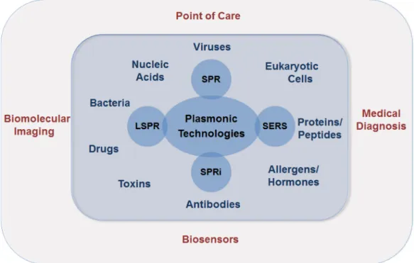
Optical sensing platforms employ various methods, including refractive index change monitoring, absorption, and spectro-scopic-based measurements.11Optical sensors that are based on refractive index monitoring cover a range of technologies, including photonic crystal fibers, nano/microring resonator structures, interferometric devices, plasmonic nano/micro arrays, and surface plasmon resonance (SPR)-based plat-forms.11,12 The latter two are plasmonic-based technologies. Plasmonics is an enabling optical technology with applications in disease monitoring, diagnostics, homeland security, food safety, and biological imaging applications. The plasmonic-based biosensor platforms along with the underlying technologies are illustrated in the Figure 1. Here, we reviewed SPR, localized surface plasmon resonance (LSPR), and large-scale plasmonic arrays (e.g., nanohole arrays).
The integration of plasmonics and microfluidic technologies can potentially serve the global health, primary care, and POC applications, offering modalities toward inexpensive, robust, and portable healthcare technologies. Convergence of optical technologies and microfluidic systems is promising for sensor applications by exploiting fluorescence detection, absorption,
transmission, and polarization measurements on lab-chip (LOC) systems.13 Microfluidics manipulates fluids on the microscale, minimizing the use of expensive reagents. Further, inexpensive microchip fabrication potentially allows mass production.3a,14 Along with the capabilities of sample enrich-ment, isolation, mixing, and sorting, microfluidics has provided applications in several fields, including molecular biology, biotechnology, and defense.15 These characteristics ideally position microfluidics in conjunction with plasmonic tech-nologies to provide medical solutions at the POC and the primary care settings.
LOC devices can potentially address the challenges encountered at POC settings.7 In these devices, single-use chips retaining the waste can be disposed of after use, avoiding contamination. The LOC system can be built from relatively inexpensive parts, specific to the disease and easy-to-operate with minimal training. The system can be designed to be portable, safe, and battery powered. Integrated microfluidic technologies with optical detection platforms such as SPR have the potential to satisfy characteristics for inexpensive, robust, and sensitive biosensors.
Here, theory and applications of plasmonic-based platforms and integration of these technologies with microfluidics are reviewed from a POC diagnostics and monitoring perspective. First, we compare the plasmonic-based biosensors with other optical, electrical, or electro-mechanical biosensor technologies. We describe the theories of SPR and LSPR and demonstrate the main experimental architecture and operational modes currently employed. We then discuss in detail the integration of microfluidic platforms, plasmonic technologies, and surface chemistry techniques leading to LOC devices. We present the current state-of-the-art plasmonic-based LOC biosensors. Finally, we provide a perspective on the future of plasmonic technologies for diagnostics and monitoring of different types of diseases, including infectious diseases and cancer, at the POC and primary care settings.
Figure 1.Plasmonic-based technologies for versatile biosensor applications. SPR stands for surface plasmon resonance, LSPR for localized surface plasmon resonance, SPRi for surface plasmon resonance imaging, and SERS for surface-enhanced Raman scattering.
2. OVERVIEW OF BIOSENSING TECHNOLOGIES Biosensors have several crucial components: (i) a recognition element that interacts with the target; (ii) a transducer that relates the interaction of the recognition element and the target to a readable electrochemical, optical, acoustic, or piezoelectric signal; and (iii) a read-out system to interface with this signal.16 SPR, surface-enhanced Raman scattering (SERS), whispering-gallery modes (WGM), reflectometric interference spectrosco-py (RIfS), and photonic crystals (PC) provide robust and sensitive optical biosensor platforms. Micro-electro-mechanical systems (MEMs) or electrical methods also reach to low detection limits. In particular, cantilever-based sensor tech-nologies, such as atomic force microscope (AFM), and electrical sensors, such as electrochemical impedance spectros-copy (EIS), are alternative detection techniques for biosensing applications.
SPR and LSPR technologies are based on the wave propagation or electromagneticfield enhancement phenomena near metal surfaces or nanoparticles. The propagating surface plasmon polaritons excited on plane metal surfaces are utilized in SPR sensors. LSPR relies on the field enhancement and confinement in close proximity to nanoparticles. The localized field oscillations around nanoparticles motivate the name “localized” in LSPR. The plasmon modes extend up to a couple of hundred nanometers into the biosensor medium in propagating surface plasmon polaritons (PSPP) and up to a few tens of nanometers in LSPR sensors, allowing sensitive subwavelength biosensors.17 SPR biosensors interrogate the resonance angle changes to detect and quantify bioagents. LSPR measurements are in the form of absorbance or spectral shift data obtained from extinction curves. In general, sensitivities of these resonance or extinction shifts to refractive index changes are used to quantifyfigure of merit parameters for SPR and LSPR sensors.18
SERS is a surface spectroscopic method providing sensitive biosensor applications, and it is also a plasmonic technique, since one of the physical mechanisms behind it is LSPR.19In SERS, the total enhancement factor arises from (i) LSPR-enhanced Raman scattering and (ii) chemical enhancement factor.20 Experimental biosensing demonstrations of SERS
include detection of bacteria,21 viruses,22 DNA,23 proteins,24 and other small biomolecules.25Single molecule detection has been achieved using SERS technology.26 The method inter-rogates Raman shifts originating from molecular vibrational energy levels, and therefore, allowing to distinguish structurally similar molecules if they have distinct vibrational spectra. Experimentally, the utilization of fiber optics and optofluidics, along with the use of portable spectrometers, holds potential for future label-free POC applications.27
RIfS, a spectroscopic method, monitors the reflected white light from thin transparent layers.28The reflected light from each consecutive thin layer acquires a phase shift and the resulting interference shows peaks and valleys as a function of wavelength. When bioagents attach to the surface, the constructive and destructive interference pattern of the reflected light changes. This effect can be used to monitor real-time binding events. This label-free technology has been used to detect cancer cells,29 oligonucleotides,30 and glyco-proteins31and to acquire kinetic analysis of binding events.32
Another label-free, sensitive optical biosensor is based on the WGM technology. In this approach, tunable laser light is usually coupled to microresonators (e.g., ∼100 μm diameter silica microspheres) through afiber. Part of the incoming light is guided along the circumference of the resonator. If the light returns back in phase after every revolution, the guided wave will drive itself coherently, resulting in a resonance that can be measured as a dip in the transmission spectra. Bioagents that are in close proximity of the sensor surface cause this spectral dip to shift. This wavelength shift can be utilized in pathogen, DNA, and protein detection applications.33 Single molecule detection is also demonstrated in modified WGM experi-ments.34
Recently, PC technology is being utilized for biosensing applications. The PCs have a photonic band gap emanating from the periodicity of the dielectric mediums.35Light cannot be coupled to the PCs in the band gap corresponding to a range of wavelengths. For instance, when light is incident on a one-dimensional PC, there will be a narrow spectral window with full reflection. Particles that are attached to the PC surface shift the position of this resonance band.36 The spectral location of the band gap can be engineered by designing the Table 1. Comparison of Biosensing Technologies Considering Their Underlying Physical Mechanisms, Multiplexing
Capabilities, and Limit-of-Detection Parameters technology
physical mechanism
portability
for POC multiplexing specificity/analyte limit of detection ref
SPR optical high sensing and imaging bulk solution (1−2.5) × 10−8RIU 54
microfluidics bacteria ∼104−107CFUs/mL 55
LSPR optical high possible to be combined with
microfluidics
human immunodeficiency
virus ∼100 copies/mL
56
SERS spectroscopic moderate possible to be combined with
spri
Rhodamine 6G and Crystal Violet dyes
single molecule 26b,
57
RIfS optical moderate multiwell plates antigen−antibody interactions 19 ng/mL 58
chemical
sensing thrombine 1.5 pg/mm
2 59
WGM optical moderate polarization multiplexing interleukin-2 (IL-2) cytokine
molecule
single molecule 34
PC optical moderate microfluidic integration porcine rotavirus 36 virus focus forming units (FFU) or
0.18× 10⁴FFU/mL 37
EIS electrical high impedance imaging proteins, antigens, nucleic
acids, antibodies 1−10 fM
44b, 60 human immuno-deficiency
virus
106copies/mL 47
AFM
electro-mechanical
moderate simultaneous imaging and
probing ligands, streptavidin−biotininteractions
periodicity of dielectric materials in the PC and by carefully selecting refractive indices of these materials. PC biosensors have been developed for label-free detection of bioagents including viruses,37,38 nucleic acids,39 proteins,40 and cancer cells.41
Electrical and micro-electro-mechanical sensors provide alternatives to optical sensing methods. EIS, an electrical sensing technology, characterizes the frequency response of the impedance of a chemical system.42 In biosensor applications, target analytes can be captured on the sensing electrode and the binding events can be recognized as capacitive changes on this electrode. Using surface modified electrodes, various EIS detection experiments have been performed, including those on cells,43 nucleic acids,44 bacteria,45 proteins,46 and DNA− analyte interactions.42 EIS was recently used in human immunodeficiency virus (HIV) detection, where viral load is a maximum (106−108copies/mL), through electrical sensing of
viral lysate.47 This label-free method selectively captures multiple HIV subtypes through anti-gp120 polyclonal antibod-ies immobilized on the surface of streptavidin-coated magnetic beads and detects the captured viruses through viral lysate impedance spectroscopy on-chip. Electro-mechanical canti-lever-based technologies are primarily used for subnanometer level imaging as well as in label-free biosensing.48 Label-free cantilever-based biosensors have been developed for detection of eukaryotic cells,49mRNA biomarkers,50protein conforma-tions,51 and DNA hybridization.52 Single bacteria and single nanoparticle detection is also shown by utilizing resonators of microfluidic channels.53
In Table 1, we review these biosensor technologies along with the SPR technology, taking into consideration the detection limit, practicality, and multimodality parameters. Many of these technologies are close to or at the single molecule detection level, and the application needs to be evaluated when choosing the appropriate biosensor platform. 3. SPR DETECTION METHODS
3.1. Fundamental Optical Mechanisms of SPR
To analyze the propagation of surface plasmons along a metal− dielectric boundary, we consider the reflection and refraction of light between two infinite media. A linearly polarized, monochromatic light propagates from the dielectric medium toward the metallic surface, as shown in Figure 2. To explain the SPR theory, transverse magnetic (TM) polarized incident light is used. TM polarization indicates that the magneticfield vector is in the plane of the metal−dielectric interface. There is no loss of generality in using TM polarization, since transverse electric modes cannot excite surface plasmons.62
Solving Maxwell’s equations for the p-polarized light for the wavevector components, one can find the surface plasmon dispersion relation,63 ω ε ε ε ε ω ε ε ε = + = + k c k c z ix i 1 1 2 1 2 2 1 2 (1)
Here, the medium is indicated by thefirst subscript (i.e., i = 1 for dielectric medium and i = 2 for metal medium), the axis is indicated by the second subscript, k is the wavevector,ω is the angular frequency of the light, c is the speed of light in vacuum, n2 and n1 are the refractive indices for the metal and the
dielectric media, respectively,ε1= n12,ε
2= n22, andε1andε2
are the dielectric constants of the media. From the boundary
conditions it also follows that k1z= k2z. Since medium 2 is a metal, the dielectric constant ε2 and kix are complex-valued
quantities, resulting in the exponential decay of the plasmon field in both media, in the direction of the x-axis. This decay results in a surface wave, confined to the metal−dielectric interface. Physically, the incident photons couple to the free electrons on the interface, resulting in a propagating surface charge-density oscillation. The k1z component of the
wave-vector defines the wavelength of the resonance oscillation and also the extent of the plasmon wave over the interface before absorption by the metal. For long-range and bound plasmon waves in an ideal, lossless medium, a real-valued k1z and an
imaginary-valued kixare required, i.e.ε1ε2< 0 andε1+ε2< 0, ignoring the imaginary parts of the dielectric constants. At optical wavelengths these two conditions are satisfied for gold and silver, which are commonly used metals in SPR experiments.64
Field components of the plasmon modes take their highest values at the interface and exponentially decay into the metal and dielectric media. The penetration depth of thefields into both mediums are given by 1/Im(k1x) and 1/Im (k2x), where Im is the imaginary part.63 The penetration depth in the dielectric media is on the order of half the wavelength of the incident light. For instance, for a gold−water interface and λ = 700 nm, the penetration depth in water can be calculated to be around 238 nm.63
When there is a local change in the dielectric constant over the metal layer, which is caused by a molecular binding event, the surface plasmon mode energy will be changed. The SPR biosensors rely on this property of the resonance. Since the total energy of the system is conserved, the change in the plasmon mode’s energy will leave a signature on the reflected or transmitted light. In SPR biosensor applications, light is monitored and analyzed to extract binding and kinetic information. This analysis is related to which method is utilized to couple the light to plasmon modes. In the following section, we overview the main light coupling methods to surface plasmon modes.
Figure 2. Plane-wave, refracting, and reflecting light at a metal− dielectric interface. n2and n1are the refractive indices of the metal and
the dielectric mediums. E is the electricfield vector, B is the magnetic field vector, and k is the wavevector. The indices i, r, and t are for incident, reflecting and refracting light. The magnetic field is perpendicular to the plane of incidence, representing transverse magnetic (p-polarized) light.
3.2. Light Coupling Methods
To excite surface plasmons on a metal−dielectric interface, the incident light needs to provide photons that would satisfy the energy and momentum conservation laws in the light−metal system. More specifically, the incident photon’s momentum and energy should match to the momentum and energy of the plasmon modes to be able to excite these charge-coupled oscillations. The preceding conditions for plasmon generation can be satisfied simultaneously only when an optical coupling element is added to the system shown in Figure 2. The common light coupling techniques utilized for this purpose are prism, grating, and waveguide coupling methods among other techniques such as waveguide, photonic crystal, andfiber-optic based coupling.65Physically, these modifications take advantage of attenuated total reflection (ATR), light diffraction, or evanescent wave coupling from waveguide modes in these applications.66
3.2.1. Prism Coupling. Otto configuration and Kretsch-mann configuration are the pioneering methods of prism coupling for SPR excitation.67In these configurations, a second dielectric layer (a prism) is added to the two-level system design considered previously, forming two interfaces. In the former case, a dielectric layer is sandwiched between a metal layer and the prism.68 In the latter case, the metal layer is sandwiched between a prism and the sensing medium.69
A biosensor setup in the Kretschmann configuration is shown in Figure 3. The addition of a prism provides the
necessary modification in the dispersion curves for photon-plasmon coupling. If the prism dielectric constantε3is chosen
such thatε3>ε1, it is possible to satisfy the energy-momentum conservation laws for the incident light and plasmon modes, allowing for surface plasmon excitation on the metal−sensing medium interface.70 The energy−momentum conserving equation in the Kretschmann configuration then takes the following form
ω
ε α =
c 3 sin( ) Re{ }kz (2)
where kzis the wavevector for the surface plasmon modes at the
metal−sensing medium interface. Analogous equations for different coupling mechanisms are summarized in Table 2. For a given light frequencyω, the incidence angle that satisfies this equation is called the plasmon resonance angle. At this particular resonance angle, the incoming light transfers most of its energy to the plasmon modes, so the reflectivity approaches to zero at this angle.71 The resonance angle is sensitive to small changes of the refractive index over the metal−dielectric interface. This property enables construction of biosensors that convert the shifts in the resonance angle to quantitative binding data. For instance, Figure 4A illustrates the use of a biosensor in the Kretchmann configuration with a microfluidic chip.
3.2.2. Waveguide Coupling. Waveguide structures can be used for coupling light to excite surface plasmons (Table 2). A generic waveguide coupling device model is shown in Figure 4B. The propagating wave intensity in the waveguide is concentrated in the planar waveguide structure, while a small portion of the light extends through the metal layer to the metal−sensing medium interface and induces surface plasmons. This phenomenon is realized in a narrow wavelength range (resonance wavelength) and presents itself in the transmitted light spectra.72Therefore, the wavelength spectra at the output port of the waveguide can be monitored for biosensing applications. The shift of the resonance wavelength will allow quantification of the captured agents.73 Fiber-optic-based coupling approach provides a special case of this method that thefiber-optic cables are cylindrical optical waveguides working with total internal reflection principle. Figure 4C shows a generic fiber-coupled SPR device. The propagating light bounces from the higher refractive index cladding while propagating in the lower refractive index core of thefiber. A portion of the cladding can be removed and coated with a thin metal layer which is in contact with the sensing layer. The incident light on the metal layer reaches to the metal−sensing layer interface as an evanescent wave and induces surface plasmons on this interface.74
3.2.3. Diffraction Grating Coupling. Two dimensional metallic gratings can be used to couple light to plasmon modes on interfaces. Figure 4D illustrates a grating coupled biosensor operating in the transmission mode. The momentum-matching condition becomes a function of the grating order m, an integer value related to the diffracted light direction (Table 2). Transmitted or reflected light from grating coupled plasmonic biosensors can be studied under intensity, wavelength, or angular interrogation.75
3.2.4. Photonic-Crystal-Based Coupling. In recent years, new coupling methods have also been attracting attention. Various PC-based sensors were realized with planar-waveguide fibers, microstructured PC fibers, and PC Bragg fibers.76
In the planar-waveguide structure, the core is covered with a periodic PC structure (Figure 4E). One side of the waveguide is gold coated and is in contact with the analyte for plasmonic detection. The microstructured PCs are being explored in several design alternatives.77 In general, the fiber is made of silica glass.78The air-filled geometric holes provide the periodic structure for the photonic crystal (Figure 4F). The semicircular shapes are analyte filled microchannels. These channels are gold-coated for surface plasmon excitation and detection. These
Figure 3. A biosensor design in the Kretschmann configuration is shown. The metal surface (e.g., gold) is functionalized with selective/ specific recognition elements, for instance with antibodies. Transverse magnetic polarized incident light is coupled to the surface plasmon modes on the metal−sensing medium interface. The plasmon waves propagate in the immediate vicinity of the interface. When the chemically activated metal surface captures biological samples, the resulting refractive index change on the surface will modify the surface plasmon modes. These binding events will leave a signature in the reflected light, which is detected by a detector [e.g., a charge-coupled device (CCD)] for analysis.
technologies can be used with microfluidics to provide integrated biosensors.
3.2.5. Combined Coupling Methods. A number of the preceding methods have been proposed to be used together for plasmonic biosensor architectures. One example is provided by the prism-coupled waveguide plasmon excitation scheme.79In this method a polymer waveguide is sandwiched between two metal layers (Figure 4G). A prism is used to couple the incident light to plasmon modes on one of the metal layers. The polymer waveguide can be electro-optically modulated to a desired refractive index. This tuning of the waveguide-coupled
surface plasmon modes makes sensitive surface plasmon angle interrogation possible by utilizing modulation and demodu-lation techniques.
3.3. Localized Surface Plasmon Resonance
LSPR sensing is a spectroscopic technique based on the strong electromagnetic response of metal nanoparticles to refractive index changes in their immediate vicinity. When light is incident on nanoparticles, particular electronic modes can be excited so that the conduction band electrons oscillate collectively.80 As a result of these resonance oscillations, also Table 2. SPR Coupling Methods and Coupling Equations
coupling method coupling equationa equation parameters
prism coupling (Kretschmann configuration)
ω
ε α =
c 3sin( ) Re{ }kz
ε3is the dielectric constant of the prism, c is the speed of light,ω is the angular frequency of the
light, andα is the incidence angle
waveguide coupling βwaveguide=Re{ }kz βwaveguideis the propagation constant for the waveguide mode.
grating coupling π
Λ + = ±
m2 k0 Re{ }kz Λ is the grating period, k0
is the component of the incident light parallel to the interface, and m is the grating order.
ak
zis the wavevector for the surface plasmon modes at the metal-sensing medium interface. Re{} indicates the real part of the argument.
Figure 4.Most common surface plasmon operation modes for light coupling: (A) Kretschmann configuration for prism coupling, (B) waveguide coupling, (C)fiber-optic based coupling, (D) grating coupling, (E) planar waveguide photonic crystal coupling, (F) honeycomb photonic crystal coupling, (G) waveguide-based coupling in Kretchmann configuration.
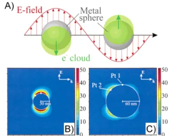
called localized surface plasmons, the nanoparticles strongly scatter light at a specific wavelength range. The plasmon oscillations obtained by solving Maxwell’s equations around metal nanoparticles are illustrated in Figure 5.81The sum of
light scattering and absorption is called extinction, and it is possible to observe the optical response of individual nanoparticles using dark-field microscopy.82 In biosensing applications, the attachment of an analyte to these nano-particles results in a refractive index change, causing a red or blue shift in the extinction peak wavelength,λmax. This shift in
λmax is given by the following equation 83 λ
Δ max ≅m nΔ [1−exp( 2 / )]− d ld (3)
where m is the sensitivity factor, Δn is the change in the refractive index, d is the effective adsorbate layer thickness, and ld is the electromagnetic field decay length. The extinction is maximized by optimizing the nanoparticle characteristics described by m and ld, and it is already well-established that
the extinction is a strong function of the nanometal’s type, size, shape, and orientation.84 Exploitation and design of these parameters will be essential for new effective LSPR applications.84
LSPR sensing experiments utilize a white light source covering the visible spectrum.85The scattered light is collected with a spectrometer, and changes in the spectra are then converted to binding data.80 In these experiments, light is directly coupled to the sample without requiring a prism or a grating, as in the SPR technique; therefore, the angle of incidence does not need to be precisely controlled. In contrast to the SPR technique, LSPR sensors based on nanoparticle surfaces are less sensitive to thermal variations.18a,65,86 In addition, with the availability of portable spectrometers, it is likely that LSPR applications can be translated for portable diagnostic applications.
3.4. Nanoplasmonic Arrays
Array-based nanoplasmonic detection is a plasmonic technique, similar to SPR imaging (SPRi) in the sense that it can be used for high-throughput biosensing applications. Various nano-plasmonic arrays have been demonstrated for refractive-index-based sensors, including nanoholes,87nanowells,88nanoposts,89 nanopillars,90nanorods,91nanodisks,92nanotubes,93and nano-pyramids.94These periodic arrays present high reproducibility in sensor fabrication, allow spectroscopic and intensity based measurements, and have a small footprint. A number of fabrication techniques are used for large-scale arrays, including focused ion beam milling and soft lithography techniques.95 However, they require expensive fabrication methods that are not suitable to create inexpensive devices.
Nanohole arrays are usually made from perforated metal films supported on substrates such as silicon nitrides. These arrays are based on the extraordinary optical transmission (EOT) effect96 and find applications as refractometric
Figure 5. Electric fields around nanoplasmonic silver particles. (A) Illustration of the plasmon oscillation and the electron cloud on metal spheres. (B) Electricfield contours of the main extinction peak 30 nm silver spheres in vacuum. Cross section of the sphere is shown with 369 nm light. (C) Electric field contours on 60 nm radius silver spheres in a vacuum. A cross section of the sphere is shown with 358 nm light and the field is from the quadropole peak. Adapted with permission from ref 81. Copyright 2003 American Chemical Society.
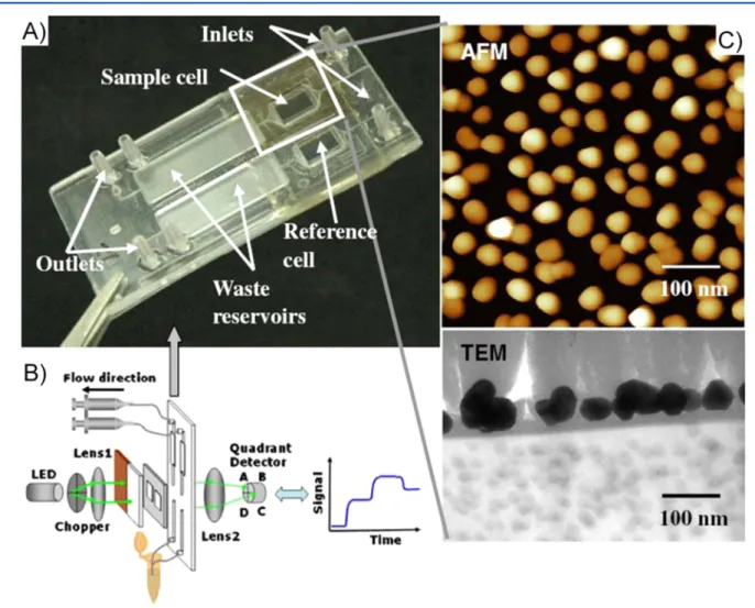
Figure 6.A nanohole array integrated withfluidics. The flow-through plasmonic nanostructure enabled local concentration of analytes. The method presented 100-fold concentration and simultaneous sensing of a protein. Further, the method presented 10-fold improvement in sensing speed in comparison to the control experiment with no analyte concentration. Reprinted with permission from ref 101a. Copyright 2012 American Chemical Society.
sensors.97 In nanohole arrays, localized and propagating SPR modes are intercoupled.98The resulting resonance modes are influenced by the periodicity, size, and shape of holes in the array and the composition of materials used in the sensor.97a,99 Extensive numerical calculations have been used to gain insight for the optimization of these parameters.100 These periodic arrays can be used in theflow-through geometry in biosensors, which enables concentrating analytes under applied electric fields (Figure 6).101
This operation mode lets analytes pass through the nanochannels, which are also forming the plasmonic structure of the sensor, and was shown to improve mass transport properties and response time.87
From a POC perspective, combination of array-based sensors with microfluidic chips can allow high-throughput sensors. Since each array can be interrogated separately and multiple arrays on parallel microchannels can be fabricated, nanohole array sensors are suitable for multiplexed on-chip detection. Light can be coupled directly to the sensor at or close to normal incidence, and the transmitted light can be interrogated with a CCD sensor.102The imaging mode permits use of high numerical aperture optics, allowing wide-field imaging from densely packed arrays.103 Microfluidic integrated nanohole arrays have been used for analyzing antibody−ligand binding kinetics104 and biomarkers.101b A recent application of nanoholes incorporated 50 microfluidic channels (30 μm width, over a 3.5× 2 mm area) and utilized high-throughput SPR imaging.105 The system was used for real-time affinity measurements.
Capture of intact viruses has been earlier shown from unprocessed whole blood on microchips.106 Nanoholes have potential to be used in optofluidic biosensor applications, as recently demonstrated in the detection of pseudoviruses (i.e., pseudotyped Ebola virus) and intact Vaccinia virus at 108pfu/
mL in PBS.107However, this platform needs to be expanded to detect viruses from bodily fluids at clinically relevant concentrations for diagnostic applications.
4. INTEGRATION OF PLASMONIC TECHNOLOGIES WITH MICROFLUIDICS
The integration of microfluidics and plasmonics brings the capability to build label-free and reliable biosensors on a LOC platform. Microfluidics has been widely used in cell separation and isolation and preparation, analysis, and delivery of samples.85,108 In particular, microfluidic technologies for blood cell separation and plasma isolation are significant for clinical applications.109 Integrated microfluidic devices with various fluid manipulators including pumps, mixing systems, separators, and valves have already been demonstrated.110 Complete processing and analysis of various bioagents have been shown on LOC microfluidic devices.111 For instance, DNA purification from bacteria on a single microfluidic chip has been shown without either pre- or post-sample processing.112
Combining microfluidics with optics provides a rapidly expanding range of applications by bringing advantages from thesefields. Microfluidic-based plasmonic devices are refractive index monitoring type devices. This class of devices can be considered as a subtype of optofluidics devices. On one hand, light can be used to direct the motion offluids in microfluidic chips. Optical-tweezers-based approaches have been used to build valves and pumps to induce flow in microfluidic channels.113 Blood cell separation has been demonstrated by utilizing optical lattices.114 On the other hand, fluids can be
used to alter optical parameters, as in plasmonics. Refractive index, absorption, polarization, and spectral properties can be monitored for various plasmonic biosensor designs. A further advantage of plasmonics is to allow label-free signal trans-duction method.
The two main optical biosensor design considerations are light−analyte interaction volume and sample delivery, which can seriously constrain the use of biosensors. The former consideration can be addressed by plasmonics taking advantage of the evanescent electromagnetic fields. For instance, the evanescent fields generated by the surface plasmon polaritons in SPR extend a few hundred nanometers into the fluid and allow low analyte densities to be detected. The latter consideration can be addressed by microfluidics, which can manipulate microliter quantities offluids.115 Further, selective delivery of analytes through multiple channels can be engineered for high-throughput microfluidic applications.
In plasmonic-based LOC biosensors, a recognition element is immobilized on the detection surface for label-free sensing. The choice of the recognition element depends on a number of factors, including the specificity and affinity toward the target molecule. Further, the complex formation between the recognition element and the target molecules should be stable in structure. Various surface functionalization techniques exist for the immobilization of these moieties for stable, high-density and efficient binding. We review these techniques in the following section.
4.1. Surface Functionalization
Biosensing platforms are comprised of a sensing support surface (e.g., gold and silver) and an immobilized biomolecular recognition element (e.g., antibodies, oligonucleic acids, peptide nucleic acids, peptides, and polymers).116The support surface enables the recognition element to be stable and allows them to interact with the target analytes. Depending on the sensitivity, specificity, and limit of detection, the recognition element is immobilized through several surface techniques, including physical adsorption, chemical adsorption, covalent binding, and affinity-based interactions.116a Besides these common methods, recent advances in surface functionalization and antifouling agents to minimize nonspecific binding are also reviewed in the following subsections.
4.1.1. Physical Adsorption. Physical adsorption technique utilizes the surface characteristics and surface charge to attach and immobilize biorecognition elements onto the surface and relies on nonspecific physical interactions between the recognition element and the support material.117 In contrast to chemical binding techniques, this method also holds a key advantage since it does not require any reagent to activate chemical groups on the surface, and thus, this technique is easy to perform, inexpensive, and reduces structural damage in biorecognition elements. The physical adsorption method particularly utilizes hydrogen bonding and van der Waals forces.118These weak interactions also allow the biorecognition elements to easily detach from the surface, and thus, the biosensing surface can be used multiple times. However, nonspecific physical interactions are closely affected by environmental changes, including temperature, ionic content, and pH. On the other hand, this technique causes nonspecific binding of other molecules and substances, resulting in a significant decrease in surface coverage of the recognition elements and sensor specificity.
The support materials can be modified to generate surface charge and reactive groups using oxidizing techniques such as oxygen plasma treatment. Oxygen-plasma-treated and un-treated polystyrene (PS) slides have been recently used as sensor substrates to detect breast cancer type 1 (BRCA1) gene mutations, and these two cases were compared in terms of uniform immobilization and binding capacity of a biorecogni-tion element (i.e., oligonucleotide−protein conjugate).119 On plasma-treated slides, the binding amount of oligonucleotide− protein conjugates significantly increased compared to un-treated slides.119Another interesting observation in this study was that plasma treatment amplified the surface area and formed nanoroughened structures that could facilitate detection of a low amount of target analyte and improve the analytical performance of the biosensing surface in microarray applications.119Although oxygen plasma treatment is a simple and effective method for many surfaces, it often causes significant damage on the biosensing support surface.120This major obstacle leads to permanent surface disruptions, which interfere with the sensor surface structure and reduce sensitivity.120 On the other hand, the surface characteristics (e.g., hydrophobicity and polarity) and the functional groups of biomolecules determine the molecular interactions for bio-molecule immobilization. Although, in some cases, the orientation of the recognition element is not critical to capture the target analyte, these molecular changes on the surface can affect biomolecular activity (e.g., denaturation of proteins) and orientation of proteins and antibodies due to the restrictions in their conformationalflexibility.121
4.1.2. Chemical Adsorption and Covalent Binding. Chemical adsorption and covalent binding techniques are most frequently combined to form chemical coupling and bond formation between support surface and biorecognition elements in three main steps: (i) support surface activation, (ii) functional group generation, and (iii) biomolecule immobilization.116a The self-assembled monolayer (SAM) technique, one of the most common chemical adsorption techniques (i.e., chemisorption), spontaneously generates self-formation of molecular assemblies on substrates.122 N-alkylthiols or disulfides are the most common SAM molecules, consisting of an alkyl backbone chain, thiol head, and functional tail groups.116a,123On these molecules, thiol head groups have strong affinity to bind to metal surfaces (e.g., gold and silver), and the alkyl backbone tethers the biomolecules from the substrate. The latter group presents a functional end to interact and covalently bind to biomolecules.116a Coupling reactions ( e . g . , N h y d r o x y s u c c i n i m i d e ( N H S ) a n d e t h y l -(dimethylaminopropyl)carbodiimide (EDC)) are the most common biomolecule immobilization methods that typically form succinimide groups that interact with amine groups of organic molecules (e.g., antibody, protein, nucleic acids, and amine-modified lipids).124
By utilizing covalent bonding, modified SAM agents (e.g., 11-mercaptoundecylamine (MUAM) and dithiobis(N-succinimidyl propionate) (DTSP)) were previously used to immobilize double-stranded DNA, peptide nucleic acid (PNA), and miRNA on SPR gold sensors for the detection of nucleic acids.125 Other than the SAM mechanism, biomolecules can be immobilized through silanization agents (e.g., (3-aminopropyl)triethoxysilane (APTES) and (3-aminopropyl)trimethoxysilane −tetramethox-ysilane (MPTMS or 3-MPS)) that cover a biosensing surface (e.g., glass, mica, metal oxides, and silica) with functional alkoxysilane molecules by forming a covalent Si−O−Si
bond.108c,126 This process can also be coupled with another reaction as performed in SAM modifications.108c,121,127On the same platform, long- and short-chained SAMs can also be utilized to block the surface from nonspecific binding.128 Further, long-chained SAMs can be used to construct artificial lipid bilayer systems using hydrophobic interactions between alkyl backbones and lipid tails.129For instance, artificial lipid bilayers are constructed by the rupture of liposomes, and self-assembled hexadecane monolayer surface assists to rupture liposomes for the formation of lipid bilayers on gold surfaces.129 Thus, self-assembled hexadecane monolayer surface provides a dynamic and stable structure to tether lipid bilayer and allows for further biomolecular analyses such as polymer−lipid bilayer interaction in vitro conditions.129 Additionally, SAMs can be used to immobilize protein conjugate, oligonucleic acids, and peptide nucleic acids for microarray analysis.130However, there are some limitations in SAM formation, including availability of substrate, low number of organic molecules for monolayer formation, the choice of anchoring groups, and limited solubility of monolayer molecules.122 Additionally, bulky monolayer molecules result in large defects in monolayer structure and lack of thermal and oxidative stability restricting their large-scale use in detection platforms.122
4.1.3. Affinity-Based Interactions. Affinity-based surface functionalization techniques address some of the current challenges in biomolecule immobilization methods mentioned above. Avidin−biotin-based interactions are commonly used to immobilize biomolecules (e.g., nucleic acids, proteins, and antibodies) on the biosensing surface without interfering with their biomolecular structure and function.106b For instance, NeutrAvidin and streptavidin are well-known members of avidin proteins, and they have high association capacity to biotinylated molecules such as antibodies, nucleic acids, peptides, and PNA. An interesting example for biotin −avidin-based surface functionalization is traptavidin, which is an engineered mutant version of streptavidin protein according to biotin−4-fluorescein dissociation rate.131 This mutant avidin protein has lowerflexibility in the biotin-binding pocket, and this structural property reduces the entropic energy required for biotin binding.131 Thus, traptavidin structurally inhibits the dissociation rate and enhances thermostability compared to native avidin proteins. Traptavidin is also a versatile protein that can bind to a range of biotin conjugates (i.e., biotin −4-fluorescein, biotin−amidocaproyl-BSA and biotinylated DNA (internal and terminal)).131 Therefore, this protein holds a great potential to replace other avidin-based proteins in nanoplasmonic detection platforms, molecular anchored arrays, and POC diagnostic technology platforms.131 However, the biotinylation site is a critical parameter for biomolecule (e.g., antibody) orientation in avidin−biotin-based surface chem-istries. Two groups of affinity-based surface chemistries (i.e., protein G- and NeutrAvidin-based) were evaluated, and the observations obtained from AFM demonstrate that protein G-based surface chemistry can efficiently immobilize the antibod-ies with their favorable orientation in microfluidic channels.106b Since protein G has a specific binding site for the fragment crystallizable region (Fc) of antibodies, it provides better control over antibody orientation.106bTo increase the number of antibody binding sites and stability, immunoglobin specific proteins are engineered using recombinant DNA technology. Protein A/G is a notable example of the recombinant antibody immobilization molecules that combines IgG binding domains of both protein A and protein G. This recombinant fusion
protein is comprised of four Fc binding domains from protein A and two from protein G, and it is more stable to pH changes compared to protein A.107,132 Overall, affinity-based surface functionalization methods increase sensitivity and capture efficiency and improve the detection limit to capture target molecules/bioagents by utilizing high binding affinity and controlling molecular orientation. Additionally, oligonucleotide immobilization for nucleic acid hybridization studies and histidine-chelated metal ion methods for protein-based detection are widely used in the immobilization of biorecognition elements.133 There are also new surface functionalization methods, including polymeric coating, lipid bilayer construction, PNA, and aptamer immobilization, to capture target analytes in POC and primary care diagnostics for various applications ranging from early cancer detection diagnosis and monitoring of infectious diseases.
4.2. Blocking of Nonspecific Binding
Nonspecific binding to biosensing surfaces is one of the major drawbacks for specific capture and quantitative analysis.134One of the challenges is the concentration of other substances being higher than target analyte since these substances can also bind/ attach to the biosensing area.135 Although the binding characteristics of nonspecific interactions is much different than that for a specific binding event, nonspecific interactions and binding still poses a significant bottleneck for limit-of-detection in biosensors. Further, nonspecific binding can occur at functionalized, passivated, and untreated regions of the biosensing area.135 Thus, these nonspecific interactions can decrease detection sensitivity. There are several antifouling agents (e.g., chemical, protein based, and polymeric agents) used to address these challenges by improving the specificity.
Thiol compounds have been commonly used as chemical blocking agents on metal surfaces. The length and terminal group of thiol compounds affect the sensitivity and detection limit.136To evaluate these parameters, a number of alkanethiol SAMs (i.e., 3-mercapto-1-propanol (3-MPL), 6-mercapto-1-hexanol (6-MHL), 8-mercapto-1-octanol (8-MOL), 9-mercap-to-1-nonanol (9-MNL), 11-mercapto-1-undecanol (11-MUL) and another blocking thiol (C11) with a −CH3 terminating
headgroup, and 1-dodecanethiol (1-DDT)) was used for the detection of the target DNA sequences using pyrrolidinyl peptide nucleotide acid (acpcPNA) probes that were immobilized via a spacer molecule.136 The blocking thiol compound with same length (9-MNL) as the total spacer molecule provided the highest sensitivity [20.4± 0.7 nF cm−2 (log M)−1] compared to the other thiol blocking agents with shorter and longer length.136 This specific length possibly arranged more favorable hybridization, resulting in the highest hybridization efficiency, whereas the blocking agent with longer length overlapped with the probe.136The terminal groups (i.e., −OH and −CH3) of thiol blocking agents were also evaluated
on the same platform, and the hydroxyl-terminated agent provided a slightly better sensitivity by increasing hydrophilicity for DNA immobilization and hybridization.136 Proteins (e.g., bovine serum albumin, casein, glycine, and gelatin) have been also used to protect the biosensing surface from nonspecific interactions. Instantized dry milk, casein, gelatins from pig and fish skin, and serum albumin were evaluated to understand the blocking capabilities, and casein and instantized milk were observed to inhibit nonspecific binding.137
In this study, porcine skin gelatin was observed to be the least effective antifouling agent.137Overall, the critical parameter for protein
blocking experiments is that blocking agent (e.g., casein) primarily interacts with the biosensing area instead of blocking the protein−protein interactions observed in porcine skin gelatin experiments.137However, the efficiency of these natural blocking agents (e.g., albumin, casein, and glycine) is not considerably satisfactory.138 In contrast, polymeric blocking agents are easily reproducible and can be modified to increase the specificity of blocking.138Polyethylene glycol (PEG) is one of the most common polymeric blocking agents, and a densely packed PEG tethered-chain surface allows one to minimize nonspecific binding.138,139 The combination of long and short PEG chains significantly reduces biofouling on the biosensing surface and increases the sensitivity.138,140Factor IX (FIX) was immobilized on glutaraldehyde-activated surface and detected via its aptamer.138A copolymer (i.e., poly(ethylene glycol)-b-poly(acrylic acid) (PEG-b-PAAc)) was used as a blocking agent to reduce nonspecific binding on untreated and glutaraldehyde-activated regions.138The limit of detection was observed to be down to 100 pM.138 The sensitivity was further improved by using dual polymers (i.e., PEG-b-PAAc and pentaethylenehex-amine-terminated PEG (N6-PEG)) on the same platform, and 1000-fold better sensitivity (100 fM) was achieved with respect to the blocking with PEG-b-PAAc.138 Here, the use of dual polymers demonstrates higher sensitivity and reliability for the biosensing platforms that detect a very small amount of target molecules from complex fluids such as whole blood.138Apart from these surface modifications, the generation of nanorough surfaces allows one to prevent the bacterial attachment on biosensing surface.141
4.3. Recent Advances in Surface Functionalization
Within the past decade, conventional surface functionalization methods have been replaced with new surface modification materials (e.g., lipids and polymers) and techniques (e.g., thiol exchange and site-specific functionalization). These innovations improve the molecular interactions between target molecule and the receptor of interest, and they enable more stable structures for biosensing platforms.142For instance, receptors (e.g., integral proteins) incorporated with cellular membranes require hydrophobic content to conserve their native structure and function in biosensing platforms.142cDynamic,flexible, and complex nature of the cellular membranes is an attractive candidate to support biorecognition elements such as receptors for these platforms.142cNoncovalent assembly of lipid bilayers on biosensing surfaces combines highly sophisticated surface modification mechanisms with a nature-synthesized functional platform.142cThe arrangements of lipid bilayers also allow one to monitor membrane-associated molecular recognition events on the close vicinity of the membrane using surface sensitive tools such as LSPR and SPR.142cTo construct lipid bilayers on biosensing platforms, specific surface immobilization strategies are employed using tethering agents that rupture lipid vesicles to form a planar lipid bilayer.142b,143This strategy can be done by utilizing a thin layer of SiO2 on plasmonic substrate.144
Other construction strategies are the use of thiolated lipids that covalently immobilize lipid membrane to metal substrate and utilization of a polymer cushion that tethers lipid membrane and forms an ionic reservoir for functional integration of receptors.145
Chemical modification and physical properties of lipid molecules have allowed lipid bilayers to be used in surface sensitive biosensing approaches, including the detection of biomolecules, proteins, and nucleic acids. Biotinylated-lipid
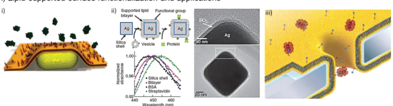
Figure 7.Recent advances in surface functionalization methods and materials. (A) Lipid-supported surface functionalization and applications: (i) Schematic of a gold nanorod coated with a biotinylated lipid membrane and interactions with streptavidin. Adapted with permission from ref 146. Copyright 2008 American Chemical Society. (ii) Schematic of supported lipid-bilayer-coated core shell nanocubes (Ag@SiO2). TEM images of
whole and select region of a Ag@SiO2nanocube. A solution-phase plasmonic sensor measures LSPR spectra of surface modification and coating on
Ag@SiO2core shell nanocubes using a standard laboratory spectroscopy. Adapted with permission from ref 147. Copyright 2012 Nature Publishing
Group. (iii) Schematic of lipid-bilayer-coated nanopore in silicon nitride substrate. This platform facilitates mobility of target molecules and minimizes clogging. Adapted with permission from ref 150. Copyright 2011 Nature Publishing Group. (B) Lipid-supported surface functionalization and applications: (i) The procedure of a gold-nanoparticle-hybridized polymerfilm for biosensing applications. Adapted with permission from ref 151. Copyright 2009 Wiley-VCH Verlag GmbH & Co. KGaA. (ii) SPR-based biosensing platform with embedded indium tin oxide microheater and rapid tuning of SPR signal using a thermoresponsive polymer (i.e., pNIPAAm). Reprinted with permission from ref 152. Copyright 2013 American Chemical Society. (C) Surface functionalization on patterned surfaces: (i) Schematic and scanning electron microscopy (SEM) image of the patterned nanostructures (i.e., nanoholes). (ii) SEM top-view image of nanholes and extinction peak values of LSPR spectra for surface
membranes were immobilized to detect streptavidin molecules on the surface of gold nanorods by monitoring the spectral shifts using a fast single particle spectroscopy (fastSPS) instrument coupled with dark-field microscopy (Figure 7A-A(i)).146 The binding of streptavidin molecules to a single nanorod resulted in a median shift of 2.9± 1.8 nm when 29 nanorods were analyzed.146 Thus, on this platform, local interactions of proteins with cellular membranes could be monitored in real-time.146Another interesting example was to assess the binding of target proteins to supported lipid-membrane-coated nanocubes (Figure 7A(ii)).147 Here, the researchers reported a solution-phase plasmonic sensor method that utilizes LSPR spectra of Ag@SiO2 core shell nanocubes
using spectroscopic measurements (Figure 7A(ii)).147 In this work, supported lipid bilayers were spontaneously formed by mixing Ag@SiO2core shell nanocubes in lipid-vesicle solution, and the plasmonic response of the platform was calibrated by examining the binding of streptavidin to biotinylated lipid molecules in the supported membrane. LSPR response was then converted to protein coverage on the nanocube surface by utilizing the LSPR shifts to protein mass change, and the limit of detection was reported as 0.191 ng/mm2 nm.147 Further, cellular-membrane-associated molecular interactions were assessed on the supported lipid-bilayer-modified gold nano-particles using a single nanoparticle tracking-based detection method.148 The binding and molecular interactions of membrane-associated molecules (i.e., cholera toxin B subunit and ganglioside GM1 pentasaccharide head-groups) were evaluated using the diffusion coefficients of gold nanoparticles on the membrane.148 The limit of detection for the cholera toxin B subunit was observed to be down to 10 pM, resulting in 100-fold improvement in the sensitivity compared to fluorophore-based methods.148
Thus, an ultrasensitive detec-tion platform was developed by utilizing the mobility of lipid molecules in membranes.148 Additionally, supported lipid membranes were employed on nanopore assays for the detection of small molecules (e.g., proteins) and the monitoring of DNA hybridization and receptor−target interactions.142c,149 The integration of lipid bilayers with nanohole platforms facilitated more frequent translocation/mobility of target molecules and, thus, introduced chemical sensitivity and avoided clogging, which are major obstacles in biosensing assays (Figure 7A(iii)).142c,150 Overall, lipid-bilayer-incorpo-rated biosensing platforms improve the molecular interactions and form a support layer for the integration of biorecognition elements. Thus, this new surface functionalization strategy can be used for membrane-associated molecule biosensors and toxin/drug screening assays in the future.
Polymer-mediated surface functionalization is another interesting strategy to generate a support layer for the immobilization of biorecognition elements. Polymers play a vital role to enhance the reliability and sensitivity of biosensors, and they are often used for hybridization with plasmonic nanoparticles (Figure 7B(i)).151 For instance, a polymer-assisted plasmonic sensor was developed by hybridizing polyelectrolyte multilayers (PEMs) with gold nanoparticles to real-time monitor the binding of antigen−antibody on
plasmonic sensors (Figure 7B(i)).151 This hybrid film presented a stable and reliable nanoporous structure under physiological conditions and enhanced the surface area for bioconjugation and recognition.151Further, PEMs exhibited an antifouling property to prevent nonspecific binding of proteins and cells that enhanced detection sensitivity.151 To utilize dynamic structure of polymers, thermoresponsive poly(N-isopropylacrylamide) (pNIPAAm)-based hydrogel was imple-mented as SPR sensors for rapid tuning of SPR signal (Figure 7B(ii)).152 Here, an indium tin oxide microheater was embedded under the SPR sensor, and thus, rapid thermal response (i.e., swelling and collapse) of pNIPAAm was evaluated (Figure 7B(ii)).152 Thermal response of pNIPAAm led to large refractive index changes and a high thermo-optical coefficient of dn/dT = 2 × 10⁻²RIU/K.152Further, polymers can be modified with biorecognition elements for specific capture of target molecules, and thus, a 3D binding matrix can be developed for biosensing applications by utilizing dynamic and functional structure of polymers.152,153,154
Recently, the researchers have taken advantage of the plasmonic surface geometry for surface functionalization. Patterned nanoplasmonic structures with specialized geo-metries (e.g., holes and edges) exhibit a potential to be used for site-specific surface modifications that can increase the utility and specificity of nanoplasmonic platforms.142b Partic-ularly, nanoplasmonic platforms employ noble metal surfaces (e.g., gold and silver) that can be modified using thiol chemistry to immobilize biorecognition elements.155Thiol chemistry also presents a broad range of variety in length, saturation degree, and terminal groups to preferably immobilize recognition elements (e.g., proteins, nucleotides, and carbohydrates) in plasmonically active zones.142b,156The thiol exchange process is an interesting strategy to selectively immobilize antibodies on the edges of triangular gold nanoplates that are used for LSPR sensing platform.142a The thiols located on the edges of the nanoplates are more attractive to exchange with the thiols in solution than the ones located on the flat surfaces of the nanoplates due to decreased steric hindrance at high-curvature sites.142b,157 Other than patterned nanoplasmonic structures, hybrid noble metal layers can also be selectively functionalized for biosensing platforms (Figure 7C). Recently, nanohole arrays consisting of TiO2/Au/TiO2 films were specifically modified
with poly-L-lysine−poly(ethylene glycol) (PLL−PEG) and
thiolated PEG (HS-PEG) molecules (Figure 7C(i,ii)).158 PLL−PEG selectively adsorbed to the TiO2 layers (i.e., top and bottom layers), and HS-PEG covalently bound to gold layer (i.e., intersectional layer) (Figure 7C(iii)).158 HS-PEG molecules were then functionalized with biotin for the selective detection of avidin on the hole sidewalls (Figure 7C(iii)).158 This site-specific functionalization mechanism enabled the increase of the signal change per unit time for avidin−biotin binding nearly 20-fold (Figure 7C(iii)).158 In the future, this functionalization strategy will play a key role for the improvement of sensitivity and the development of multiplex assays by enabling specific modifications on plasmonically active sites. Overall, surface functionalization is one of the key
Figure7. continued
modifications. (iii) Nanoholes arrays consisting of TiO2/Au/TiO2films are specifically modified with poly-L-lysine−poly(ethylene glycol) (PLL−
PEG) and thiolated PEG (HS-PEG) molecules for site-specific surface functionalization. Adapted with permission from ref 158. Copyright 2010 American Chemical Society.
parameters to develop a sensitive, reliable, and accurate biosensing platform.
5. APPLICATIONS OF PLASMONIC-BASED
TECHNOLOGIES FOR POC: SPR, LSPR, AND SPRi
5.1. SPR
SPR has been used in a broad range of biosensing applications, including detection of bacteria, viruses, eukaryotic cells, nucleic acids, peptide nucleic acids, proteins, and drugs, and in monitoring of biomolecular interactions such as nucleic acid hybridization or protein−ligand interaction.55
Another important potential clinical application of SPR is in cancer diagnosis. Cancer is a significant problem both in the developed and developing world.159 In 2008, ∼12.7 million cancer cases and 7.6 million cancer deaths occurred worldwide, and 56% of the cases and 64% of the deaths were reported in developing countries.159 Although overall cancer incidence rates in developed countries are higher than those observed in developing countries, cancer mortality rates are usually similar between developed and developing countries.159,160 Some of the most critical cancers in the developing world are female breast (27.3%), stomach (15.3%), lung (19.1%), colorectal (10.7%), and cervical (17.8%) cancers.161 Early detection of cancer is a critical need in medicine, especially for cancer types such as breast, cervical, ovarian, and colorectal cancers.160,162 Rapid available technologies to monitor early cancer markers supported by our advanced understanding of cancer and discovery of specific biomarkers will enhance the capabilities in cancer detection. Detection platform technologies could serve diverse clinical needs in early cancer detection and diagnosis (for instance by detecting circulating tumor cells163) or monitoring cancer treatment. In addition, these platforms
could be inexpensive, rapid, portable, and easy to operate in developing countries as well as in developed settings, creating potentially broad screening tools for applicable cancer types. From a diagnostic perspective, detection of circulating biomarkers for cancer diagnosis is an interesting application of SPR-based detection platforms. For instance, cytokine interleukin-8 (IL-8) plays a crucial role in human cancer.164 The differentiations in IL-8 expression level result in multiple human cancers, such as breast cancer, Hodgkin’s lymphoma, and prostate cancer.164IL-8 concentration in saliva was shown to be elevated in oropharyngeal squamous cell carcinoma (OSCC) patients.165 To detect the IL-8 concentrations in human saliva, a microfluidic SPR-based immunoassay platform was developed.166 For this experiment, two monoclonal antibodies were used as a sandwich assay to detect different epitopes on the antigen (IL-8) in either buffer or saliva samples. This platform presented a 250 pM limit-of-detection in saliva environment.166IL-8 levels in healthy individuals saliva are 30 pM whereas the levels in oral cancer patients’ saliva are 86 pM.166 By preconcentrating the saliva in sample preparation steps, the system could potentially be used in diagnostics as well.
Another attractive biomarker detection experiment was performed for prostate-specific antigen (PSA). The increase in the levels of PSA (>4 ng/mL) in patient samples is one of the symptoms for possible prostate malignancy.167 On the other hand, PSA has been reported as a potential marker for breast cancer in women.168 To detect PSA levels a sandwich bioassay was developed.169Basically, anti-PSA antibodies were immobilized on the Au layer of the sensor surface. After the sampling, Au nanoparticles coated with a secondary antibody were applied to increase the SPR signal levels. In one
Figure 8.Portable SPR biosensor platforms. (A) A multichannel cartridge to be used for on-site antibiotic detection in milk samples. Reprinted with permission from ref 171. Copyright 2010 Elsevier. (B) Portable SPR biosensor prototype to be used with microfluidic chips. Reprinted with permission from ref 172. Copyright 2009 Elsevier. (C) Portable microfluidic-based device for cardiac marker detection. Reprinted with permission from ref 173. Copyright 2006 American Chemical Society.