triggers degradation by the Hrd1 ubiquitin ligase
The MIT Faculty has made this article openly available.
Please share
how this access benefits you. Your story matters.
Citation
Rubenstein, E. M. et al. “Aberrant Substrate Engagement of the
ER Translocon Triggers Degradation by the Hrd1 Ubiquitin Ligase.”
The Journal of Cell Biology 197.6 (2012): 761–773. © 2012 by The
Rockefeller University Press
As Published
http://dx.doi.org/10.1083/jcb.201203061
Publisher
Rockefeller University Press, The
Version
Final published version
Citable link
http://hdl.handle.net/1721.1/74669
Terms of Use
Creative Commons Attribution-Noncommercial-Share Alike 3.0
Unported
The Rockefeller University Press $30.00
Correspondence to Mark Hochstrasser: mark.hochstrasser@yale.edu
S.G. Kreft’s present address is Dept. of Biology, University of Konstanz, 78457 Konstanz, Germany.
W. Greenblatt’s present address is Harvard-MIT Division of Health Sciences and Technology, Harvard Medical School, Boston, MA 02115.
Abbreviations used in this paper: ERAD, ER-associated degradation; PTM, post-translational modification; PTT, postpost-translational translocation; TM, transmem-brane; WT, wild type.
Introduction
The ubiquitin–proteasome system accounts for a major
por-tion of eukaryotic protein degradapor-tion. Proteins destined for
proteolysis by this pathway are covalently modified with
chains of the protein ubiquitin through the sequential action
of ubiquitin-activating enzymes (E1s), ubiquitin-conjugating
enzymes (E2s), and ubiquitin ligases (E3s; Ciechanover et al.,
1980; Scheffner et al., 1995). Ubiquitylated substrates are
degraded by the 26S proteasome (Ravid and Hochstrasser, 2008).
In the yeast Saccharomyces cerevisiae, the transmembrane (TM)
RING domain proteins Hrd1 and Doa10 are the predominant
ER-associated degradation (ERAD) E3s that target misfolded
and regulatory proteins for proteolysis (Hampton et al., 1996;
Plemper et al., 1999; Wilhovsky et al., 2000; Bays et al., 2001;
Swanson et al., 2001). With a few exceptions, Hrd1 and Doa10
recognize unique substrate classes depending on degron
dis-position relative to the ER membrane (Vashist and Ng, 2004;
Carvalho et al., 2006). Specifically, Hrd1 recognizes proteins
with ER luminal or intramembrane degrons, called ERAD-L
(lumen) and ERAD-M (membrane) substrates, respectively
(Carvalho et al., 2006; Gauss et al., 2006; Sato et al., 2009).
In contrast, Doa10 promotes degradation of proteins with
cyto-plasmically disposed degrons, ERAD-C (cytoplasm) substrates
(Huyer et al., 2004; Ravid et al., 2006; Metzger et al., 2008).
Hrd1 and Doa10 exhibit overlapping E2 requirements. Hrd1
mediates ubiquitin transfer from Ubc7 and, to a lesser extent,
Ubc1 (Plemper et al., 1999; Bays et al., 2001), whereas Doa10
functions with Ubc6 and Ubc7 (Swanson et al., 2001).
The prototypical Doa10 substrate is the transcriptional
repressor MAT2, the proteolytic control of which is critical
for regulating the yeast-mating phenotype (Swanson et al.,
2001; Laney and Hochstrasser, 2003). The N-terminal 67
resi-dues of MAT2, Deg1, contain a degron for Doa10-dependent
degradation (Hochstrasser and Varshavsky, 1990; Johnson
et al., 1998; Swanson et al., 2001). Importantly, Deg1 confers
Doa10-dependent instability when fused to the cytoplasmic
N termini of heterologous soluble proteins (Hochstrasser and
Varshavsky, 1990; Chen et al., 1993; Johnson et al., 1998;
L
ittle is known about quality control of proteins that
aberrantly or persistently engage the endoplasmic
reticulum (ER)-localized translocon en route to
mem-brane localization or the secretory pathway. Hrd1 and
Doa10, the primary ubiquitin ligases that function in
ER-associated degradation (ERAD) in yeast, target
dis-tinct subsets of misfolded or otherwise abnormal proteins
based primarily on degradation signal (degron)
loca-tion. We report the surprising observation that fusing
Deg1, a cytoplasmic degron normally recognized by
Doa10, to the Sec62 membrane protein rendered the
protein a Hrd1 substrate. Hrd1-dependent
degrada-tion occurred when Deg1-Sec62 aberrantly engaged
the Sec61 translocon channel and underwent
topologi-cal rearrangement. Mutations that prevent translocon
engagement caused a reversion to Doa10-dependent
degradation. Similarly, a variant of apolipoprotein B,
a protein known to be cotranslocationally targeted for
proteasomal degradation, was also a Hrd1 substrate.
Hrd1 therefore likely plays a general role in targeting
proteins that persistently associate with and potentially
obstruct the translocon.
Aberrant substrate engagement of the ER translocon
triggers degradation by the Hrd1 ubiquitin ligase
Eric M. Rubenstein,
1Stefan G. Kreft,
1Wesley Greenblatt,
1Robert Swanson,
2and Mark Hochstrasser
11Department of Molecular Biophysics and Biochemistry, Yale University, New Haven, CT 06520 2Department of Biology, Valparaiso University, Valparaiso, IN 46383
© 2012 Rubenstein et al. This article is distributed under the terms of an Attribution– Noncommercial–Share Alike–No Mirror Sites license for the first six months after the pub-lication date (see http://www.rupress.org/terms). After six months it is available under a Creative Commons License (Attribution–Noncommercial–Share Alike 3.0 Unported license, as described at http://creativecommons.org/licenses/by-nc-sa/3.0/).
THE
JOURNAL
OF
CELL
BIOLOGY
on October 2, 2012
jcb.rupress.org
Downloaded from
http://jcb.rupress.org/content/suppl/2012/06/07/jcb.201203061.DC1.html Supplemental Material can be found at:Ravid et al., 2006; Scott and Schekman, 2008). As expected
for a Doa10 substrate, Deg1-Sec62 is stabilized in cells
lack-ing both Ubc6 and Ubc7 (Mayer et al., 1998). Because
elimi-nating Ubc7 inactivates both Doa10 and Hrd1 pathways,
however, it remained formally possible that Deg1-Sec62 is a
Hrd1 substrate. We show that this is indeed the case and that
aberrant engagement of the translocon by Deg1-Sec62 is
respon-sible for this E3 switch. This engagement triggers a topological
Swanson et al., 2001). Besides soluble Deg1 fusions, we have
fused Deg1 to the ER-resident TM protein Vma12 to study
Doa10-dependent ERAD (Ravid et al., 2006). Deg1 has also
been fused to the topologically similar ER protein Sec62
(Mayer et al., 1998). Like Vma12, Sec62 has two TM
seg-ments, with both protein termini on the cytoplasmic side of the
membrane (Deshaies and Schekman, 1990). Attachment of
Deg1
destabilizes both Sec62 and Vma12 (Mayer et al., 1998;
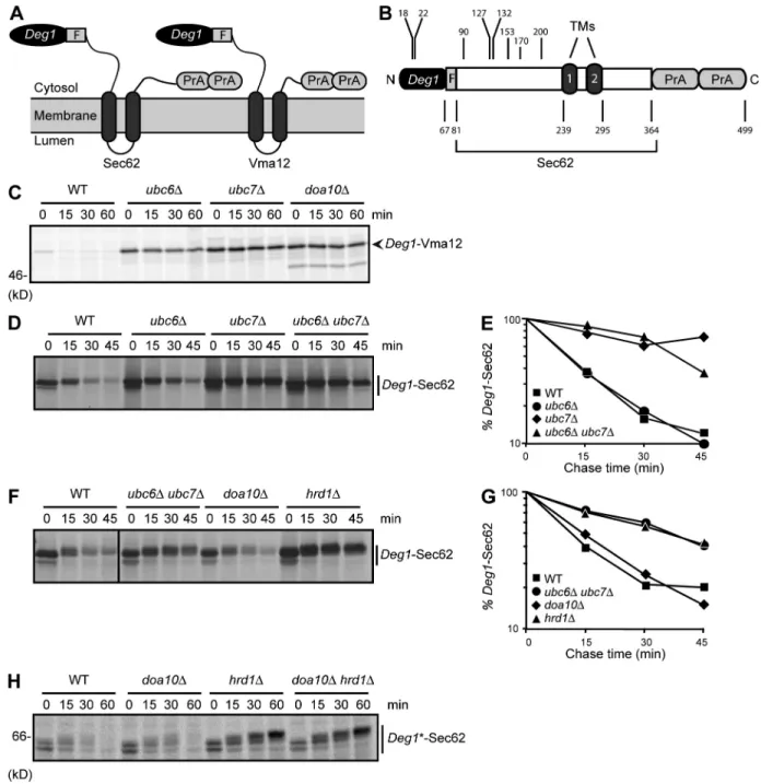
Figure 1. Deg1-Sec62 is a Hrd1 substrate. (A) Schematic diagram of Deg1 fusion proteins (with predicted topologies) used in this study. For each con-struct, the N-terminal Deg1 is followed, in sequence, by the Flag (F) epitope, the 2-TM ER protein (Sec62 or Vma12), and two copies of the Protein A (PrA) tag. For clarity, the fusion proteins are referred to as Deg1-Sec62 or Deg1-Vma12. Any additional alterations to protein sequence will be noted. (B) Linear representation of Deg1-Sec62, drawn to scale. Positions of boundaries between elements within the fusion protein and residue numbers mentioned in the text are highlighted. (C) Pulse-chase analysis of Deg1-Vma12 in the indicated yeast strains. (D and F) Pulse-chase analysis of Deg1-Sec62 in the indicated yeast strains. Molecular weight markers for these autoradiographs are not available; however, migration of the same protein can be seen in Fig. 4 B. (E and G) Quantification of autoradiographs in D and F. Data are representative of three experiments. The black line indicates that intervening lanes have been spliced out. (H) Pulse-chase analysis of Deg1*-Sec62 in the indicated yeast strains. Deg1 fusions in C, D, and F were precipitated with anti-Deg1 antibodies. Deg1*-Sec62 in H was precipitated with anti-Flag antibodies. Cycloheximide was included in the chase depicted in H. Deg1*, F18S/I22T double mutant.
on October 2, 2012
jcb.rupress.org
apparent exception to the division of ERAD-L/M and ERAD-C
substrates between Hrd1 and Doa10, respectively (Vashist and
Ng, 2004; Carvalho et al., 2006).
Doa10-mediated degradation of Deg1-harboring proteins
depends on the hydrophobic face of a predicted amphipathic
helix within Deg1 (Johnson et al., 1998). When mutations in
this helix that stabilize Deg1-containing Doa10 substrates were
introduced into Deg1-Sec62, the resultant protein
(Deg1*-Sec62) was still degraded in a Hrd1-dependent manner (Fig. 1 H),
unlike Deg1*-Vma12, which is strongly stabilized (Ravid
et al., 2006). Thus, the details underlying molecular
recog-nition of Deg1-containing substrates differ between the two
ERAD E3s.
Posttranslational modification (PTM) of Deg1-Sec62
Deg1
-Sec62 migrated as multiple species by SDS-PAGE and
became increasingly modified with time after its synthesis
(Fig. 1, D and F). It was previously noted that Deg1-Sec62 is
N-glycosylated, in contrast to wild-type (WT) Sec62, which
is not typically posttranslationally modified (Kim et al., 2006;
Scott and Schekman, 2008). Immediately after radiolabeling,
Deg1
-Sec62 migrated as two distinct species (Fig. 2 A, lane 1).
After treatment with Endoglycosidase H (Endo H), which
re-moves all N-glycans from yeast proteins, the slower-migrating
band collapsed to the faster migrating species (lane 2).
Mu-tational analysis revealed that N153 was the major site of
N-glycosylation (with a possible minor contribution by N90;
lanes 5–12), which is consistent with an earlier study (Scott
and Schekman, 2008). After 60 min, the higher mass species
appeared as multiple diffuse bands (lane 3). Endo H treatment
only partially increased the mobility of these poorly resolved
bands (lane 4), which indicates additional PTM.
Deg1
-Sec62 is also modified by O-mannosylation, an
ER-specific modification with a role in the ERAD of some Hrd1
substrates (Vashist et al., 2001; Hirayama et al., 2008). We
per-formed pulse-chase analysis of doa10 hrd1 yeast expressing
Deg1
-Sec62-N90D/N153D cultured in the presence of a global
change in Deg1-Sec62 that is closely associated with
Hrd1-dependent degradation.
We extended our investigation to a known
cotranslo-cationally degraded protein. Apolipoprotein B (apoB), the
major protein component of low- and very-low-density
lipo-proteins, stalls in the translocon if it cannot associate with
its lipid ligand and is subsequently degraded by the
pro-teasome (Yeung et al., 1996; Fisher et al., 1997; Pariyarath
et al., 2001). We found that, like Deg1-Sec62, an apoB variant
expressed in yeast that is also associated with the translocon
(Hrizo et al., 2007) is targeted by the Hrd1 ubiquitin ligase.
These results suggest that Hrd1 may play a general role in the
degradation of proteins that aberrantly or persistently engage
the translocon.
Results
Deg1-Sec62 is a Hrd1 substrate
We used pulse-chase analysis to examine the metabolic
stabil-ity of the ER-resident TM proteins Sec62 and Vma12 when
the Deg1 degron was appended to their N termini (depicted
in Fig. 1, A and B). Deg1 destabilized both proteins (Fig. 1,
C and D). As previously demonstrated (Ravid et al., 2006),
Deg1
-Vma12 was strongly stabilized in cells lacking Doa10.
It was also stabilized when either of the E2s that function with
Doa10 (Ubc6 or Ubc7) were absent (Fig. 1 C).
Unexpect-edly, Deg1-Sec62 was only stabilized in the absence of Ubc7,
which works with either Doa10 or Hrd1, but not in cells
lack-ing Ubc6, which functions exclusively in Doa10-dependent
ERAD (Fig. 1, D and E).
We directly tested which ERAD E3s were responsible for
Deg1
-Sec62 degradation. Surprisingly, deletion of DOA10 did
not impair Deg1-Sec62 degradation, whereas the Deg1 fusion
protein was strongly stabilized in the absence of Hrd1 (Fig. 1,
F and G). The Hrd1 dependence for Deg1-Sec62 degradation
was unanticipated because Deg1 typically targets proteins to
which it is fused for Doa10-dependent proteolysis. Moreover,
the predicted cytoplasmic disposition of Deg1 represents an
Figure 2. N-glycosylation of Deg1-Sec62. (A) doa10 hrd1 yeast cells expressing Deg1-Sec62 or Deg1-Deg1-Sec62 with the indicated con-sensus N-glycosylation sites mutated were pulse labeled for 10 min and lysed immediately or after 60 min in the presence of excess nonradio-active amino acids and cycloheximide. Deg1 fusion proteins were precipitated with anti-Flag antibodies and incubated in the presence or absence of Endo H. (B) Pulse-chase analysis of
Deg1-Sec62-N90D/N153D in the indicated
yeast strains. Deg1-Sec62 was precipitated with anti-Flag antibodies. Cycloheximide was included in the chase.
on October 2, 2012
jcb.rupress.org
(Fig. 1, A and B) of Deg1-Sec62 remain on the
cytoplas-mic face of the ER membrane (Scott and Schekman, 2008).
To confirm the cytoplasmic disposition of Deg1, ER-derived
microsomal membranes were incubated with Proteinase
K followed by anti-Deg1 immunoblotting (Fig. 3 A). The
Deg1
-dependent topological rearrangement that allows
gly-cosylation did not protect Deg1 from protease exposure to
the external (cytoplasmic) face of microsomal membranes in
the absence of detergent (lane 2). Cytoplasmic exposure of
Deg1
is particularly intriguing given that Doa10 is incapable
of targeting Deg1-Sec62.
The susceptibility of the Deg1-Sec62 N terminus to
proteolysis despite ER luminal access of N153 could be
ex-plained in two ways. Deg1 fusion to Sec62 might stimulate
stable movement of a portion of the normally cytoplasmic
N-terminal domain downstream of Deg1 into the ER lumen.
Alternatively, Deg1 attachment may allow reversible
move-ment of the entire N-terminal Deg1-Sec62 domain into and
out of the ER lumen. Return of the N terminus to the
cyto-plasmic face of the ER would explain protease accessibility.
Consistent with the first alternative, we found that
micro-somal Deg1-Sec62 was sensitive to Endo H only when
deter-gent was added (Fig. 3 B, lane 3). Therefore, fusion of Deg1
to Sec62 triggers the stable movement of a portion of the
N-terminal Sec62 tail into the ER lumen while Deg1 remains
on the cytoplasmic side. These experiments are consistent
with two models for the final topology of Deg1-Sec62, which
differ in how much of the N-terminal domain is dislocated
into the ER lumen (Fig. 3 C; see Discussion).
inhibitor of O-mannosylation (Orchard et al., 2004). Under
these conditions, the more slowly migrating species of
Deg1-Sec62-N90D/N153D exhibited enhanced mobility but did not
obtain the mobility of the fastest migrating, presumably
un-modified species (
Fig. S1 A
). The full complement of PTMs
decorating Deg1-Sec62 and the substrate residues that they
modify remain to be characterized.
Neither N-glycosylation nor O-mannosylation was
re-quired for Hrd1-mediated degradation of Deg1-Sec62 (Fig. 2 B
and Fig. S1 B). Preventing glycosylation also did not
re-store Doa10-mediated targeting of Deg1-Sec62 (Fig. 2 B and
Fig. S1 B). Conversely, N-glycosylation at engineered sites in
Deg1
-Vma12 did not prevent its Doa10-dependent
degrada-tion (
Fig. S2 A
). Nonetheless, Deg1-Sec62 PTMs serve as a
convenient indicator of a topological transition that, as
subse-quent experiments will demonstrate, plays an important role
in the switch from Doa10 to Hrd1 dependency.
Topology of Deg1-Sec62
Strikingly, N-glycosylation of Deg1-Sec62 occurred on
resi-dues in the predicted cytoplasmic N-terminal domain (Fig. 1 B),
whereas the glycosylation machinery resides in the ER lumen.
Therefore, fusion of Deg1 to Sec62 must stimulate dislocation
of at least a portion of the cytoplasmically disposed N-terminal
domain into the ER lumen. As judged by immunoblot analysis,
the majority of Deg1-Sec62 at steady state was
posttranslation-ally modified, and therefore topologicposttranslation-ally altered (Fig. S1 C).
Previous work indicated that both the Flag epitope
immedi-ately downstream of Deg1 and the C-terminal Protein A tag
Figure 3. Membrane topology of Deg1-Sec62. (A) Intact microsomal membranes prepared from doa10 hrd1 cells expressing Deg1-Sec62 and ER luminal control protein CPY* were treated with 5 µg/ml Proteinase K (or mock-treated) in the presence or absence of 1% Triton X-100. Samples were sepa-rated by SDS-PAGE and detected by immunoblotting with antibodies against Deg1 (top) or CPY (bottom). Diamonds denote nonspecific bands. Partially protease-resistant, anti–Deg1-reactive species are seen between 37 and 50 kD when detergent is excluded (lane 2). Species of similar size and intensity are
observed when Deg1-Vma12 is subjected to the same treatment (Fig. S3, lane 8). Thus, the Deg1 moiety of Deg1-Sec62 exhibits protease accessibility
similar to that of Deg1-Vma12. (B) The same as in A, but microsomal preparations were incubated with Endo H instead of Proteinase K. Deg1-Sec62 was detected by immunoblotting with peroxidase anti-peroxidase antibody, which recognizes the Protein A epitope. (C) Models for topological rearrangement of Deg1-Sec62 with respect to the ER membrane. In each case, both N-terminal Deg1 and C-terminal Sec62 tail remain on the cytoplasmic face of the ER membrane. In the 4-TM model, the normally cytoplasmic sequence of Deg1-Sec62 downstream of Deg1 loops into the ER lumen, flanked by two novel membrane-spanning segments. In the 2-TM model, the first membrane-spanning segment of Deg1-Sec62 is significantly upstream of the first normally used Sec62 TM. The approximate position of the N-glycosylated N153 residue is indicated with a cartoon representation of the N-glycan.
on October 2, 2012
jcb.rupress.org
in doa10 cells and remained unstable in hrd1 cells. Full
metabolic stabilization was not observed unless both E3s were
absent, which is consistent with the incomplete ablation of
sec62† interaction with Sec63 (Wittke et al., 2000).
The dramatically reduced Hrd1 dependence of
Deg1-sec62† degradation coincided with significantly delayed
gly-cosylation (which requires topological rearrangement of the
Sec62 N-terminal domain), which suggested a mechanistic link
between the two (Fig. 4 A, compare doa10 hrd1 cells, top
and bottom panels; and Fig. S1 D). The remaining trace of
post-translationally modified Deg1-sec62† was stabilized in hrd1
cells (Fig. 4 A). As with the Deg1-sec62† mutant, the sec63-201
mutant, which interacts poorly with WT Sec62 (Ng and Walter,
1996; Wittke et al., 2000), caused an increase in the
degrada-tion rate, a switch in E3 specificity, and a delay in
modifica-tion of Deg1-Sec62 (Fig. 4 B). Complementamodifica-tion of sec63-201
cells with WT SEC63 caused all these changes to be reverted
(Fig. 4 C). When the sec62† mutation was introduced in the
con-text of Deg1*, which is not recognized by Doa10, the protein
was almost completely stabilized (Fig. 4 D). These data strongly
suggest that persistent interaction of Deg1-Sec62 with the
trans-locon stimulates both Deg1-Sec62 domain dislocation into the
ER lumen and its switch to Hrd1-dependent degradation.
Impaired translocon binding to Deg1-Sec62 allows Doa10-dependent degradation
Based on genetic and physical interactions, Sec61 has been
proposed to function as a retrotranslocation channel for Hrd1
substrates, although this remains controversial (de Virgilio
et al., 1998; Willer et al., 2008; Schäfer and Wolf, 2009). Sec62
is a component of the Sec61 complex involved in
posttrans-lational translocation (PTT; Deshaies et al., 1991; Ng et al.,
1996). In a strain background that limits its degradation,
Deg1-Sec62 complements sec62 mutants, which suggests that it
as-sociates with Sec61 in a manner similar to WT Sec62 (Mayer
et al., 1998). We hypothesized that recognition of Deg1-Sec62 as
a Hrd1 substrate is mediated by its incorporation into the
trans-locon complex. To test this, we introduced a point mutation into
Deg1
-Sec62 (G127D, Deg1-sec62†) that partially impairs Sec62
association with Sec63, its interacting partner within the
trans-locon complex (Deshaies and Schekman, 1990; Deshaies et al.,
1991; Wittke et al., 2000), and assayed protein degradation.
Deg1
-sec62† exhibited a shorter half-life than
Deg1-Sec62 (Fig. 4 A, compare WT cells, top and bottom).
Strik-ingly, this single point mutation directed a switch in the E3
dependence of degradation (Fig. 4 A, top). Unlike Deg1-Sec62,
Deg1
-sec62† was strongly (though incompletely) stabilized
Figure 4. Inhibiting Deg1-Sec62 association with the translocon delays its PTM and restores Doa10-dependent degradation. (A–D) Pulse-chase analysis of Deg1-Sec62 variants in the indicated yeast strains. Where noted, yeast harbor a plasmid encoding WT Sec63 or an empty vector.
Deg1 fusions in A and D were precipitated with anti-Deg1
antibodies. Deg1 fusions in B and C were chased in the presence of cycloheximide and precipitated with anti-Flag antibodies. Molecular weight markers for the autoradio-graph in A are not available; however, migration of the
same protein can be seen in Fig. S1 D. sec62†, G127D
of Deg1-Sec62, equivalent to G37D of untagged Sec62. sec63-201, 27-residue C-terminal truncation of Sec63.
Deg1*, F18S/I22T double mutant.
on October 2, 2012
jcb.rupress.org
The gradually appearing, posttranslationally modified species
of Deg1-Sec62 were cleared in a Hrd1-dependent manner.
A similar delay in PTM and reversion to Doa10-dependent
deg-radation were observed in cells lacking the BiP co-chaperone
Lhs1, which is required for efficient PTT of many proteins
(Fig. 5 B; Hamilton et al., 1999; Tyson and Stirling, 2000; Steel
et al., 2004). Passage of a portion of Deg1-Sec62 through the
translocon is consistent with the reported formation of a
tran-sient disulfide linkage between a cysteine within the channel
interior of Sec61 and a cysteine in the N-terminal domain of
Deg1
-Sec62 (but not WT Sec62; Scott and Schekman, 2008).
Indeed, mutating either participating cysteine accelerated
Deg1-Sec62 degradation and partially switched E3 dependence from
Hrd1 to Doa10 (Fig. 5, C and D).
In summary, these data strongly suggest that topological
rearrangement of Deg1-Sec62 takes place through the Sec61
translocon by a PTT mechanism and that this is necessary for
Hrd1 recognition.
Degradation of Deg1-Sec62 differs from that of previously characterized ERAD-L and ERAD-M substrates
Deg1
-Sec62 domain dislocation moves normally cytoplasmic
residues of Sec62 to the ER lumen and membrane. We therefore
determined if Deg1*-Sec62 degradation requires Hrd1
cofac-tors required for ERAD-L and ERAD-M substrate degradation.
Topological rearrangement of Deg1-Sec62 via PTT
Most N-glycosylated proteins are modified while they are being
translocated (Whitley et al., 1996; Popov et al., 1997; Scheper
et al., 2003). Like WT Sec62, Deg1-Sec62 is likely
cotransla-tionally translocated into the ER membrane. The gradual
acqui-sition of N-glycosylation, especially when translocon binding
is partially impaired (Fig. 4 and Fig. S1 D), suggested that the
topological transition allowing such modification occurs after
the initial “normal” membrane insertion, possibly through the
Sec61 channel itself. We hypothesized that appending Deg1
to Sec62 triggers insertion of a portion of the cytoplasmic
N-terminal domain through the Sec61 translocon via a PTT
mechanism after initial cotranslational translocation of
Deg1-Sec62 (Zimmermann et al., 2011). Moreover, we speculated
that this insertion is critical for triggering Hrd1-dependent
deg-radation and preventing Doa10 recognition of Deg1-Sec62.
To test these ideas, we analyzed Deg1-Sec62 E3
specific-ity and PTM kinetics in cells expressing a variant of Sec61—
sec61-L7B(ala)—which harbors four mutations in a luminal
loop and is predominantly defective for PTT (Trueman et al.,
2011). Strikingly, in the context of sec61-L7B(ala),
Deg1-Sec62 degradation reverted to Doa10 dependence (Fig. 5 A).
A delay in PTM coincided with impaired degradation in doa10
cells expressing sec61-L7B(ala), likely signifying delayed
movement of the Sec62 N-terminal domain into the ER lumen.
Figure 5. Deg1-Sec62 topological rearrangement and Hrd1 targeting depend on the PTT pathway. (A–D) Pulse-chase analy-sis of denoted Deg1-Sec62 variants in the indicated yeast strains. Where noted, yeast harbor a plasmid encoding a vari-ant of Sec61. Cycloheximide was included in the chase. Deg1 fusion proteins were precipitated with anti-Flag antibodies.
on October 2, 2012
jcb.rupress.org
mammalian ERAD components, including HRD1, Derlin-1,
and p97 (Fisher et al., 2008; Rutledge et al., 2009). Moreover,
overexpression of the mammalian E3 gp78 (a HRD1 paralogue)
stimulates degradation of apoB (Liang et al., 2003).
Knock-down of gp78 expression decreases apoB ubiquitylation and
enhances apoB secretion (Fisher et al., 2011). A model apoB
derivative (apoB29) expressed in yeast copurifies with the
translocon and is partially stabilized in ubc7 or doa10 hrd1
cells but not ubc6 cells, which is consistent with a wider
role for Hrd1 in degradation of proteins persistently
associ-ated with the translocon (Hrizo et al., 2007). We therefore
per-formed cycloheximide-chase/immunoblot analysis to test directly
whether apoB29 degradation requires this E3. ApoB29 was in
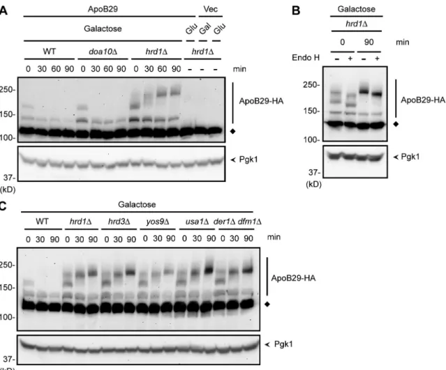
fact significantly stabilized in hrd1 cells (Fig. 7 A).
ApoB29 appeared as multiple bands when separated
by SDS-PAGE, with a gradual conversion of lower to higher
molecular weight species over time. This is most apparent
under stabilizing conditions (i.e., in hrd1 cells). ApoB29
is a translocon-engaged protein with its N terminus exposed
to the ER lumen. These slower migrating bands of apoB29
were sensitive to Endo H, which indicates N-glycosylation of
apoB29 (Fig. 7 B). A complete collapse to the fastest
migrat-ing species was not observed, however, which suggests that
apoB29, like Deg1-Sec62, acquires additional PTMs distinct
from N-glycosylation. These additional PTMs became more
prominent after inhibition of protein synthesis, as has
previ-ously been observed for other ERAD substrates (Wilhovsky
et al., 2000; Sato and Hampton, 2006).
Finally, we analyzed the cofactor requirements for apoB29
degradation. In contrast to Deg1-Sec62, apoB29 was markedly
sta-bilized in cells lacking Hrd3, Yos9, Usa1, or the Derlins (Fig. 7 C).
Thus, although both proteins may be targeted by Hrd1 by virtue of
their aberrant or prolonged translocon engagement, they differ with
respect to the exact cofactors required for their degradation.
Discussion
Many proteins pass through the Sec61 translocon en route
to their ultimate subcellular or extracellular destinations.
Deg1
*-Sec62 was efficiently degraded in the absence of
Yos9 and the Derlins (Der1 and Dfm1), proteins with roles in
ERAD-L, and only marginally stabilized in the absence of Usa1,
which functions in both ERAD-L and ERAD-M (Fig. 6 A;
Knop et al., 1996; Buschhorn et al., 2004; Bhamidipati et al.,
2005; Kim et al., 2005; Szathmary et al., 2005; Carvalho
et al., 2006; Goder et al., 2008; Horn et al., 2009; Carroll and
Hampton, 2010; Carvalho et al., 2010; Kanehara et al., 2010).
Deg1
*-Sec62 was significantly, but incompletely, stabilized
in the absence of Hrd3, which is required for degradation of
both ERAD-L and ERAD-M substrates (Hampton et al., 1996;
Carvalho et al., 2006; Gauss et al., 2006). However, this effect
may be indirect, as Hrd1 levels are markedly reduced in the
absence of Hrd3 (Plemper et al., 1999). A control ERAD-L
substrate, CPY*-HA, was significantly stabilized in these mutant
strains (Fig. 6 B). Degradation requirements for Deg1*-Sec62
bear some resemblance to those for the ERAD-M substrate
Hmg2. However, Hmg2 degradation displays a stronger
de-pendence on Hrd3 (Wilhovsky et al., 2000) and potentially
Usa1 (Horn et al., 2009; Carroll and Hampton, 2010) for its
degradation. Together, these results suggest that recognition
of Deg1-Sec62 occurs through a novel mechanism requiring
the minimal ERAD machinery consisting of Hrd1, Ubc7,
pre-sumably Cue1 (which anchors Ubc7 to the ER membrane),
and possibly Hrd3. Novel cofactors might also be involved.
Degradation of an apolipoprotein B derivative requires Hrd1
We hypothesized that the mechanism used by Hrd1 to target
Deg1
-Sec62 at the translocon may be related to that of a known
cotranslocationally degraded protein with intriguing links to
human health. Under conditions in which lipid binding of apoB
(a major component of low-density and very-low-density
lipo-proteins) is impaired, apoB translocation is arrested and the
protein remains associated with the translocon (Fisher et al.,
1997; Pariyarath et al., 2001). Incompletely translocated apoB
is redirected to proteasomal degradation in the cytoplasm
(Yeung et al., 1996), perhaps via a mechanism similar to that
of Deg1-Sec62 degradation. ApoB copurifies with several
Figure 6. Hrd1 cofactor requirements for Deg1-Sec62 degra-dation. (A) Pulse-chase analysis of Deg1*-Sec62 in the indicated yeast strains. The percentage of input protein remaining at each time-point is indicated below the autoradiograph. Cyclohexi-mide was included in the chase. Deg1*-Sec62 was precipitated with anti-Flag antibodies. Deg1*, F18S/I22T double mutant. The black line indicates that intervening lanes have been spliced out. (B) Cycloheximide chase analysis of CPY*-HA in the indi-cated yeast strains. CPY*-HA was detected by anti-HA immuno-blotting. Pgk1 serves as a loading control and was detected by anti-Pgk1 immunoblotting.
on October 2, 2012
jcb.rupress.org
Although glycosylation is not required for Hrd1-mediated
degradation, the switch in degradation dependence from
Doa10 to Hrd1 tracks very closely with acquisition of these
PTMs. Our data strongly suggest that the topological
rear-rangement across the ER membrane is the crucial event
dic-tating E3 specificity (Fig. 8).
Other work supports the 4-TM model of rearranged
topology (Fig. 3 C). Potential O-mannosylation sites (serines)
were systematically introduced throughout Deg1-Sec62 (Scott
and Schekman, 2008). We inferred that residues that became
O-mannosylated reached the ER lumen; nonmodified serine
substitutions were judged to be in the membrane-spanning or
cytoplasmic segments. O-mannosylation of serines inserted
at positions 132 and 170 confirmed that this span is
pres-ent within the ER lumen (see Fig. 1 B). Substitutions at other
positions, such as immediately upstream of the first known
TM, did not result in O-mannosylation. The most
straightfor-ward interpretation of these data is the 4-TM model of
Deg1-stimulated topological rearrangement (Fig. 3 C), although the
failure to observe O-mannosylation of a particular serine does
not exclude its luminal residence (Lommel and Strahl, 2009).
Potentially, ERAD substrates may also pass through this
chan-nel in a retrograde fashion en route to proteasomal
degrada-tion. Aberrant, translocationally stalled proteins might limit
flux through the channel in either direction, and eukaryotes
may therefore have evolved mechanisms for eliminating such
obstructions. Although Deg1-Sec62 (like many heavily
stud-ied model ERAD substrates) is an artificial protein, it may
illuminate a previously unappreciated protein quality-control
pathway mediated by the Hrd1 ubiquitin ligase and serve as a
prototype for proteins that aberrantly engage or occupy the
translocon. Indeed, we find that clearance of apoB, a protein
previously reported to be cotranslocationally degraded, also
depends on Hrd1.
Deg1
-Sec62 inserts into the ER membrane in two
dis-tinct steps. Initially, the two TM segments of Deg1-Sec62
are probably cotranslationally translocated to yield the same
topology as WT Sec62. In a second, Deg1-dependent step,
a loop within the normally cytoplasmic N-terminal domain
of Deg1-Sec62 penetrates the ER membrane. This
rearrange-ment allows access of previously cytoplasmic residues to
the N- and O-glycosylation machinery of the ER lumen.
Figure 7. ApoB29 is a Hrd1 substrate. (A–C) Cycloheximide chase analysis of ApoB29-3HA, expressed under the control of the GAL1 promoter in the indicated yeast strains, which were grown for 5 h in SD medium containing 2% galactose. ApoB29-3HA was detected by anti-HA immunoblotting. Pgk1 serves as a loading control and was detected by anti-Pgk1 immunoblotting. The diamonds denote a nonspecific band. (A) As controls, hrd1 yeast harbor-ing a plasmid with GAL1-driven ApoB29-3HA were grown in 2% glucose (repressharbor-ing) medium, and hrd1 yeast harborharbor-ing an empty vector were grown in 2% glucose (repressing) or 2% galactose (inducing) medium and processed similarly. (B) Lysates prepared immediately or 90 min after cycloheximide addition were incubated in the presence or absence of Endo H.
on October 2, 2012
jcb.rupress.org
Lateral release of true TM segments from the translocon
into the plane of the lipid bilayer depends strongly on the
hy-drophobic character of the TM helices (Shao and Hegde, 2011;
Zimmermann et al., 2011). The N-terminal loop of Deg1-Sec62
does not include bona fide TM domains; thus, we speculate
that the membrane-spanning polypeptide segments flanking
the N-glycosylated luminal element resist lateral release from
the translocon and persistently occupy the Sec61 channel. We
propose that Hrd1 recognizes persistent translocon
engage-ment by Deg1-Sec62 after the aberrant translocation event.
In this respect, Deg1-Sec62 may resemble a translocationally
stalled or arrested protein. Hrd1, by virtue of its reported
as-sociation with the translocon (Schäfer and Wolf, 2009), may be
ideally poised to target such incompletely translocated proteins
for ubiquitin-mediated degradation. It is also possible that the
Sec61-dependent topological change in Deg1-Sec62 generates
a permanently misfolded TM protein or misassembled
trans-locon complex that is recognized by Hrd1. Nevertheless, the
cofactor requirements for Deg1-Sec62 differ from other
well-characterized Hrd1 substrates (Fig. 8), which is consistent with
a unique mechanism for recognition and degradation.
Deg1
has been found to destabilize other TM proteins in
a partially Hrd1-dependent manner. Like Deg1-Sec62, both
Deg1
-Hmg1 and Deg1-Hmg2 fusion proteins are strongly
sta-bilized in the absence of Ubc7. Deletion of HRD1 partially
impairs turnover of these proteasome substrates (Wilhovsky
et al., 2000). Whether these proteins are targeted in a mechanism
similar to that targeting Deg1-Sec62 remains to be determined.
The factors preventing Doa10 from recognizing
Deg1-Sec62 remain enigmatic. After topological rearrangement
of Deg1-Sec62, Deg1 remains on the cytoplasmic face of
the ER membrane (Fig. 3). However, this posttranslationally
translocated form of the fusion protein is resistant to
Doa10-mediated proteolysis. The translocon-dependent rearrangement
might alter the position or conformation of Deg1 such that it is
inaccessible to the Doa10 complex. Preventing interaction of
Deg1
-Sec62 with the translocon causes a strong reversion to
Topological rearrangement of Deg1-Sec62 likely occurs
via Sec61-mediated PTT. Disrupting contacts of Deg1-Sec62
with the translocon or impairing PTT by Sec61 mutation
pre-vents domain dislocation and significantly reverts degradation
dependence to Doa10 (Figs. 4 and 5). Additionally, a cysteine
found within the initially cytoplasmic N-terminal domain of
Deg1
-Sec62 forms a transient disulfide linkage with a cysteine
on the interior of the Sec61 channel (Scott and Schekman,
2008). This bond appears to facilitate the altered topology of the
Sec62 N-terminal domain, as mutation of either participating
cysteine delays Deg1-Sec62 PTM and causes a partial switch
to Doa10-dependent degradation (Fig. 5, C and D).
Fusion of Deg1 to Sec62 was not anticipated to trigger
PTT of the normally cytoplasmic Sec62 N-terminal domain.
MAT2, from which Deg1 is derived, is a soluble nuclear
pro-tein, and Deg1 fused to the isolated soluble Sec62 N-domain
(residues 1–149) does not trigger membrane translocation
(Scott and Schekman, 2008). Rather, Deg1-Sec62
1–149is
de-graded in a Doa10-dependent manner, behaving like other
characterized soluble Deg1 fusions (Fig. S2 B). Potentially,
fusing any protein sequence to the Sec62 N terminus could
promote the observed topological rearrangement. This is not
the case, however, as Deg1 bearing a 20-residue internal
dele-tion does not promote PTM (or degradadele-tion) when fused to
Sec62 (Scott and Schekman, 2008). Deg1, by virtue of novel
protein–protein and/or protein–membrane interactions
stimu-lated by enforced proximity to the translocon, may
conforma-tionally alter the N-terminal portion of Sec62 or cause it to
linger persistently near the opening of the Sec61 translocon
channel. The translocon could respond by attempting to
con-duct the Sec62 N-terminal tail into the ER lumen. The
disul-fide linkage between Deg1-Sec62 and the Sec61 channel may
strengthen the interaction of the Sec62 N-domain with a signal
sequence-binding site within the channel. Consistent with this,
the cysteine cross-link is not strictly necessary for topological
rearrangement but significantly accelerates it (Fig. 5, C and D;
and Fig. S1 D).
Figure 8. Model for Hrd1-dependent ERAD of Deg1-Sec62. (A) After its initial cotranslational translocation, Deg1-Sec62 aberrantly engages the Sec61 translocon, resulting in PTT of the normally cytoplasmic N-terminal Sec62 tail. After membrane penetration, Deg1-Sec62 becomes N-glycosylated. In this rearranged conformation, Doa10 does not recognize Deg1-Sec62 as an ERAD-C substrate. Deg1-Sec62 remains trapped in this orientation unless Hrd1 targets it for degradation, thereby restoring functionality to the Sec61 translocon. With the exception of the E2 Ubc7 (and presumably its membrane-anchoring binding partner Cue1; Biederer et al., 1997), and potentially Hrd3 (not depicted), well-characterized Hrd1 cofactors that function in ERAD-L and ERAD-M are dispensable for ERAD of Deg1-Sec62. The approximate position of N-glycosylated N153 residue is indicated with a cartoon representation of the N-glycan. (B) When translocon engagement is prevented by disrupting Deg1-Sec62 interaction with the translocon or impairing PTT, Deg1-Sec62 retains its original topology and is targeted by Doa10 as an ERAD-C substrate. Black circles, ubiquitin.
on October 2, 2012
jcb.rupress.org
process provisionally called ERAD-T (for “translocon
associ-ated”). ERAD-T substrates may include proteins that aberrantly
stall in the translocon either because of an abnormality in the
translocating protein (such as may be the case for Deg1-Sec62)
or because of malfunction in the translocation process itself.
Such substrates may be difficult to identify experimentally,
as they likely represent a small subpopulation of any given
translocated gene product. Artificial translocon-occupying
substrates, such as Deg1-Sec62, may serve as a model for
un-derstanding such mechanisms. Other cellular pathways may
have co-opted this quality-control pathway to mediate the
regulated destruction of otherwise normal proteins. The
low-density and very-low-low-density lipoprotein biosynthesis
path-way provides a likely example of a protein—apoB—whose
regulated degradation utilizes a basic cellular quality control
mechanism. Future studies will seek to understand the
mecha-nisms by which Hrd1 recognizes this potentially distinct class
of substrates that persistently engage the translocon.
Materials and methods
Yeast and bacterial methods
With the exception of cells prepared for cycloheximide chase/immunoblot analysis of apoB29, yeast were grown in synthetic defined (SD) medium that was prepared as described previously (Guthrie and Fink, 1991). For experiments involving apoB29, yeast cells were cultured in modified SD medium with 5 mg/ml of acid-hydrolyzed casein and 0.08 mg/ml tryp-tophan as the amino acid source. Standard techniques were used for ge-netic manipulation of yeast strains (Guthrie and Fink, 1991). To introduce gene deletions, selection markers were PCR-amplified from donor strains with flanking sequences containing homology upstream and downstream of target gene start and stop codons. PCR products were transformed into yeast to replace genes by homologous recombination. Gene disruptions were confirmed by PCR. Standard techniques were used for plasmid ma-nipulation in Escherichia coli (Ausubel et al., 1989). Site-directed muta-genesis was used to introduce point mutations (Zheng et al., 2004). Yeast
strains and plasmids used in this study are presented in Tables S1 and S2,
respectively. Yeast strains and plasmids not constructed in our labora-tory were provided by C. Andreasson (Stockholm University, Stockholm, Sweden), J. Brodsky (University of Pittsburgh, Pittsburgh, PA), E. Craig (University of Wisconsin, Madison, Madison, WI), R. Gilmore (University of Massachusetts Medical School, Boston, MA), Y. Jigami (National Insti-tute for Advanced Industrial Science and Technology, Tsukuba, Japan), N. Johnsson (Ulm University, Ulm, Germany), D. Ng (National Univer-sity of Singapore, Singapore), R. Schekman (UniverUniver-sity of California, Berkeley, Berkeley, CA), D. Scott (St. Jude Children’s Research Hospital, Memphis, TN), and T. Yoko-o (National Institute for Advanced Industrial Science and Technology, Tsukuba, Japan).
Pulse-chase analysis
Pulse-chase analysis was performed essentially as described previously
(Chen et al., 1993). Yeast cells were labeled with 20 µCi TRAN35S-LABEL
(MP Biomedicals) per 1 OD600 unit of cells at 30°C for 10 min in SD
me-dium lacking methionine and cysteine. Chases with excess unlabeled Met and Cys were performed in the absence of cycloheximide, except where noted in the figure legends. Inclusion of 500 µg/ml cycloheximide had no detectable impact on degradation kinetics, modification kinetics, or E3 specificity. Immunoprecipitation of Deg1 fusion proteins was performed with anti-FLAG M2 affinity resin (Sigma-Aldrich) or sequential incubation with anti-Deg1 antibody (Hochstrasser and Varshavsky, 1990) and aga-rose cross-linked to recombinant Protein A (Repligen), as indicated in the figure legends. Immunoprecipitated proteins were separated by SDS-PAGE and analyzed by autoradiography, using a Storm 860 Phosphorimager sys-tem and ImageQuant 5.2 software (Molecular Dynamics).
Cycloheximide chase and standard immunoblot analyses
Cycloheximide was added to a final concentration of 250 µg/ml to loga-rithmically growing cells. For analysis of CPY*-HA, cells were lysed
es-sentially as described by Kushnirov (2000). 2.5 OD600 units of cells were
Doa10-dependent degradation (Fig. 4). Therefore, Deg1-Sec62
is not inherently unrecognizable by Doa10. Interfering with
events downstream of translocon binding (e.g., inhibiting PTT
or preventing Deg1-Sec62–Sec61 disulfide formation) also
causes a significant switch to Doa10-dependent degradation
(Fig. 5). Thus, Doa10 is capable of recognizing Deg1-Sec62
even when conditions allow initial interaction with the
trans-locon. In these cases, Doa10 might target Deg1-Sec62 that is
in complex with the translocon but has not yet undergone PTT
or might recognize Deg1-Sec62 that has transiently dissociated
from Sec61.
The discovery that abnormal translocon engagement
precedes Deg1-Sec62 degradation led us to hypothesize that
cotranslocational proteolysis of apoB might occur by a similar
mechanism. ApoB uses the translocon as a platform for
pro-gressive lipid binding. When lipid binding is inhibited,
translo-cation into the ER lumen is slowed, and the protein is destroyed
by the cytoplasmic proteasome (Fisher et al., 1997; Pariyarath
et al., 2001). Earlier studies implied that degradation of a model
variant of apoB occurred via the ERAD system in yeast (Hrizo
et al., 2007). Our results directly demonstrate Hrd1 dependence
for apoB29 degradation in yeast (Fig. 7 A). The cofactor
re-quirements for Deg1-Sec62 and apoB Hrd1-dependent
degra-dation, however, differed, which suggests that the details of
E3-substrate recognition diverge for these substrates.
A previous study had led to the suggestion that
Deg1-Sec62 glycosylation and disulfide formation with Sec61 occur
during Sec61-dependent retrotranslocation of a Doa10 substrate
(Scott and Schekman, 2008). Our results, however, strongly
suggest that these events occur in the process of Hrd1 substrate
generation. Given the general dependence of substrate
ret-rotranslocation upon functional ubiquitin conjugation
machin-ery (Biederer et al., 1997; de Virgilio et al., 1998), N-glycosylation
of Deg1-Sec62 in doa10 hrd1 cells (Fig. 2 A) indicates that
such modification happens upstream of substrate selection. The
disulfide cross-link between Deg1-Sec62 and Sec61 was
preted as representing a briefly stalled retrotranslocation
inter-mediate, as preventing this disulfide from forming accelerated
degradation. However, we found that the increased degradation
rate of Deg1-Sec62 in the absence of the disulfide linkage is
caused by a switch from Hrd1-dependent degradation to
com-paratively rapid Doa10-mediated proteolysis (Fig. 5, C and D).
Therefore, although our data establish a role for Sec61 in the
biogenesis of a Hrd1 substrate, they do not directly implicate
Sec61 as a retrotranslocon for Deg1-Sec62, although this
re-mains possible. It is tempting to speculate that, in a manner
mir-roring its membrane insertion, retrotranslocation of Deg1-Sec62
occurs in two steps: extraction of the aberrantly rearranged
por-tion from the Sec61 channel followed by removal of the normal
TM segments, potentially also through Sec61. Similarly, the
precise mechanism by which apoB is extracted from the ER
membrane remains to be elucidated. Translocationally stalled
apoB might simply be extruded back into the cytoplasm via
ret-rograde transport through the translocon in which it stalls, or it
might be transferred to another retrotranslocating complex.
We speculate that Hrd1 may play an important role in
removing proteins that persistently engage the translocon in a
on October 2, 2012
jcb.rupress.org
phosphate, pH 7.5, 150 mM NaCl, and 2 mM dithiothreitol). Cells were
digested in 140 µg zymolyase 100T (MP Biomedicals)/10 OD600 units of
cells in spheroplast buffer for 20 min at 30°C. Spheroplasts were har-vested by centrifugation (5 min at 600 g at 4°C) and washed in sphero-plast buffer containing 20 µg/ml pepstatin A and 1 mM EDTA (for samples destined for protease protection analysis) or spheroplast buffer containing 1× EDTA-free Complete Protease Inhibitor Cocktail (Roche) and 1 mM EDTA (for samples destined for Endo H protection analysis). Spheroplasts were centrifuged again (5 min at 600 g at 4°C) and
resus-pended in fractionation buffer (200 mM d-mannitol, 20 mM sodium
phos-phate, pH 7.5, and 150 mM NaCl) containing appropriate protease inhibitors and lysed by vortexing in the presence of glass beads for three 30-s pulses with 1 min on ice between pulses. Unbroken cells and cellular debris were centrifuged (5 min at 600 g at 4°C), and the supernatant was used as the microsomal preparation. For Proteinase K protection
ex-periments, microsome preparations corresponding to 3.75 OD600 units
were incubated with or without 1% Triton X-100 and treated with
Protein-ase K (Roche) in storage buffer (20 mM Tris, pH 7.5, 1 mM CaCl2, and
50% glycerol) at a final concentration of 5 µg/ml or mock-treated with storage buffer alone. Reactions were performed on ice for 30 min. Pro-teinase K activity was quenched with 10 mM PMSF. For Endo H
protec-tion experiments, microsome preparaprotec-tions corresponding to 3.75 OD600
units were supplemented with potassium acetate, pH 5.6, to a final con-centration of 80 mM. Samples were incubated with or without 1% Triton X-100 and treated with 0.004 U Endo H or mock-treated with water. Re-actions were performed at 37°C for 2 h with gentle mixing every 20–30 min. Reactions were terminated with ice-cold TCA added to a final concentra-tion of 10%.
For both Proteinase K and Endo H protection experiments, proteins were precipitated with 10% ice-cold TCA. Proteins were pelleted by cen-trifugation at 4°C. Pellets were washed with 2% TCA and resuspended in 50 µl TCA sample buffer (Loayza and Michaelis, 1998). Acidic samples (which caused the bromophenol blue indicator to turn yellow) were neutral-ized, and samples were boiled for 8 min.
Inhibition of O-mannosylation
Yeast cells were cultured for 3 h at 30°C in the presence of 1 µM 5-[[3-(1-phenylethoxy)-4-(2-phenylethoxy)phenyl]methylene]-4-oxo-2-thioxo-3-thiazolidineacetic acid (“Compound 5a”; Biotrend Chemicals; Orchard et al., 2004), provided by S. Strahl (University of Heidelberg, Heidelberg, Germany), a global inhibitor of O-mannosylation in S. cerevisiae (Strahl, S., personal communication; Arroyo et al., 2011).
Online supplemental material
Fig. S1 shows PTM of Deg1-Sec62. Fig. S2 shows Doa10-dependent degradation of Deg1 fusion proteins. Fig. S3 shows membrane topol-ogy of Deg1-Sec62, Deg1-sec62†, and Deg1-Vma12. Table S1 shows yeast strains used in this study. Table S2 shows plasmids used in this study. Online supplemental material is available at http://www.jcb.org/ cgi/content/full/jcb.201203061/DC1.
We thank Claes Andreasson, Jeffrey Brodsky, Elizabeth Craig, Reid Gilmore, Yoshifumi Jigami, Nils Johnsson, Davis Ng, Randy Schekman, Danny Scott, Nava Segev, and Takehiko Yoko-o for yeast strains, plasmids, and antibodies. We also thank Sabine Strahl for the O-mannosylation inhibitor and Davis Ng, Sabine Strahl, and David Andrews for technical advice. We thank Christopher Hickey, Robert Tomko, Dimitrios Zattas, and Christian Schlieker for critical reading of the manuscript.
This work was supported by National Institutes of Health (NIH) grant GM046904 to M. Hochstrasser and NIH National Research Service Award postdoctoral fellowship F32 GM088995 to E.M. Rubenstein.
Submitted: 12 March 2012 Accepted: 10 May 2012
References
Ausubel, F.M., R. Brent, R.E. Kingston, D.D. Moore, J.G. Seidman, J.A. Smith, and K. Struhl. 1989. Current protocols in molecular biology. Wiley, New York.
Arroyo, J., J. Hutzler, C. Bermejo, E. Ragni, J. García-Cantalejo, P. Botías, H. Piberger, A. Schott, A.B. Sanz, and S. Strahl. 2011. Functional and genomic analyses of blocked protein O-mannosylation in baker’s yeast. Mol. Microbiol. 79:1529–1546. http://dx.doi.org/10.1111/j.1365-2958.2011.07537.x
collected at each time point, pelleted by centrifugation, resuspended in 400 µl of 0.2 N NaOH, and incubated for 5 min at room temperature. Cells were pelleted by centrifugation and resuspended in sample loading buffer (Laemmli, 1970). Samples were boiled for 5 min before separation by SDS-PAGE and immunoblotting.
For analysis of apoB29, cells were lysed essentially as described
by Loayza and Michaelis (1998). 2.5 OD600 units of cells were collected
at each time point, pelleted by centrifugation, and resuspended in 1 ml of ice-cold water. Cells were lysed by the addition of NaOH (to 0.26 N final concentration) and -mercaptoethanol (to 0.13 M final concentration) and allowed to incubate on ice for 15 min. TCA was added to 5% final concentration to precipitate proteins. Proteins were pelleted by centrifuga-tion at 4°C. Pellets were washed with 2% TCA and resuspended in 50 µl TCA sample buffer (3.5% SDS, 0.5 M DTT, 80 mM Tris [not pH adjusted], 8 mM EDTA, 15% glycerol, and 0.1 mg/ml of bromophenol blue) before separation by SDS-PAGE and immunoblotting.
For analysis of Deg1-Sec621–149, 2.5 OD600 units of cells were
col-lected at each time point, pelleted by centrifugation, resuspended in 100 µl of 0.2 N NaOH, and incubated for 5 min at room temperature. Cells were pelleted by centrifugation and resuspended in 30 µl of urea lysis buffer (1% NP-40, 1% sodium deoxycholate, 1% SDS, and 8 M urea). Laemmli sample buffer was added and samples were boiled for 5 min before sepa-ration by SDS-PAGE and immunoblotting.
Proteins were immunoblotted to polyvinylidene fluoride mem-branes (Millipore), which were blocked with 5% skim milk in TBS/T (20 ml Tris, pH 7.6, 136 mM NaCl, and 0.1% Tween 20) for 60 min at room temperature or overnight at 4°C. All antibody incubations were con-ducted for 45–90 min at room temperature in 1% skim milk in TBS/T followed by three 5-min washes in TBS/T. The following mouse mono-clonal antibodies were used at the indicated dilutions: anti-FLAG M2 (Sigma-Aldrich) at 1:10,000, HA 16B12 (Covance) at 1:2,000, anti-yeast Pgk1 (Molecular Probes) between 1:20,000 and 1:40,000, and anti–yeast CPY at 1:5,000. Anti-Deg1 (MAT2) rabbit polyclonal antibodies were used at a dilution of 1:5,000. The anti–yeast CPY anti-body is a monoclonal mouse antianti-body raised against yeast CPY and was a gift of N. Segev (University of Illinois at Chicago, Chicago, IL). Anti-Deg1 antibodies are polyclonal rabbit anti–2 antiserum raised against a full-length 2--galactosidase fusion protein (Hochstrasser and Varshavsky, 1990). Mouse primary antibody incubations were fol-lowed by incubation with peroxidase-coupled sheep anti–mouse IgG (GE Healthcare) at a dilution of 1:5,000; anti-Deg1 antibody incubation was followed by incubation with peroxidase-coupled donkey anti–rabbit IgG (GE Healthcare). The peroxidase anti-peroxidase (PAP) antibody (Sigma-Aldrich) was used to detect the Protein A epitope at a dilution of 1:8,000 and required no secondary antibody. Bound antibody was visualized using Enhanced Chemiluminescence (GE Healthcare). Endo H treatment of cellular proteins
After pulse chase, Deg1-Sec62 variants were immunoprecipitated. After five washes with wash buffer (150 mM NaCl, 50 mM Hepes, pH 7.5, 5 mM EDTA, 1% Triton X-100, and 0.1% SDS), two additional equilibrat-ing washes were performed with 1× NEBuffer 4 (New England Biolabs). The resin was resuspended in 50 µl of 1× NEBuffer 4 that had been supplemented with potassium acetate, pH 5.6, to an 80-mM final con-centration. After adding 0.005–0.0075 U of Endo H (Roche) to the resin suspension, samples were incubated at 37°C for 2 h with gentle mixing every 20–30 min. Laemmli sample buffer was added, and samples were boiled for 8 min.
For treatment of whole cell lysates, lysates were prepared by glass-bead vortexing 3 × 5.0 m/s for 20 s (with 5 min on ice between pulses) using a FastPrep-24 bead beating machine (MP Biomedicals) in a buffer containing 110 mM Hepes, pH 7.4, (adjusted with KOH),
2 mM MgCl2, 0.1% Tween 20, and 1× Complete Protease Inhibitor
Cocktail (Roche). Lysate volumes corresponding to 0.075 OD600 units
of cells were incubated with 0.005 U of Endo H (or mock-treated with water) at 378 for 2 h. Laemmli sample buffer was added, and samples were boiled for 8 min.
Proteinase K and Endo H protection of microsomal proteins
Yeast microsomal membranes were prepared essentially as described
previously (Kreft et al., 2006; Scott and Schekman, 2008). 15 OD600
units of cells were harvested by centrifugation, resuspended in 1 ml resus-pension buffer (10 mM Tris, pH 9.4, and 10 mM DTT), and incubated at room temperature for 10 min. Cell pellets were harvested by centrifuga-tion and washed with spheroplast buffer (1 M sorbitol, 20 mM sodium
on October 2, 2012
jcb.rupress.org
Saccharomyces cerevisiae. J. Biochem. 143:555–567. http://dx.doi.org/ 10.1093/jb/mvm249
Hochstrasser, M., and A. Varshavsky. 1990. In vivo degradation of a transcrip-tional regulator: the yeast alpha 2 repressor. Cell. 61:697–708. http:// dx.doi.org/10.1016/0092-8674(90)90481-S
Horn, S.C., J. Hanna, C. Hirsch, C. Volkwein, A. Schütz, U. Heinemann, T. Sommer, and E. Jarosch. 2009. Usa1 functions as a scaffold of the HRD-ubiquitin ligase. Mol. Cell. 36:782–793. http://dx.doi.org/10.1016/ j.molcel.2009.10.015
Hrizo, S.L., V. Gusarova, D.M. Habiel, J.L. Goeckeler, E.A. Fisher, and J.L. Brodsky. 2007. The Hsp110 molecular chaperone stabilizes apo-lipoprotein B from endoplasmic reticulum-associated degradation (ERAD). J. Biol. Chem. 282:32665–32675. http://dx.doi.org/10.1074/ jbc.M705216200
Huyer, G., W.F. Piluek, Z. Fansler, S.G. Kreft, M. Hochstrasser, J.L. Brodsky, and S. Michaelis. 2004. Distinct machinery is required in
Saccharomyces cerevisiae for the endoplasmic reticulum-associated degradation of a multispanning membrane protein and a soluble luminal protein. J. Biol. Chem. 279:38369–38378. http://dx.doi.org/10.1074/ jbc.M402468200
Johnson, P.R., R. Swanson, L. Rakhilina, and M. Hochstrasser. 1998. Degradation signal masking by heterodimerization of MATalpha2 and MATa1 blocks their mutual destruction by the ubiquitin-proteasome pathway. Cell. 94:217–227. http://dx.doi.org/10.1016/S0092-8674(00) 81421-X
Kanehara, K., W. Xie, and D.T. Ng. 2010. Modularity of the Hrd1 ERAD com-plex underlies its diverse client range. J. Cell Biol. 188:707–716. http:// dx.doi.org/10.1083/jcb.200907055
Kim, W., E.D. Spear, and D.T. Ng. 2005. Yos9p detects and targets misfolded glycoproteins for ER-associated degradation. Mol. Cell. 19:753–764. http://dx.doi.org/10.1016/j.molcel.2005.08.010
Kim, I., J. Ahn, C. Liu, K. Tanabe, J. Apodaca, T. Suzuki, and H. Rao. 2006. The Png1-Rad23 complex regulates glycoprotein turnover. J. Cell Biol. 172:211–219. http://dx.doi.org/10.1083/jcb.200507149
Knop, M., A. Finger, T. Braun, K. Hellmuth, and D.H. Wolf. 1996. Der1, a novel protein specifically required for endoplasmic reticulum degra-dation in yeast. EMBO J. 15:753–763.
Kreft, S.G., L. Wang, and M. Hochstrasser. 2006. Membrane topology of the yeast endoplasmic reticulum-localized ubiquitin ligase Doa10 and com-parison with its human ortholog TEB4 (MARCH-VI). J. Biol. Chem. 281:4646–4653. http://dx.doi.org/10.1074/jbc.M512215200
Kushnirov, V.V. 2000. Rapid and reliable protein extraction from yeast. Yeast. 16:857–860. http://dx.doi.org/10.1002/1097-0061(20000630)16:9<857:: AID-YEA561>3.0.CO;2-B
Laemmli, U.K. 1970. Cleavage of structural proteins during the assembly of the head of bacteriophage T4. Nature. 227:680–685. http://dx.doi .org/10.1038/227680a0
Laney, J.D., and M. Hochstrasser. 2003. Ubiquitin-dependent degradation of the yeast Mat(alpha)2 repressor enables a switch in developmental state.
Genes Dev. 17:2259–2270. http://dx.doi.org/10.1101/gad.1115703 Liang, J.S., T. Kim, S. Fang, J. Yamaguchi, A.M. Weissman, E.A. Fisher,
and H.N. Ginsberg. 2003. Overexpression of the tumor autocrine motility factor receptor Gp78, a ubiquitin protein ligase, results in increased ubiquitinylation and decreased secretion of apolipoprotein B100 in HepG2 cells. J. Biol. Chem. 278:23984–23988. http://dx.doi .org/10.1074/jbc.M302683200
Loayza, D., and S. Michaelis. 1998. Role for the ubiquitin-proteasome sys-tem in the vacuolar degradation of Ste6p, the a-factor transporter in
Saccharomyces cerevisiae. Mol. Cell. Biol. 18:779–789.
Lommel, M., and S. Strahl. 2009. Protein O-mannosylation: conserved from bacteria to humans. Glycobiology. 19:816–828. http://dx.doi.org/10 .1093/glycob/cwp066
Mayer, T.U., T. Braun, and S. Jentsch. 1998. Role of the proteasome in mem-brane extraction of a short-lived ER-transmemmem-brane protein. EMBO J. 17:3251–3257. http://dx.doi.org/10.1093/emboj/17.12.3251
Metzger, M.B., M.J. Maurer, B.M. Dancy, and S. Michaelis. 2008. Degradation of a cytosolic protein requires endoplasmic reticulum-associated degra-dation machinery. J. Biol. Chem. 283:32302–32316. http://dx.doi.org/10 .1074/jbc.M806424200
Ng, D.T., and P. Walter. 1996. ER membrane protein complex required for nuclear fusion. J. Cell Biol. 132:499–509. http://dx.doi.org/10.1083/ jcb.132.4.499
Ng, D.T., J.D. Brown, and P. Walter. 1996. Signal sequences specify the target-ing route to the endoplasmic reticulum membrane. J. Cell Biol. 134:269– 278. http://dx.doi.org/10.1083/jcb.134.2.269
Orchard, M.G., J.C. Neuss, C.M. Galley, A. Carr, D.W. Porter, P. Smith, D.I. Scopes, D. Haydon, K. Vousden, C.R. Stubberfield, et al. 2004. Bays, N.W., R.G. Gardner, L.P. Seelig, C.A. Joazeiro, and R.Y. Hampton.
2001. Hrd1p/Der3p is a membrane-anchored ubiquitin ligase required for ER-associated degradation. Nat. Cell Biol. 3:24–29. http://dx.doi .org/10.1038/35050524
Bhamidipati, A., V. Denic, E.M. Quan, and J.S. Weissman. 2005. Exploration of the topological requirements of ERAD identifies Yos9p as a lectin sen-sor of misfolded glycoproteins in the ER lumen. Mol. Cell. 19:741–751. http://dx.doi.org/10.1016/j.molcel.2005.07.027
Biederer, T., C. Volkwein, and T. Sommer. 1997. Role of Cue1p in ubiquitination and degradation at the ER surface. Science. 278:1806–1809. http://dx.doi .org/10.1126/science.278.5344.1806
Buschhorn, B.A., Z. Kostova, B. Medicherla, and D.H. Wolf. 2004. A ge-nome-wide screen identifies Yos9p as essential for ER-associated degradation of glycoproteins. FEBS Lett. 577:422–426. http://dx.doi .org/10.1016/j.febslet.2004.10.039
Carroll, S.M., and R.Y. Hampton. 2010. Usa1p is required for optimal func-tion and regulafunc-tion of the Hrd1p endoplasmic reticulum-associated deg-radation ubiquitin ligase. J. Biol. Chem. 285:5146–5156. http://dx.doi .org/10.1074/jbc.M109.067876
Carvalho, P., V. Goder, and T.A. Rapoport. 2006. Distinct ubiquitin-ligase com-plexes define convergent pathways for the degradation of ER proteins.
Cell. 126:361–373. http://dx.doi.org/10.1016/j.cell.2006.05.043
Carvalho, P., A.M. Stanley, and T.A. Rapoport. 2010. Retrotranslocation of a misfolded luminal ER protein by the ubiquitin-ligase Hrd1p. Cell. 143:579–591. http://dx.doi.org/10.1016/j.cell.2010.10.028
Chen, P., P. Johnson, T. Sommer, S. Jentsch, and M. Hochstrasser. 1993. Multiple ubiquitin-conjugating enzymes participate in the in vivo degra-dation of the yeast MAT alpha 2 repressor. Cell. 74:357–369. http://dx.doi .org/10.1016/0092-8674(93)90426-Q
Ciechanover, A., H. Heller, S. Elias, A.L. Haas, and A. Hershko. 1980. ATP-dependent conjugation of reticulocyte proteins with the polypeptide required for protein degradation. Proc. Natl. Acad. Sci. USA. 77:1365– 1368. http://dx.doi.org/10.1073/pnas.77.3.1365
de Virgilio, M., H. Weninger, and N.E. Ivessa. 1998. Ubiquitination is required for the retro-translocation of a short-lived luminal endoplas-mic reticulum glycoprotein to the cytosol for degradation by the pro-teasome. J. Biol. Chem. 273:9734–9743. http://dx.doi.org/10.1074/jbc .273.16.9734
Deshaies, R.J., and R. Schekman. 1990. Structural and functional dissection of Sec62p, a membrane-bound component of the yeast endoplasmic reticu-lum protein import machinery. Mol. Cell. Biol. 10:6024–6035. Deshaies, R.J., S.L. Sanders, D.A. Feldheim, and R. Schekman. 1991. Assembly
of yeast Sec proteins involved in translocation into the endoplasmic re-ticulum into a membrane-bound multisubunit complex. Nature. 349:806– 808. http://dx.doi.org/10.1038/349806a0
Fisher, E.A., M. Zhou, D.M. Mitchell, X. Wu, S. Omura, H. Wang, A.L. Goldberg, and H.N. Ginsberg. 1997. The degradation of apolipoprotein B100 is mediated by the ubiquitin-proteasome pathway and involves heat shock protein 70. J. Biol. Chem. 272:20427–20434. http://dx.doi .org/10.1074/jbc.272.33.20427
Fisher, E.A., L.R. Lapierre, R.D. Junkins, and R.S. McLeod. 2008. The AAA-ATPase p97 facilitates degradation of apolipoprotein B by the ubiquitin- proteasome pathway. J. Lipid Res. 49:2149–2160. http://dx.doi.org/10 .1194/jlr.M800108-JLR200
Fisher, E.A., N.A. Khanna, and R.S. McLeod. 2011. Ubiquitination regulates the assembly of VLDL in HepG2 cells and is the committing step of the apoB-100 ERAD pathway. J. Lipid Res. 52:1170–1180. http://dx.doi .org/10.1194/jlr.M011726
Gauss, R., T. Sommer, and E. Jarosch. 2006. The Hrd1p ligase complex forms a linchpin between ER-lumenal substrate selection and Cdc48p recruitment. EMBO J. 25:1827–1835. http://dx.doi.org/10.1038/sj.emboj .7601088
Goder, V., P. Carvalho, and T.A. Rapoport. 2008. The ER-associated degra-dation component Der1p and its homolog Dfm1p are contained in complexes with distinct cofactors of the ATPase Cdc48p. FEBS Lett. 582:1575–1580. http://dx.doi.org/10.1016/j.febslet.2008.03.056 Guthrie, C., and G.R. Fink. 1991. Guide to yeast genetics and molecular biology.
Academic Press, San Diego. 735 pp.
Hamilton, T.G., T.B. Norris, P.R. Tsuruda, and G.C. Flynn. 1999. Cer1p functions as a molecular chaperone in the endoplasmic reticulum of
Saccharomyces cerevisiae. Mol. Cell. Biol. 19:5298–5307.
Hampton, R.Y., R.G. Gardner, and J. Rine. 1996. Role of 26S proteasome and HRD genes in the degradation of 3-hydroxy-3-methylglutaryl-CoA re-ductase, an integral endoplasmic reticulum membrane protein. Mol. Biol.
Cell. 7:2029–2044.
Hirayama, H., M. Fujita, T. Yoko-o, and Y. Jigami. 2008. O-mannosylation is required for degradation of the endoplasmic reticulum-associated degradation substrate Gas1*p via the ubiquitin/proteasome pathway in