Publisher’s version / Version de l'éditeur:
Canadian Journal of Chemistry, 56, 6, pp. 824-830, 1978
READ THESE TERMS AND CONDITIONS CAREFULLY BEFORE USING THIS WEBSITE.
https://nrc-publications.canada.ca/eng/copyright
Vous avez des questions? Nous pouvons vous aider. Pour communiquer directement avec un auteur, consultez la
première page de la revue dans laquelle son article a été publié afin de trouver ses coordonnées. Si vous n’arrivez pas à les repérer, communiquez avec nous à PublicationsArchive-ArchivesPublications@nrc-cnrc.gc.ca.
Questions? Contact the NRC Publications Archive team at
PublicationsArchive-ArchivesPublications@nrc-cnrc.gc.ca. If you wish to email the authors directly, please see the first page of the publication for their contact information.
NRC Publications Archive
Archives des publications du CNRC
This publication could be one of several versions: author’s original, accepted manuscript or the publisher’s version. / La version de cette publication peut être l’une des suivantes : la version prépublication de l’auteur, la version acceptée du manuscrit ou la version de l’éditeur.
Access and use of this website and the material on it are subject to the Terms and Conditions set forth at
Structure and properties of a trans-anti photodimer of 5-methylorotate
Huber, Carol P.; Birnbaum, George I.; Post, Michael L.; Kulikowska, Ewa;
Gajewska, Lucyna; Shugar, David
https://publications-cnrc.canada.ca/fra/droits
L’accès à ce site Web et l’utilisation de son contenu sont assujettis aux conditions présentées dans le site LISEZ CES CONDITIONS ATTENTIVEMENT AVANT D’UTILISER CE SITE WEB.
NRC Publications Record / Notice d'Archives des publications de CNRC:
https://nrc-publications.canada.ca/eng/view/object/?id=2dfc5a7c-f1f1-4058-aecc-db10e57b04e0
https://publications-cnrc.canada.ca/fra/voir/objet/?id=2dfc5a7c-f1f1-4058-aecc-db10e57b04e0
Structure and properties of a
traizs-anti
photodimer of 5-methylorotate'
CAROL
P. H U B E R , ?
GEORGE
I .
B I R N B A U M ,
A N DMICHAEL
L. P O S ,
fli~.i.siotr c~/'Biologiccrl Sr,ietrc,c.s, Ncr/iotrcrl Rc.secrr.cIr Cororc.il c~/'Cot~trtln, O//crn~cr, Otrr., Cotrcrrla K I A OR6A N D
EWA K U L I K O W S K A ,
L U C Y N A
GAJEWSKA,
A N DD A V I D
S H U G A R
D c p t r t ~ ~ t r r ~ t r ~ c?/'Bio/~lly,sic.s, ltr.s/i/r~/c c:/'E.v/~rritt~o~ltrl Plry.sic,s, U t r i ~ ~ c ~ r s i ~ y Wor:sni~~, 02-089 Wal:c-:tr\~~cr. Polotlrl Received September 19, 1977
CAROL P. HUBER, GEORGE I. BIRNBAUM, MICHAEL L. POST, EWA KULIKOWSKA, LUCYNA GAJEWSKA, and DAVID SHUGAR. Can. J. Chem. 56,824 (1978).
5-Methylorotate is relatively radiation resistant in aqueous fluid medium, but readily photodilnerizes in an ice matrix. Rapid for~nation of such a matrix made possible the prepara- tive isolation of the photodi~iier in good yield. The potassii~m salt of the photodinier crystal- lizes as the hexahydrate, C 1 2 H , o N , 0 , K 2 ~ 6 H 2 0 . The crystals are triclinic with space group P i , o = 8.139(3), b = 9.759(3), c = 7.398(3)
A,
a = 100.28(7), = 74.22(5), y = 108.67(7)", and V = 533.0.A3,
Z = I. The structure was solved by Patterson and direct mcthods; refine- ment by block-diagonal least-squares converged at R = 0.041 for all 1671 observed reflec- tions. The pyrimidine rings of the centrosymmetric photodimer are arranged in /rntrs-crtrli configuration across the planar cyclobutane ring. The potassium ion is seven-coordinated. In aqueous medium the photodimer exhibits a pK,, of 12.8 due to dissociation of the ring N(3) hydrogen. Irradiation in aqueous medium at 254 nm leads to quantitative regeneration of the monomer with a quantum yield of 0.8. Thermal regeneration of the monomer also occurs in neutral and acid aqueous media, but at a slower rate than for the trnt~s-syt~ orotate photodinier. In contrast to the orotate photodimer, which undergoes alkali-catalyzed opening of the 3,4 bonds of the pyrimidine rings, the 5-methylorotate photodimer under these conditions dissociates to the parent monomer.CAROL P. HUBER, GEORGE I. BIRNBAUM, MICHAEL L. POST, EWA KULIKOWSKA, LUCYNA GAJEWSKA et DAVID SHUGAR. Can. J. Chem. 56,824(1978).
Dans I'eau liquide, le n~ethyl-5 orotate est assez resistant aux radiations; toutefois il subit facilement une photodinlerisation dans une matrice de glace. La formation rapide d'une telle niatrice permet d'isoler le photodimtre avec de bons rendements a I'echelle preparative. Le sel du potassium du photodimere cristallise sous fornie d'hexahydrate, C 1 2 H , o N ~ O ~ K 2 ~ 6 H 2 0 . Les cristaux sont tricliniques avec un groupe d'espace P i , n = 8.139(3), b = 9.759(3),
c = 7.398(3)
A,
a = 100.28(7),B
= 74.22(5), y = 108.67(7)" et V = 533.0.A3,
Z = 1. On a resolu la structure par les methodes directes et de Patterson; un affinement par la mtthode des moindres carrCs (bloc diagonal) converge vers une valeur de R de 0.041 pour les 1671 rtflexions observkes. Les cycles de pyrimidine du photodimtre centrosynietrique sont orientts dans une configuration tt.ntrs-anti a travers le cycle planaire du cyclobutane. L'ion potassium est hepta- coordonnt. En solution aqueuse, le photodimere prtsente un pK, de 12.8 qui est d i ~ k une dissociation a I'hydrogene attach6 en position N(3) du cycle. L'irradiation d'une solution aqueuse a 254 nm conduit a une regheration quantitative du monomtre avec un rendement quantique de 0.8. La regentration therrnique du n~ononitre se produit aussi en milieu aqueux acide et neutre mais a une vitesse plus lente que pour le photodimere orotate trnt~s-s~~tl. Par opposition avec le photodimere orotate qui subit une ouverture catalyste par les bases des liens 3 et 4 du cycle pyrimidine, le photodimire du mtthyl-5 orotate se dissocie, dans ces condi- tions, pour rtgentrer le monomere de base.[Traduit par le journal]
Introduction
almost exclusively the
tran.r-syi~cyclobutane photo-
Various cyclobutalle photodimers of 2,4-diketo- dimer (2, 3), 5-methylorotate under these conditions
pyrimidil~es
are involved in the lethal and mutagenic is only slightly susceptible to photodi~nerization at
effects of ultraviolet irradiation, and orotic acid
analogues have been extensively used as model
systems in studies of the photodimerization reac-
tions. Whereas orotic acid
(I), in aqueous medium
over the concentration range
l o p 2 M to
lo-"
M,
readily photodimerizes via a triplet state (1) to yield
\
'NRCC No. 16389.
'Author to whom correspondence should be addressed.
H U B E R ET AL. 825
irradiation wavelengths to the red of 270 nni, and
virtually fully resistant when irradiated a t 254 nm (4).
This situation is reversed in aqueous frozen medium,
or in a KBr matrix. In the latter case (5) the quantum
yields for photodimer formation from orotate and
5-methylorotate are 0.12 and 0.90, and probably
depend on the extent of base stacking in the matrix.
We have now found that the degree of photodimeri-
zation of 5-methylorotate in a n ice matrix inay be
considerably enhanced by increasing the rate of
formation of the matrix from the liquid phase. This
has, in turn, made it possible t o isolate the main
photoproduct in adequate yield for crystallization o n
a preparative scale, and to examine some of its addi-
tional properties in solution, as well as t o determine
its structure by X-ray diffraction. A different (cis-
anti) photodimer of 5-methylorotate has also been
isolated, in very low yield, from frozen aqueous
medium and analyzed crystallographically (6).
Experimental
5-Methylorotic acid was a product of Signla Chemical CO. The photodi~ner of orotic acid was prepared as previously described (4).
Ultraviolet absorption spectra and spectral titrations niade use of Zeiss (Jena, GDR) and Hilger UVISPEK instruments with 10-mm pathlength quartz cuvettes. ' H nmr spectra were run on Varian 60 and 220 MHz instruments, using solutions in D,O with sodium 2,2'-dimethyl-2-silapentane-5-sulphonate as internal standard. Infrared spectra were recorded on a Zeiss (Jena, GDR) UR-10 spectrophotometer, using variable path- length cuvettes fitted with CaF, windows. Thin-layer chroma- tography made use of Merck cellulose F plates with the follow- ingsolvent systems: (A) MeOH -conc. NH,OH - H,O (7: 1 : 2), (B) i-ProOH - MeOH - conc. NH,OH - H 2 0 (3 : 4 : 1 : 2), (C) tr-BuOH - acetone
-
ether - H,O (8:4.5:4.5:8), (D) EtOH -I M ammoniuln acetate (4: 3). Quantunl yields for photodinler dissociation were estimated by chenlical actinometry, with the aid of the photohydration reaction of uridine, for which the quantum yield is 0.021.
Preparatiorz of 5-Methylorotate Plzotoditner
A M solution of 5-n~ethylorotate in 0.01 N HCI was gently sprayed onto the surface of a 70 cm x 15 cm glass plate, the lower surface of which was in contact with finely powdered dry ice. Spraying was continued until an approximately 1-nun thick layer of frozen solution had been formed. This layer was then irradiated for a total of about 10-12 mi11 with a Westing- house 40-W germicidal lamp supported about 2 cm from the surface of the layer; at intervals of 2-3 nlin during irradiation, the surface of the layer was gently brushed to remove the opaque film for~ned by condensation from the atmosphere. Upon subsequent thawing of the irradiated frozen solution, a precipitate formed, due to the low solubility of the dimcr photoproduct. The precipitate was collected by centrifugation, and the supernatant (the optical density of which was reduced to half of its initial level) concentrated twofold under reduced pressure and further irradiated as above. The final pooled product was extracted several times with snlall volunies of cold water to remove traccs of monomer, dissolved in the minimum volunle of 0.02 N NH,OH and centrifuged to remove dust and other impurities. The clear solution was then brought to dry- ness and the a~nmonium salt of the 5-n~ethylorotic acid d i ~ n e r crystallized from water in the form of colourless needles (over-
all yield 30-35%). The ammonium salt was soluble in water, less so in ethanol and methanol. Suitable crystals for X-ray diffraction could not be obtained with this product, and it was therefore converted to the potassiunl salt by passage through a colunln of Dowex 50W-X8. Exposure of an aqueous solution of the potassii~nl salt to ethanol vapour in a desiccator at atniospheric pressure led to precipitation of satisfactory crystals.
Neither the a ~ n m o n i u ~ n nor potassiunl salts exhibited clearly defined melting points, both undergoing initial thermal conversion to the parent 5-methylorotate. For the potassium salt, 20 min heating at 305"C, followed by thin-layer chroma- tography, showed 907, conversion to monomer, which in- cluded some thyminc resulting from decarboxylation of the 5-n~ethylorotate. Attempts to thermally decarboxylate the product to thymine photodinler were unsucccssful.
Thin-layer chromatography of the crude animonium and potassiunl salts of 5-nletliylorotate photodinier with solvent systenls A, B, C, and D sliowecl one majoi. component with I<, values of 0.44, 0.10, 0.08, and 0.21, respectively. The corre- sponding R, values for the parent 5-methylorotatc were 0.56, 0.37, 0.24, and 0.50. I n addition, there were up to three minor spots, alnounti~lg to less than 10% of the total, presurnably photodinier isomers. Attempts to isolate these by gas chroma- tography of the trimethylsilylated derivatives were unsuccess- ful. However, all four iso~ners in aqueous medium exhibited similar spectral and photochemical characteristics (see below). Collectiotr atzd Reclrtctior~ of X-Ray Drrtrr
Preliminary precession photographs indicated triclinic symmetry. The crystal used for data collection was a prism with approxinlately hexagonal cross section, 0.25 mm across, was 0.57 nlnl long, and was mounted with b+ as rotation axis; the direction of elongation was parallel to (101). Cell-parani- eter and intensity measurements were niade with a card- controlled Picker four-circle diffractometer equipped with a scintillation coilnter and pulse-hcight analyser, using Ni- filtered Cu radiation. Cell dimensions were calculated froni 20 values of high-angle reflections.
[C,2H,oN408]Z-.2K+'6Hz0 f~ = 524.5 Pi (confirmed by the structure analysis), cr = 8.139(5), b = 9.759(5), c = 7.398(5) A, a = 100.28(7),
0
= 74.22(5), y = 108.67(7)'. (Delaunay reduced cell: 0 = 9.393, b = 10.518, c = 7.398 A, r = 92.57,B
= 123.50, y = 112.20°.) V = 533.0 A3, po = 1.632(5),Z
= 1, p, = 1.634 g ~ m - ~ . (Cu Ka,,h = 1.54051, Cu K g Z r h = 1.54433 A ; 201C.)
One asy~nnletric unit contains half a dimer anion, one
Kf
ion, and three water niolecules.
Moving-crystal moving-counter (0120) scans were used for the intensity measuremenls; a 10-s background count was accumulated at thc end of each scan. All 1818 independent reflections accessible to the diffractometer (20 I 130") were measured, and of these, 1671 had net counts above a threshold level which was detcrmined as 20 (deca) counts or 10% of the total background count, whichever was greater. The 147 reflections considered as 'i~nobservably weak' were excluded from the structure deterlnination and refinement, but included in the final structure-factor calculation. The intensities of two check reflections were monitored periodically during the data collection; fluctilations were less than 2% and appeared to be random. Absorption corrections were applied using a Gaussian integration formula; maximunl and ~ninilnuni transmission factors were 0.428 and 0.1 36 respectively for p(Cu Kcr) = 46.2 c n i r L . The usual Lorentz and polarization corrections were also applied to obtain structure-factor amplitudes.
Detemzitzatiotz atzcl Refino~zozt of'tlze Crystal Strrrctrrre Positions for eight a t o ~ i ~ s were deduced from a sharpened
826 C A N . J. CHEM. VOL. 56. 1978 Patterson synthesis. Extension from this partial structure by
tangent refinement (7) gave 17 more atom sites, and a difference synthesisrevealed therest ofthestructure.1ntensity statisticshad been inconclusive regarding the presence o r absence of a centre of symmetry, and therefore the analysis was carried out in space group PI until the existcnce of the symmetry centres became obvious. Refinement by block-diagonal least-squares con- verged at a final R value of 0.041 for all observed reflections. Scattering factors for
K+,
0 , N , and C,~,,,,,, were taken from International Tables for X-ray Crystallography, Vol. IV (8), and the real part (Af' = 0.365) of the anomalous dispersion correction was applied to theK +
curve. Scattering factor values for bonded H were taken from Stewart et 01. (9). Non- hydrogen atoms were refined anisotropically and hydrogen atoms, which were all located o n differencc maps, were refined with isotropic thernial parameters. The function minimized wasZtv(F,I - jF,l)2, and the final weighting scheme used was W = MJ1W2
where
and
\v2
=(sin2 0/0.65)2
if sin2 0 < 0.65, otherwise kv2 = 1.0. An extinction correction (10) was applied near the end of the refinement. T h e largest (shiftlestimated standard deviation) ratio in the final least- squares cycle was 0.30, and the mean (shiftlo) was less than 0.10. A difference synthesis computed3 from the final structure factorsJ showed two small peaks and a trough (maxiniun~ height
k
0.45 e/A3) near theK +
ion site plus a peak (0.38 e/A3) near the mid-point of the C(6)-C(5)' bond, the latter possibly attributable to bonding electron density.Results
Properties of 5-Methyloiotate Pliotodin7ei
I n neutral aqueous medium the photodirner
exhibited only end absorption in the ultraviolet.
Spectral titration in alkaline medium, accompanied
by the appearance of a clearly defined band a t 240 nm,
demonstrated the existence of a pK;, of 12.8, corre-
sponding t o the known dissociation of the N(3)
hydrogen of 2,4-diketopyrimidines with a saturated
5,6 bond (12, 13).
The infrared spectrum of the photodimer in a KBr
matrix in the 1600-1700cm-' region showed two
intense bands a t 1690 and 1710 c m - ' . Similar
carbonyl bands were exhibited by the orotate photo-
diiner.
The 60 M H z 'H nmr spectrum of $-methylorotate
3Unless otherwise indicated, all con~putations were made with the N R C C set of Crystallographic Programs for the 1BM1360 system (11).
4Copies of tables of observed and calculated structure factors and of interatomic distances and angles in the K-0 coordination polyhedron are available, at a nominal charge, from the Depository of Unpublished Data, CISTI, National Research Council of Canada, Ottawa, Ont., Canada
KIA
OS2.in D 2 0 showed a single sharp singlet a t 2.00 ppm,
ascribed to the 5-methyl protons. The 220 M H z
spectrum of the photodimer in D 2 0 showed a unique
singlet a t 1.44 ppm. This shift to higher field is similar
t o that observed for the 5-methyl protons in thymine
photodimers (14) and is consistent, as are the other
spectral properties, with a cyclobutane photodiiner
structure. However, the only noncrystallographic
evidence regarding the nature of the isomer present
comes from the titration in acid medium of the
photodimers of orotate and of 5-methylorotate
against the corresponding parent monomers as
controls. Each photodimer gave a single titration
curve, indicating that each photodimer must have the
tratls
configuration.'
Lability it1 Aqlreolrs Medirul?
Irradiation of a
M
solution a t 254 nm led t o
quantitative photodissociation to the parent inono-
iner with a quantum yield of 0.8, as observed for
other 2,4-diketopyrimidine cyclobutane photodimers
(4). A t neutral p H the photoproduct underwent
thermal dissociation t o the parent monomer. Heating
for 15 h a t 10O0C, a t a concentration of l o p 4
M ,
led
to 8% conversion t o monomer a t p H 6.5, and 22%
conversion in 1 N HC1. Under analogous conditions
the
trails-syn
orotate photodimer proved more labile;
conversion t o the monomer was 24% and 85% in
neutral and acid media respectively. In strongly
alkaline medium the 5-methylorotate photodimer
was slowly converted to the parent monomer. I n
1 N N a O H a t 60°C the rate constant for this reaction
was 6.3
x
min-', so that the
t , , ,was about
11 min. Under such conditions orotate photodiiner
is more reactive, but its reactivity is manifested by
disappearance of the uv absorption band at about
240 nm (4), as in the case of 5,6-dihydro-2,4-diketo-
pyrimidines (12), and appears t o be due to opening
of the 3,4 bonds in the pyriinidine rings, with reten-
tion of the cyclobutane ring as in the case of a
thymine photodimer (15).
X-Rc[y At7nlysis Resrrlts
Final fractional coordinates and thermal parain-
meters are listed with their esd's in Table
1. Bond
lengths and bond angles are shown for the organic
anion in Fig. 1, and interatomic dista~lces
and angles
for the coordination polyhedron around the potas-
sium ion have been d e p o ~ i t e d . ~
The anisotropic
thermal vibrations of the atoms comprising the
anion were analysed (16) for rigid-body motion, and
the translational and librational motion of the group
was found t o be very small. Bond length corrections
5 A
cis configuration would have led to the known splitting ofthe pK values for neighbouring carboxyl groups in dicarboxylic acids.
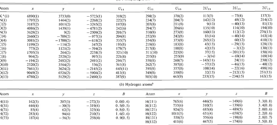
TABLE 1. Final parameters and their standard deviations
(0) Nonhydrogen atoms"
Atom x Y z U I I u22 u3 3 2 u z 3 2u13 2u12
(b) Hydrogen atoms"
Atom x 11 z B Atom x 1' B
.These parameters were multiplied by 101. T h e thermal parameters a r e expressed as exp [-2rr2(U, lhzo*' -4- U22k2b' -i- U 3 3 1 z ~ * z -i- 2U2'klb*c* i- Z U 1 3 h / u * ~ * -I- ~ u ~ , / l k ~ * b * ) I . bThe number of the a t o m t o which the hydrosen is bonded is obtained by dropping the leirsl significant digit ol' the hydrogen a t o m number. T h e coordinates were multipl~ed by 10'.
828 C A N . J. C H E M . VOL. 5 6 . 1978
TABLE 2. Torsional angles in the photodimer
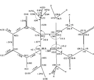
FIG. 1. Bond distances and angles in the 5-methylorotate photodimer anion. The esd's for the distances involving only nonhydrogen atoms are 0.002-0.003
A ;
esd's for the corres- ponding angles are 0.13-0.19". When hydrogen atoms are involved the esd's are about 10 times larger.derived from the libration tensor were less than the
individual esd's and were therefore not applied.
Discussion of the Crystal Structure
In this photodinier a crystallographic centre of
symmetry lies in the centre of the cyclobutane ring,
constraining it t o be planar. The two pyrimidine
rings are fused t o the cyclobutane ring in
tt.ans-anti
configuration and the dihedral angle between the
cyclobutane ring and the meal1 pyrimidine ring
plane is 121.6". Each pyrimidine ring is significantly
nonplanar, and the endocyclic torsional angles, given
in Table 2, indicate that the distortions from
planarity are in the direction of a slight twist-boat
shape (17). Of the six atoms, C(4) deviates inost
(0.088 A) from the mean pyrimidine ring plane,
carrying O(4) 0.181
A
out of the plane, thereby
increasing the separation between O(4) and C(7)
which is, even so, only 2.84 A . The C(7)...C(S) and
C(7)...N(1)' nonbonded distances, 2.83 and 2.81
A
respectively, are also rather short, indicating the
severe overcrowding caused by the full substitution
of C(5), C(6), C(5)', C(6)'.
The bond lengths and angles in this structure are,
in general, equal within experimental error t o those
in the
cis-anti
photodimer of 5-methylorotate (6).
The few significant deviations are shown in Table 3,
and it is interesting t o note that where parameters
involving only pyrimidine ring atoms differ, i.e. the
N(1)-C(2)
bond length and C(6)-N(1)-C(2),
C(5)-C(6)-N(1)
bond angles, the values found in
the present
trrrns-anti
structure compare rather better
with the values in the thymine
trans-rrnti
photodimer
(18). Other significant deviations, involving the
( a ) Endocyclic torsional angles in the pyrimidine ring
(b) Other torsional angles
C(4)-C(5)-C(7),
N(
I )-C(6)-C(8),
and C(5)'-
C(6)-C(8)
angles, probably can be explained as the
result of the different distortions necessary to mini-
mize interatomic repulsion in the two isomers.
Partial relief of these forces of interatomic repul-
sion is also achieved by the lengthening of the
C(5)-C(6)
and C(6)-C(8)
bonds. The C(5)-C(6)
bond length in the present structure (1.577 A), while
essentially equal t o the corresponding value (1.57 A)
in the
cis-anti
isomer, is significantly longer than in
most other pyrimidine photodimers (3, 18-21). T h e
C(6)-C(8)
bond lengths in this structure (1.555 A)
and the
cis-anti
isomer (1.56 A) are significantly
longer than corresponding values in the orotic acid
monohydrate structure (22) (1.498 A) and in the
trans-syn
photodimer of methylorotate (3) (1.522 A).
Differences in the interpyrimidine bond lengths in
the present structure and in the
tt.ans-syn
orotate
photodimer help partially t o explain the differences
in lability of the two din~ers,
In the
trans-sy17
crystal
structure (3), the C(6)-C(6)'
bond, to which both
carboxyl groups are bonded, is abnormally length-
ened t o 1.628 A, while the C(5)-C(5)'
bond, 1.556 A,
is close t o normal length, a s are the intrapyrimidine
bonds, 1.544 A. In the present structure the strain
induced by overcrowding is distributed more
equally; the interpyrimidine bonds (1.585A) are
essentially the same length as the intrapyrimidine
bonds (1.577 A). If we assume that the C(6)-C(6)'
bond in (nonesterified)
trans-syn
orotate photodinier
has a fairly similar length t o that observed in the
crystal structure of the methyl ester (and the con-
figurations of the dimer acid and ester have been
shown t o be identical
(2)), then the increased
lability in neutral and acid media of the orotate
photodimer relative to the 5-methylorotate dimer
H U B E R ET A L
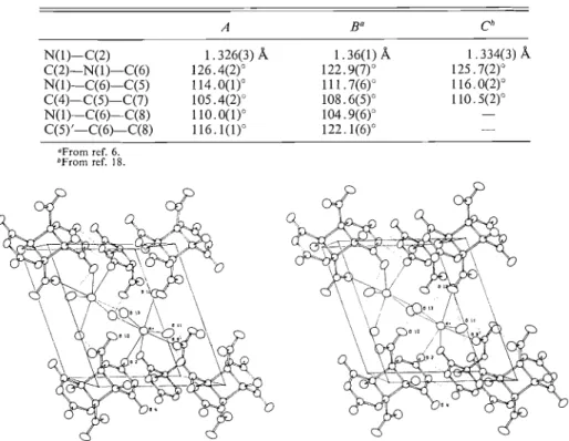
TABLE 3 . Comparison of molecular geometry in the trans-anti ( A ) and cis-ar~ti ( B ) photodimers of 5-methylorotate. Bond length and bond angle values given here are those showing significant differences in the two structures. Con~parable values
are shown also from the trans-anti thymine photodimer ( C )
OFrorn ref. 6. bFrorn ref. 18.
FIG. 2. Stereoscopic view of the packing arrangement drawn by the ORTEP program ( 2 3 ) . K + . - O contacts are indicated by single solid lines, hydrogen bonds by dotted lines. Axial directions: s - t , y t , z pointing out of the plane toward the reader.
can be correlated with this longer bond. It is not so
ueasy to understand why, in alkaline solution, the
cyclobutane ring is preserved in the case of orotate
photodimer (and also a
cis-syn
thymine photodirner
(15)),
and cleaved in the case of 5-methylorotate
photodimer.
The carboxyl groups in the present diiner are both
ionized, balancing the two K + ions per unit cell, and
one would therefore expect delocalization to make
the C-0
bond l'engths equal. The small difference,
0.009
A,
between the observed C(8)-O(9)
and
C(8)-O(10) bond lengths is possibly not significant,
but could be ascribed to different hydrogen bonding
patterns at O(9) and O(10) (see below).
The potassiunl ion is seven-coordinated, by three
oxygen atoms from the diiner and by four contacts
to water-molecule oxygens. Six of the K+...O dis-
tances are in the range 2.67-2,94A, while the
seventh is 3.057
A.
There are no other K+...O
distances less than 3.50 A, and no short contacts to
nonoxygen atoms. The six closer oxygens can be
described as forming a distorted octahedron, with
the seventh oxygen acco~nmodated outside the
largest octahedral face. Both the sevenfold coordina-
tion of the
K + ion and the range of K+...O distances
are unexceptional.
Figure 2 gives a stereoscopic view of the packing
arrangement and hydrogen bonding, and further
geometrical details about the hydrogen bonds are
listed in Table 4. All of the available hydrogen atoms
are involved in hydrogen bond formation.
The dimes anions are linked into infinite chains
along [IOi] by pairs of hydrogen bonds joining N(3)
and O(2) of one dimer to O(2) and N(3) respectively
of an adjacent dimer. The pairs of pyrimidine rings
joined by hydrogen bonds are coplanar within 0.2
A.
Diiner anions within a chain are also linked through
participation of 0(2), 0(4), and 0(9), from adjacent
dimers, in each potassium coordination polyhedron.
The coordination polyhedra occur in pairs, sharing
an edge of which the mid-point is a centre of sym-
metry. The only linkage of one dimer chain to
another by the potassium coordination polyhedron
is indirect, via centrosy~n~netrically
related 0(2),
0(4), and O(9) atoms at opposite ends of each
coordination pair.
830 C A N J CHEh4 VOL 56. 1978
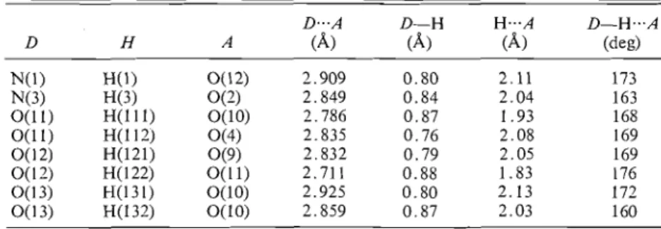
TABLE 4. Geometrical details of the hydrogen bonds, D-H...A
D . . . A D-H H...A D - H , . , A
D H A
(A)
(A)
(A)
(deg)Other cohesive forces are provided by the hydro-
gen bonds involving water molecules. Adjacent
dimer chains In the
xz
plane are linked via 0(12),
while connections in the
ydirection are via O(l1) and
O(13).
The appearance of the report (6) on the
cis-nnti
photodimer of 5-methvlorotate, which followed corn-
pletion of the present inalysis, raises the question of
why two different isomers were obtained under
apparently very similar experimental conditions.
(Both photodimers were prepared by irradiation of
frozen aqueous solutions of 5-methylorotic acid, and
then converted to the particular salts.) It seems
possible that the different dinler may have resulted
from differences in the procedure employed for
preparation of the ice matrix, o r that crystallization
of the barium salt led t o preferential isolation of one
o f the minor isonlers referred to above. Unfor-
tunately, Cheng
et
a/.(6) give no quantitative data
o n yields o r chromatographic behaviour of the crude
photodimer solution prior to crystallization.
I
Acknowledgments
We thank Dr.
R.
W. Dabeka and
H.
Seguin for
chemical analyses.
1. M. CH,\RLIER. C. HLLLNE, and M. DOURLEN-I.. J . Chilli. Phys. Phys.-Chim. Biol. 66,700 (1969).
2. G . I. B I K N B A U M , J. M . DUNSION. and A . G . SZABO. 'Tet- rahedron Lert. 947 (1971).
3. G . I . B I R N B A U M . ActaCrystallogl-. Sect. B, 28. 1248(1972). 4. E. SZ-I.UMPF iind D. S H U G A R . Pliotoctiern. Photobiol. 4,719
(1965).
5 , R. L I S E W S K I aricl K. L. W I E K Z C ~ I O W S K I . Photochem. Photobiol. 11.327 (1970).
6. P.-I'. C H E N G , V. H O R N B Y , W. WOWG-Nc;, S . C . N Y B U R G ,
and D. W E I S B L U M . Actil Crystitllogr. Sect. B, 32, 2251 (1976).
7. J . KARLE. Acta Crystallogr. Sect. B , 24, 182 (1968). 8. Interna~ional Tables for X-ray Crystallog~xphy, Vol. 1V.
Kynoch Press, Birrninghiim, Engl2uid. 1974. p. 72. 9. R. F. S-I-EWARI-, E. R. DAVII)SON, i~ncl W. '1'. S I M P S O N . J .
Chem. Phys. 42.3 175 (1965).
10. P. R. P I N N O C K , C. A. T A Y L O R , iind I H . LII'SON. Aclit CI'YS- tallogr. 9. 173 (1956).
I I. F. R. A H M C D , S. R. HALL, M. E. PIPPY. ;i1ic1 C. P. H U B E R .
J . Appl. Crystnllogr. 6.309(1973).
12. C . J A N I O N id D. S H U G A R . Actit Biochim. Pol. 7. 309 (1960).
13. K. L. W I E R Z C H O W S K I , E. LI-IONSKA, alicl D. S I I U G A R . J . Am. Chem. Soc. 87,4621 (1965).
14. D. L. WULI'F itlid G. FIIAENKIL. Biochilli. Biophys. Acta. 51. 332(1961).
15. G. M. B L A C K B U R N ancl R. J . H . DAVIES. J. Chem. Soc. C, 2239 (1966).
16. V. S C ~ ~ O M A K E K and K. N. TRUEBLOOD. Acta C~.ystallOg~.. Sect. B, 24,63 (1968).
17. R. BUCOUR'I- and D. H A I N A U T . Bull. Soc. Chim. Fr. 1366 (1965).
18. N. C A M E K ~ I A N itlid S . C. NYBURG. ActaCrvstallogr. Sect. -
B, 25,388 (19693.
19. N. C A M E R M A N . D. W E I N B L U M , itnd S. C. NYBURG. J . Am. Chem. Soc. 91.982 (1969).
20. E. ADMAN and L. H. J ~ N S E N . ActaCrystallogr. Sect. B,26. 1326 (1970).
21. J . K O N N E R T and I . L. KARLE. J . Crvst. Mol. Str~lct. 1 , 107 (1971).
22. F. TAKUSAGAWA and A . S H I M A D A . Bull. Ctle111. SOC. Jpn.
46,201 1 (1973).
23. C. K. JOHNSON. OR'TEP, Report ORNL-3794. 2nd rev. Oitk Ridge National Laboratory, Oitk Ridge, Tennessee. 1970.