similar suites of dissolved organic matter
The MIT Faculty has made this article openly available.
Please share
how this access benefits you. Your story matters.
Citation
Becker, Jamie W., Paul M. Berube, Christopher L. Follett, John B.
Waterbury, Sallie W. Chisholm, Edward F. DeLong, and Daniel J.
Repeta. “Closely Related Phytoplankton Species Produce Similar
Suites of Dissolved Organic Matter.” Frontiers in Microbiology 5
(March 28, 2014).
As Published
http://dx.doi.org/10.3389/fmicb.2014.00111
Publisher
Frontiers Research Foundation
Version
Final published version
Citable link
http://hdl.handle.net/1721.1/88049
Terms of Use
Article is made available in accordance with the publisher's
policy and may be subject to US copyright law. Please refer to the
publisher's site for terms of use.
Closely related phytoplankton species produce similar
suites of dissolved organic matter
Jamie W. Becker1,2
, Paul M. Berube2
, Christopher L. Follett3
, John B. Waterbury1
, Sallie W. Chisholm2,4, Edward F. DeLong2,5and Daniel J. Repeta3*
1Department of Biology, Woods Hole Oceanographic Institution, Woods Hole, MA, USA
2
Department of Civil and Environmental Engineering, Massachusetts Institute of Technology, Cambridge, MA, USA
3
Department of Marine Chemistry and Geochemistry, Woods Hole Oceanographic Institution, Woods Hole, MA, USA
4
Department of Biology, Massachusetts Institute of Technology, Cambridge, MA, USA
5Department of Biological Engineering, Massachusetts Institute of Technology, Cambridge, MA, USA
Edited by:
Jennifer Pett-Ridge, Lawrence Livermore National Lab, USA Reviewed by:
Tom Metz, Pacific Northwest National Laboratory, USA Xavier Mayali, Lawrence Livermore National Laboratory, USA *Correspondence:
Daniel J. Repeta, Department of Marine Chemistry and Geochemistry, Woods Hole Oceanographic Institution, Watson Building, MS #51, 266 Woods Hole Rd., Woods Hole, MA 02543, USA e-mail: drepeta@whoi.edu
Production of dissolved organic matter (DOM) by marine phytoplankton supplies the majority of organic substrate consumed by heterotrophic bacterioplankton in the sea. This production and subsequent consumption converts a vast quantity of carbon, nitrogen, and phosphorus between organic and inorganic forms, directly impacting global cycles of these biologically important elements. Details regarding the chemical composition of DOM produced by marine phytoplankton are sparse, and while often assumed, it is not currently known if phylogenetically distinct groups of marine phytoplankton release characteristic suites of DOM. To investigate the relationship between specific phytoplankton groups and the DOM they release, hydrophobic phytoplankton-derived dissolved organic matter (DOMP) from eight axenic strains was analyzed using high-performance liquid
chromatography coupled to mass spectrometry (HPLC-MS). Identification of DOM features derived from Prochlorococcus, Synechococcus, Thalassiosira, and Phaeodactylum revealed DOMPto be complex and highly strain dependent. Connections between DOMP
features and the phylogenetic relatedness of these strains were identified on multiple levels of phylogenetic distance, suggesting that marine phytoplankton produce DOM that in part reflects its phylogenetic origin. Chemical information regarding the size and polarity ranges of features from defined biological sources was also obtained. Our findings reveal DOMPcomposition to be partially conserved among related phytoplankton species, and
implicate marine DOM as a potential factor influencing microbial diversity in the sea by acting as a link between autotrophic and heterotrophic microbial community structures.
Keywords: dissolved organic matter, untargeted metabolomics, marine phytoplankton, exometabolome,
Prochlorococcus, Synechococcus, Thalassiosira, Phaeodactylum
INTRODUCTION
Extracellular release of dissolved organic matter (DOM) by marine phytoplankton fuels secondary production in the sea (Pomeroy, 1974; Mague et al., 1980; Fogg, 1983; Baines and Pace, 1991). As much as 50% of the total carbon fixed by photoau-totrophs may be released into seawater as a by-product of normal metabolism, or through active processes for waste removal, sub-strate acquisition, defense, or communication (Bjornsen, 1988; Carlson, 2002; Bertilsson and Jones, 2003). The amount of extracellular DOM released and its composition depend on the organism and its physiological state (Myklestad, 1995; Meon and Kirchman, 2001; Wetz and Wheeler, 2007; Romera-Castillo et al., 2010), as well as additional factors including: temperature, light, growth phase, availability of inorganic nutrients, and the pres-ence of other organisms (Hirt et al., 1971; Obernosterer and Herndl, 1995; Grossart and Simon, 2007; Barofsky et al., 2009; Engel et al., 2011). Factors affecting DOM uptake are less clear, but recent evidence suggests that DOM resource partitioning within microbial communities may impact the ecological suc-cess and community structure of heterotrophic bacterioplankton
(McCarren et al., 2010; Poretsky et al., 2010; Nelson and Carlson, 2012; Sarmento and Gasol, 2012). Substrate specificity may be of particular importance for heterotrophs that have evolved streamlined genomes and are therefore fine-tuned to particular resources (Giovannoni et al., 2005, 2008).
Genomic and physiological differences among marine phy-toplankton likely influence the nature of the organic matter they produce, and consequently, what substrates are available to sympatric heterotrophic communities. If different phytoplank-ton groups release DOM of varying composition and lability, then DOM could provide a direct link between autotrophic and heterotrophic microbial diversity and community structure. For example, Prochlorococcus is frequently the numerically dominant photoautotroph in oligotrophic surface waters and is an essential source of organic matter used by heterotrophic bacterioplankton in this environment (Partensky et al., 1999; Bertilsson et al., 2005; Flombaum et al., 2013). It has been suggested that DOM released by Prochlorococcus could be responsible for supporting 12–41% of bacterial production in tropical and subtropical regions (Bertilsson et al., 2005). If DOM released by Prochlorococcus
is distinct in composition, metabolic specialization to utilize Prochlorococcus-derived DOM within associated heterotrophic populations may influence bacterioplankton community struc-ture in these regions.
Several recent studies have employed phytoplankton-derived DOM for use in the study of microbial DOM consump-tion (Romera-Castillo et al., 2011; Nelson and Carlson, 2012; Sarmento and Gasol, 2012; Landa et al., 2013a,b; Sharma et al., 2013), with the implied assumption that DOM suites derived from individual phytoplankton strains represent compositionally distinct organic substrates. However, a comparative assessment of phytoplankton-derived DOM has not been conducted at the molecular level and it is not yet clear how much chemical varia-tion exists in DOM produced by different marine phytoplankton types. Furthermore, it is not known how DOM composition varies as a function of taxonomic or phylogenetic relatedness among different phytoplankton species. Investigating how DOM produced by marine phytoplankton varies with phylogenetic relatedness may help us to better understand marine microbial community structure, allow us to target important components of DOM in future studies, and potentially improve our predictive powers regarding the composition of DOM produced by different phytoplankton communities.
To investigate the relationship between phylogenetic related-ness and DOM composition, we used HPLC-MS to detect chem-ical features in DOM released by laboratory cultures of marine phytoplankton. While the determination of empirical formulae within DOM is now possible through advances in ultrahigh-resolution mass spectrometry, definitive chemical identifications in untargeted metabolomic footprinting studies remain a chal-lenge due to structural complexities including isomerization and the magnitude of unknown metabolites that comprise both
marine (Nebbioso and Piccolo, 2013) and phytoplankton-derived DOM (Schwarz et al., 2013). Rather than attempt chemical iden-tifications with no a priori knowledge regarding compounds of interest, we chose to sacrifice mass resolution for chromato-graphic separation, providing us with two independent param-eters (i.e., polarity and mass) to detect and compare chemical features in DOM released by eight marine phytoplankton strains. These strains were selected to encompass both the Bacteria and Eukarya domains. Cyanobacterial strains within the genera Prochlorococcus and Synechococcus were chosen to represent cells with different adaptations to light and temperature. Thalassiosira and Phaeodactylum were chosen to represent different groups of eukaryotic diatoms (centric and pennate, respectively). Selection of these strains provided both broad phylogenetic coverage as well as examples of closely related organisms with unique physiolo-gies. Hundreds to thousands of features detected in each culture sample were used to chemically compare DOM produced by phy-toplankton strains with differing degrees of relatedness. The suite of features associated with each strain was then compared with phylogenies deduced from both rRNA and whole genome com-parisons to uncover novel connections between phytoplankton phylogeny and DOM composition.
MATERIALS AND METHODS
PHYTOPLANKTON CULTURES
Eight species of marine phytoplankton were selected for this study to investigate variability in the composition of organic matter released by unicellular photoautotrophs at various taxonomic lev-els (Table 1). Individual strains were chosen on the criteria that a published sequenced genome is, or will soon be available, and that axenic strains were capable of growth to high cell densities (ca. 107cells/ml) in natural seawater-based media. All media was
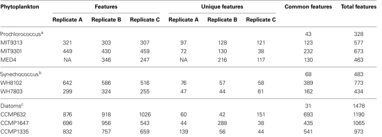
Table 1 | DOMPfeatures detected using HPLC-ESI-MS after blank subtraction.
Phytoplankton Features Unique features Common features Total features Replicate A Replicate B Replicate C Replicate A Replicate B Replicate C
Prochlorococcusa 43 328 MIT9313 321 303 307 97 128 121 123 577 MIT9301 449 430 459 72 130 38 232 673 MED4 NA 346 247 NA 216 117 130 463 Synechococcusb 68 483 WH8102 642 586 516 76 57 58 389 773 WH7803 299 324 255 47 44 61 162 434 Diatomsc 31 1478 CCMP632 876 918 1026 60 42 151 693 1190 CCMP1647 696 956 543 44 288 38 435 1065 CCMP1335 832 757 659 139 56 44 541 973
Feature totals for individual biological replicates of each phytoplankton strain are shown along with the sum of all features for each strain. The number of features unique to an individual replicate as well the number of features common to all replicates for a given strain are also included. Total and common features (given that each feature was shared among biological replicates) are provided for each organism group.
aDescribed inMoore and Chisholm (1995); Moore et al. (1998); Rubio et al. (1999); Rocap et al. (2002, 2003).
bDescribed inWaterbury et al. (1986); Rocap et al. (2002); Palenik et al. (2003).
prepared by the National Center for Marine Algae and Microbiota (NCMA) using filtered, autoclaved surface water collected from the Sargasso Sea. Media amendments were added aseptically after sterilization. Triplicate 1 L batch cultures of each strain were prepared alongside triplicate 1 L controls containing all media amendments, but no cell additions. Cells were pre-conditioned in 60–400 ml of the appropriate medium prior to inoculation in 1 L. Thalassiosira pseudonana (CCMP str. 1335), Thalassiosira rotula (CCMP str. 1647), Phaeodactylum tricornutum (CCMP str. 632), and both Synechococcus strains (WH8102 and WH7803) were grown at the NCMA in L1 medium prepared according to existing protocols (Guillard and Hargraves, 1993). Diatom strains were grown at 20◦C and Synechococcus strains at 24◦C. All five strains were grown on a 13/11 light/dark cycle with gentle periodic mixing. Synechococcus str. WH7803 was grown under 10–20µmol photons/m2/s illumination, while the remain-ing strains were grown at 100–120µmol photons/m2/s. Diatom cells were enumerated using a Palmer-Maloney counting chamber at 40X magnification on a Zeiss light microscope. Synechococcus cells were enumerated via epifluorescence microscopy. Three Prochlorococcus strains (MED4, MIT9301, and MIT9313) were grown at 22◦C in Pro99 medium prepared by the NCMA accord-ing to existaccord-ing protocols (Moore et al., 2002, 2007) with the addi-tion of 5 mM sodium bicarbonate. All three strains were grown on a 14/10 light/dark cycle. Prochlorococcus str. MIT9313 was grown under low light conditions at ca. 20µmol photons/m2/s, while MIT9301 and MED4 were grown at ca. 40µmol photons/m2/s. Prochlorococcus cultures were monitored for growth using total fluorescence and for changes in pH, which were mitigated by additions of 2 mM HEPES buffer and 5 mM sodium bicarbon-ate 6 days after inoculation (sensuMoore et al., 2007). Samples for direct cell counts by flow cytometry were fixed with 0.125% final concentration of grade I glutaraldehyde (Sigma) and stored at –80◦C prior to analysis. Fixed samples were diluted in 0.2µm filtered seawater and Prochlorococcus were enumerated using an Influx Cell Sorter (BD Biosciences) as previously described (Olson et al., 1985; Cavender-Bares et al., 1999).
ORGANIC MATTER PRODUCTION
Samples for cell enumeration and particulate and dissolved organic carbon (POC and DOC, respectively) quantification were taken at the onset of stationary phase growth to evaluate organic carbon production by each strain. An exception was that DOC data for Prochlorococcus strains was not acquired due to the addi-tion of HEPES buffer during growth to mitigate pH changes (sensu Moore et al., 2007). All organic carbon samples were processed using combusted (450◦C for 8 h) glassware. Duplicate samples for POC analysis were taken by vacuum filtering 25 ml of sample onto a combusted 25 mm 0.7µm glass fiber filter (Whatman GF/F). Filters were then placed inside a combusted glass petri dish, wrapped in foil, and immediately frozen. A blank filter was also prepared at each sampling point. Filters were later thawed and dried (60◦C; overnight) before encapsulation into 9× 10 mm tin capsules. Measurements for POC quantifi-cation were performed by the University of California Davis Stable Isotope Facility. POC filtrates were transferred into com-busted glass vials, acidified with 150µl of a 25% phosphoric acid
solution and measured for DOC using high temperature catalytic oxidation on a Shimadzu TOC-VCSHwith platinized aluminum
catalyst. Sample concentrations were determined alongside potas-sium hydrogen phthalate standards and consensus reference materials provided by the DOC-CRM program (www.rsmas. miami.edu/groups/biogeochem/CRM.html).
EXTRACTION OF DOMP
Upon entering stationary phase, cells were removed by cen-trifugation at either 878 RCF (diatom cultures) or 10,751 RCF (cyanobacteria cultures) for 15 min, followed by gentle filtration (0.1µm; Whatman Polycap 36 TC capsule filter). Filtrates were then stored briefly in the dark at 4◦C until solid-phase extrac-tion (SPE). Material obtained via SPE constitutes 20–60% of the total DOC in marine DOM (Dittmar et al., 2008), encompassing a large amount of organic substrate available to heterotrophic bac-terioplankton. Media controls were processed and stored along-side the culture samples. Filtrates were acidified to pH 2–3 by addition of trace metal grade hydrochloric acid and organic mat-ter was extracted onto ISOLUTE C18(EC) SPE columns (0.5 g, Biotage) at a rate of 1 ml/min. SPE columns were precondi-tioned with 5 ml HPLC-grade methanol followed by 10 ml ultra-pure water. After sampling, mineral salts were washed from the columns with acidified ultrapure water (pH 2–3) at a flow rate of 1 ml/min. Organic matter was recovered by gravity elution using 10 column volumes of acidified HPLC-grade methanol (pH 2–3). Samples were concentrated to a small volume by rotary evap-oration, and then taken to dryness under filtered, high purity nitrogen. Samples were resuspended in 156µl of methanol, and stored briefly in combusted amber vials at 4◦C in the dark prior to chemical analysis. A 3µl subsample of each was placed onto a combusted 25 mm, 0.7µm glass fiber filter (Whatman GF/F), and submitted for POC analysis to quantify the organic carbon recovered by SPE.
CHROMATOGRAPHY AND MASS SPECTROMETRY
Chromatographic and spectrometric analyses were performed using an Agilent 1200 series high-performance liquid chromato-graph coupled to an Agilent 6130 (single quadrupole) mass spec-trometer. Organic extracts were separated on a ZORBAX SB-C18 column (Agilent; 3.5µm 4.6 × 150 mm) eluted at 0.8 ml min−1 using a linear gradient (% solvent A, % solvent B, minutes): 100, 0, 0; 20, 80, 31.25; 0, 100, 43.75; 0, 100, 64, where solvent A is aqueous formic acid (0.1%) and solvent B is methanolic formic acid (0.1%). Mass spectrometry was performed using an atmo-spheric electrospray ionization (ESI) source. Drying gas was set at 11.5 l min−1and 300◦C, the nebulizer was at 60 psig, and capil-lary voltage was set to 4000 V. Data was obtained in the positive ion mode from 100–2000 m/z with a 4.0 fragmentor, 150 thresh-old, and 0.1 step size. A tune solution containing five standard compounds (117–2122 m/z) was used to determine mass accu-racy of the instrument prior to analysis. Mass accuaccu-racy in this size range was found to always be within a tolerance of 0.2 Da.
Mass spectral data was analyzed using MZmine 2 molecular profiling software (Pluskal et al., 2010). Ions with signal inten-sity at least 5-fold greater than the maximum noise level were identified using a centroid mass detector. Chromatograms were
then built from the raw data using a retention time tolerance of±5 s, and a mass tolerance of ±0.3 m/z. Alignments were made to correct for small shifts in retention times between samples and individual peaks were identified by setting a minimum accept-able intensity (5-fold greater than the noise level) and duration (5 s) to remove noise within each chromatogram, and also by searching for local minima within each chromatogram. Adduct peaks ([M+Na]+, [M+2Na-H]+, [M+H2PO4]+, [M+HSO4]+)
and isotopic peaks were identified and removed within a reten-tion time tolerance of±1.42 s, and a mass tolerance of ±0.2 m/z. Additional adduct peaks for Prochlorococcus ([M+NH4]+) and
both Synechococcus and diatoms ([M+K]+) were identified and removed due to the presence of these chemical species in the media types used to grow these strains. Constraints were placed on the intensity ratio of identified isotope pairs based on the min-imum ((M–1)/30) and maxmin-imum ((M–3)/14) number of carbon atoms for a given mass. Sample feature lists were then aligned and gaps were filled in using a secondary threshold of 3-fold greater than the noise to identify common peaks that fell just below the initial strict threshold. A feature was defined as a unique m/z at a specific retention time, thus multiple features could share a specific m/z or retention time, but never both. While the possi-bility of distinct metabolites sharing identical retention times and m/z values within the tolerance levels mentioned above cannot be ruled out, the use of two independent parameters (polarity and mass) to designate DOMPfeatures reduces the likelihood of
falsely identifying common features.
Biological triplicates of each strain were first compared against triplicate media controls to distinguish material produced by the organism from any background material, including DOM present in the seawater medium used to cultivate each strain. Features were considered to be associated with a particular organism only if they were present in all three biological replicates of that strain and absent in all three replicate controls of the appropriate media type. Removal of features present in sterile media controls reduced the possibility of differences arising due to variations in growth media (i.e., Pro99 medium vs. L1 medium). Features were also removed if they were present in any blank samples, including triplicate instrument blank injections of pure methanol and trip-licate processing blanks created by rinsing and eluting pre-cleaned resins without any prior sample loading. Feature lists for each organism were then aligned to identify both common and unique features among the eight strains tested. Aligned feature lists were also used to investigate intensity level differences among features common to multiple samples.
To provide a rigorous analysis of the similarity between all cul-tures, an aligned feature list containing all the features detected in every replicate was created and converted into a binary matrix where each column represented an individual culture and each row a feature present in at least one culture. Multiplying this matrix by its transpose created a second matrix where each ele-ment was the number of features common to each pairwise culture comparison. The matrix was then normalized by the total number of features present in each pairwise comparison. This normalized similarity matrix was used to quantify the similarity of feature lists and their parent strains, and to generate a cluster diagram as a visual representation of DOMP similarity among
strains. The number of features shared by any two cultures was divided by the sum of features present in those cultures to generate a percent similarity value for all pairwise sample combinations.
RESULTS
We employed liquid chromatography coupled to low-resolution mass spectrometry to characterize DOM produced by eight marine phytoplankton strains grown in axenic batch cultures. We determined this approach to be of sufficient mass resolution based on elemental analyses of DOM using ultrahigh-resolution mass spectrometry that show the following four prominent, reoccur-ring mass differences: 14.00156 Da (CH2), 2.0157 Da (double
bond/ring series), 1.0034 Da (13C and12C), and 0.0364 Da (C=O
vs. CH-CH3). All of these mass differences can be distinguished
using the mass resolution of the instrument in our study, except for 0.0364 Da. This final mass difference would also likely lead to shifts in retention time that can be resolved by HPLC how-ever, and would therefore be detected as distinct features in our data set. Indeed, we analyzed DOM isolated from Prochlorococcus str. MED4 and its corresponding medium control using identical chromatography coupled to Fourier-transform ion cyclotron res-onance mass spectrometry (FT-ICR-MS). Processing the resulting data using an appropriate mass resolution (2 ppm) revealed only a modest increase in the number of total DOM features iden-tified in this sample when compared to results obtained using low-resolution mass spectrometry (364 features compared to the 346 features reported in this study; FT-ICR-MS data not shown). Equal sample volumes were injected to minimize differences in signal-to-noise between these analyses.
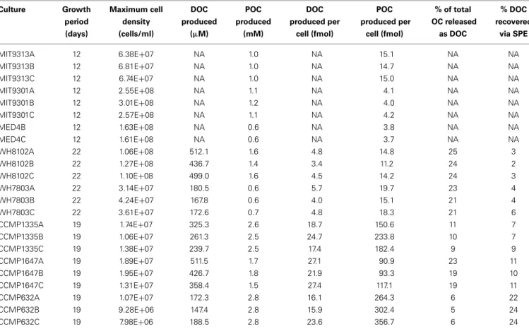
All eight of the marine phytoplankton strains analyzed pro-duced DOM containing hundreds of extracellular hydrophobic features recovered by SPE and detected by HPLC-ESI-MS (Table 1 and Supplementary File 1). Final cell densities and organic carbon production for each replicate are detailed in Table 2. A variable portion (5–25%) of the organic carbon produced by each phyto-plankton strain was released as extracellular DOM where it might be accessible to heterotrophic microbes in a natural setting. The majority of the features recovered were consistently associated with only one of the strains tested, while a relatively small sub-set appeared to be produced by a diverse range of organisms. Of the 2032 features detected in culture samples, ranging from 104– 1466 m/z, the majority (1633 features, or 80%) were unique to a particular strain; only a few (6 features, or 0.3%) were found in all eight. Some variation in feature composition was also found among biological replicates for all strains (Table 1)—possibly a result of uncontrollable differences in growth conditions and/or small variations in growth stage at the time of processing. On average, 43% of the features for a given set of triplicates were found in all three, while 24% were found in two out of the three, and the remaining 33% were unique to one replicate. On average, 61% of the features in a given replicate were also present in the other two replicates. DOMPcomposition of the different strains
was compared using two approaches. First, only those features common to all three replicates for a given strain were consid-ered produced by that strain. These features were then compared among all eight strains to stringently identify both common and
Table 2 | Biomass and organic carbon production by phytoplankton cultures tested in this study.
Culture Growth Maximum cell DOC POC DOC POC % of total % DOC period density produced produced produced per produced per OC released recovered (days) (cells/ml) (µM) (mM) cell (fmol) cell (fmol) as DOC via SPE MIT9313A 12 6.38E+07 NA 1.0 NA 15.1 NA NA MIT9313B 12 6.81E+07 NA 1.0 NA 14.7 NA NA MIT9313C 12 6.74E+07 NA 1.0 NA 15.0 NA NA MIT9301A 12 2.55E+08 NA 1.1 NA 4.1 NA NA MIT9301B 12 3.01E+08 NA 1.2 NA 4.0 NA NA MIT9301C 12 2.57E+08 NA 1.1 NA 4.2 NA NA MED4B 12 1.63E+08 NA 0.6 NA 3.8 NA NA MED4C 12 1.61E+08 NA 0.6 NA 3.7 NA NA WH8102A 22 1.06E+08 512.1 1.6 4.8 14.8 25 3 WH8102B 22 1.27E+08 436.7 1.4 3.4 11.2 24 2 WH8102C 22 1.10E+08 499.0 1.6 4.5 14.2 24 3 WH7803A 22 3.14E+07 180.5 0.6 5.7 19.7 23 4 WH7803B 22 4.24E+07 167.8 0.6 4.0 15.1 21 4 WH7803C 22 3.61E+07 172.6 0.7 4.8 18.3 21 6 CCMP1335A 19 1.74E+07 325.3 2.6 18.7 150.6 11 7 CCMP1335B 19 1.06E+07 261.3 2.5 24.7 233.8 10 7 CCMP1335C 19 1.38E+07 239.7 2.5 17.4 182.4 9 9 CCMP1647A 19 1.89E+07 511.5 1.7 27.1 90.9 23 11 CCMP1647B 19 1.95E+07 426.7 1.8 21.9 93.3 19 10 CCMP1647C 19 1.31E+07 358.4 1.5 27.4 117.1 19 11 CCMP632A 19 1.07E+07 172.3 2.8 16.1 264.3 6 22 CCMP632B 19 9.28E+06 147.4 2.8 15.9 302.4 5 24 CCMP632C 19 7.98E+06 188.5 2.8 23.6 356.7 6 24
Organic carbon values are background subtracted using media controls.
“NA” denotes data not available. Organic carbon data for Prochlorococcus strains was not acquired due to the addition of HEPES buffer during growth to mitigate pH changes.
unique features that were consistently associated with each strain. A second approach employed all features detected in any replicate to generate a similarity matrix for running all possible pairwise comparisons, treating all replicates as individual samples. Both of these approaches identified a similar connection between the phy-logenetic relationship of the phytoplankton strains tested and the chemical composition of the DOM they produced.
PROCHLOROCOCCUS
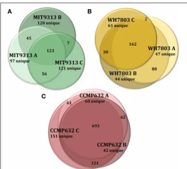
HPLC-ESI-MS detected 577, 673, and 463 features in samples from strains MIT9313, MIT9301, and MED4, respectively. Purity broth tests revealed subsequent heterotrophic contamination in one of the MED4 replicate cultures; therefore this sample was removed from all analyses (see N/A in Table 1). Approximately 40% of the features associated with each Prochlorococcus strain were detected in all replicates of that strain (Table 1). MIT9313 replicates had the least agreement of all the strains in this study (Figure 1A). Prochlorococcus-derived DOMPgenerally
con-sisted of small, non-polar material, with the majority of fea-tures detected between 200–500 m/z and 20–50 min, when the mobile phase composition was between 51 and 100% methanol (Figure 2).
Of the total Prochlorococcus-derived features detected in this study, 13% were produced by all three strains. High-light adapted
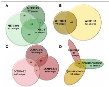
strains MIT9301 and MED4 were the most similar Prochlorococcus strains, sharing 34% of their features. Low-light adapted strain MIT9313 shared approximately 22% of its features with both high-light adapted strains (Figure 3A). Unique features (as much as 52% of the total) were identified in all three strains. Comparing all replicates as individual samples revealed that biological repli-cates were generally more alike (27–67% similar) than cultures of different strains (12–41% similar), although both MED4 sam-ples (MED4B and MED4C) had greater similarity to replicates of MIT9301 than to each other (Table 3).
SYNECHOCOCCUS
HPLC-ESI-MS detected 773 and 434 features in samples from strains WH8102 and WH7803, respectively. Approximately one half to three quarters of the features associated with each Synechococcus strain were detected in all three replicates of that strain (Table 1). The agreement among biological replicates of WH7803 was representative of the average agreement among replicates for all of the strains tested (Figure 1B). The majority of Synechococcus-derived features were detected between 150– 500 m/z and 12–50 min, when the mobile phase composition was between 31 and 100% methanol (Figure 2). Of the total Synechococcus-derived features detected in this study, 14% were found associated with both strains. Unique features (as much
FIGURE 1 | Venn diagrams showing the degree to which triplicate cultures of representative phytoplankton strains share DOMPfeatures. Each culture is represented by a circle, the area of which is proportional to the total number of features identified in that sample. The degree of overlap between circles is proportional to the number of shared features. Prochlorococcus str. MIT9313 replicates had the most variation of any strain tested (A), while P. tricornutum (CCMP632) replicates had the least variation (C). Synechococcus str. WH7803 replicates exhibited an average degree of variation indicative of most strains tested in this study (B). For more detailed information regarding replicate correlations, see Table 3.
as 83% of the total) were identified in both strains (Figure 3B). Comparing all replicates as individual samples revealed that all biological replicates were more alike (42–72% similar) than cul-tures of different strains (13–21% similar) (Table 3).
CYANOBACTERIA
Among the features consistently detected in Prochlorococcus and Synechococcus samples, 14% were found associated with all five cyanobacteria strains. The most similar strains between the two groups were Prochlorococcus str. MIT9301 and Synechococcus str. WH8102, sharing 20% of their features. All Prochlorococcus strains shared more features with each other than with either Synechococcus strain. Synechococcus str. WH7803 shared more features with the other Synechococcus strain tested (WH8102) than with any of the Prochlorococcus strains; however the reverse was not true. Synechococcus str. WH8102 had the most over-lap with Prochlorococcus str. MIT9301, sharing 20%, followed by MIT9313 (15%) and then WH7803 (14%). Low-light adapted Synechococcus str. WH7803 was more similar to the low-light adapted Prochlorococcus str. MIT9313 than it was to either of the high-light adapted Prochlorococcus strains. The percent of fea-tures shared between all cyanobacteria strain combinations are summarized in Table 4.
Comparing all replicates as individual samples confirmed that Synechococcus str. WH8102 and Prochlorococcus str. MIT9301 were the most similar Synechococcus and Prochlorococcus strains (15–23% similar) with respect to the DOM features analyzed,
and also revealed that WH7803 and the high-light adapted Prochlorococcus strains (MIT9301 and MED4) were the least sim-ilar cyanobacteria tested (6–14% simsim-ilar). In general, all of the Prochlorococcus strains were more similar to WH8102 (11–23% similar) than WH7803 (6–14% similar) (Table 3).
THALASSIOSIRA sp.
T. pseudonana (CCMP1335) and T. rotula (CCMP1647) were chosen to represent different species of the Thalassiosira genus. HPLC-ESI-MS detected 973 and 1065 features in samples from T. pseudonana and T. rotula, respectively. Approximately one half to five sixths of the features associated with each Thalassiosira strain were detected in all three replicates of that strain (Table 1). The majority of Thalassiosira-derived features were detected between 150–700 m/z and 20–45 min, when the mobile phase composi-tion was between 51 and 100% methanol (Figure 2). Of the total Thalassiosira-derived features detected in this study, 11% were found associated with both strains. Unique features (as much as 82% of the total) were identified in both strains (Figure 3C). Comparing all replicates as individual samples revealed that all biological replicates were more alike (47–71% similar) than cul-tures of different strains (12–15% similar) (Table 3).
PHAEODACTYLUM TRICORNUTUM
HPLC-ESI-MS detected 1190 features in P. tricornutum sam-ples. Approximately three fourths of the features associated with each P. tricornutum culture were detected in all three repli-cates (Table 1). P. tricornutum replirepli-cates had the most agreement of all the strains in this study (Figure 1C). Phaeodactylum-derived DOMP generally consisted of larger, more polar
mate-rial when compared to cyanobacteria DOMP, with the
major-ity of features detected between 150–1000 m/z and 12–45 min, when the mobile phase composition was between 31 and 100% methanol (Figure 2). Comparing all replicates as individual sam-ples revealed that the biological replicates were more alike (66– 73% similar) than cultures of any other strain (2–10% similar) (Table 3).
DIATOMS
In general, DOMP composition was more diverse among the
diatom strains than among the cyanobacteria. Among the fea-tures consistently detected in the diatom strains tested, only 2% were found associated with all three strains. T. pseudonana and T. rotula were the most similar diatoms, sharing 11% of their fea-tures. T. rotula and P. tricornutum shared 6% of their features, while T. pseudonana and P. tricornutum shared 5% (Figure 3C). A large number of unique features (as much as 87% of the total) were detected in all three diatom strains. T. pseudonana and T. rotula were both more similar to each other than to any of the cyanobacteria strains tested and P. tricornutum was more similar to both Thalassiosira diatoms than to any cyanobacteria strain. Comparing all replicates as individual samples revealed that biological replicates were more alike (47–73% similar) than cultures of different strains (7–15% similar), and confirmed that T. pseudonana and T. rotula are the most similar diatoms (12– 15% similar), followed by T. pseudonana and P. tricornutum (7–10% similar), and T. rotula and P. tricornutum (7–9% similar) (Table 3).
FIGURE 2 | Histograms comparing the size (A) and polarity (B) distributions of DOMP derived from representative strains from
each major phytoplankton group. Bars indicate the percentage of
features that fall within each bin labeled on the X-axes and
moving average lines are shown to highlight trends. In general,
diatom-derived DOMP had a more even size distribution and a
greater abundance of larger, more polar features than
cyanobacteria-derived DOMP.
DOMAIN-LEVEL COMPARISONS
The 2032 DOMP features detected in this study were
orga-nized into three groups: those detected in all Prochlorococcus strains, those detected in all Synechococcus strains, and those
detected in all diatom strains. Of the 122 features that can be classified this way, only 5% (6 features) were common to all three groups. Prochlorococcus and Synechococcus were the most similar groups, sharing 14% of their features, followed by
FIGURE 3 | Venn diagrams comparing DOMPcomposition at several
levels of phylogenetic variation for all eight phytoplankton strains tested in this study. Features common to all replicate cultures for a given
strain are represented by circles or ellipses, the area of which is proportional to the total number of features identified as common for that strain. The degree of overlap between shapes is proportional to the number of shared features between strains of Prochlorococcus (A), Synechococcus
(B), and diatoms (C). Features common to each of these 3 broad
phytoplankton groupings were also compared (D) to examine variation at the genus and domain levels. For more detailed information regarding
percentages of shared DOMPfeatures among different strains, see Table 4.
diatoms and Prochlorococcus sharing 10%, and finally diatoms and Synechococcus sharing 8% (Figure 3D). All three groups con-tained a large quantity of features that were unique to that group; the Synechococcus group had the most with 54 unique features, or 79% of its total. Combining the Prochlorococcus and Synechococcus groups into one cyanobacteria group and comparing this with the diatom group reduced the number of total classifiable features to 39. The same 6 features (now 15% of the total) mentioned above were common to both groups, while 8 features (21%) were unique to the cyanobacteria group and 25 features (64%) were unique to the diatom group.
Comparing all replicates as individual samples revealed that all five cyanobacteria strains were more similar to each other (12–41% similar) than to any diatom strain tested (2–12% sim-ilar). Comparisons between cyanobacteria and diatom strains revealed Prochlorococcus str. MIT9301 and T. rotula, and Synechococcus str. WH8102 and T. rotula to be the most similar strains between these two groups, both sharing 8% of their fea-tures. Prochlorococcus str. MED4 and P. tricornutum were the least similar strains, sharing only 1% of their features. Comparing all replicates as individual samples confirmed that all five cyanobac-teria strains were most like T. rotula (4–12% similar), followed by T. pseudonana (3–8% similar) and finally P. tricornutum (2–6% similar). T. pseudonana and P. tricornutum were both more simi-lar to all diatom strains than to any cyanobacteria tested. While T. rotula was most like T. pseudonana (11% similar), it had more features in common with Prochlorococcus str. MIT9301 (8% similar) and Synechococcus str. WH8102 (8% similar) than
with P. tricornutum (6% similar). All three diatom strains were more like Synechococcus str. WH8102 (4–12% similar) than any other cyanobacteria strain (2–10% similar) and more like Prochlorococcus str. MIT9301 (3–10% similar) than any other Prochlorococcus strain (2–7% similar). Similarity values are sum-marized in Table 3, while the percent of features shared between strains are shown in Table 4, alongside their rRNA gene sequence distances. Unique features were detected in all eight phytoplank-ton strains that were not found in any other strain. P. tricornutum was the most distinct strain with 587 unique features (49% of its total), while Prochlorococcus str. MED4 was the least distinct with 30 unique features (6% of its total). DOMPcomposition was most
similar at the clade-level (14–34% of features in common) and least similar at the domain-level (1–8% of features in common).
Molecular weight and polarity distributions of DOMP were
analyzed for additional connections between DOMP
composi-tion and phytoplankton phylogeny. While all eight strains pro-duced low-molecular-weight (≤1466 m/z) material over a broad polarity spectrum, cyanobacteria-derived DOMP was generally
comprised of smaller, less polar features when compared to diatom-derived DOMP(Figure 2). P. tricornutum cultures in
par-ticular produced large polar DOM, while T. rotula produced smaller DOM that more closely resembled cyanobacterial pro-files. The low-light adapted Synechococcus str. WH7803 produced slightly larger DOM, resembling centric diatom profiles.
ORIGIN OF ISOLATION
While much of the observed variation in DOMP composition
appeared related to producer phylogeny, the data also suggested connections between the composition of DOMP and the
loca-tions from which these strains were originally isolated (Table 5). Among the diatoms tested, the DOMP composition of P. tri-cornutum was found to be more similar to that of T. pseudo-nana, than to that of T. rotula. Both P. tricornutum and T. pseudonana were isolated from the North Atlantic and under lower temperature conditions than T. rotula, which was iso-lated from the Mediterranean Sea. We also found that DOMP
derived from Prochlorococcus str. MED4 was more composi-tionally similar to that of T. rotula (the only other strain isolated from the Mediterranean Sea) than to either of the other diatom strains tested. Additionally, DOMP derived from Prochlorococcus str. MIT9313 as well both of the Synechococcus strains tested was more similar to that of Prochlorococcus str. MIT9301 than to that of Prochlorococcus str. MED4. MIT9301, MIT9313, and both Synechococcus strains were all isolated from the Sargasso Sea and MIT9313 and MIT9301 were both isolated from deeper depths than MED4. Prochlorococcus str. MIT9301 and Synechococcus str. WH8102 had the highest correlation in DOMP composition between the two cyanobacteria groups.
These two strains were originally isolated from the lowest lati-tudes and most pelagic locations of all the strains tested in this study (Table 5).
ABUNDANCE OF COMMON FEATURES
Although strictly quantitative assessments within a sample are not feasible due to uncertainties in ionization efficiency in ESI, if we assume that matrix effects are minimal and that abundance
T a ble 3 | P e rc ent similar ity v a lues fo r a ll pairwise compar isons. MIT9313 MIT9313 MIT9313 MIT930 1 M IT930 1 M IT930 1 M ED4 M ED4 W H81 0 2 W H81 0 2 W H81 0 2 W H7803 WH7803 WH7803 CCMP632 CCMP632 CCMP632 CCMP1647 CCMP1647 CCMP I647 CCMP1335 CCMP1335 CCMPI335 A B CA BC B C A B C A B C A B C A B C A B C MI T931 3 A 1 0 0 3 7 4 0 2 4 2 0 2 5 1 6 2 5 2 0 1 9 2 0 1 3 11 8 3 4 3 6 7 6 5 5 4 MI T931 3 B 1 0 0 2 7 2 1 2 9 2 4 2 6 2 2 1 5 1 4 1 6 1 0 1 0 8 3 3 3 5 6 6 4 4 4 MI T931 3 C 1 0 0 1 4 1 4 1 6 1 2 1 9 1 4 1 3 1 3 1 0 9 7 2 3 3 4 5 4 3 4 4 MI T930 1 A 1 0 0 3 8 6 7 2 4 3 3 2 1 1 9 2 2 1 2 9 8 4 5 5 9 1 0 9 6 5 5 MI T930 1 B 1 0 0 4 8 4 1 3 4 1 7 1 5 1 8 1 0 8 7 3 4 4 7 8 8 5 4 5 MI T930 1 C 1 0 0 2 5 3 6 2 3 2 1 2 3 1 4 11 8 4 4 5 9 1 0 1 0 6 6 6 M E D 4 B 1 0 0 2 8 11 11 1 2 7 6 6 3 3 3 5 553 33 MED4 C 1 0 0 1 5 1 4 1 7 1 4 1 0 8 3 3 3 6 663 33 WH81 02 A 1 0 0 7 2 6 2 1 91 91 3 5 6 6 11 1 2 11 8 8 7 WH81 02 B 10 0 5 8 2 1 2 1 1 4 4 5 5 9 1 0 9 6 8 7 WH81 02 C 10 0 2 0 2 0 1 4 4 5 5 10 11 1 2 6 8 8 WH7803 A 1 0 0 6 7 4 2 4 5 5 5 665 55 WH7803 B 1 0 0 5 0 3 4 4 5 564 54 WH7803 C 1 0 0 6 3 4 4 454 44 CCMP632 A 1 0 0 7 3 6 6 7 877 77 CCMP632 B 1 0 0 7 2 8 988 98 CCMP632 C 10 0 8 9 8 9 10 9 CCMP1 647 A 10 0 6 1 5 9 1 5 1 5 1 3 CCMP1 647 B 10 0 4 7 1 4 1 3 1 2 CCMP1 647 C 10 0 1 3 1 4 1 4 CCMP1 335 A 10 0 7 1 6 3 CCMP1 335 B 10 0 7 0 CCMP1 335 C 10 0 Met a bolites present in an y replicate media c ontrol o r b lank sample w ere remo v e d p rior to generation o f the normaliz ed similarit y matrix u sed to gener ate these v alues.
Table 4 | The percent of features shared among all pairwise strain comparisons (top) alongside sequence distances based on small subunit rRNA gene sequences (bottom in bold) for the eight phytoplankton strains tested in this study.
MIT9301 MED4 MIT9313 WH8102 WH7803 CCMP1647 CCMP1335 CCMP632 MIT9301 × 33.6 21.6 20.3 8.5 7.8 3.6 2.3 MED4 0.0081 × 22.2 11.4 8.6 3.9 2.0 1.4 MIT9313 0.0223 0.0240 × 15.3 10.0 3.7 2.5 2.3 WH8102 0.0322 0.0322 0.0232 × 14.1 8.0 4.4 3.1 WH7803 0.0320 0.0319 0.0176 0.012246 × 2.8 2.5 2.0 CCMP1647 0.7499 0.7510 0.7566 0.7350 0.7489 × 11.4 5.5 CCMP1335 0.7631 0.7641 0.7719 0.7462 0.7602 0.0366 × 5.4 CCMP632 0.7754 0.7771 0.7812 0.7678 0.7826 0.0916 0.0883 ×
Features used in percentage calculations were considered to be produced by a strain only if they were present in all replicates for that strain and absent in all media controls and blanks. Comparisons between Prochlorococcus strains are highlighted in green, comparisons between Synechococcus strains are highlighted in yellow, and comparisons between diatom strains are highlighted in red.
Aligned 16S and 18S rRNA gene sequences were obtained from the SILVA SSURef database release 108, 19.08.2011. The distance matrix was constructed from
1,349 sites using the unweighted LogDet algorithm in Phylip version 3.6.8. SeeRocap et al. (2002);Sorhannus (2004)andKettler et al. (2007)for more information
regarding phylogenetic relationships within these groups.
scales linearly with signal intensity, then the intensity level of features detected in multiple samples offers semi-quantitative information regarding common features associated with differ-ent phytoplankton strains. Comparing a particular strain with its respective medium control can identify material present in the seawater-based medium that were also produced by the organism as well as highlight material consumed by phytoplankton during growth.
Scatter plots of signal intensity provide a visual representa-tion of semi-quantitative differences among features common to multiple samples (Figure 4). Intensity comparisons above and below a threshold of 4-fold were chosen because the major-ity of common features (93% on average) among all replicate samples and blanks were within a 4-fold intensity difference (Figure 4A). A representative comparison of common features found in a culture sample (Prochlorococcus str. 9301 replicate C) and its respective media control (Pro99 medium replicate C) demonstrates that while the majority of features (95% on average) are within a 4-fold intensity difference, there was mate-rial present in the medium produced (dots above the upper 4:1 line) and consumed (dots below the lower 4:1 line) dur-ing phytoplankton growth (Figure 4B). Pairwise comparisons of different strains exhibited greater intensity differences than among replicates, and the degree of intensity differences between strains varied widely. On average, 91% of common features among different Prochlorococcus strains were within a 4-fold intensity difference (Figure 4C). An average of 82% of com-mon features acom-mong the different Synechococcus strains were within a 4-fold intensity difference, while 77% of common fea-tures were within a 4-fold intensity difference among the dif-ferent diatom strains. On average, 84% of features common to Prochlorococcus and Synechococcus were within a 4-fold inten-sity difference, while an average of 75 and 68% of common features were within a 4-fold intensity difference when compar-ing Prochlorococcus to diatoms and Synechococcus to diatoms, respectively (Figures 4D–F).
DISCUSSION
Untargeted metabolomic profiling of DOMPderived from eight
model marine phytoplankton strains suggests that producer phy-logeny may be an important factor in determining the chemical composition of marine DOM. While a large proportion (80%) of the features detected in this study were unique to a par-ticular strain, cluster analyses based on phylogeny and DOMP
composition (by either pooling common features among repli-cates or analyzing each replicate separately) revealed that phy-logenetically related strains tended to produce organic com-pound suites of a more similar chemical composition (Figure 5). This relationship was found to exist at the domain, order, genus, species, and clade level, and indicates that variations in DOMP composition can reflect phylogenetic relationships
among the producing organisms, a potential consequence of con-nections between their genomes and exometabolomes (defined here as DOMP). Chemical trends in the DOMP composition
of the phytoplankton strains indicate that eukaryotic phyto-plankton may contribute a greater amount of higher molecular weight material over a broad polarity range when compared to cyanobacteria, and that both the molecular weight and polar-ity of DOMPcomponents may also vary according to biological
origin.
The degree of variation in ion intensity among common fea-tures was also found to parallel phylogenetic variation. Under the growth conditions tested here, closely related strains not only produced DOM of similar composition, but also produced common components in similar relative abundance (Figure 4). Semi-quantitative analysis in this fashion also provides an oppor-tunity to examine growth dynamics and mixotrophic activity of marine phytoplankton. Many marine phytoplankton have been shown to exhibit mixotrophic tendencies (Cerón García et al., 2006; Bronk et al., 2007; Baran et al., 2011; Gómez-Pereira et al., 2012) and features found in greater abundance in media con-trols compared to their respective DOMP provide interesting
Table 5 | Isolation and niche information for the eight phytoplankton strains tested in this study.
Strain Isolation Isolation Growth Light site depth (m) temp. (◦C) optima
Prochlorococcus MIT9313 37.5002N 68.2334W Sargasso Sea 135 18–22 Low MIT9301 34.1667N 66.3000W Sargasso Sea 90 18–22 High MED4 35.0000N 20.0000Ea Mediterranean Sea 5 18–22 High Synechococcus WH8102 22.4950N 65.6000W Sargasso Sea
Near surface 22–26 High WH7803 33.7423N 67.4913W Sargasso Sea 25 22–26 Low Diatoms P. tricornutum 54.0000N 04.0000Wa Eastern N. Atlantic
Near surface 18–22 High
T. rotula 40.7500N 14.3300E
Mediterranean Sea
Near surface 18–22 High
T. pseudonana 40.7560N 72.8200W
Western N. Atlantic
Near surface 11–16 High
aApproximate isolation coordinates.
Data compiled from ncma.bigelow.org.
represent compounds present in the growth medium that were consumed by phytoplankton cells during growth (e.g., features below the 4:1 line in Figure 4B).
Our data also lends some support for a possible connec-tion between DOMPcomposition and habitat. Mass
spectromet-ric analysis revealed similarities in DOMP composition among
strains isolated from similar habitats, suggesting that adaptations to a particular environment may also influence DOM produc-tion by marine phytoplankton. Although many of the strains employed here have been in laboratory culture for many years, one strain (MED4) has been shown to have an extremely stable genome (Osburne et al., 2010), which we suspect is a character-istic of this phytoplankton group. Thus, these adaptations have likely been retained in current isolates. Genomic and metage-nomic studies have shown that environmental conditions can shape the gene content of marine bacteria (Martiny et al., 2009; Coleman and Chisholm, 2010). Potential connections between habitat and DOMP composition appear to be worthy of
addi-tional investigation in the field, and many more samples will need to be analyzed to adequately test this hypothesis.
The protocols described here allow for the creation of a spec-tral database of DOMP characteristics and for identification of
target features of interest for additional chemical analyses, such as nuclear magnetic resonance spectroscopy and high-resolution mass spectrometry (Aluwihare et al., 1999; Soule et al., 2010). Spectral data can be compared against existing metabolomic databases for compound identification (Tautenhahn et al., 2012a,b). Each phytoplankton strain that we tested produced and released a complex suite of DOMP containing hundreds
to thousands of hydrophobic features. As the effort required to
FIGURE 4 | Scatter plots illustrating variations in signal intensity among features common to multiple samples. Signal intensity is used
as a semi-quantitative proxy for feature abundance. Features are indicated by dots and their location is a function of their signal intensity in the 2 samples labeled on the axes for each plot. Solid lines indicate 1:1 and broken lines indicate 4:1. Variations in common feature intensities are shown for biological replicates (A) indicating some abundance variation among replicate samples. Comparing a culture sample to its respective media control (B) reveals features present in the medium that are produced (above the upper broken line) and consumed (below the lower broken line) by the phytoplankton strain during growth in culture. Variations in common feature intensities can also be compared at multiple levels of phylogenetic variation including at the clade-level (C), genus-level (D), and domain level
(E,F), indicating that the quantity of features produced is also related to
producer phylogeny.
definitively identify a single feature derived from an untargeted metabolomics approach is not trivial (Bowen and Northen, 2010; Schwarz et al., 2013), it is useful to target organic compounds worthy of additional attention. One way to accomplish this task is to organize features into categories based on their presence or absence in particular samples according to phylogenetic group-ing. For instance, only six features were found to be common to all of the strains tested here, and likely represent metabo-lites that are produced by a diverse array of phytoplankton. This relatively short list of features could be likely targets for fur-ther chemical characterization. The coupling of DOMP analysis
as described here with DOM consumption experiments involv-ing heterotrophic bacterioplankton has the potential to yield novel information regarding chemical transformations of DOM and interactions between autotrophic and heterotrophic marine microorganisms.
Differences in DOMP composition were generally much
greater between strains than among biological replicates of the same strain. However, significant variation was found to exist among replicates (Figure 1). While some of these differences may be attributed to variables in sample processing, this finding more likely indicates that DOMPcomposition is also influenced
FIGURE 5 | Phylogeny of the eight model phytoplankton strains and the chemical similarity of the DOM they produce show similar relationship patterns. At left is shown a dendrogram created from the normalized
similarity matrix of all DOMPfeatures identified in each culture replicate after
media subtraction using the unweighted pair group method average (UPGMA). The scale bar corresponds to percent similarity values of DOM
composition for all pairwise comparisons as given in Table 3. At right is a schematic representation of the phylogenetic relationships between the 8 organisms based on previous work with ribosomal gene sequences and
whole genome comparisons (Rocap et al., 2002; Sorhannus, 2004; Kettler
et al., 2007). Branch lengths do not correspond to phylogenetic distances. See Table 4, for information regarding rRNA gene sequence distances.
growth. Growth phase in particular has been shown to affect the composition of DOM released by several diatom strains in culture (Barofsky et al., 2009). Our results support this con-clusion, and suggest that even subtle shifts in physiology and other (as yet unidentified) parameters may significantly impact the composition of phytoplankton-derived DOM. To provide suf-ficient material for analysis, it was necessary to grow the cells to late exponential phase; while all replicates behaved similarly and yielded similar cell densities, slight variations in the final growth phase among replicates may have influenced DOMp
com-position. The five cyanobacteria strains tested in this study all displayed greater DOMP variation among replicates than the
diatom strains, suggesting that cyanobacteria, and Prochlorococcus in particular, may have been more susceptible to these factors. Under dynamic in-situ conditions, producer phylogeny is likely to be only one of several factors (temperature, light, nutrient availability, growth rate, etc.) influencing the composition of phytoplankton-derived DOM. As the eight strains tested here represent only a small fraction of eukaryotic algal and cyanobac-terial diversity grown under controlled conditions, additional studies on an extensive array of marine phytoplankton using continuous cultures or fed batch reactors under a variety of con-ditions are needed. Further experimentation along these lines will help to determine if marine phytoplankton typically pro-duce DOM suites that reflect their phylogenetic relationships and evaluate how phytoplankton-derived DOM composition varies in response to growth rate and changes in environmental conditions. Initial results from this study of DOMP show that marine
phytoplankton can release characteristic suites of organic mate-rial and support the notion that phytoplankton phylogeny is a factor in determining DOM composition. Spatial and temporal
variations in phytoplankton community structure (over smaller scales such as seasonal phytoplankton blooms or larger scales including gradual shifts due to global climate conditions) may therefore be accompanied by predictable variations in the com-position of marine DOM. Comcom-positional differences in DOM have been shown to affect both the relative abundance and activity of particular heterotrophic bacteria groups, suggesting a relationship between DOM composition and heterotrophic bac-terioplankton diversity (Cottrell and Kirchman, 2000; Romera-Castillo et al., 2011; Nelson and Carlson, 2012; Sarmento and Gasol, 2012). If additional studies confirm the relationship between DOM composition and producer phylogeny presented here, then phytoplankton diversity could directly impact het-erotrophic bacterioplankton diversity in the marine environment via a DOM link.
ACKNOWLEDGMENTS
We gratefully acknowledge the National Center for Marine Algae and Microbiota, particularly C. Schuman and T. Riggens, for providing phytoplankton strains and media. We also thank V. Starczak, K. Longnecker, M. Bhatia, and R. Boiteau for insightful discussions regarding data analysis, M. Rappé and two review-ers for helpful comments on the manuscript, and C. Johnson for technical assistance with mass spectrometry method devel-opment. This research was supported by grants to Daniel J. Repeta and Sallie W. Chisholm from the Gordon and Betty Moore Foundation and funding to Daniel J. Repeta, Edward F. DeLong, and Sallie W. Chisholm from the National Science Foundation Science and Technology Center Award 0424599. This article is a contribution from the Center for Microbial Oceanography: Research and Education (C-MORE).
SUPPLEMENTARY MATERIAL
The Supplementary Material for this article can be found online at: http://www.frontiersin.org/journal/10.3389/fmicb.2014. 00111/abstract
REFERENCES
Aluwihare, L. I., Repeta, D. J., and Chen, R. F. (1999). A comparison of the chemical characteristics of oceanic DOM and extracellular DOM produced by marine algae. MEPS 186, 105–117. doi: 10.3354/meps186105
Alverson, A. J., Beszteri, B., Julius, M. L., and Theriot, E. C. (2011). The model marine diatom Thalassiosira pseudonana likely descended from a freshwater ancestor in the genus Cyclotella. BMC Evol. Biol. 11:125. doi: 10.1186/1471-2148-11-125
Baines, S. B., and Pace, M. L. (1991). The production of dissolved organic matter by phytoplankton and its importance to bacteria: patterns across marine and freshwater systems. Limnol. Oceanogr. 36, 1078–1090. doi: 10.4319/lo.1991.36. 6.1078
Baran, R., Bowen, B. P., and Northen, T. R. (2011). Untargeted metabolic footprinting reveals a surprising breadth of metabolite uptake and release by Synechococcus sp. PCC 7002. Mol. Biosyst. 7, 3200–3206. doi: 10.1039/c1mb05196b
Barofsky, A., Vidoudez, C., and Pohnert, G. (2009). Metabolic profiling reveals growth stage variability in diatom exudates. Limnol. Oceanogr. Methods 7, 382–390. doi: 10.4319/lom.2009.7.382
Bertilsson, S., Berglund, O., Pullin, M. J., and Chisholm, S. W. (2005). Release of dissolved organic matter by Prochlorococcus. Vie et Milieu 55, 225–232. Bertilsson, S., and Jones, J. B. Jr. (2003). “Supply of dissolved organic matter
to aquatic ecosystems: autochthonous sources,” in Aquatic Ecosystems, eds S. E. G. Findlay and R. L. Sinsabaugh (Burlington: Academic Press), 3–24. doi: 10.1016/B978-012256371-3/50002-0
Bjornsen, P. K. (1988). Phytoplankton exudation of organic matter: why do healthy cells do it? Limnol. Oceanogr. 33, 151–154. doi: 10.4319/lo.1988.33.1.0151 Bowen, B. P., and Northen, T. R. (2010). Dealing with the unknown: metabolomics
and metabolite atlases. J. Am. Soc. Mass Spectrom. 21, 1471–1476. doi: 10.1016/j.jasms.2010.04.003
Bronk, D. A., See, J. H., Bradley, P., and Killberg, L. (2007). DON as a source of bioavailable nitrogen for phytoplankton. Biogeosciences 4, 283–296. doi: 10.5194/bg-4-283-2007
Carlson, C. A. (2002). “Production and removal processes,” in Biogeochemistry
of Marine Dissolved Organic Matter, eds D. A. Hansell and C. A. Carlson
(San Diego, CA: Academic Press), 91–151. doi: 10.1016/B978-012323841-2/ 50006-3
Cavender-Bares, K. K., Mann, E. L., Chisholm, S. W., Ondrusek, M. E., and Bidigare, R. R. (1999). Differential response of equatorial Pacific phytoplankton to iron fertilization. Limnol. Oceanogr. 44, 237–246. doi: 10.4319/lo.1999.44.2.0237
Cerón García, M. C., Camacho, F. G., Mirón, A. S., Sevilla, J. M. F., Chisti, Y., and Grima, E. M. (2006). Mixotrophic production of marine microalga Phaeodactylum tricornutum on various carbon sources. J. Microbiol. Biotechnol. 16, 689–694.
Coleman, M. L., and Chisholm, S. W. (2010). Ecosystem-specific selection pressures revealed through comparative population genomics. Proc. Natl. Acad. Sci. U.S.A. 107, 18634–18639. doi: 10.1073/pnas.1009480107
Cottrell, M. T., and Kirchman, D. L. (2000). Natural assemblages of marine proteobacteria and members of the Cytophaga-Flavobacter cluster consum-ing low-and high-molecular-weight dissolved organic matter. Appl. Environ.
Microbiol. 66, 1692–1697. doi: 10.1128/AEM.66.4.1692-1697.2000
De Martino, A., Meichenin, A., Kehou Pan, J. S., and Bowler, C. (2007). Genetic and phenotypic characterization of Phaeodactylum tricornutum (Bacillariophyceae) accessions. J. Phycol. 43, 992–1009. doi: 10.1111/j.1529-8817.2007.00384.x Dittmar, T., Koch, B., Hertkorn, N., and Kattner, G. (2008). A simple and
efficient method for the solid-phase extraction of dissolved organic mat-ter (SPE-DOM) from seawamat-ter. Limnol. Oceanogr. Methods 6, 230–235. doi: 10.4319/lom.2008.6.230
Engel, A., Handel, N., Wohlers, J., Lunau, M., Grossart, H. P., Sommer, U., et al. (2011). Effects of sea surface warming on the production and composi-tion of dissolved organic matter during phytoplankton blooms: results from a mesocosm study. J. Plankton Res. 33, 357–372. doi: 10.1093/plankt/fbq122
Flombaum, P., Gallegos, J. L., Gordillo, R. A., Rincón, J., Zabala, L. L., Jiao, N., et al. (2013). Present and future global distributions of the marine Cyanobacteria, Prochlorococcus and Synechococcus. Proc. Natl. Acad. Sci.
U.S.A. 110, 9824–9829. doi: 10.1073/pnas.1307701110
Fogg, G. E. (1983). The ecological significance of extracellular products of phyto-plankton photosynthesis. Bot. Mar. 26, 3–14. doi: 10.1515/botm.1983.26.1.3 Giovannoni, S. J., Hayakawa, D. H., Tripp, H. J., Stingl, U., Givan, S. A., Cho, J.-C.,
et al. (2008). The small genome of an abundant coastal ocean methylotroph.
Environ. Microbiol. 10, 1771–1782. doi: 10.1111/j.1462-2920.2008.01598.x
Giovannoni, S. J., Tripp, H. J., Givan, S., Podar, M., Vergin, K. L., Baptista, D., et al. (2005). Genome streamlining in a cosmopolitan oceanic bacterium. Science 309, 1242–1245. doi: 10.1126/science.1114057
Gómez-Pereira, P. R., Hartmann, M., Grob, C., Tarran, G. A., Martin, A. P., Fuchs, B. M., et al. (2012). Comparable light stimulation of organic nutrient uptake by SAR11 and Prochlorococcus in the North Atlantic subtropical gyre. ISME J. 7, 603–614. doi: 10.1038/ismej.2012.126
Grossart, H. P., and Simon, M. (2007). Interactions of planktonic algae and bacte-ria: effects on algal growth and organic matter dynamics. Aquat. Microb. Ecol. 47, 163–176. doi: 10.3354/ame047163
Guillard, R. R. L., and Hargraves, P. E. (1993). Stichochrysis immobilis is a diatom, not a chrysophyte. Phycologia 32, 234–236. doi: 10.2216/i0031-8884-32-3-234.1 Hirt, G., Tanner, W., and Kandler, O. (1971). Effect of light on the rate of gly-colysis in Scenedesmus obliquus. Plant Physiol. 47, 841–843. doi: 10.1104/pp.47. 6.841
Kettler, G. C., Martiny, A. C., Huang, K., Zucker, J., Coleman, M. L., Rodrigue, S., et al. (2007). Patterns and implications of gene gain and loss in the evolution of Prochlorococcus. PLoS Genet. 3:e231. doi: 10.1371/journal.pgen.0030231 Landa, M., Cottrell, M. T., Kirchman, D. L., Blain, S., and Obernosterer, I. (2013a).
Changes in bacterial diversity in response to dissolved organic matter supply in a continuous culture experiment. Aquat. Microb. Ecol. 69, 157–168. doi: 10.3354/ame01632
Landa, M., Cottrell, M. T., Kirchman, D. L., Kaiser, K., Medeiros, P. M., Tremblay, L., et al. (2013b). Phylogenetic and structural response of heterotrophic bacteria to dissolved organic matter of different chemical composition in a continuous culture study. Environ. Microbiol. doi: 10.1111/1462-2920.12242. [Epub ahead of print].
Mague, T., Friberg, E., Hughes, D., and Morris, I. (1980). Extracellular release of carbon by marine phytoplankton; a physiological approach. Limnol. Oceanogr. 25, 262–279. doi: 10.4319/lo.1980.25.2.0262
Martiny, A. C., Huang, Y., and Li, W. (2009). Occurrence of phosphate acquisition genes in Prochlorococcus cells from different ocean regions. Environ. Microbiol. 11, 1340–1347. doi: 10.1111/j.1462-2920.2009.01860.x
McCarren, J., Becker, J. W., Repeta, D. J., Shi, Y., Young, C. R., Malmstrom, R. R., et al. (2010). Microbial community transcriptomes reveal microbes and metabolic pathways associated with dissolved organic matter turnover in the sea. Proc. Natl. Acad. Sci. U.S.A. 107, 16420–16427. doi: 10.1073/pnas.1010732107
Meon, B., and Kirchman, D. L. (2001). Dynamics and molecular composition of dissolved organic material during experimental phytoplankton blooms. Mar.
Chem. 75, 185–199. doi: 10.1016/S0304-4203(01)00036-6
Moore, L. R., and Chisholm, S. W. (1995). Comparative physiology of
Synechococcus and Prochlorococcus: influence of light and temperature on
growth, pigments, fluorescence and absorptive properties. MEPS 116, 259–275. doi: 10.3354/meps116259
Moore, L. R., Coe, A., Zinser, E. R., Saito, M. A., Sullivan, M. B., Lindell, D., et al. (2007). Culturing the marine cyanobacterium Prochlorococcus. Limnol.
Oceanogr. Methods 5, 353–362. doi: 10.4319/lom.2007.5.353
Moore, L. R., Post, A. F., Rocap, G., and Chisholm, S. W. (2002). Utilization of different nitrogen sources by the marine cyanobacteria Prochlorococcus and Synechococcus. Limnol. Oceanogr. 47, 989–996. doi: 10.4319/lo.2002.47.4.0989 Moore, L. R., Rocap, G., and Chisholm, S. W. (1998). Physiology and
molecu-lar phylogeny of coexisting Prochlorococcus ecotypes. Nature 393, 464–467. doi: 10.1038/30965
Myklestad, S. M. (1995). Release of extracellular products by phytoplankton with special emphasis on polysaccharides. Sci. Total Environ. 165, 155–164. doi: 10.1016/0048-9697(95)04549-G
Nebbioso, A., and Piccolo, A. (2013). Molecular characterization of dissolved organic matter (DOM): a critical review. Anal. Bioanal. Chem. 405, 109–124. doi: 10.1007/s00216-012-6363-2


