Causal Control of the Thalamic Reticular Nucleus Using
Optogenetic And Novel Chemogenetic Approaches
By
Bryan T. Higashikubo
B.S. Psychology
Duke University, 2004
ARPHNVES
SUBMITTED TO THE DEPARTMENT OF BRAIN AND COGNITIVE SCIENCES IN
PARTIAL FULFILLMENT OF THE REQUIREMENTS FOR THE DEGREE OF
DOCTOR OF PHILOSOPHY IN NEUROSCIENCE
AT
THE
MASSACHUSETTS INSTITUTE OF TECHNOLOGY
JUNE 2016
C
Se e e
-f 2
20
@
2016 Massachusetts Institute of Technology. All rights reserved.
Signature
Signature of Author:
Department
of
Bra'I" and Ognitive Sciences
June 1, 2016
Signature redacted
Certified by:
Christopher I. Moore
Associate Professor of Neuroscience
Thesis Supervisor
Accepted by:
-Sianature redacted
Matthew A.
Ison
MASSACHUSETS INSTITUTE OF TECHNOLOGYDEC 202015
LIBRARIES
redacted
MITLibraries
77 Massachusetts Avenue Cambridge, MA 02139 http://Iibraries.mit.edu/ask
DISCLAIMER NOTICE
Due to the condition of the original material, there are unavoidable
flaws in this reproduction. We have made every effort possible to
provide you with the best copy available.
Thank you.
The images contained in this document are of the
Abstract
Incoming sensory information from all modalities, with the exception of olfaction, synapses
in the thalamus on the way to neocortex. This sensory relay is uniquely positioned to act as
a gate, to determine which inputs from the periphery are processed by the neocortex. A key
'guardian' of the gate may be the thalamic reticular nucleus (TRN). The TRN is a primary
source of GABAergic input to thalamic relay nuclei. The TRN projects directly to the rest of
thalamus, generating feedforward and feedback inhibition. It is therefore positioned to
mediate forebrain function, and specifically the computations of the neocortex-thalamic
loop. Accordingly, failures of the normal dynamics of the TRN are prominent in disease
Thalamocortical and corticothalamic projections synapse within this nucleus, and it is
subject to a variety of neuromodulatory influences. Depolarization of TRN neurons, and
their subsequent firing, is driven by a variety of sources on a range of time scales. The TRN
receives excitatory inputs ranging from single spikes to sustained tonic firing to bursting in
thalamic relay neurons or layer 6 of neocortex. The temporal dynamics of these inputs, and
their spatial organization, can drive different types of firing behavior in TRN. Layer 6 cells
form strong synapses in the TRN and even sparse activity in this layer would be predicted
to drive substantial inhibition in vivo. Primary thalamocortical relay projections branch
into the TRN on their way to sensory cortices, and the nature of this excitatory input
reflects the functional modes of the relay nuclei. Inputs include tonic firing that reflects
high fidelity to peripheral input, as well as extended bouts of bursting, similar to that seen
in TRN itself. In sum, a variety of inputs can excite TRN neurons on different time scales.
Understanding how these different patterns may regulate excitability in general, and burst
activity specifically, is key to understanding thalamocortical function.
The Moore laboratory previously showed that TRN activation could modulate firing and
bursting in relay neurons, and induce spindles in the neocortex. In these experiments, the
activity of TRN cells during stimulation could only be inferred from downstream effects on
spiking and spindle rhythms. Characterizing responses within TRN using a similar
stimulation protocol provided a more complete view of the circuit activity underlying this
evoked behavior.
In Chapter 2 I provided optogenetic input while characterizing multi-unit responses in the
TRN and well-sorted single units. I found that longer duration activation drove enhanced
bursting and decreased latency to bursting. I also discovered two new -types of cell
responses, a more sensitive 'non-linear' cell type that was prone to sustained responses
and to bursting, and a more 'linearly' responsive cell class that fired in direct proportion to
the duration of stimulation. These findings provide direct predictions as to the behavior of
TRN neurons in response to a range of natural depolarizing inputs, and a guide for the
optical control of this key structure in studies of network function and behavior.
As indicated by the availability of neuromodulatory inputs to TRN, and its apparent role in
basic state changes such as sleep and wakefulness, long-term shifts in its depolarization are
also likely essential to normal brain function. Optogenetics has rapidly become a standard
technique in systems neuroscience, and its genetic specificity and rapid development of
new compounds has revolutionized our ability to causally manipulate neural circuits. While the use of light to drive cellular reactions brings a number of advantages when compared to electrical stimulation, there are still many limitations, especially in vivo. Light delivery through tissue is problematic in the intact brain, so targeting deep structures relies on implanted fiber optics and/or LEDs. These methods are not ideal for illuminating large or irregularly-shaped regions without using high light intensity or large arrays of invasive devices. I have been key in inventing a new approach using bioluminescent light to drive optogenetic responses ('BL-OG'). This approach leverages the variety of light sensitive molecules and bioluminescent emitters while providing a means of chemical control. BL-OG combines the cell-type specificity of conventional optogenetics with the potential for non-invasive, system-wide activation. In Chapter 3, I review both this new method and some of my contributions to its realization, specifically demonstrating its functionality in the TRN in vitro and in vivo.
Acknowledgements
I would like to thank the members of the Moore lab, past and present, with whom I worked
at first as a technician and then as a graduate student. First, I want to acknowledge my advisor, Chris Moore, who I've worked with for the last -10 years. Chris has been a great mentor, and even with many changes of personnel, the lab reflects his collaborative nature and wide range of intellectual interests. My whole academic path owes a lot to Chris taking a chance on hiring me as a tech-I sent him an email while looking for interesting neurophysiology labs and it turned into an exciting job and then a PhD.
My first work in the lab was done with Mark Andermann, who showed me the proverbial
ropes and taught me techniques that I use to this day. Jason Ritt and Dominique Pritchett were there when I got started, and were great sources of insight as well as good people to have around if you're doing late-night science at the MBL. Ulf Knoblich, Chris Deister, and Tyler Brown were people who I learned a lot from, both scientifically and culturally-they helped to ensure that the Moore lab was the place to be for neuroscience discussions as well as solid recommendations for music and/or food. I spent a lot of time in dark rooms with Ethan Skowronski-Lutz and Rosa Cao while doing imaging, and I later shared a rig with Katy Thorn, all of which worked out incredibly well even when things needed to be fixed. I'd also like to thank Jakob Voigts and Nathan Vierling-Claassen, for their help building stuff, debugging stuff, and conversations on the long commute. Finally, no mention of the Moore lab would be complete without thanking the lab managers who kept everything going. In my tenure with the lab, Cheryl Cheney, Alexis Bradshaw, Qian Wan, Cassie Burley, Kia Salehi, and Emma Lehmann have all been essential for anything to get done.
I'd next like to specifically thank collaborators and people whose work directly influenced
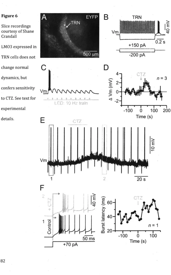
my thesis. For chapter 2, a key inspiration for my experiments was work done previously in lab by Mike Halassa and Josh Siegle. For chapter 3, a number of people were essential for the whole project. Ute Hochgeschwender and her group were responsible for designing the luminopsin constructs that I used and also provided huge amounts of guidance and preliminary data. Initial testing of LMO3 in slice was made possible by the help of Shane Crandall and the lab of Barry Connors. Liz McDonnell was a collaborator for both in vivo and in vitro work.
A special thanks to the administrative staff for the BCS graduate program, who were
knowledgeable advocates for us students, even if we were at different universities and forgot to check our email sometimes.
I have to thank my family, who has supported me throughout my whole graduate career. My parents provided love, support, and a desire for knowledge that brought me to where I
am today. My brother is a great friend who can now truly understand the ups and downs of grad school.
Finally, I'd like to thank Katie Wu, who supported me through random work hours, frantic thesis writing, and general PhD madness. I love you and couldn't have done it without you. 4
Table of Contents
Ch ap ter 1 : In tro d u ctio n ... 7
O verview ... 8
T R N and sensory gating ... 8
T R N and rhythm icity ... 10
Chapter 2: Background and M otivation ... 12
Chapter 3: Next Step Methods for Long Time Scale TRN Regulation- B i olumines cent O ptogenetics ... 14
R eferences ... 16
Chapter 2: Systematic Examination of the Impact of Depolarization Duration on Thalam ic Reticular Nucleus Firing in vivo ... I ... 20
A uthor contributions: ... ... 21
A bstract: ... 22
Introduction ... 24
M aterials and M ethods ... 29
Results ... 31
D iscussion ... -36
References ... 43
Figures ... 49
Chapter 3: Development of Novel Bioluminescent Methods For Non-Invasive O p togen etic C o n tro l In Vivo ... 6 0 A uthor Contributions: ... 61
Background ... 61
M aterials and m ethods ... r ... 68
R esults ... 69
Conclusions ... 72
R eferences ... 74
Figures ... 77
Ch ap ter 4 : C o n clu sio n s ... 8 5 Future experim ents: ... ... 86
Chapter 1
Overview
My primary research interests are in the neural circuit mechanisms that underlie sensory
perception-given the massive amount of data taken in from a constantly changing outside
world, the brain is capable of forming a seemingly cohesive representation of our physical
reality that highlights behaviorally relevant information.
My thesis research approached these questions at the level of the thalamus, which is
central to the flow of sensory signals to cortex and is profoundly important in the
regulation of brain state. My interest in the thalamic reticular nucleus is tied to its position
within the thalamocortical pathway and its particular importance in the rodent
somatosensory system, the primary model used in the Moore lab. The use of optogenetics
in TRN by Halassa and Siegle (Halassa et al., 2011) showed the utility of the technique for
targeting such a small subcortical structure in the mouse, and my first project was to
characterize the response properties of TRN cells through multielectrode recording and
optical stimulation. In addition to general parameterization of optogenetics in this
population, I found evidence for functional heterogeneity within TRN that became apparent
with modulation of stimulus duration. My subsequent work used these data as a baseline
for investigating a novel technique for brain stimulation that is particularly relevant for
clinical and research use in the thalamus.
Incoming sensory information from all modalities, with the exception of olfaction, synapses
in the thalamus on the way to neocortex (Pinault, 2004). This sensory relay is uniquely
positioned to act as a gate, to determine which inputs from the periphery are processed by
the neocortex (Crick, 1984). A gating mechanism has been hypothesized to underlie
attentional processes, which are often conceived as a 'spotlight' that selects behaviorally
relevant information through a spatially delimited/topographic enhancement or
suppression of information. While effects of attention on behavior are well characterized,
and much has been studied about attentional modulation of cortical activity (ie V4 in the
monkey) (Desimone and Duncan, 1995; Reynolds and Chelazzi, 1999; Treue and Maunsell,
1999), less is known about its role earlier in sensory processing. Inhibitory circuits provide
a way for the thalamus to act as more than a passive relay that routes peripheral signals to
the appropriate higher areas-it could be used to filter out irrelevant information before it
competes for resources further along the pathway.
A primary source of GABAergic input to thalamic relay nuclei that send information to
neocortex is the thalamic reticular nucleus (TRN), a thin shell of neurons enveloping the
thalamus (Jones, 1975). Projections from thalamocortical and corticothalamic cells synapse
within this nucleus, and the TRN projects directly to the rest of thalamus, generating
feedforward and feedback inhibition in this structure. This source of inhibition is
particularly important in rodents, which lack local inhibitory interneurons in
These properties of the TRN led Francis Crick to postulate that it may be key to the
attentional spotlight, stating that the TRN might be described as the "guardian of the
gateway" (Crick, 1984). Recent monkey neurophysiological studies support this idea,
showing that TRN activity in the representation where selective attention is allocated are
modulated in cross-modal and within-modality tasks (McAlonan et al., 2000; McAlonan and
Brown, 2002; McAlonan et al., 2006; 2008). Under this framework, the TRN could exert an
influence on attention at very early levels of primary sensory pathways, as topographically
specific inhibition of relay nuclei is a potent mechanism for blocking inattended stimuli
before even reaching the cortex. Studies in the rodent somatosensory thalamus have
shown the appropriate anatomical substrate for this level of spatial control, as well as
patterns of interconnection that could allow for complex lateral interaction (Bourassa et al.,
1995; Pinault et al., 1995; Deschenes et al., 1996; Pinault and Deschenes, 1998a; 1998b;
Desilets-Roy et al., 2002). The degree of innervation in TRN from both TC and CT fibers
positions the nucleus to respond to feedback from primary sensory cortices, while input
from the prefrontal cortex (as observed in monkeys) (Zikopoulos and Barbas, 2006)could
provide top-down control of the attentional spotlight, as determined by behavioral state
and cognitive demand.
TRN and rhythmicity
A well-known property of thalamic projection neurons is their ability to fire action
potentials in different modes, tonic or bursting, depending on the state of the network. A
burst consists of multiple spikes fired in rapid succession (e.g., with an interval <4ms)
widely applied descriptor of neural activity, but in thalamic nuclei, this term specifically
refers to a period of high frequency action potential firing mediated by a large underlying
calcium event (a calcium "spike") (Crunelli et al., 1989; Huguenard and Prince, 1992;
Coulon et al., 2009). In vitro studies have shown that many thalamic neurons (including
thalamocortical projection cells and TRN cells) express T-type calcium channels that cause
calcium spikes and bursts after a period of hyperpolarization. The TRN inhibits
thalamocortical neurons, causing a hyperpolarization that de-inactivates T-channels. Upon
release of inhibition, these channels are available to open, leading to a calcium spike with a
burst of multiple concurrent sodium action potentials. The activity of these relay cells in
turn provides an excitatory input to the TRN, forming an oscillatory loop.
The change from tonic to burst firing denotes a shift in thalamic network dynamics, but the
role of bursting in information processing is debated. Some studies have argued that a
burst is highly efficient at relaying information by virtue of the postsynaptic impact of
several spikes fired in short succession (Swadlow and Gusev, 2001). In a contrasting view
(Llinas and Steriade, 2006), bursting represents a state of inattention and impoverished
signal relay. In support of this view, recent studies have shown that alpha rhythms-widely
believed to emerge from thalamic bursting at 5-15 Hz-occur in specific parts of sensory
maps that are disattended during task performance (Worden et al., 2000; Banerjee et al.,
2011; Foxe and Snyder, 2011). In either case-whether bursting is an optimal signal relay
or the signature of a decreased probability of information relay-understanding any
As stated previously, the rapid firing of multiple action potentials in a burst can be highly
effective for driving activity in post-synaptic targets, potentially enhancing signal relay in
the brain (Lesica, 2004; Alitto and Usrey, 2005; Lesica et al., 2006; Stanley, 2013).
Excessive expression of this powerful activity pattern in thalamus is hypothesized to be
causal in several diseases (Llinas et al., 1999). As a cardinal example, multiple types of
epileptic seizures are believed to be driven by hyper-synchronized rhythmic bursting in the
thalamic reticular nucleus (TRN) and in thalamic relay neurons that project to the
neocortex (Beenhakker and Huguenard, 2009). The TRN is a shell of inhibitory neurons
that target and hyperpolarize thalamic relay cells, generating rebound bursts. Epileptic
seizures emerging from neocortical injury, and absence seizures, the most common form in
children, are both believed to emerge from overly robust rates and synchrony of thalamic
bursting (Avanzini et al., 1996; Beenhakker and Huguenard, 2009; Paz et al., 2013).
Thalamic burst-driven seizure activity is hypothesized to be an altered form of endogenous
patterns observed in slow-wave sleep called spindles, bouts of rhythmic thalamic bursting
typically lasting -. 5-1.5 seconds (Kostopoulos, 2000; Beenhakker and Huguenard, 2009).
Chapter 2: Background and Motivation
The depolarization of TRN neurons, and their subsequent firing, is driven by a variety of
sources on a range of time scales. The TRN receives excitatory inputs ranging from single
spikes to sustained tonic firing to bursting in thalamic relay neurons or layer 6 of
neocortex(de Curtis et al., 1989; Contreras et al., 1992; Bourassa et al., 1995; Avanzini et al.,
al., 2015). The temporal dynamics of these inputs, as well as their spatial organization, can
drive different types of firing behavior in TRN(Lam and Sherman, 2010; 2011). Layer 6
cells form strong synapses in the TRN(Lam and Sherman, 2010; Crandall et al., 2015), and
even sparse activity in this layer would be predicted to engage substantial inhibition in
vivo. Primary thalamocortical relay projections branch into the TRN on their way to sensory cortices, and the nature of this excitatory input reflects the functional modes of the
relay nuclei. Inputs include tonic firing that reflects high fidelity to peripheral input, as well
as extended bouts of bursting, similar to that seen in TRN itself. In sum, a variety of inputs
can excite TRN neurons on different time scales. Understanding how these different
patterns may regulate excitability in general, and burst activity specifically, is key to
understanding thalamocortical relay and function. In addition to the basic science
questions associated with this variability, characterization of optogenetic responses in TRN
is vital for future experimental planning. The TRN is an appealing target for research into
sensory perception and neurological disease, and understanding how different modes of
stimulation affect its activity is crucial to interpreting its role in these broader contexts.
In our lab, we previously showed that putative TRN activation could modulate firing and
bursting in relay neurons, and induce spindles in the neocortex (Halassa et al., 2011). In
these experiments, stimulation was confined to the TRN using implanted optical fibers in
the VGAT-ChR2 mouse, but electrophysiological recordings were targeted to the
somatosensory relay thalamus and the cortex. As a result, the activity of TRN cells during
provided a more complete view of the circuit activity underlying evoked burst and spindle
behavior.
As such, my work in Chapter 2 was important at several levels. I provided optogenetic
input while characterizing multi-unit responses in the TRN and well-sorted single units. As
reviewed below, I found that longer duration activation drove enhanced bursting and
decreased latency to bursting. I also discovered two new types of cell responses, a more
sensitive 'non-linear' cell type that was prone to sustained responses and to bursting, and a
more 'linearly' responsive cell class that fired in direct proportion to the duration of
stimulation. These findings provide direct predictions as to the behavior of TRN neurons in
response to a range of natural depolarizing inputs, and a guide for the optical control of this
key structure in studies of network function and behavior.
Chapter 3: Next Step Methods for Long Time Scale TRN Regulation-Bioluminescent Optogenetics
The TRN is believed to be crucial to the regulation of long time scale state transitions, e.g.,
between sleep and wakefulness. As such, developing methods for minimally-invasive
control of this structure on such time scales has high utility to my research goals.
Optogenetics has rapidly become a standard technique in systems neuroscience, and its
genetic specificity and rapid development of new compounds has revolutionized our ability
to causally manipulate neural circuits. While the use of light to drive cellular reactions
brings a number of advantages when compared to electrical stimulation, there are still
tissue is problematic in the intact brain, so targeting deep structures relies on implanted
fiber optics and/or LEDs. These methods are not ideal for illuminating large or
irregularly-shaped regions without using high light intensity or large arrays of invasive devices. I have
been key in inventing a new methodological approach using bioluminescent light to drive
optogenetic responses ('BL-OG'; (Berglund et al., 2013; Tung et al., 2015; Berglund et al.,
2016a; 2016b)). This approach leverages the revolution in new light sensitive molecules,
and the wide array of bioluminescent options, while providing a means of
chemiluminescent control. BL-OG combines the cell-type specificity of conventional
optogenetics with the potential for non-invasive, system-wide activation of opsins. In
References
Alitto HJ, Usrey WM. Dynamic properties of thalamic neurons for vision. Prog Brain Res
149:83-90,2005.
Avanzini G, de Curtis M, Franceschetti S, Sancini G, Spreafico R. Cortical versus thalamic
mechanisms underlying spike and wave discharges in GAERS. Epilepsy Res 26: 37-44, 1996.
Banerjee S, Snyder AC, Molholm S, Foxe JJ. Oscillatory alpha-band mechanisms and the
deployment of spatial attention to anticipated auditory and visual target locations: supramodal or sensory-specific control mechanisms?JNeurosci 31: 9923-9932, 2011.
Beenhakker MP, Huguenard JR. Neurons that fire together also conspire together: is
normal sleep circuitry hijacked to generate epilepsy? Neuron 62: 612-632, 2009.
Berglund K, Birkner E, Augustine GJ, Hochgeschwender U. Light-emitting
channelrhodopsins for combined optogenetic and chemical-genetic control of neurons. PLoS ONE 8: e59759, 2013.
Berglund K, Clissold K, Li HE, Wen L, Park SY, Gleixner J, Klein ME, Lu D, Barter JW, Rossi MA, Augustine GJ, Yin HH, Hochgeschwender U. Luminopsins integrate opto- and
chemogenetics by using physical and biological light sources for opsin activation. Proceedings of the National Academy of Sciences 113: E358-67, 2016a.
Berglund K, Tung JK, Higashikubo B, Gross RE, Moore CI, Hochgeschwender U.
Combined Optogenetic and Chemogenetic Control of Neurons. Methods Mol Biol 1408:
207-225, 2016b.
Bourassa J, Pinault D, Deschenes M. Corticothalamic projections from the cortical barrel
field to the somatosensory thalamus in rats: a single-fibre study using biocytin as an anterograde tracer. EurJ Neurosci 7: 19-30, 1995.
Contreras D, Dossi RC, Steriade M. Bursting and tonic discharges in two classes of reticular thalamic neurons.J Neurophysiol 68: 973-977, 1992.
Coulon P, Herr D, Kanyshkova T, Meuth P, Budde T, Pape H-C. Burst discharges in
neurons of the thalamic reticular nucleus are shaped by calcium-induced calcium release. Cell Calcium 46: 333-346, 2009.
Crandall SR, Cruikshank SJ, Connors BW. A corticothalamic switch: controlling the
thalamus with dynamic synapses. Neuron 86: 768-782, 2015.
Crick F. Function of the thalamic reticular complex: the searchlight hypothesis. Proc Natl Acad Sci USA 81: 4586-4590, 1984.
Crunelli V, Lightowler S, Pollard CE. A T-type Ca2+ current underlies low-threshold Ca2+
543-561, 1989.
de Curtis M, Spreafico R, Avanzini G. Excitatory amino acids mediate responses elicited in
vitro by stimulation of cortical afferents to reticularis thalami neurons of the rat. Neuroscience 33: 275-283, 1989.
Deschenes M, BOURASSA J, Doan VD, Parent A. A single-cell study of the axonal
projections arising from the posterior intralaminar thalamic nuclei in the rat. EurJ Neurosci
8: 329-343, 1996.
Desimone R, Duncan
J.
Neural mechanisms of selective visual attention. Annu Rev Neurosci18: 193-222, 1995.
Desilets-Roy B, Varga C, Lavallee P, Deschenes M. Substrate for cross-talk inhibition
between thalamic barreloids.J Neurosci 22: RC218, 2002.
Foxe
JJ,
Snyder AC. The Role of Alpha-Band Brain Oscillations as a Sensory SuppressionMechanism during Selective Attention. Front Psychol 2: 154, 2011.
Halassa MM, Siegle JH, Ritt JT, Ting JT, Feng G, Moore CI. Selective optical drive of
thalamic reticular nucleus generates thalamic bursts and cortical spindles. Nat Neurosci
(July 24, 2011). doi: 10.1038/nn.2880.
Huguenard JR, Prince DA. A novel T-type current underlies prolonged Ca(2+)-dependent
burst firing in GABAergic neurons of rat thalamic reticular nucleus. Journal of Neuroscience
12: 3804-3817, 1992.
Jones E. Some aspects of the organization of the thalamic reticular complex.J Comp Neurol.
Jones EG. Some aspects of the organization of the thalamic reticular complex.j Comp
Neurol 162: 285-308, 2004.
Kostopoulos GK. Spike-and-wave discharges of absence seizures as a transformation of
sleep spindles: the continuing development of a hypothesis. Clin Neurophysiol 111 Suppl 2:
S27-38, 2000.
Lam Y-W, Sherman SM. Functional organization of the somatosensory cortical layer 6
feedback to the thalamus. Cereb Cortex 20: 13-24, 2010.
Lam Y-W, Sherman SM. Functional organization of the thalamic input to the thalamic
reticular nucleus.JNeurosci 31: 6791-6799, 2011.
Lesica NA, Weng C, Jin J, Yeh C-I, Alonso J-M, Stanley GB. Dynamic Encoding of Natural
Luminance Sequences by LGN Bursts. PLoS Biol 4: e209, 2006.
Llinas RR, Ribary U, Jeanmonod D, Kronberg E, Mitra PP. Thalamocortical dysrhythmia:
A neurological and neuropsychiatric syndrome characterized by magnetoencephalography.
Proc NatlAcad Sci USA 96: 15222-15227, 1999.
Llinas RR, Steriade M. Bursting of thalamic neurons and states of vigilance.JNeurophysiol
95: 3297-3308, 2006.
McAlonan K, Brown VJ, Bowman EM. Thalamic reticular nucleus activation reflects
attentional gating during classical conditioning.J Neurosci 20: 8897-8901, 2000. McAlonan K, Brown VJ. The thalamic reticular nucleus: more than a sensory nucleus? Neuroscientist8: 302-305, 2002.
McAlonan K, Cavanaugh
J,
Wurtz RH. Attentional modulation of thalamic reticularneurons. J Neurosci 26: 4444-4450, 2006.
McAlonan K, Cavanaugh
J,
Wurtz RH. Guarding the gateway to cortex with attention invisual thalamus. Nature 456: 391-394, 2008.
Paz JT, Davidson TJ, Frechette ES, Delord B, Parada I, Peng K, Deisseroth K,
Huguenard JR. Closed-loop optogenetic control of thalamus as a tool for interrupting
seizures after cortical injury. Nat Neurosci 16: 64-70, 2013.
Pinault D, Bourassa
J,
Deschenes M. The Axonal Arborization of Single Thalamic ReticularNeurons in the Somatosensory Thalamus of the Rat. European Journal of Neuroscience 7:
31-40, 1995.
Pinault D, Deschenes M. Anatomical evidence for a mechanism of lateral inhibition in the
rat thalamus. EurJ Neurosci 10: 3462-3469, 1998a.
Pinault D, Deschenes M. Projection and innervation patterns of individual thalamic
reticular axons in the thalamus of the adult rat: a three-dimensional, graphic, and morphometric analysis. J Comp Neurol 391: 180-203, 1998b.
Pinault D. The thalamic reticular nucleus: structure, function and concept. Brain Research
Reviews 46: 1-31, 2004.
Reynolds JN, Chelazzi L. Competitive mechanisms subserve attention in macaque areas V2
and V4. Journal of Neuroscience.
Sherman SM, Guillery RW. Functional organization of thalamocortical relays.J
Neurophysiol 76: 1367-1395, 1996.
Stanley GB. Reading and writing the neural code. Nat Neurosci 16: 259-263, 2013. Swadlow HA, Gusev AG. The impact of "bursting" thalamic impulses at a neocortical
Treue S, Maunsell JHR. Effects of attention on the processing of motion in macaque middle temporal and medial superior temporal visual cortical areas.JNeurosci 19: 7591-7602,
1999.
Tung
JK,
Gutekunst C-A, Gross RE. Inhibitory luminopsins: genetically-encodedbioluminescent opsins for versatile, scalable, and hardware-independent optogenetic
inhibition. Sci Rep 5: 14366, 2015.
Worden MS, Foxe
JJ,
Wang N, Simpson GV. Anticipatory biasing of visuospatial attentionindexed by retinotopically specific alpha-band electroencephalography increases over occipital cortex. J Neurosci 20: RC63, 2000.
Zikopoulos B, Barbas H. Prefrontal projections to the thalamic reticular nucleus form a
Chapter 2
Systematic Examination of the Impact of
Depolarization Duration on Thalamic Reticular
Nucleus Firing in vivo
Author contributions:
BTH and CIM designed experiments, BTH collected and analyzed data, BTH and CIM drafted and edited the manuscript and approved the final version.
A version of this chapter is under review as:
Systematic Examination of the Impact of Depolarization Duration on Thalamic Reticular Nucleus Firing in vivo
Abstract:
The thalamic reticular nucleus (TRN) is optimally positioned to regulate information
processing and state dynamics in dorsal thalamus. Distinct inputs depolarize TRN on a
variety of time scales, including thalamocortical afferents, corticothalamic 'feedback', and
neuromodulatory influences. Here, we systematically tested the immediate and sustained
effects of depoiarization duration on TRN firing in vivo using selective optogenetic drive. In
VGAT-ChR2 mice, well-isolated TRN single units (SU: N=100 neurons) responded at brief
latency (<5 millisecond) to optical pulses. These units, and multi-unit activity (MUA) on
corresponding electrodes, were analyzed in detail. Burst-like events occurred after light
cessation in 74% of MUA sites, and 16% of SU. Increasing optical duration from 2 to 330
milliseconds enhanced burst probability, and decreased the latency to the first burst after
stimulation. All neurons demonstrated a 'plateau' firing response lasting 20-30
milliseconds in response to light, but significant heterogeneity existed in the minimal
stimuli required to drive this response. Two distinct types were' evident, more sensitive
'non-linear' neurons that were already driven to the plateau response by 2 or 5 millisecond
light pulses, versus 'linear' neurons that fired in proportion to optical duration, and
reached the plateau with -20 millisecond optical drive. Non-linear neurons showed higher
evoked firing rates and burst probability, but spontaneous rate did not differ between
types. These findings provide direct predictions as to TRN responses to a range of natural
depolarizing inputs, and a guide for the optical control of this key structure in studies of
network function and behavior.
New and Noteworthy: The thalamic reticular nucleus receives depolarizing inputs on a
to the TRN in vivo and cell activity was monitored during and after stimulation. Longer
periods of stimulation were associated with increased post-stimulus activity. Direct
responses t6 stimuli revealed two categories: units with a response duration matched to
Introduction
Thalamic reticular nucleus (TRN) neurons play a key role in thalamocortical dynamics. The
TRN makes GABAergic projections to first and higher-order thalamic nuclei, providing a
major source of hyperpolarization to relay cells (Pinault, 2004). The TRN is therefore
positioned to regulate the flow of signals between the periphery and neocortex, potentially
gating information during active processing (Crick, 1984; Weese et aL, 1999; Yu et al.,
2009). Primate and mouse studies have shown modulation of TRN with allocation of
selective attention (McAlonan et al., 2000; McAlonan and Brown, 2002; McAlonan et al.,
2006;.Wimmer et al., 2015), and TRN receives connections from brain regions involved in
decision making, such as the prefrontal cortex, that could direct its activity based on
behavioral context (Zikopoulos and Barbas, 2006). The TRN is also widely implicated in
regulating dorsal thalamic and neocortical dynamics during less active states including in
sleep (Steriade, 1985; Steriade and Llinas, 1988; Steriade et al., 1993; Huguenard and
McCormick, 2007).
The duration and firing pattern of TRN neurons are likely both key determinants in
controlling its downstream partners. Both TRN and thalamic relay neurons express T-type
low threshold calcium channels that cause bursting following hyperpolarization (Crunelli
et al., 1989; Huguenard and Prince, 1992). Inhibition generated by TRN, particularly the
robust hyperpolarization generated by TRN burst firing, can drive rebound bursting in
relay cells that feed back to TRN (Bal and McCormick, 1993; Bal et al., 1995). This loop is
believed important in establishing sustained rhythmic activity, including repeated bursting
Changes in factors that regulate excitatory dynamics in TRN cells are also key to brain
health. Abnormalities in TRN firing and sleep spindles are observed in schizophrenia
(Austin et al., 2004; Ferrarelli et al., 2007; 2010; Pinault, 2011; Pratt and Morris, 2015), and
a mutation of the CAv 3.3 T-type channels expressed in TRN alters TRN firing patterns and
predicts schizophrenic phenotype (Liu et al., 2011; Astori et al., 2011; Schizophrenia
Working Group of the Psychiatric Genomics Consortium, 2014). Altered patterns of TRN
firing are also implicated in absence epilepsy, where seizures are associated with
spike-wave discharges driven by thalamic bursting (Pinault et al., 1998; LlinA's et al., 1999; Slaght
et al., 2002; Crunelli et al., 2011; Tringham et al., 2012; Paz et al., 2013).
Depolarization of TRN neurons, and their subsequent firing, is driven by a variety of
sources on a variety of time scales. The TRN receives synaptic input from both
thalamocortical and corticothalamic axons that pass through its extent. While relatively few
studies have recorded from identified corticothalamic cells, these layer 6 neurons fire are
known to fire in a highly sparse manner, with low spontaneous and sensory-evoked firing
rates, and a low probability of firing to sensory stimuli (< 10% of identified neurons), even
in topographically aligned positions in primary sensory neocortex (Temereanca and
Simons, 2004; Kwegyir-Afful and Simons, 2009). However, brief activity in the layer 6 input
to TRN is particularly effective in driving depolarization. Large excitatory post-synaptic
potentials with rapid rise times are observed in TRN neurons after single spikes or brief
Correspondingly, brief corticothalamic input also generates robust suppression in relay neurons (Landisnan and'Connors, 2007; Mease et al. 2014).
Primary thalamocortical relay neurons show activity on a wide variety of time scales, firing single spikes, sustained trains of single action potentials and bursts in response to sensory
drive, and revealing a range of patterns of afferent adaptation (Diamond et al., 1992;
Alamancos and -Connors, 1996; Alamancos, 2C02; Rigas and Castro-Alamancos, 2007; Lan) nd Sherman, 2011). Relay thalamic inputs to the TRN car, in concept, provide a variety of time scales of depolarization dependent on state and context.
In addition to the single spikes or epochs of bursting provided by cortical and thalamic inputs, the TRN receives modulatory inputs from a number of different brain structures. Cholinergic projections to the TRN have multiple origins, including projecticns from the brainstem that are associated with arousal (Boucefta et al., 2014) as well as a basal forebrain projection that can act on a synaptic timescale to drive firing followed by desychronization (Pita-Almenar et al., 2014). In addition to connections between individual inhibitory cells within the TRN, the nucleus also receives inhibitory inputs from the basal ganglia, with projections from the substantia nigra pars reticulata (SNr) and the globus pallidus (Gandia et al., 1993) that could be a substrate for disinhibition of reticular targets or for burst regulation. The neuropeptide somatostatin is expressed throughout the TRN (Graybiel et al. 1983), and has been shown to influence oscillatory activity in slice preparations (Sun et al., 2002).
In sum, a variety of inputs can excite TRN neurons on different time scales. Understanding
how these different patterns may regulate excitability in general, and burst activity
specifically, is key to understanding thalamocortical relay and function.
In addition to diversity in types of depolarization the TRN may receive, TRN cells
themselves exhibit functional and anatomical heterogeneity (Lee et al., 2007; Halassa et al.,
2014; Lee et al., 2014; Kimura and Imbe, 2015). Axonal arborization of TRN neurons
projecting to first- and higher-order thalamic nuclei in the same sensory modality varies by
target (Cox et al., 1996). In rodent somatosensory thalamus, projections to VPM are well
defined and correspond to individual barreloids, while projections to POm are more
diffuse(Pinault and Deschenes, 1998b; 1998a; Desilets-Roy et al., 2002). Within the TRN,
somata of cells targeting different regions are biased to occur in distinct tiers(Clemence
and Mitrofanis, 1992; Pinault and Deschenes, 1998a; 1998b; Desilets-Roy et al., 2002).
At a functional level, Contreras et al. (Contreras et al., 1992) described two types of cells in
the reticular nucleus of the anesthetized cat, type I cells that exhibit standard T-current
mediated bursting, and type II cells that did not burst and showed less adaptation when
driven at high frequencies. Recent work from Halassa et al. (Halassa et al., 2014) also found
evidence for functional distinction between two types of TRN cells in mice. In that study,
firing rates were either negatively or positively correlated with spindle and delta
oscillation expression. Cells negatively correlated with spindles were more active during
To understand the potential role of this key node in controlling thalamocortical dynamics, and the potential for heterogeneity in response types in response to diverse inputs, we studied TRN cell responses to changes in stimulus duration in an intact, in vivo network. A powerful tool for manipulation of an identified cell population is optogenetics. Consistent with the hypothesized role of TRN cells, direct optogenetic drive of this structure causes bursting in relay neurons and sleep spindles in vivo (Halassa et al., 2011). While showing that bursting can result from TRN activation, this prior study did not assess the impact of optogenetic drive on TRN neurons themselves, nor did it seek to systematically understand the impact of the duration of stimulation on network dynamics.
To directly address these questions, we used an acute preparation to study TRN units activated at short latencies after light onset (i.e., those cells presumably expressing ChR2). Here we report that bursting probability in TRN neurons depends systematically on, and varies positively with, the duration of stimulation. We further discovered two classes of TRN neurons by their responses to direct optical activation. One group showed greater sensitivity to brief light pulses, including a greater propensity to bursting, but also showed greater response saturation with increasing stimulus duration. In contrast, the second cell type showed a near linear increase in firing with increased stimulus duration, but a lower propensity to bursting. These findings provide direct predictions as to the behavior of TRN neurons in response to a range of natural depolarizing inputs, and a guide for the optical control of this key structure in studies of network function and behavior.
Materials and Methods
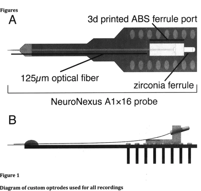
Optrode construction
Prefabricated 16-channel linear electrode arrays (Neuronexus technology,
A1x16-5mm-25-177, with 25 ptm spacing between 177 ptm2
contacts) were chosen as the basis for thalamic
optrodes. Multimode optical fiber cannulae (105ptm core diameter, coating stripped,
Thorlabs) were positioned using a surgical microscope to rest -200ptm above the top
electrode contact of the array without touching the probe shank, and fixed in place with a
silicone elastomer. The back end of each fiber was terminated in a 1.25mm ceramic ferrule
that was anchored to the probe PCB by a 3D printed bracket (Fig 1A schematic, Fig lB side
view showing fiber orientation). This configuration provided a stable attachment point for
an optical patch cable and strain relief for the fiber during connection and disconnection.
Designs are available at Open Ephys (www.open-ephys.org).
Animal preparation and electrophysiology
All recordings were performed in lightly anesthetized (0.75 - 1.1% isoflurane) adult
VGAT-ChR2 mice (2-4 weeks old) fixed in a stereotaxic frame with an integrated nosecone. A
craniotomy was made at approximate coordinates 1.9mm lateral and 1.7mm posterior to
bregma, and custom optrodes were lowered to -3mm below the cortical surface
obtained with the Neuralynx Cheetah data acquisition system, with 16 amplified electrode
channels sampled at 30.3kHz and additional analog inputs used to register the timing of
stimulus delivery.
Optical stimulus delivery
Light pulses were delivered from a 100mW, 472nm DPSS laser coupled to a multimode
patch cable. Stimulus intensity was measured as 7-8mW at the end of the optrode. Stimulus
durations of 1, 2, 5, 10, 20, 100, and 330ms were presented randomly using custom
MATLAB code to drive square pulses from a National Instruments data acquisition card.
Laser power was held constant throughout each trial, and was calibrated manually in each
session to drive robust multi-unit spiking at short latency from TRN units. Each recording
session consisted of 300-600 stimuli delivered at 0.5 Hz, lasting 5-10 minutes.
Data processing and analysis
Neural data was filtered for spikes at 300-6000Hz. These signals were processed offline
using custom MATLAB code. Spikes were sorted using simpleclust
(https://github.com/open-ephys/simpleclust), and light-evoked artifacts were identified
and rejected during this process based on characteristic shape and amplitude.
Multi-unit activity (MUA) was characterized by taking the RMS power of the filtered
to detect events. MUA activity peaks with a duration greater than 16.5 ms (500 Neuralynx
samples) was defined as a 'burst', and shorter events were excluded. Single-unit bursts
were defined as three or more spikes separated by 4 ms or less, and preceded by a
non-firing period of at least 100 ms (Swadlow and Gusev, 2001).
The durations of spiking responses to stimuli were determined from mean PSTHs using a
morphologically inspired filter to define epochs of firing above baseline while ignoring
brief (<3 ms) fluctuations. The duration of a PSTH response was determined by first
detecting response epochs that were 3 standard deviations above mean pre-stimulus
activity (20 ms). The duration was then defined as sustained increases with no longer than
a 3 millisecond period without a threshold crossing.
Results
Longer optical stimulations increased burst probability and decreased burst latency
Successful optogenetic activation of TRN was defined as single unit spikes driven within 5
ms of stimulus onset, with spiking probability above 3 SD from the baseline mean of the
PSTH. Electrode channels containing at least one well-defined single unit activated in this
way were considered located in TRN for subsequent multi-unit activity (MUA) analyses (N
Changing the duration of light stimulation increased the likelihood of subsequent bursts after the initial MUA response. An MUA 'burst' was defined as a peak in MUA power at least
3 SD above the mean, excluding the time of stimulation to avoid light artifacts, and lasting
at least 16.5 ms. As shown in the example plot in Figure 2, subsequent MUA bursts were observed following a pause of 100-300 ms after the initial light-driven response. In some cases, multiple subsequent MUA bursts were observed. In Figure 2, blue periods indicate initial firing, green indicates the first post-light burst, orange the second post-light burst, and red the third post-light burst.
With increasing optical duration, the likelihood of a subsequent burst and the probability of observing more than one subsequent burst were increased, and the latency to the first burst after light cessation was decreased. Figure 3A-B shows quantification of these dependencies. Chi-squared tests were performed for each pair of stimulus durations of most similar length (e.g., 2 versus 5 ms duration), and in each case the increase in proportion of trials exhibiting bursts following longer stimulation was statistically significant (p < .05). Linear regression of event probability on log stimulus duration was
also statistically significant (r2 = .15, p < .0 1).
Increased stimulus duration was also associated with a weak but steady decrease in the latency of MUA bursts from time of stimulation, as modeled by a linear regression on log stimulus duration (Figure 4, slope = -1796, r2 = .152, p <.05 versus the constant model). In sum, longer TRN depolarizations increase the likelihood and decrease the delay before an
We also analyzed single unit responses to light presentation. We isolated 100 single units
that showed short-latency evoked activity (<5 milliseconds following light onset). To
analyze single unit burst probability, bursting was defined similarly to Swadlow (Swadlow
and Gusev, 2001) as a unit firing multiple spikes separated by less than 4 ms with a
preceding quiescent period of at least 100 ms. An example raster plot from one recorded
unit is shown in Figure 5, with detected bursts indicated using the same color scheme as
Figure 2. Highly synchronized firing at stimulus onset was selected for by our rules for the
inclusion of responsive units, as with the MUA analyses, following analyses exclude the
initial response in quantification of 'second' bursts in single units.
Burst probability increased with stimulus duration in isolated single units (Figure 6), and
these differences were characterized by a linear regression of burst frequency versus log
stimulus duration (r2 = .09, p <.01). Single unit burst latency was highly variable, but there
was a weak linear relationship between increased stimulus duration and lower latency to
bursts across single units (r2 = .01, P <.01).
Distinct response types emerge with increasing duration of TRN depolarization
Inspection of the PSTHs evoked by light stimuli ranging from 2-100 ms duration for all
activated single units suggested the existence of distinct sub-populations. This response
the cell on the left showed robust firing lasting -20 milliseconds for all light durations of 5 ms or greater. In contrast, the cell on the right showed a basically linear increase in the duration of firing with the duration of light, reaching a 20 ms response duration when a 20 ms light pulse was applied. To visualize these data, we calculated the duration of firing as contiguous activity 3SD or greater over baseline (ignoring gaps of up to 3 ms) and plotted the data as a line plot connecting the responses of each individual cell. All neurons showed sustained firing (> 20 ms duration) following sustained optical drive (Figure 7B). However, as indicated by the examples, there was significant diversity as to the light duration required to reach this response. The red and blue lines in Figure 7B correspond to the example cells shown in Figure 7A.
To visualize this diversity in the data in a more reduced format, we plotted response duration histograms for all neurons as a function period of light drive (Figure 7C). In the range from 2-20 milliseconds, a clearly bimodal distribution is observed, with a prominent peak of neurons with 20 millisecond durations of firing across stimulus conditions, and a second contingent with shorter sustained responses for shorter stimuli.
The summary plots revealed a separation into two main groups in response to stimulus durations. Due to large variability and difficulty of distinguishing evoked from spontaneous spikes in the 2 ms condition, the range between 5 ms and 20 ms was chosen for further analysis. Linear regressions were fit for the response lengths of each cell to quantify the relationship between stimulus and response durations. We measured the relative slope of responses in this region of the curve and found that these formed an apparently bimodal
distribution (Figure 8A), which we separated into two categories using a Gaussian mixture
model. This distribution was modeled as a combination of two Gaussians in one dimension,
with the best fit containing the following components (distribution 1: mean = -. 13, standard
deviation = .07; distribution 2: mean = .90, standard deviation = .10; slope at crossover =
.35).
We next plotted the firing rate PSTHs for each group, and these are shown in Figure 8B.
This population level spiking format reveals the main effects observed in the rest of the
data, that linear cells fire less to briefer stimuli, whereas non-linear cells show more
sustained responses up to a duration of -20 milliseconds.
We next investigated whether intrinsic activity patterns and/or excitability separated these
groups (Figure 8C-D). Spontaneous firing rate was statistically indistinguishable between
these groups (linear = 0.47 spikes/s, SD = 1.0; nonlinear = 0.85, SD = 2.3; two-tailed
Wilcoxon ran-sum test, p = .56). In contrast, mean evoked firing during light presentation
was higher for the non-linear cells (summed across all light intensity levels; linear = 2.2
spikes/s, nonlinear = 4.8 spikes/s, two-tailed Wilcoxon rank-sum test, p < .05). Significant
differences were also found in evoked burst rate, as non-linear cells were approximately
two times more likely to burst during stimulation than linear cells (linear = 0.15 bursts/s,
nonlinear = 0.31 bursts/s, two-tailed Wilcoxon rank-sum test, p<.05).
these burst rate differences were a product of overall excitability, subgroups of each unit
class were matched for driven spike rate and reanalyzed (Figure 8C, third panel). Cells in
both groups were sorted in order of mean evoked spike rate - linear cells had a wider
range, so the minimum and median values of the nonlinear group were taken as boundaries
for extracting a subset of the linear group. The mean evoked firing rates of these extracted
subgroups were statistically indistinguishable (N = 18 linear cells, 22 nonlinear; linear =
2.8 sp/s, nonlinear = 3.0 sp/s, two-tailed Wilcoxon rank-sum test, p = 0.78). These subsets
maintained a significant distinction in burst firing (linear = 0.15 bursts/s, nonlinear = 0.28
bursts/s, two-tailed Wilcoxon rank-sum test, p<.05), and the behavior of these
sub-populations was indistinguishable from the values in each category (linear subset vs linear
population, p = .13, nonlinear subset versus population, p = .30).
Discussion
In this study, we characterized the response of TRN cells to optogenetic stimuli of varying
durations in vivo. In the lightly-anesthetized mouse, TRN was mostly quiescent in
spontaneous spiking and oscillatory activity. Optical drive not only directly stimulated
firing, but also post-stimulus activity that persisted for up to hundreds of milliseconds. We
found that this response was dependent on total stimulus duration, as stimulation for 100
ms or longer consistently evoked subsequent bursting, while less than 10 ms stimulation
was ineffective at post-stimulus burst induction. In contrast, persistence of directly driven
firing could be observed after stimulus offset in a subset of recorded units. These
The steep increase in the probability of evoking a subsequent burst after light cessation at
durations of 10 ms or longer (Figure 3A) suggests that endogenous depolarizing events of
at least this duration may be key to triggering rhythmic TRN activity. As previously
described, the TRN receives input from multiple structures across a wide range of
durations. Such depolarization could be driven by burst input from relay cells, or by
sustained tonic relay firing reflecting strong bottom-up sensory input. Projections from the
prefrontal cortex to the TRN are widespread with large axon boutons in the primate
(Zikopoulos and Barbas, 2006), and recent work from Wimmer et al. (Wimmer et al., 2015)
described potent modulation of TRN in mice following optogenetic stimulation of the
rodent analogue to PFC. This projection could induce lasting activity in the TRN as a
function of behavioral demands. Biophysically, Neyer et al. (Neyer et al., 2016) have
demonstrated that mGluR agonists can trigger the release of calcium from intracellular
stores in TRN, leading to long-lasting depolarization. In sum, our data predict that
minimally 10 milliseconds of depolarization is required to evoke bursting, but that there
are many possible candidates for generating this input.
The two types of responses observed here may reflect the basic physiological observations
of Contreras et al. (Contreras et al., 1992) in urethane-anesthetized cats. Their type I cells
showed bursting, much as our non-linear cells showed a greater propensity to burst. In
contrast, their type II cells did not burst but did show a linear increase in firing with
sustained depolarization, and our linear cells showed a diminished burst rate relative to
Contreras data, - 20% of recorded cells were classified as 'type II', but we found
approximately the same number of linear and nonlinear cells. This difference could reflect
the respective sampling biases for intracellular versus extracellular recording, or could
indicate that these are separate phenomena. More importantly, type II neurons were not
observed to fire in bursts, either on release from hyperpolarization or from depolarizing
pulses given to cells held at extremely hyperpolarized levels. This behavior suggests a lack
of t-type calcium channels, as opposed to our linear cells that have a lower burst rate, but
do display this pattern of activity to some extent.
Different modes of burst firing could arise from the intrinsic mechanisms underlying
membrane bistability observed during spindles in a subpopulation of TRN cells
(Fuentealba et al., 2005). This effect was hypothesized to require voltage-gated Na+
channels, and did not include abolition of burst firing. In our preparation, however,
sustained spindle-like activity was not observed. These observations are consistent with
earlier work using isoflurane during dLGN and V1 recordings in mice (Denman and
Contreras, 2015). That study used an isoflurane concentration of -1.2% during recordings
to produce an anesthetized state with minimal slow oscillations in cortex and strong L6
responses to stimulation. As the slow oscillation is thought to be key for synchronizing
thalamically-generated spindles (Steriade, 2001), the absence of obvious rhythmicity in our
data may be a product of the relatively light (0.9%-1.1%) isoflurane levels during recording
The cell classes observed here may also reflect those reported by Halassa et al. (Halassa et
al., 2014). The more readily-bursting 'non-linear' cells that fired at higher evoked rates in
our anesthetized preparation may correspond to those units previously reported with high
firing rates positively correlated to spindles and delta oscillations. In contrast, the 'linear'
neurons observed here that did not burst as readily may correspond to those TRN neurons
more active in aroused states, potentially indicative of more tonic firing patterns. One
caveat to this interpretation is that the populations described by Halassa et al. were
distinguished by projection targets, and were also distributed unevenly along the
anterior-posterior axis of the TRN. The segment of the TRN targeted in our study was consistently
posterior to the area of overlap between limbic- and sensory-projecting TRN cells
described in this previous work.
There are a number of potential explanations for distinct classes of activity in TRN units.
One possibility is that the differences observed reflect subtle shift in state of the animal
within the regime of anesthesia employed here. We do not favor this view, as animals
showed little variation in basic measures of state while under a fairly tight range of
administered anesthetic levels, which were consistent with levels used previously to obtain
an EEG-activated pattern (Denman and Contreras, 2015). More importantly, 'linear' and
nonlinear' cells were observed during the same recordings, suggesting that their behavior
does not reflect an overall shift in arousal or baseline state between sessions or animals.
Additionally, matching cells from each group for evoked firing rates did not change the
Functional differences of this sort could be explained by intrinsic biophysical heterogeneity
in the somatosensory TRN. Differences in responses could also be due to differing patterns
of connectivity between groups of TRN cells. Anatomical evidence of heterogeneous
morphology (Pinault and Deschenes, 1998a; 1998b; Desilets-Roy et al., 2002) and laminar
distribution of cells based on thalamic target (Clemence and Mitrofanis, 1992; Pinault and
Deschenes, 1998a; 1998b; Desilets-Roy et al., 2002) suggests the possibility of neurons
within the same area of TRN participating in different thalamocortical circuits.
We also observed suppressed units on TRN responsive contacts. Out of 186 total recorded
single units, 60 showed initial response suppression with light drive, and 26 showed no
response. Because suppressive and absent responses could not be classified as emerging
from TRN cells with the same certitude as quickly responding activated units, they were
excluded from the current analysis. These units may represent a mixture of TRN cells and
axons of passage from relay nuclei. Because all TRN cells are inhibitory and should express
ChR2 in the VGAT mouse, we would expect suppression of TRN neurons could result from
the activation of intra-TRN connections or inhibitory axon terminals for exogenous sources
(e.g., the basal ganglia). The strength and timing of inhibitory input combined with
differential levels of direct optogenetic drive (due to ChR2 expression levels or light
propagation) could generate a primarily suppressive effect or excitation followed by rapid
suppression, both of which we observed in a subset of recordings. If these units represent
relay axons, there could be more complex circuit interactions, but the most straightforward