DNA Hybridization: Fundamental Studies and
Applications in Directed Assembly
by Manish G. Bajaj
B. Chem. Engg. (2000)
University of Mumbai, Department of Chemical Technology, Mumbai M. S. in Chemical Engineering Practice (2002)
Massachusetts Institute of Technology, Cambridge Submitted to the Department of Chemical Engineering in partial fulfillment of the requirements for the degree of
Doctor of Philosophy in Chemical Engineering at the
. A CC A nTT TCT"TC TTCT' TTT r ' T T''rThT TTT _f'v
IV1-u'fl3 _ 1 ;1 11111 U lU I.
'Jr
ILIl NLU I-April, 2005
CJe zoos]
© Massachusetts Institute of Technology, 2005. All rights reserved.
Signature of Author ...
Manish G. Bajaj Department of Chemical Engineering
April 11, 2005
Certified
by ... ...
... ...
~/
/
,~-Payl
E.
Laibinis
Asso e Professor of Chemical Engineering, vice University /Thesis Advisor
Certified by ...
Certified by7
/.
- .regory StephanopoulosBayer Profeslor of Chemical Engineering& Biotechnology Thesis Co-Advisor Accepted by
...
Daniel Blankschtein Professor of Chemical Engineering Chairman, Committee for Graduate Students
*ARCHIves
MASSACHUSETTS INSTITUTE OF TECHNOLOGY
JUN 0 1 2005 LIBRARIES
_··___
DNA Hybridization: Fundamental Studies and
Applications in Directed Assembly
by Manish G. Bajaj
Submitted to the Department of Chemical Engineering on April 11, 2005 in partial fulfillment of the requirements for the degree of
Doctor of Philosophy of Chemical Engineering
Abstract
Programmed self-assembly using non-covalent DNA-DNA interactions is a promising technique for the creation of next-generation functional devices for electronic, optical, and magnetic applications. This thesis develops the ability to tailor surfaces for the DNA-driven assembly of molecular, nano-, and micron-sized objects. Specifically, DNA hybridization was employed to direct the regiospecific assembly of DNA molecules onto substrates and in the targeted assembly of supraparticulate structures from nanoparticles and microparticles that express DNA molecules on their surfaces. These studies provide fundamental information needed for deploying a programmable process for the 'bottom-up' assembly of smaller species into large aggregates.
DNA-based assembly spans areas of molecular biology and nanotechnology. In the former area, DNA microarrays have become a standard tool for gene expression analysis. In spite of the large number of studies that employ DNA microarrays, fundamental aspects of DNA
hybridization on these platforms have been largely unexplored. In this thesis, the effects of immobilized probe density on DNA hybridization were examined by employing a mixed silane chemistry to systematically control the density of immobilized probe DNA strands (0.2 x 10¹³ probes/cm2 to 5.2 x 10¹³ probes/cm2) on glass surfaces. The surface density of the immobilized
species was found to significantly affect the hybridization yields; the equilibrium dsDNA amounts being highest on surfaces with ss-DNA probe densities corresponding to average inter-strand distances of 18
A.
The strong effects of surface probe density on hybridization performance indicate that it can be a useful parameter for improving the signal-to-noise ratios for assays performed on microarrays.A target in nanotechnology is the generation of larger functional units from smaller nanoscale objects. Using a mixed silane chemistry, the DNA-directed assembly of gold nanoparticles was investigated on surfaces with different probe densities. Gold nanoparticles could be assembled at a dense coverage of -28% corresponding to a density of -1070 particles/pm2. As with DNA-DNA hybridization, particle coverage was reduced at high probe
densities due to strong steric and electrostatic hindrances. Non-specific adsorption-crucial for the creation of defect-free assembled devices-was three orders of magnitude lower than the specific adsorption of nanoparticles demonstrating the effectiveness of the surface chemistry in blocking extraneous particle-substrate interactions. The effect of probe density on the
thermodynamics of nanoparticle adsorption was found to be fundamentally different than that on the thermodynamics of molecular DNA adsorption due to the multivalent nature of nanoparticle attachment.
Asymmetric building blocks can substantially broaden the creation of novel self-assembled devices because of their morphological and/or chemical asymmetry. In this thesis, DNA-based recognition was employed to achieve orthogonal self-assembly on asymmetric microspheres. Dual-functional microspheres with two different DNA sequences were made by a shadow deposition of gold onto silica microspheres in conjunction with DNA immobilization procedures using thiol and silane chemistries. The prepared microspheres were used as templates for the selective orthogonal assembly of fluorophore-tagged target oligonucleotides and for the regiospecific assembly of nanoparticles of two different sizes. The selective attachment of nanoparticles and DNA molecules onto different specified regions of the building block was achieved solely by the sequence complementarity of the various components. Extending the shadow deposition technique a step further, tri-functional particles were formed by the shadow deposition of gold and aluminum. After functionalizing the silica and gold surfaces with two different DNA sequences and passivating the aluminum surface with stearic acid, an orthogonal assembly of DNA molecules was successfully performed within specified regions on these tri-functional particles. The flexibility for specifying the regio-selective attachment of DNA molecules and nanoparticles onto these building block objects will be important for the modular creation of a variety of novel self-assembled devices.
In order to expand the assembly to other asymmetric structures and to understand the effect of shape on mediated attachment, microrods were selectively assembled via DNA-DNA interactions on complementary surfaces. Because of the weak nature of the DNA-DNA-DNA-DNA interactions, a large contact area between the building block and substrate-as made possible by the microrod geometry-was essential in ensuring robust assembly. Further, dual-functional microrods were prepared by a shadow deposition of gold and could be assembled on flat surfaces in an orientation-specific manner highlighting another advantage of DNA-directed assembly beyond regiospecificity. In essence, employing DNA as the linker molecule and a robust chemistry for DNA attachment, asymmetric multi-functional particles were assembled into novel configurations, which would be difficult to realize using symmetrical building blocks. This programmable self-assembly approach exploits the multiplicity and specificity of DNA-DNA interactions and provides a powerful strategy for the generation of novel l-D, 2-D, and 3-D functional devices.
Thesis Advisors:
Paul E. Laibinis, Associate Professor of Chemical & Biomolecular Engineering, Rice University Gregory Stephanopoulos, Bayer Professor of Chemical Engineering & Biotechnology, MIT
To mvfilmily andfriends
Acknowledgements
My thesis advisor, Prof. Paul Laibinis, for all his mentoring and guidance throughout the course of my PhD research. It was a great experience working with him. He was always available for discussions on scientific matters and was instrumental in my personal and professional development.
Members of the Laibinis lab: Ivan Lee, Jiehyun Seong, Tseh-Hwan Yong, Shinae Jun, and Hyun-Goo Choi for their inputs during the various brainstorming sessions to find solutions to vexing research problems. They made my time in 66-425 enjoyable and memorable. Ivan Lee was a great mentor for me without whose help I wouldn't have been able to accomplish as much as I did in the early years of my PhD research.
Prof. Gregory Stephanopoulos for accepting me as his co-advised student and for all his support and help. Members of the Stephanopoulos lab for helping me in my research progress by their constructive feedback and for listening to my research queries and offering valuable advice.
My office mates - Bharath, Sara, Nina, and Jeremy - for their good humor and friendship during the final years of my PhD. My other friends at MIT without whose help and camaraderie MIT wouldn't have been such a lovely place for me.
6
Table of Contents
Abstract ... Abs c . ... 3 Acknowledgements ... 6 List of Figures... 11 List of Tables ... 14Chapter 1. Introduction - DNA Surface Hybridization ... 15
1.1 Introduction ... 15
1.1.1 DNA microarrays... 16
1.1.2 Immobilizing probe DNA molecules on the substrate ... 17
1.1.3 Surface hybridization of probe and target DNA molecules ... 17
1.1.4 Detection ... 18
1.2 Array formats used currently ... 19
1.2.1 Problems with currently available formats ... 20
1.3 Techniques for DNA immobilization ... ... 21
1.3.1 End-immobilization chemistries ... ... 22
1.4 DNA surface hybridization ... 25
1.4.1 Factors affecting DNA surface hybridization ... 27
1.5 Chemistries for controlling probe densities ... ... 28
1.5.1 Our approach... 29
1.6 Previous W ork ... 32
1.7 References & Footnotes ... 34
Chapter 2. Introduction - DNA-Directed Assembly ... 39
2.1 Self-assembly ... 39
2.2 Properties of DNA ... 41
2.2.1 DNA immobilization ... . ... 43
2.3 Advantages of DNA-directed assembly... 44
2.4 DNA-directed assembly of biomolecular fragments ... ...44
2.5 DNA-directed assembly of solid-state systems ... 51
2.6 DNA-directed meso-to-micro-scale assembly ... 60
2.7 Motivation ... ... 61
2.8 References & Footnotes ... 62
Chapter 3. Effect of Probe Density on DNA Surface Hybridization ... 67
3.1 Introduction ... 67
3.2 Experimental Section ... 69
3.3 Results and Discussion ... 75
3.3.1 Surfaces with controllable probe densities ... 75
3.3.2 Hybridization kinetics ... . ... 83
3.3.3 Adsorption models ... 91
3.3.4 Effect of probe density on thermodynamic variables ... 94
3.3.5 Duplex stability and probe density ... 97
3.4 Conclusions ... 100
3.5 Appendix: Second-order diffusion-limited Langmuir isotherm ... 101
3.6 References & Footnotes ... 102
Chapter 4. Modulating Nanoparticle Attachment by DNA Probe Density ... 105
4.1 Introduction ... 105
4.2 Experimental Section ... ... 107
4.3 Results and Discussion ... 110
4.3.1 Surfaces with varying probe densities ... ... 110
4.3.2 DNA-driven nanoparticle adsorption... 112
4.3.3 Comparison with other studies... 117
4.3.4 Thermodynamic analysis ... 118
4.4 Conclusions ... 121
4.5 References & Footnotes ... 122
Chapter 5. Effect of Shape on DNA-Directed Assembly - Assembly of Microparticles ... 125
5.1 Introduction... 125
5.1.1 Forces involved in the assembly process ... ... 127
5.2 Experimental Section ... ... 130
5.3 Results & Discussion ... 133
5.3.1 Functionalization of the microparticles with DNA ... 133
5.3.2 DNA-driven assembly of microparticles on flat surfaces . ... ... 136
5.3.3 Formation of dual-functional microrods ... ... 137
5.3.4 Orientation-specific assembly of dual-functional microrods ... 140
5.4 Conclusions ... 142
5.5 References & Footnotes ... 145
Chapter 6. DNA-Directed Assembly on Dual- and Tri-Functional Particles ... 147
6.1 I ntroduction ... 147
6.2 Experimental Section ... 149
6.3 Results and Discussion ... 153
6.3.1 Formation of dual-functional microparticles ... 153
6.3.2 Separation of the dual-functional particles ... 156
6.3.3 Orthogonal assembly of DNA molecules on dual-functional particles ... 158
6.3.4 Formation and functionalization of tri-functional particles ... 162
6.3.5 Orthogonal assembly on the tri-functional particles ... 165
6.4 C onclusions ... ... 168
6.5 References & Footnotes ... 169
Chapter 7. Orthogonal Assembly of Nanoparticles on Dual-Functional Microspheres.... 171
7.1 Introduction ... 171
7.2 Experimental Section ... 173
7.3 Results & Discussion ... 176
7.3.1 Nanoparticle adsorption onto silica microspheres ... 176
7.3.2 Nanoparticle adsorption onto Au surfaces ... 179
7.3.3 Orthogonal nanoparticle assembly onto dual-functional microspheres... 181
7.4 Conclusions ... ... 182
7.5 References & Footnotes ... 185
Chapter 8. Summary and Future Directions ... ... 187
8.1 Summary ... 187
8.2 Future Directions ... 190
8.3 References & Footnotes ... 192
List of Figures
Figure 1-1 Figure 1-2 Figure 2-1 Figure 2-2 Figure 2-3 Figure 2-4 Figure 2-5 Figure 2-6 Figure 2-7 Figure 2-8 Figure 2-9 Figure 3-1 Figure 3-2 Figure 3-3 Figure 3-4 Figure 3-5 Figure 3-6 Figure 3-7 Figure 3-8Steps involved in the surface hybridization process ... 26
Schematic illustration of formation of mixed self-assembled monolayers... 31
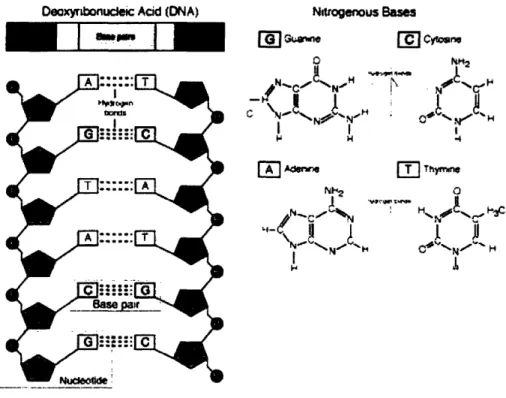
Structure of double stranded DNA and the hydrogen binding characteristics of the bases G, C , A, and T ... 42
Schematic representation of the DNA-directed assembly of biomolecules to probe oligonucleotides immobilized on a surface ... 48
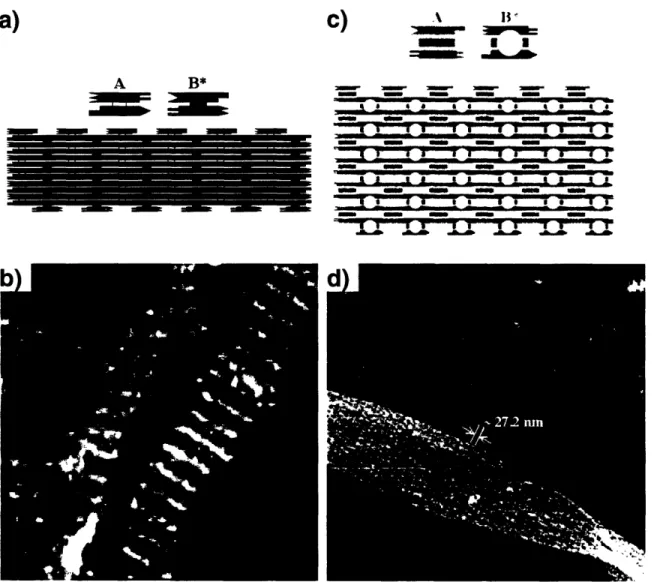
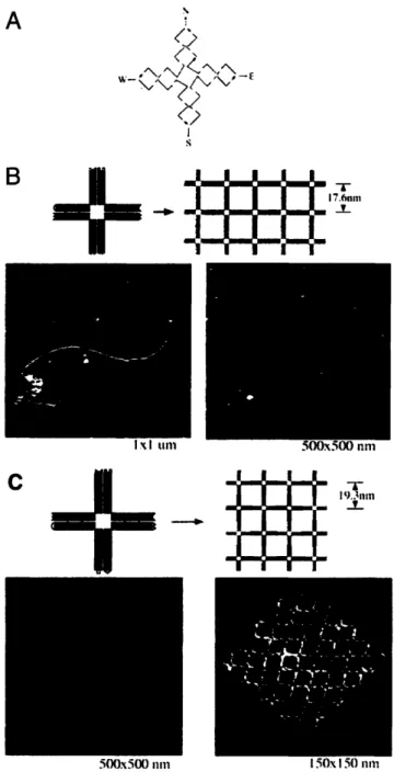
Double crossover (DX) and triple crossover (TX) molecules and their assemblies 49 Self-assembly of DNA nanoribbons and nanogrids using a 4 x 4 DNA tile ... 50
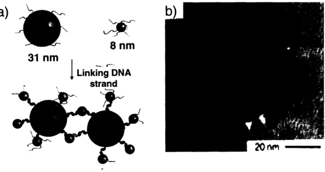
Schematic illustration of the DNA-directed assembly of colloidal nanoparticles... 55
Schematic representation of the DNA-directed binary nanoparticle assembly ... 56
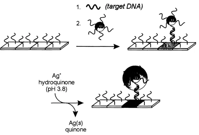
Schematic illustration of the scanomteric detection of DNA hybridization ... 57
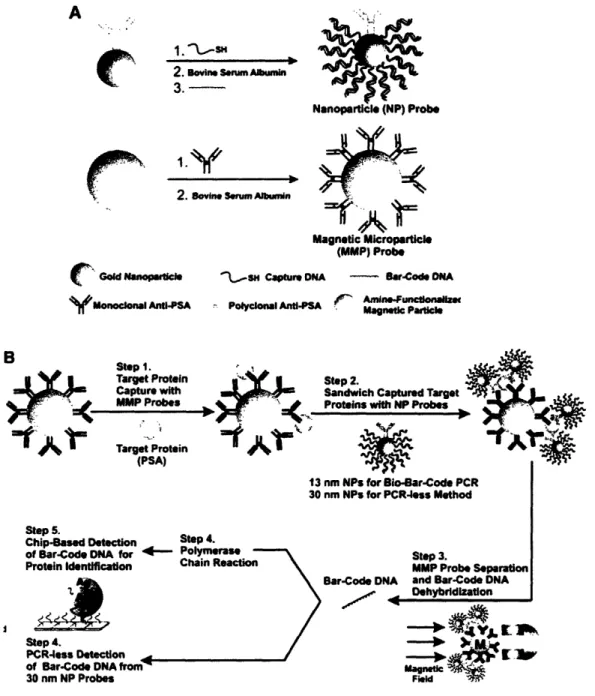
Schematic illustration of the ultrasensitive detection of proteins using DNA-tagged nanoparticles ... 58
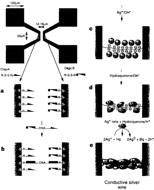
Scheme showing the metallization of dsDNA to construct a conductive silver wire connecting two gold electrodes... 59
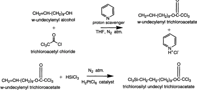
Two-step reaction scheme for the synthesis of the trichloroacetate (TCA)-protected C I silane (TCA silane). ... 74
Schematic illustration of the procedure used to form end-immobilized oligonucleotides at controlled surface densities ... 79
Custom-built flow cell for facilitating oligonucleotide synthesis on flat microscope glass slides within an ABI 392 DNA synthesizer ... 80
Variation in the density of the first nucleotide and a 16-mer probe with the composition of the silane solution ... 81
Uniformity of oligonucleotide synthesis on flat microscope glass slides ... 82
Calibration plot of normalized fluorescence intensity vs. target density ... 85
Fluorescence images obtained from a GenePix®scanner ... 86
Hybridization kinetics for various probe densities ... 87
Figure 3-9 Figure 3-10 Figure 3-11 Figure 3-12 Figure 4-1 Figure 4-2 Figure 4-3 Figure 4-4 Figure 4-5 Figure 5-1 Figure 5-2 Figure 5-3 Figure 5-4 Figure 5-5 Figure 5-6 Figure 6-1 Figure 6-2 Figure 6-3
Variation of dsDNA densities with the surface probe density ... 88
Variation in hybridization efficiency with surface probe density ... 89
Variation of equilibrium binding constant with surface probe density ... 96
Effect of surface probe density on duplex stability ... 99
Schematic illustration of the surface preparation to form end-immobilized oligonucleotides of controlled surface densities ... 111
Schematic illustration of nanoparticle functionalization and subsequent adsorption via DNA-DNA hybridization... 113
SEM images of nanoparticle adsorption on Si/SiO2 surfaces ... 114
High resolution Au (4f) XPS spectra of DNA-tagged gold nanoparticles adsorbed on complementary and non-complementary surfaces ... 115
Comparison of thermodynamics of nanoparticle adsorption vs. molecular D N A adsorption... 120
Schematic illustrating the functionalization and DNA-directed assembly of microparticles onto a flat surface ... 134
Confocal microscopy images of DNA-functionalized cylindrical microparticles after hybridization... 135
Effect of shape on the directed assembly of DNA-functionalized (oligo A) microrods and microspheres onto surfaces containing complementary (oligo cA) and non-complementary (oligo cB) sequences ... 138
Dual-functional microrods ... 139
Schematic illustrating the orientation-specific assembly of dual-functionalized microrods and the use of fluorescent tags to characterize their orientation ... 143
Orientation-specific assembly of dual-functionalized microrods ... 144
Schematic illustration of coating the silica particles (30-50 gm) with gold using shadow deposition and SEM images of gold-coated particles ... 155
Images of the dual-functional particles taken under an optical microscope showing their unique wetting properties ... 157
Schematic illustration of the overall process of making the dual-functional particles and demonstrating the orthogonal assembly of fluorophore-tagged target DNA molecules onto C-C particles ... 160
2-D projection of the confocal microscopy images collected at various
cross-sections of the dual-functional particles ... 161
Schematic illustration of the process used to generate microparticles with three different surfaces... 163
Energy dispersive X-ray analysis (EDX) images of a silica microsphere after shadow deposition of Au and Al ... 164
Schematic illustration of the overall process for making the tri-functional particles and demonstrating the orthogonal assembly of fluorophore-tagged target DNA molecules onto their surfaces ... 166
DNA-directed assembly onto a tri-functional particle ... 167
DNA-driven nanoparticle adsorption onto silica microspheres ... 178
DNA-driven nanoparticle adsorption onto flat gold-coated substrates ... 180
Selective DNA-driven nanoparticle adsorption onto the SiO2side of dual-functional microspheres ... 183
Orthogonal DNA-driven nanoparticle assembly onto dual-functional silica m icrospheres ... 184 13 Figure 6-4 Figure 6-5 Figure 6-6 Figure 6-7 Figure Figure Figure Figure 6-8 7-1 7-2 7-3 Figure 7-4
List of Tables
Table 1-1 Comparison of various immobilization chemistries ... 38 Table 3-1 Deprotection schemes to cleave the trichloroacetyl (TCA) groups and expose
the hydroxyl groups on silane films formed from 100% TCA silane and 0% C8
silane solution ... 78 Table 3-2 Parameters obtained by fitting a second-order Langmuir model to the lowest
probe density and a second-order, diffusion-limited Langmuir model to the
rem aining probe densities ... 93 Table 4-1 Nanoparticle area coverages computed from SEM & XPS measurements ... 116 Table 5-1 Effect of shape on the forces acting during the self-assembly process ... 129
14
Chapter 1.
Introduction - DNA Surface Hybridization
1.1 Introduction
The vast amount of genomic information made available by the Human Genome Project has spurred the development of solid-state DNA based assays. These assays are based on the principle of selective hybridization between two DNA strands according to the Watson-Crick base pairing of adenine with thymine (A-T) and cytosine with guanine (C-G). This selective binding process can be used to identify DNA strands with different sequences and assemble them to specific locations. These assays have been used to measure the mRNA or gene expression levels in cells,' to determine the polymorphisms in various alleles,2 to detect pathogens in water,3' to elucidate biochemical pathways,5 and to perform transcript profiling of tumors6 7. Over the last decade or so, there has been an explosion in the use of DNA-based assays for obtaining genetic information. Advances in gene expression monitoring and the establishment of their correlations to disease phenotypes have the potential to expand clinical abilities in prognosis, diagnosis, and treatment of diseases.8 The genome-wide expression profiles made possible by the annotation of the Human Genome and the availability of whole-genome based DNA assays will play a vital role in developing a more holistic view of biology.9
The application of DNA assays to combinatorial studies was brought about by the development of the DNA microarrays. Southern and coworkerst°were the first to form arrays of nucleic acids, in their case oligonucleotides, on a surface for the purpose of large-scale genetic studies. Since then, DNA microarrays have been improved to yield systems capable of high-throughput analysis of genetic material."
1.1.1 DNA microarrays
DNA microarrays consist of a solid substrate such as a microscopic glass slide, a silicon wafer, or a polypropylene sheet, that is patterned with thousands of different DNA sequences (probes) in an array format at defined locations on its surface. In a typical use of a DNA microarray, messenger RNA (mRNA) is extracted from pathological and normal samples. The mRNA is then converted to cDNA (complementary DNA) using the enzyme reverse transcriptase (RT) and amplified using the polymerase chain reaction (PCR). The cDNA strands are tagged either by incorporating fluorophore-tagged nucleotides into their sequences during the reverse transcription/amplification process or by chemically coupling fluorophores to the modified nucleotides added during the reverse transcription/amplification process. These fluorophore-tagged cDNA samples are then brought in contact with the DNA microarray. After the requisite time for hybridization and the necessary washing steps, the microarray is scanned for fluorescence by laser excitation of the fluorophores. Image analysis and statistical tools are used to convert the fluorescence signal intensities into gene expression levels. The differences in the expression levels of different genes between the normal and pathological samples are used to correlate their link to the disease phenotype. DNA microarrays provide the platform for performing these nucleic acid analyses on a combinatorial scale. The important issues in constructing microarrays are the design of the probe sequences and their appropriate immobilization. For the DNA assays to be useful, each of the following steps-immobilization, hybridization, and detection-has to be properly designed and optimized. Surface issues play a key role in the first two steps involving immobilization and hybridization.
16
1.1.2 Immobilizing probe DNA molecules on the substrate
Immobilization can be achieved either by synthesizing oligonucleotides in a combinatorial fashion on the array surface or by individually spotting pre-synthesized oligonucleotides or cDNAs (complementary DNAs) sequentially onto the array surface. This process requires the substrate and/or the oligonucleotides to be suitably functionalized so that selective coupling can occur. Ultra high densities of probes (as high as 1,000,000 different probes on a 1 cm x 1 cm slide'2) can be achieved by employing sophisticated robotic and fluidic delivery equipment or photolithographic tools along with photo-labile reagents. After probe immobilization, the microarrays are ready to be used for hybridization with target DNA. The sensitivity and selectivity of hybridization depends on the characteristics of the probe surface, the composition of the hybridizing medium, and the hybridization conditions.
1.1.3 Surface hybridization of probe and target DNA molecules
The hybridizing solution that is put in contact with a DNA microarray usually contains genomic fragments (for polymorphism studies) or a mixture of cDNAs (for gene expression studies) that have been amplified by PCR (polymerase chain reaction) and include labels useful for detection. The solution also contains salt to stabilize the negatively charged phosphodiester backbone of the DNA strands and other additives like surfactants to enhance hybridization and to reduce non-specific adsorption. The substrate should be derivatized such that it does not promote the non-specific DNA binding to its probe sites. For example, a positively charged substrate would readily adsorb polyanionic target in the absence of immobilized DNA probes. After hybridization has occurred, the substrate is washed with a buffer solution to remove any non-specifically adsorbed DNA molecules. Hybridization efficiencies typically vary from I to 100%
depending on the surface packing density of the immobilized probes, and the length of the target DNA, the hybridization conditions (time, temperature, salt concentration) employed.
The detection of hybridization event and quantification of its extent has been performed using labeling or non-labeling techniques. For analysis of high-density microarrays, constraints on the detection scheme require that it should have a spatial resolution of a few microns and should be able to distinguish changes even at the pico gram level.
1.1.4 Detection
The most popular method for detecting hybridization events is the introduction of either a fluorescent, chemiluminescent, or radioactive tag in the target DNA molecules. After hybridization, the chips are scanned for the presence of the signals from the tags using either an optical or a radioactivity scanner. The location and intensity of the acquired signals give information about the identity of and the approximate quantity of the target DNA. Various label-free techniques have been used for DNA detection and these methods take advantage of the change in the charge, mass, or refractive index at particular surface sites in order to detect a hybridization signal. These methods include electrochemical detection,3 quartz crystal microbalance,l4 and surface plasmon resonances (SPR) and allow monitoring the hybridization process in real-time. For example, Krull et al.'6 have immobilized oligonucleotides on fused silica optical fibers, and used these fibers as a medium to transfer the fluorescence of the hybridized targets by a total internal reflection process in order to generate denaturation profiles for the hybrid duplexes.
18
1.2 Array formats used currently
Currently available DNA microarrays or "DNA chips" can be broadly classified into two categories depending on whether their probe DNA molecules are synthesized on- or off-chip:
i) On-chip oligonucleotide synthesis:
On-chip synthesis of probes is accomplished by solid-state phosphoramidite chemistry where the bases are added stepwise to prepare the attached oligonucleotides with lengths of 15-70 bases. The solid substrates in this case are often derivatized with molecules that expose hydroxyl groups as reaction sites. Arrays of thousands of probes can be created by either ink-jet techniques"7 or by the Affymetrix's photolithographic technique'8 using photo-labile deprotecting groups on the phosphoramidites. The stepwise coupling yield for the photo-deprotection technique is 195%19, whereas it is 98-99% for the conventional phosphoramidite
chemistry. For comparison, the photo-deprotection technique would have a 35% yield for a 20-mer while the conventional chemistry will form a 65-20-mer at greater than 35% yields.
ii) Spotting of cDNAs / oligonucleotides:
Another method for preparing microarrays is to spot pre-synthesized oligonucleotides or cDNAs directly onto a functionalized surface. Patrick Brown of Stanford developed one of the most commonly used protocols for spotting'l . Here, cDNAs are first produced by reverse transcription from mRNAs, followed by amplification by PCR and purification, and then robotically spotted in nanoliter quantities onto a glass slide coated with polylysine or amino-terminated organic moieties. The system is subsequently illuminated with UV light to effect crosslinking between the thymine residues on the cDNAs and the positively charged amine groups on the functionalized surface. As each DNA strand is attached via various sites along its backbone, the length and the sequences available for subsequent hybridization can vary with the
hybridization conditions. Other immobilization chemistries for this format involve derivatizing the oligonucleotide at one end with a selective linking agent so that it can attach directly to a surface functionality surface or another agent that has been immobilized on the surface.
1.2.1 Problems with currently available formats
Although the currently available formats are becoming widely used in the research community, nonetheless, they are plagued with shortcomings that are being increasingly realized to hamper their reliability. For example, poor control over immobilization densities, lack of reproducibility, on-chip heterogeneity, lack of flexibility, high background adsorption levels, and lack of reusability are some of the problems that need attention and improvement. Some of the most important are discussed below in detail.
Lack of reproducibility - The results obtained by using commercially available systems
lack reproducibility from one batch to other because of the variability caused by the present surface derivatization chemistries and immobilization techniques. The lack of control over the orientation and density of the reactive sites that are the agent for immobilization compounds the problem of reproducibility. Previously, most of the experiments conducted were based on single measurements because of the prohibitive cost for repeating microarray studies. As the microarrays have gotten cheaper, multiple experiments for the same measurements have been made possible and have revealed the extent of chip-to-chip deviation for these measurements. These deviations raise questions about the fidelity of the data and the validity of the conclusions drawn from them.
Lack of reusability - The assay systems are irreversibly changed once they pass through
one cycle of hybridization, denaturation and washing. Thus reduced signals are obtained if the chips are used again for the next round of hybridization. The surface chemistry employed is not
20
robust enough to sustain the washing and denaturation treatments. The lack of reusability has made these systems too expensive for frequent use and beyond the reach of smaller labs.
Loaw signal-to-noise ratios - Low signal-to-noise ratios result from lower density of
hybridization sites and /or large amount of non-specific hybridization. Most of the chemistries employed do not have enough reactive probes on the surface to guarantee large signals after hybridization. Others have too much of non-specific adsorption which is not removed even after stringent washing conditions.
Most of these problems stem directly from the surface derivatization and immobilization chemistry used for making these chips. A targeted design of the surface derivatization and immobilization techniques could help to improve the performance of these systems and improve the reliability of the data obtained from these assays.
1.3
Techniques for DNA immobilization
DNA immobilization on solid surfaces can be achieved by a variety of methods. An ideal immobilization scheme should enable the immobilized nucleic acids to mimic their solution phase behavior. Also such a scheme should ensure the operational effectiveness of these systems including issues of specificity, reproducibility, and reusability. The factors that must be taken into consideration while designing a DNA immobilization scheme are: type of immobilization (covalent, non-covalent etc.), point of attachment, linker length, and linker characteristics.
Non-covalent immobilization schemes such as immobilization on nitrocellulose filters:-and association on lipid bilayer23 generally result in poorly defined strand orientations, low
packing densities, low mobilities, and regions of the nucleic acid sequence being unavailable for hybridization due to the immobilization. Gel entrapment of DNA2 4leads to excessive diffusional
limitations for the target DNA and thus the kinetics of hybridization are slow. Also in these cases, the DNA molecules are susceptible to removal from the surface under high salt or high temperature conditions. Covalent attachment provides far more stable situation for the experimental conditions employed for hybridization.
Regarding the site for attachment, end immobilization through either the 3' or 5' end seems to be the best option as they allow almost all the bases to be available for hybridization. Covalent immobilization either through the backbone or the bases increases the chances of non-specific adsorption because of poor accessibility of the entire sequence for hybridization. It has been found that DNA could become totally inaccessible for hybridization when only 3% of its bases are involved in the covalent linkage.2 5
The hybridization rates on the surface are much slower than those in solution. By having a long enough linker to distance the molecule from any interactions with the surface, it is possible to have a sequence mimic its solution phase behavior even while being immobilized. Various research groups have suggested different required linker lengths between the support and the DNA for their examined experimental conditions. Kawasaki et al.26report that a linker at least 28
A
in length when fully extended between the support and the DNA is required to approach solution phase hybridization rates. Southern et al.27suggest an optimal length of at least40 atoms for best hybridization yields. In contrast, Beattie and coworkers28 have found that such linker arms were not necessary for achieving the efficient hybridization of long PCR products (> 500 bases).
1.3.1 End-immobilization chemistries
Over the years, many alternative immobilization chemistries have been proposed because of inherent problems with the commercially available systems. A number of these have been
22
commercialized. The general approach is to end-immobilize the DNA/oligonucleotide through its 3' or 5' end. A few systems have also been constructed to address issues of density control and non-specific adsorption. In most of these studies, the substrates used are glass, silicon, fused
silica optical fibers, and polypropylene.
Reactive groups can be added to the 3' or 5' end of the DNA/oligonucleotides when phosphoramidite synthesis or PCR is employed for generation of the DNA sequences. These end-functionalized DNA/oligonucleotides can then be attached to functionalized surfaces directly or through a crosslinker. DNA has been end -functionalized with amino-4, carboxylic29, phosphate3 0, silyl31, acrylic31, and thiol3 2 groups. Amino terminal oligonucleotides have been bound to isothiocyanate-activated 3 3 glass, to aldehyde-activated 34 glass and to glass modified with epoxide3 5 without the use of a crosslinker. Thiol-terminated 3 6 and disulfide-terminated oligonucleotides have been bound to aminosilane derivatized glass using a heterobifunctional crosslinker. Disulfide-modified3 7 and acrylic-modified oligonucleotides have been immobilized onto thiol-functionalized surfaces directly. Amino-modified oligonucleotides have been attached to amine-terminated surfaces using glutaraldehyde3 8 as a crosslinker. Alternatively, because of its intrinsic stability the highly specific biotin-avidin3 9 interaction has also been used for DNA immobilization. However, since avidin is a protein, there is a large possibility for non-specific adsorption. Also, the surface densities with this system have been an order of magnitude lower than for other systems. In an interesting departure from other methods, Kumar et al.3 have utilized an immobilization chemistry wherein they attach silanized DNA to unmodified glass. They have demonstrated different procedures to covalently conjugate an active silyl moiety onto the oligonucleotides or cDNAs in solution thereby forming a new class of modified nucleic acids, namely silanized nucleic acids.
The specificity of the immobilization chemistry must compete with the inherent reactivity contained in DNA. Specifically, the nucleic acids contain many reactive functionalities: the negatively charged phosphates, the exocyclic amines on the bases, the enolizeable carbonyl groups on the bases, and cleavable glycosidic bonds. In most of the immobilization schemes described above, the crosslinkers employed for specific attachments can react with the other reactive sites on the nucleic acids and cause unwanted side reactions. Further, competing side reactions with water due to the large amount of water molecules (55 M) relative to probe oligonucleotides (m or less) presents an important challenge as it can reduce the amount of surface-active groups such as activated ester or isocyanate groups that provide sites for oligonucleotide immobilization only when in an active, non-hydrolyzed form. Zammatteo et al.40 while comparing different strategies for covalently attaching DNA to glass surfaces found that the best immobilization and hybridization results occurred for fixing aminated DNA to an aldehyde-modified glass. Lindroos et al.3 4, on the other hand, found that disulfide-modified oligonucleotides immobilized onto thiol-terminated glass work better than the aldehyde-amine immobilization chemistry as regards background fluorescence and signal-to-noise ratios. The literature is filled with competing claims revealing that a flexible, reliable procedure for DNA immobilization is not yet available.
Methods for controlling probe density are also beset with problems. For example, most of the chemistries that use glass and silicon as substrates employ short chain silanes such as the glycidoxy propyl triethoxysilane (GOPS) (an epoxy silane that yields -OH terminations), mercaptopropyl trimethoxysilane (MPS) (-SH terminated), aminopropyltriethoxy silane (APTES) (-NH2terminated) to generate reactive surface sites for DNA attachment. A problem is
that these silanes have a tendency to form multilayers of poorly controlled structures and these
films can be hydrolytically removed from surfaces. Also, crosslinking reactions between adjacent immobilized molecules can reduce the number of reactive sites available for the immobilization of the oligonucleotide/DNA thereby causing difficulties in controlling the density of reactive sites. As such, only a few research groups have tried to address the issue of controlling the surface density of the immobilized probes and have been successful in tackling it (see Chapter 3).
1.4 DNA surface hybridization
The process of surface hybridization can be schematically pictured to begin with the diffusion of the target DNA molecules from the solution phase to the probe layer (Figure 1-1). The diffusion coefficient of DNA in solution is in the range of 10-6- 1 0-8 cm2/s. In a second step, the diffusion of the DNA in the probe layer is affected by hindrance due to steric and electrostatic repulsions offered by the immobilized probe strands. The higher the packing density of the immobilized DNA strands, the slower is the diffusion in this layer. Hindered diffusion in the immobilized layer can cause the target DNA to collide with the surface where it can be non-specifically adsorbed or with the several immobilized probe strands. Collisions with a probe strand at the correct base site could lead to nucleation of hybridization. The nucleation process can be severely impeded by secondary structures found in long target DNA molecules. Once nucleated at the correct spot, the duplex would form by zippering complementary bases together at a rapid rate. The resulting duplex formation between the target DNA and the probe DNA is an equilibrium process since thermal energy can later lead to dissociation of the non-covalently H-bonded DNA duplexes.
0
©
Diffusion Nucleation/
in solution dissociation
Diffusion in After
oligo layer zippering
Non-specific
/
,
()
adsorption/ desorption
CHCH3OH OH C H H OH CH3CH3OH OH OHOH CH3CH3CH3OH OH OH OH CH3CH3O OH
Figure 1-1 Steps involved in the surface hybridization process. 1) The target strands have to undergo diffusion in the bulk solution, followed by 2) diffusion in the probe layer. Next, 3) the target molecules collide with the immobilized probes leading to a nucleation event followed by 4) a quick zippering step to form the DNA duplex. Competing processes include non-specific adsorption and duplex dissociation.
26
1.4.1 Factors affecting DNA surface hybridization
Hybridization on surfaces is influenced by factors such as surface chemistry and surface probe density as well as those factors that influence hybridization in solution: pH, salt concentration, temperature, solvent properties, GC content, and DNA length. Higher GC content increases the stability of DNA duplexes since a GC base pair produces three hydrogen bonds resulting in greater stability than does a AT base pair that produces just two hydrogen bonds. The addition of salt stabilizes DNA by screening the charges between two polyanionic oligonucleotide strands. Further, higher salt concentration helps in improving the kinetics of the process, but at the same time reduces the stringency of discriminating between mismatch sequences. Higher temperatures help to provide better discrimination between mismatches by denaturing the less stable mismatched duplexes. On the downside, higher temperatures may also
cause the dissociation rate to increase and thus decrease equilibrium-hybridized duplex amounts. Surfactants like SDS (sodium dodecyl sulphonate) help in improving the signal-to-noise ratios from hybridization experiments by reducing non-specific adsorption events. However, the addition of too much surfactant can inhibit the hybridization process by interacting with the charged polyanionic backbones.
The length of the complementary DNA sequences plays an important role in setting the stability of the DNA duplex. Longer the length of the oligonucleotide the higher is the melting point of the duplex as a result of a greater enthalpic driving force for duplex formation. In contrast, the greater restrictions to the degrees of freedom for these longer oligonucleotide upon hybridization leads to larger entropic losses. Thus, a balance of these two factors defines the overall equilibrium hybridization levels. Beyond these factors, longer DNA molecules have
larger diffusional limitations in accessing the probe oligonucleotides and mismatch discrimination also gets more difficult with longer oligonucleotide sequences.
At surfaces, probe density is one of the important parameters affecting the process of DNA-DNA hybridization. Generally, it is desired that the number of surface-bound DNA probes be substantially higher than the number of targets in solution in order to achieve large hybridization signals even for low target concentrations. However, higher surface probe densities can increase the diffusional limitations on the incoming target DNA molecules by providing steric as well as electrostatic repulsion to the target molecules. The interplay of these opposing effects leads to an optimal surface probe density where the availability of the probes, the hybridization equilibrium and kinetics are within acceptable limits. Any study on the effects of probe density on the thermodynamics and kinetics of hybridization requires a system where it is possible to systematically vary the surface density of the reactive groups and thus vary the amount of immobilized oligonucleotides in a controlled and measurable manner.
1.5 Chemistries
for controlling probe densities
The packing density of the probes determines the surface charge and steric crowding at the hybridization sites. It has important implications in the thermodynamics and kinetics of hybridization and also in those of denaturation when used for certain SNP analysis. The few successful attempts at achieving this control are discussed below.
Tarlov et al.32'41have used a system wherein they deposit thiol-modified oligonucleotides
from a micromolar solution onto gold surfaces and then immerse the substrate in a millimolar solution of 6-mercapto- 1 l-hexanol to achieve a range of densities for the immobilized oligonucleotides. In this system, the thiol-Au bond responsible for oligonucleotide attachment is heat sensitive. A limitation to this chemistry is that the amount of immobilized oligonucleotides
would be reduced after going through one complete cycle of hybridization, washing, and denaturation thereby making these systems useful generally for single investigations.
Smith et al.42have used mixtures of t-butyloxycarbonyl (t-BOC)-protected 10-aminodec-1-ene and dodecene to derivatize the surface of hydrogen-terminated silicon (001) with controlled densities of amine groups. They used UV light to remove the t-BOC protecting groups. Although this substrate has a highly defined crystalline structure and presents a homogeneous surface, it oxidizes under ambient conditions. To protect the substrate from possible side reactions, experiments must be performed under controlled atmospheres in order to control the density of reactive groups. This system allows density control but is handicapped somewhat by its lack of operational flexibility.
Other groups such as Krull et al.43 have tried to modulate the surface probe density by changing the delivery times of linker molecules to substrates within an oligonucleotide synthesizer. In another approach, Gou et a133 have tried to change surface densities by spotting different concentrations of oligonucleotide solution to a substrate. A comprehensive comparison of various immobilization chemistries with regards to packing density, density control, and hybridization time is provided in Table 1.4 3'4 4
1.5.1 Our approach
The surface immobilization chemistry developed in our laboratory by a previous graduate student (Ivan Lee) meets most of the above requirements. Specifically, the immobilization process utilizes the concept of mixed monolayers4 5 where two silane compounds with different
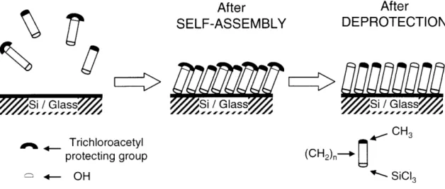
end groups-one with a protected reactive (hydroxyl) end group and the other with an inert (methyl) end group-are mixed in desired proportions to obtain controlled densities of reactive groups on surface.46 Figure 1-2 shows an illustration of the self-assembly of two silanes onto a
surface yielding film of mixed composition. Experimentally, the surface composition is related to the silane composition in solution. Once covalently attached, the end groups of the silanes are base deprotected to expose the reactive hydroxyl functional groups. These functionalized surfaces are then used for synthesis of oligonucleotides on the surface. The inert end groups ensure that the non-specific adsorption of DNA is small. Our approach is compatible with soft lithography techniques such as micro-contact printing thereby allowing surfaces with regions of high reactive group density and other regions with inert groups to be obtained. These patterned surfaces provide a substrate for array construction as spots are defined by differences in their reactivity and hydrophilicity. In this process, the silane compounds form covalent bonds with the surface and are more stable to heat treatments than are systems constructed using thiol-Au bonds. Our chemistry provides a fairly good control over surface composition as needed to achieve a wide range of probe densities. The resulting system is reliable, flexible, reproducible, and generates probe surfaces that yield hybridization results with high signal-to-noise ratios. The thermodynamic and kinetic study of DNA-DNA surface hybridization can be accomplished in a more systematic manner with access to such an immobilization chemistry.
30
After SELF-ASSEMBLY
C
< Trichlorc protectin( *--4 OH After DEPROTECTION //,S i / as Glass/, i / A C H3 )acetyl g group (CH2)- - SiCI3Figure 1-2. Schematic illustration of formation of mixed self-assembled monolayers. Two silanes-one with a protected-hydroxyl group and other with an inert methyl group-are first brought in contact with the glass or Si/SiO2 surface. The two silanes assemble on the surface
based on their solution composition. After the assembly, the protecting trichloroacetate groups are removed using a basic solution to expose the hydroxyl groups.
1.6
Previous Work
Although the use of microarrays is increasing rapidly, there has been little in-depth theoretical or experimental research on the factors that influence DNA-DNA surface hybridization. For example, in diagnostic applications, a system with fast kinetics is desired so that a measurable signal can be obtained within a reasonable time. In gene-expression studies, reliable quantitative data is desired even if experiments require longer hybridization times. Thus, there is a need to better understand factors that influence both the kinetics and thermodynamics of surface hybridization in order to provide improvements for this process. Such a study necessitates the availability of a well-defined system with controllable immobilization chemistry and also effective detection techniques to characterize the system at various points during its operation. Georgiadis et al.47 have explored the effects of probe density for the hybridization of 25-mer DNA strands to surfaces. They observed decreasing hybridization efficiencies with increases in probe density. Also, they observe two different kinetic profiles for hybridization onto surfaces with probe densities below and above a particular threshold value. Krull et al.4 8 immobilized 20-mers onto fused silica optical fibers and used a total internal reflection fluorescence instrument to monitor the hybridization in real time. They observed that the standard enthalpic changes for the hybridization process on surface were 2-3 fold lower than the values obtained in a bulk solution. They also observed a decrease in the surface TM values with
increases in probe density.
On the theoretical side, only a few research groups have tried to describe the surface hybridization process via fundamental equations. Chan et al.49 have tried to model the system based on a Brownian dynamics model. However, they failed to take any ionic effects into consideration. Also, they have modeled only a system with a low probe density. The current
32
I __I____ ··_I__·
-L·-trend in experiments is to have systems at higher surface densities so that the number of probes is greater than the number of targets, but low enough to avoid detrimental electrostatic effects on surface hybridization reactions. Wong et al.50 have tried to model the hybridization phenomenon using molecular dynamics. But due to the enormity of atoms even in a single 10-mer oligonucleotide and the number of degrees of freedom in its 3-D structure, not to mention the electrostatic effects due to the phosphate backbone, they have been able to simulate the system only for few nanoseconds despite using a very powerful computing facility. As the phenomenon of hybridization takes place on a time scale of a few microseconds it is difficult to characterize the validity of these hybridization results based on simulations of only a few nanoseconds. Thus, for molecular dynamics simulations to gain more insight into such systems, they would require tremendous increase in computing power. As such these approaches do not appear useful at this time. Less detailed models like Brownian dynamics or simpler diffusion-limited reaction models would be easier to implement. In another approach, Pettitt et al.51 55 have modeled the target DNA molecules as ion-penetrable charged spheres and the probe surface as a charged plate. They employed the linearized Poisson-Boltzmann theory of the electric double-layer interaction in order to model the electrostatic interactions. Their estimations suggest the presence of strong electrostatic forces even at high salt concentrations and predict a 'Coulomb blockage' of the target hybridization at high surface probe densities.
The current state of research in this field suggests that more effort is required into understanding the fundamentals of hybridization both at the kinetic and thermodynamic level so as to achieve better control over the behavior of these systems. Based on the previous work, the main motivation for this part of this thesis was to systematically explore the effects of surface density on the DNA surface hybridization process. I was particularly interested in understanding
the trade-offs involved with low and high probe densities. Additionally I wanted to use our immobilization chemistry to also explore nanoparticle adsorption on surfaces via DNA-DNA hybridization, a promising technique for ultra-sensitive detection of DNA fragments and a potential assembling strategy for materials synthesis.
1.7 References & Footnotes
(1) Duggan, D. J.; Bittner, M.; Chen, Y. D.; Meltzer, P.; Trent, J. M. Nat Genet 1999, 21, 10-14. (2) Kwok, P.-Y. Annual Review s in Genomics and Human genetics 2001, 2, 235-258.
(3) Wang, J. L. Progress in Biochemistry cnd Biophysics 2001, 28, 125-128.
(4) Cheng, J. F., P. Surrey, S. Kricka, L. J. Wilding, P. Mol. Diagn. 1996, 1, 183-200.
(5) Ideker, T.; Thorsson, V.; Ranish, J. A.; Christmas, R.; Buhler, J.; Eng, J. K.; Bumgarner, R.; Goodlett, D. R.; Aebersold, R.; Hood, L. Science 2001, 292, 929-934.
(6) Dhanasekaran, S. M.; Barrette, T. R.; Ghosh, D.; Shah, R.; Varambally, S.; Kurachi, K.; Pienta, K. J.; Rubin, M. A.; Chinnaiyan, A. M. Nature 2001, 412, 822-826.
(7) Welsh, J. B.; Zarrinkar, P. P.; Sapinoso, L. M.; Kern, S. G.; Behling, C. A.; Monk, B. J.; Lockhart, D. J.; Burger, R. A.; Hampton, G. M. Proc Natl Acad Sci U S A 2001, 98,
1176-1181.
(8) Sevenet, N.; Cussenot, O. Clin Exp Med 2003, 3, 1-3.
(9) Shoemaker, D. D.; Linsley, P. S. Curr Opin Microbiol 2002, 5, 334-337. (10) Southern, E. M.; Maskos, U.; Elder, J. K. Genomics 1992, 13, 1008-1017. (11) Heller, M. J. Annu Rev Biomned Eng 2002, 4, 129-153.
(12) Lipshutz, R. J.; Fodor, S. P. A.; Gingeras, T. R.; Lockhart, D. J. Nat Genet 1999, 21, 20-24. (13) Steel, A. B.; Herne, T. M.; Tarlov, M. J. Anal Chem 1998, 70, 4670-4677.
(14) Caruso, F.; Rodda, E.; Furlong, D. F.; Niikura, K.; Okahata, Y. Anal Chem 1997, 69, 2043-2049.
(15) Thiel, A. J.; Frutos, A. G.; Jordan, C. E.; Corn, R. M.; Smith, L. M. Anal Chem 1997, 69, 4948-4956.
34
(16) Piunno, P. A. E.; Krull, U. J.; Hudson, R. H. E.; Damha, M. J.; Cohen, H. Anal Chim Acta 1994, 288, 205-214.
(17) Hughes, T. R.; Mao, M.; Jones, A. R.; Burchard, J.; Marton, M. J.; Shannon, K. W.;
Lefkowitz, S. M.; Ziman, M.; Schelter, J. M.; Meyer, M. R.; Kobayashi, S.; Davis, C.; Dai, H. Y.; He, Y. D. D.; Stephaniants, S. B.; Cavet, G.; Walker, W. L.; West, A.; Coffey, E.; Shoemaker, D. D.; Stoughton, R.; Blanchard, A. P.; Friend, S. H.; Linsley, P. S. Nat Biotechnol 2001, 19, 342-347.
(18) Fodor, S. P. A.; Read, J. L.; Pirrung, M. C.; Stryer, L.; Lu, A. T.; Solas, D. Science 1991, 251, 767-773.
(19) McGall, G. H.; Barone, A. D.; Diggelmann, M.; Fodor, S. P. A.; Gentalen, E.; Ngo, N. J
Am Chem Soc 1997, 119, 5081-5090.
(20) Shalon, D.; Smith, S. J.; Brown, P. O. Genome Res 1996, 6, 639-645. (21) Schena, M. Bioessays 1996, 18, 427-431.
(22) Southern, E. M. J Mol Biol 1975, 98, 503-&.
(23) Schouten, S.; Stroeve, P.; Longo, M. L. Langmuir 1999, 15, 8133-8139.
(24) Proudnikov, D.; Timofeev, E.; Mirzabekov, A. Anal Biochem 1998, 259, 34-41. (25) Bunemann, H. Nucleic Acids Res 1982, 10, 7181-7196.
(26) Zhang, Y.; Coyne, M. Y.; Will, S. G.; Levenson, C. H.; Kawasaki, E. S. Nucleic Acids Res
1991, 19, 3929-3933.
(27) Shchepinov, M. S.; CaseGreen, S. C.; Southern, E. M. Nucleic Acids Res 1997, 25, 1155-1161.
(28) Beattie, W. G.; Meng, L.; Turner, S. L.; Varma, R. S.; Dao, D. D.; Beattie, K. L. Mol
Biotechnol 1995, 4, 213-225.
(29) Kumar, A.; Advani, S. Nucleosides Nucleotides 1992, 11, 999-1002.
(30) Henke, L.; Piunno, P. A. E.; McClure, A. C.; Krull, U. J. Anal Chim Acta 1997, 344, 201-213.
(31) Kumar, A. L., O. Parodi, D. Liang, Z. Nucleic Acids Res 2000, 28, e71. (32) Heme, T. M.; Tarlov, M. J. JAm Chem Soc 1997, 119, 8916-8920.
(33) Guo, Z.; Guilfoyle, R. A.; Thiel, A. J.; Wang, R. F.; Smith, L. M. Nucleic Acids Res 1994, 22, 5456-5465.
(34) Lindroos, K., Liljedahl, U. Raitio, M. Syvanen, A.C. Nucleic Acids Res 2001, 29, e69. (35) Maskos, U.; Southern, E. M. Nucleic Acids Res 1992, 20, 1679-1684.
(36) Chrisey, L. A.; Lee, G. U.; Oferrall, C. E. Nucleic Acids Res 1996, 24, 3031-3039.
(37) Rogers, Y. H.; Jiang-Baucom, P.; Huang, Z. J.; Bogdanov, V.; Anderson, S.; Boyce-Jacino, M. T. Anal Biochem 1999, 266, 23-30.
(38) Yang, M. S.; McGovern, M. E.; Thompson, M. Anal Chim Acta 1997, 346, 259-275. (39) Nikiforov, T. T.; Rendle, R. B.; Goelet, P.; Rogers, Y. H.; Kotewicz, M. L.; Anderson, S.;
Trainor, G. L.; Knapp, M. R. Nucleic Acids Res 1994, 22, 4167-4175.
(40) Zammatteo, N.; Jeanmart, L.; Hamels, S.; Courtois, S.; Louette, P.; Hevesi, L.; Remacle, J. Anal Biochem 2000, 280, 143-150.
(41) Levicky, R.; Herne, T. M.; Tarlov, M. J.; Satija, S. K. JAm Chem Soc 1998, 120, 9787-9792.
(42) Strother, T.; Hamers, R. J.; Smith, L. M. Nucleic Acids Res 2000, 28, 3535-3541. (43) Henke, L.; Krull, U. J. Can. J. Anal. Sci. Spectrosc. 1999, 44, 61-70.
(44) Cavic, B. A.; McGovern, M. E.; Nisman, R.; Thompson, M. Analyst 2001, 126, 485-490. (45) Prime, K. L.; Whitesides, G. M. Science 1991, 252, 1164-1167.
(46) Lee, I. H. In Chemical Engineering; Massachusetts Institute of Technology: Cambridge, 2001.
(47) Peterson, A. W.; Heaton, R. J.; Georgiadis, R. M. Nucleic Acids Res 2001, 29, 5163-5168. (48) Watterson, J. H.; Piunno, P. A. E.; Wust, C. C.; Krull, U. J. Langmuir 2000, 16, 4984-4992. (49) Chan, V.; Graves, D. J.; McKenzie, S. E. Biophys J 1995, 69, 2243-2255.
(50) Wong, K. Y.; Pettitt, B. M. Theor. Chem. Acc. 2001, 106, 233-235. (51) Vainrub, A.; Pettitt, B. M. Chem. Phys. Lett. 2000, 323, 160-166. (52) Vainrub, A.; Pettitt, B. M. Biophys J 2000, 78, 404A-404A.
36
(53) Vainrub, A.; Pettitt, B. M. Phys. Rev. E 2002, 66.
(54) Vainrub, A.; Pettitt, B. M. JAm Chem Soc 2003, 125, 7798-7799. (55) Vainrub, A.; Pettitt, B. M. Biopolymers 2003, 68, 265-270.
(56) Piunno, P. A. E.; Watterson, J.; Wust, C. C.; Krull, U. J. Anal Chim Acta 1999, 400, 73-89.
Table 1-1 Comparison of various immobilization chemistries
Immobilization Probe Probe Density Hybrid. Hybridization Group Chemistry Substrate Target, Density Control Time Conditions
Length Molecule/cm:
Kumar et Silanized DNA Glass 20 nt / 2x10'3 No 30 min 37 C, 20 nM to
al.31 slide 20 nt - 12 h I jiM oligo, 750
mM NaCI, 125
mM Na citrate,
0.1% Tween
McGall et OH-silane + oligo Glass 20-25 nt - No 4 h 35-40 C, 6x SSPE,
al.'9 synthesis slide / PCR 0.001% Triton
product X-100
Shalon et Poly-lysine + Glass PCR / - No 14-18 h 65 C, 0.5 M NaC1,
al.-° PCR product slide PCR 0.05 M Na citrate,
products 0.3% SDS
Piunno et Glycidoxy propyl Fused 20 nt / 9x101° Some 40 min 90 C, 0.62 M
al.5 6 silane + DMT- silica 20 nt to oligo, 1 MNaCI,
HEG linker - 4.6x10¹² 50 mM NaH2PO4
oligo synthesis
Shchepinov Glycidoxy propyl Glass 12 nt / 6x10' 2 Some 2 h 30 C, 3 nM oligo,
et al.27 silane + linker + slide 12 nt 0.1 M NaCI
oligo synthesis
Guo et al.33 NH7 propylsilane Glass 157 nt / 6x101 Some 3 h 30 C, 20-50 nM
+ PDC + 5' NH,- slide 182 nt to PCR product, 5x
PCR product 3x10'3 SSPE, 0.5% SDS
Graves et NH: propylsilane Glass 15 nt / Ix101- Some 3-6 h 46 C, 2 iM oligo,
al.4 9 + PDC + 5' slide 15 nt (appx.) 0.9 M NaCI, 0.06
amino oligo/ PCR M NaH2PO4, 6
product mM EDTA, pH
7.4
Chrisey et NH2 silane + Si/SiO., 20 nt/ 1.2x10¹³ Some 2 h 25°C, ltiM oligo,
al.3 6 SMPB + 3' Fused 20 nt 10 mM HEPES, 5
SH-oligo silica mM EDTA buffer
Strother et (NH2-decene + Si (001) 16 nt / 2.3x101- Yes 30 min 25 C, 2 tIM oligo,
al.42 dodecene) + 16 nt 2x SSPE, 0.2%
SSMCC +- SDS
SH-oligo
Cavic et (SH-silane + Si/SiO 25 nt/ 2x10¹³ Yes 1.5 h 25 °C, 20 LM
al.44 Alkane silane) + 25 nt oligo, 10 mM
Tris-BMH + 3' HC1, 1 mM EDTA,
SH-oligo 1.5 M NaCl
Tarlov et (5' SH-oligo + Au- 25 nt / 5.7x101- Yes 90 min 24°C, ltaM oligo,
al.3 2 4' mercapto coated 25 nt 10mM Tris-HCl, 1
hexanol) silicon mM EDTA, 1 M
NaCI
Our (OH-silane + Si/SiO7, 12 nt / -3x10 3 Yes 24 h 4°C, 0.5 ,uM oligo,
system4 6 alkane silane) + Glass 12 nt (optim.) 1 M NaCI, 0.1 M
oligo synthesis slide Na citrate, 0.1%
SDS PDC- Phcnylcncdiisothiocyanatc
HEPES- N-2-hydroxyethylpiperazine-N'-2-ethanesulfonic acid Twccn- Polyoxycthylcnc sorbitan monolaurcatc
DMT-HEG- Dimethoxy trityl hexaethylene glycol
SSMCC- Sulfo-succinimidyl 4-(N-malcimidomcthyl) cyclohcxanc- -carboxylatc
SMPB- Succinimidyl 4-[malcmidophcnyl] butyratc EDTA- (Ethylenedinitrilo) tetraacetic acid SDS- Sodium dodccyl sulphonatc BMH- Bis(maleimido) hexane nt - nuclcotidc
38
Chapter 2.
Introduction - DNA-Directed Assembly
2.1 Self-assembly
Self-assembly-the formation of often complex supramolecular structures by non-covalent interactions-remains a powerful strategy for manipulating chemical systems and their properties. The broad use of surfactants, the generation of micro-domains within block co-polymers, and the folding of polypeptide chains into proteins are examples where local interactions between individual molecules or segments produce such assembled structures. The strength of the specific interactions that drive self-assembly within a system play a key role in defining the effects that temperature and concentration have on the integrity of such structures and their tolerance of other present chemical species. In many cases, self-assembly is guided by the generic preference for one species type (charged, polar, non-polar) for its counterpart (as in the case of charged species'-5) or its kin (as for polar and non-polar species6), with these
interactions being quite general. Indeed, most synthetic self-assembled systems are based on the inclusion of two interactions where a molecular fragment or polymer segment A strongly prefers to interact with A over B and similarly B prefers to interact with B over A. Synthetic systems that expand this local complexity of interactions to include a third or more components are limited by mutual complementarity for specificity or preference. Candidate possibilities for providing this driving force for self-assembly include the use of metal ligation 7-10 along with
highly specific biomolecular systems using proteins"1, antibodies12, DNA or other species.
The richness present in biology provides motifs for self-assembly that are compatible with water and that themselves can operate independently and in the presence of a wide range of other species. Of these, oligonucleotides offer the specific advantage that their intermolecular self-assembly depends directly on primary sequence information, in contrast with that for
polypeptide-based systems that involve secondary and tertiary structures resulting from specific folding events that rely on polar and non-polar interactions. Further, the rules and energetics for their assembly are straightforward and much better understood than are de novo approaches to predicting peptide folding and recognition based on primary sequence. To be useful, a critical element for the consideration, adoption, and broader inclusion of such motifs within synthetically prepared self-assembling systems is their widespread availability and easy tailorability. Advances in molecular biology have led to the development of routine methods for the synthetic preparation of oligonucleotides by automated means. In fact, the on-demand custom synthesis of oligonucleotides by commercial vendors has become a routine part of many genetic investigations and has been a key element underlying the rapid growth in this area. Their availability has led to the design of unnatural self-assembling systems that incorporate oligonucleotide sequences.
The top-down methods of device manufacture are now approaching their physical limits as far as the lateral resolution of functional components is concerned. E-beam lithography and extreme UV lithography (EUVL) may enhance the resolution further, but entirely different ways of manufacturing devices need to be invented to maintain further pace with Moore's law. Researchers are now increasingly looking at novel bottom-up ways to create faster, denser, and more energy efficient devices.'3 One of the key areas-directed assembly-involves the assembly of nano-sized components in a highly parallel fashion into predetermined forms, based on the instructions programmed onto their surfaces. DNA hybridization has features which will be beneficial for use in bottom-up assembly techniques.
40