Publisher’s version / Version de l'éditeur:
Molecular Biosystems, 6, 7, pp. 1131-1142, 2010-03-01
READ THESE TERMS AND CONDITIONS CAREFULLY BEFORE USING THIS WEBSITE. https://nrc-publications.canada.ca/eng/copyright
Vous avez des questions? Nous pouvons vous aider. Pour communiquer directement avec un auteur, consultez la première page de la revue dans laquelle son article a été publié afin de trouver ses coordonnées. Si vous n’arrivez pas à les repérer, communiquez avec nous à [email protected].
Questions? Contact the NRC Publications Archive team at
[email protected]. If you wish to email the authors directly, please see the first page of the publication for their contact information.
Archives des publications du CNRC
This publication could be one of several versions: author’s original, accepted manuscript or the publisher’s version. / La version de cette publication peut être l’une des suivantes : la version prépublication de l’auteur, la version acceptée du manuscrit ou la version de l’éditeur.
For the publisher’s version, please access the DOI link below./ Pour consulter la version de l’éditeur, utilisez le lien DOI ci-dessous.
https://doi.org/10.1039/b924668c
Access and use of this website and the material on it are subject to the Terms and Conditions set forth at
Host–virus interactions during hepatitis C virus infection : a complex
and dynamic molecular biosystem
Pezacki, John Paul; Singaravelu, Ragunath; Lyn, Rodney K.
https://publications-cnrc.canada.ca/fra/droits
L’accès à ce site Web et l’utilisation de son contenu sont assujettis aux conditions présentées dans le site LISEZ CES CONDITIONS ATTENTIVEMENT AVANT D’UTILISER CE SITE WEB.
NRC Publications Record / Notice d'Archives des publications de CNRC:
https://nrc-publications.canada.ca/eng/view/object/?id=e420e8d8-28fb-42ea-bc0b-837845637722
https://publications-cnrc.canada.ca/fra/voir/objet/?id=e420e8d8-28fb-42ea-bc0b-837845637722
Host–virus interactions during hepatitis C virus infection: a complex
and dynamic molecular biosystem
John Paul Pezacki,*
abcRagunath Singaravelu
aband Rodney K. Lyn
acReceived 23rd November 2009, Accepted 8th February 2010 First published as an Advance Article on the web 12th March 2010 DOI: 10.1039/b924668c
The hepatitis C virus (HCV) is a global health issue with no vaccine available and limited clinical treatment options. Like other obligate parasites, HCV requires host cellular components of an infected individual to propagate. These host–virus interactions during HCV infection are complex and dynamic and involve the hijacking of host cell environments, enzymes and pathways. Understanding this unique molecular biosystem has the potential to yield new and exciting strategies for therapeutic intervention. Advances in genomics and proteomics have opened up new possibilities for the rapid measurement of global changes at the
transcriptional and translational levels during infection. However, these techniques only yield snapshots of host–virus interactions during HCV infection. Other new methods that involve the imaging of biomolecular interactions during HCV infection are required to identify key interactions that may be transient and dynamic. Herein we highlight systems biology based strategies that have helped to identify key host–virus interactions during HCV replication and infection. Novel biophysical tools are also highlighted for
identification and visualization of activities and interactions between HCV and its host hepatocyte. As some of these methods mature, we expect them to pave the way forward for further exploration of this complex biosystem and elucidation of mechanisms for HCV pathogenesis and carcinogenesis.
Introduction
Hepatitis C virus (HCV) infection is a rapidly increasing global health problem affecting approximately 3% of the global population.1,2Currently there is no vaccine available
and the clinical efficacy of modern therapeutics is limited. HCV infection is one of the leading cause of chronic hepatitis, aSteacie Institute for Molecular Sciences, National Research Council
of Canada, 100 Sussex Dr., Ottawa, Ontario, Canada K1A 0R6. E-mail: [email protected]
b
Department of Biochemistry, Microbiology & Immunology and Ottawa Institute of Systems Biology, University of Ottawa, Health Sciences Campus, 451 Smyth Road, Ottawa, Canada K1H 8M5
cDepartment of Chemistry, University of Ottawa, 10 Marie Curie,
Ottawa, Canada K1N 6N5
John Paul Pezacki
John Paul Pezacki (b. 1969) was born in Cambridge, England, and grew up in Toronto, Canada. He graduated with a BSc in chemistry from the University of Toronto in 1994, and received his PhD in organic chemistry from McMaster University in 1998 for his work with Professor John Warkentin. He conducted postdoctoral studies at the University of Toronto on enzyme engineering and signal transduction with R. Kluger and at the Scripps Research Institute with P. G. Schultz on various topics encompassing chemical biology and genomics of HCV. He returned to Canada to the Steacie Institute for Molecular Sciences of the National Research Council of Canada where he is currently a Senior Research Officer and also holds adjunct professor appoint-ments at the University of Ottawa in the Departappoint-ments of Chemistry and Biochemistry, Microbiology and Immunology while being a member of the Ottawa Institute for Systems Biology. His research interests include chemical biology, virology, proteomics, screening technologies, and bioimaging.
Ragunath Singaravelu
Ragunath Singaravelu (b. 1986) graduated from University of Ottawa with an honours BSc in biochemistry and BASc in chemical engineering in 2009. He joined Dr Pezacki’s group as an undergraduate student in 2007 and is continuing his postgraduate research there. The projects he has been involved in deal with the design of novel small RNA profiling assays, activity-based protein profiling, methods of live cell small RNA imaging, and probing the miRNA signature associated with hepatitis C infection, replication, and carcinogenesis.
Downloaded by CISTI Archive Access on 08 March 2011
cirrhosis and hepatocellular carcinoma (HCC) worldwide. There are six genotypes of HCV with genotype 1a being the most prevalent in North America while 1b is most prevalent in Europe and Asia.3 HCV infection remains a major medical challenge to the world because of the prevalence of HCV infection, the severity of the associated HCC, the lack of good diagnostic and prognostic markers, and the absence of broadly effective treatment strategies.4 Currently clinical treatments involve combination therapy with modified interferons (pegylated), which stimulate an immune response, and ribavirin, an antiviral nucleoside analog. Although much effort has been put into the development of antiviral therapeutics specifically targeting HCV proteins and some of these lead molecules have made it to clinical trials, none are currently in use as a clinical alternative to the combination of interferon (IFN) and ribavirin. This treatment has limited efficacy with only B50% of patients with genotype 1a achieving sustained virological response. It is not clear how easily HCV will develop resistance to such therapeutics, although this is a distinct concern. On the other hand, interactions between HCV and its host cell environment may bring about new strategies and leads for therapeutic development that will be less susceptible to viral adaptations. This review will highlight some of the recent approaches that have been successfully employed to identify interactions between HCV and host cell hepatocytes and how these novel targets may be exploited for therapeutic applications.
The hepatitis C virus is a positive-sense RNA virus of the Flaviviridae family that replicates through double-stranded (ds) RNA intermediates in the cytoplasm of host cells, primarily, if not exclusively, in hepatocytes.5,6It is an icosahedral virus that is enveloped with host cell derived membrane components.6 Post-translational modifications of the HCV polyprotein (Fig. 1) are essential to the viral lifecycle and, thus, represent potential events for therapeutic intervention.6 Processing is critical at an early stage in the HCV replication cycle since its 9.6 kb RNA genome encodes an B3000 amino acid polyprotein that is cleaved into three structural proteins (core, E1, and E2) and seven non-structural proteins
(p7, NS2, NS3, NS4A, NS4B, NS5A, and NS5B) (Fig. 1).7,8 Enzymatic cleavage of the polyprotein into functional components is mediated by at least one cellular signal peptidase and one signal peptide peptidase, and two viral proteases, NS2 and NS3.7 The host signal peptidases cleave the structural components of HCV9 while processing of the NS3-5B fragment is catalyzed by NS3 and its cofactor NS4A.9Kinetics
studies indicate that cleavage between NS3 and NS4A occurs early, although small amounts of an NS3-5A intermediate suggest that alternative cleavage occurs simultaneously.5,10–14 In addition to proteolytic cleavage, phosphorylation by kinases and glycosylation are other host contributions important to the HCV lifecycle. Phosphorylation of NS5A mediates the viral protein’s effects in cells harboring HCV.6,15,16Different host cellular kinases are responsible for phosphorylation of NS5A including members of the CMGC and CSK1 kinase families.16–19Glycosylation of HCV proteins is also essential for its proliferation and virulence.20At least
three HCV proteins, namely the envelope glycoproteins E1 and E2, which comprise an important part of the viral coat, and the serine protease NS3, are known to be glycosylated.21–24 Since HCV does not produce its own glycosyl transferase or glycosidase enzymes, it must rely on host proteins to appropriately glycosylate these proteins.20The glycosylation
of the E1 and E2 envelope proteins is of particular importance to HCV virulence since it is required for both efficient host-cell recognition and viral entry.21–24 The identity of all host glycosidases that could participate in HCV glycosylation and how their activity could be affected by the high-level expression of viral envelope proteins is not yet known.
While the basic stages of the HCV lifecycle are known (Fig. 1), many of the details regarding the molecular inter-actions between the host cell and the virus remain elusive. Viral entry into the cell marks the primary event leading to infection. Recently it has been established that the E1 and E2 viral proteins interact with four known host receptors CD81, claudin-1, SRB-1 and occludin and their interaction can also be mediated by GAGs, the LDL receptor, and both DC and liver cell specific-SIGN.25–42The next step involves the fusion
of the virion particle with membranes of the host cell and subsequent uncoating and release of the components of the virion particle that include the positive strand HCV genomic RNA, some HCV non-structural proteins, including NS5A, and, in some cases, host proteins, including low density lipoproteins (LDLs).25,28,29 The HCV RNA then moves to
ribosomes where the HCV polypeptide is translated and processed as previously mentioned.
Subsequent to the maturation of the individual HCV proteins, the virus induces modified membranes in the host cell. These are often observed as membranous webs that are ER-derived.5,11–14
The non-structural HCV protein NS4B is thought to induce these changes as it is sufficient to generate membranous webs that are visualized using electron microscopy.5,11–14 These altered membranes then become the sites for HCV replication, which is performed by the RNA-dependent RNA polymerase NS5B. Negative strand RNAs are generated which then act as templates for the synthesis of new positive strand HCV genomic RNA. Replication sites are situated on the ER near where lipid droplets (LDs) are formed.5,43Viral assembly then Rodney K. Lyn
Rodney K. Lyn (b. 1984) is currently a doctoral candidate in chemistry at the University of Ottawa. In the Steacie Institute for Molecular Sciences at the National Research Council of Canada (NRC) he is currently utilizing CARS microscopy and multi-photon fluorescence to demonstrate the ability to screen for antivirals and small molecule inhibitors against HCV. By combining novel molecular imaging techniques, his work also focuses on understanding the dynamics of host–pathogen interactions of human hepatoma cells expressing HCV viral proteins on the surface of the lipid droplet.
Downloaded by CISTI Archive Access on 08 March 2011
proceeds and is initiated by the HCV core protein, which is known to strongly interact with LDs and even cause them to change localization by trafficking along microtubules.44,45The site of viral particle assembly is thought to occur on LDs adjacent to the ER, and this process is in conjunction with VLDL particle synthesis.46,47 Although it is not precisely known, secretion is thought to then involve components of the VLDL secretion pathway.47
Since its discovery approximately 20 years ago, there has been a tremendous amount of progress made on understanding the HCV genome especially with regards to variability in genomic sequences and the structure and function of the individual proteins of HCV. However, HCV requires many specific biomolecular interactions with host hepatocyte cells in order to proliferate. These host–pathogen interactions can be difficult to identify as many are dynamic and/or involve host cell enzymes that are only transiently hijacked. The virus requires the host cellular factors’ enzymatic activity for post-translational modifications, structural elements for trafficking,
and unique pathways for cell entry and secretion. Along with glycosylation of the envelope proteins and phosphorylation of NS5A, additional examples of host interactions include direct interaction with host cell geranylgeranylated FBL2 protein48 and a liver specific microRNA, miR-12249,50—both of which
are deemed essential for viral protein maturation and proper viral infectivity. Much work remains in order to fully understand the virus’ interactions with the host; consequently, there exists an imminent need for novel experimental approaches to discover these interactions and determine their importance in HCV pathogenesis and persistence.
Identifying host–virus interactions
Biochemical methods
By far the most common and, arguably, the most successful approaches to identifying host–pathogen interactions for HCV have come about through fundamental hypothesis
Fig. 1 Schematic of the lifecycle of HCV. (a) The HCV viral particles enter through endocytosis that is mediated by a cascade of interactions with cell surface receptors (claudin-1, occludin, CD81, and SRBI); (b) after entry, viral RNA is released and (c) translated whereby the polyprotein is translocated to the ER membrane. Both host and viral proteases post-translationally process the polyprotein into the individual HCV proteins; (d) viral replication occurs on ER-derived membranes that undergo alterations induced by the HCV non-structural protein NS4A. These sites include the presence of HCV non-structural proteins, LD-associated membranes, and the dsRNA intermediates. A negative-sense RNA strand is created and then used as a template to make more positive-sense strand HCV RNA during replication; (e) the viral particle is then formed by budding into the ER lumen adjacent to LD associated membranes which then interacts with VLDL particles and (f) is released from the cell. The blue shading in the enlarged images highlighting particular steps in the pathway represents the ER lumen.
Downloaded by CISTI Archive Access on 08 March 2011
driven research in model systems using modern biochemical and cell biological methods. These methods have uncovered various aspects of the entire lifecycle of HCV including inter-actions of envelope proteins E1 and E2 with various receptors that are required for HCV entry,25–29,33–42association of HCV proteins and replication complexes with modified membrane compartments,5,13 modulation of host cell function including
misfolded protein response pathway51and lipid metabolism,48,52–56 interactions of HCV with PKR and the RIG-I pathway for evasion of an immune response,57–60 association of the core protein with lipid droplets,45,52,61 and interactions of HCV with components of the VLDL pathway for viral assembly and secretion.46,47All of these discoveries help us to better
under-stand the viral lifecycle, the evolution of HCV, and the reasons for target specificity or tissue tropism; moreover, they point to novel strategies for combating HCV. Over the past decade, there have been hundreds of papers published on potential host–HCV interactions, most of which have as of yet to be validated as physiologically or clinically relevant interactions. Some of these have been the subjects of other reviews and, in total, there are too many to be covered comprehensively in this review. Rather, here we will use specific examples to highlight current approaches and how modern advances in high-throughput biochemistry are changing the strategies that researchers are employing to identify important interactions.
Affinity-based approaches for determining molecular associa-tions. Affinity-based approaches have been very successful at identifying host–virus interactions for HCV. These experiments typically involve the genetic fusion of HCV proteins with affinity tags such as poly-histidine tags, glutathione S-transferase (GST) tags, FLAG tags, or Myc tags. Limitations of these approaches include that these tags may interfere with the normal function or localization of the fused protein and also that the overexpression of the fusion proteins may identify interactions that aren’t physiologically relevant during an HCV infection. Yeast two hybrid (Y2H) screening represents another approach that has been applied to HCV for identification of protein–protein and protein–oligonucleotide interactions. The technique tests for physical interactions by linking the interactions with a downstream reporter gene. The inter-actions then control the binding of a transcription factor onto the transcriptional regulator sequences upstream of the reporter gene. This is usually accomplished by splitting the transcription factor into DNA binding and activation domains, with one fragment fused to the ‘bait’ protein which in this case would be an HCV protein while the other domain is fused to ‘prey’ proteins that the ‘bait’ is screened against.
Affinity-based approaches have been used to identify inter-actions between HCV non-structural proteins themselves as well as with host cell proteins.62–65The identification of higher order multi-protein complexes that are likely to be involved in the replication of the HCV genome has been achieved by means of affinity-based methods as well as biochemical and immunohistochemical investigations.5,12–14,42,62Many of these
interactions have been summarized elsewhere.65 Recently,
Y2H approaches have established interactions of NS5A, NS5B and HVAP-33, a SNARE-like protein, which together form replication complexes that aid in RNA polymerase
activity of NS5B.66,67Affinity and Y2H approaches have also identified HCV core and NS5A interactions with apolipo-proteins and components of lipid droplets.52 Also of note, HCV proteins were also found to interact with proteasome components,68ubiquitin-like proteins,69host cell RNA binding proteins,70 helicases,71 DEAD box proteins,72,73 and lymphotoxin-beta receptor74,75—the latter interaction aiding
in evasion of the host immune response. Experiments involving pull-down methods have been applied more generally in the determination of proteome-wide interaction networks.76 A brief summary of progress towards understanding the interaction networks during HCV infection will be discussed in the Proteomics approaches section of this article.
Genomic approaches
Transcriptional profiling. Transcriptional profiling is a powerful approach for determining changes in mRNA levels on a genome-wide scale during cellular events. Typical transcriptional profiling experiments involve high-density microarrays that contain probes for all genes within the genome of a target organism. Typical experiments distinguish the mRNA profiles in normal and diseased states with changes reported as a relative fold change. Transcriptional profiling has been extensively applied to HCV in studies of various model systems including HCV replicons77,78 and the over-expression of individual viral proteins79–82in hepatocytes, as well as in small83,84 and large56,85 animal models for HCV infection. Additionally, studies have been performed on patient biopsies to investigate links between HCV infection and the onset of HCC86–93 as well as the effects of HIV co-infection94or pegylated interferon and ribavirin therapies95,96 on gene expression profiles.
In a seminal study, Bigger et al. applied transcriptional profiling to the investigation of an HCV infection in a chimpanzee.85The authors described the changes in mRNA levels during the course of an acute-resolving HCV infection giving insights into the requirements of the host for successful clearance of the virus. Most notably, interferon stimulated genes (ISGs) increased significantly in correlation with viral load. The data also suggest that HCV clearance requires the innate immune response to limit viral replication until the adaptive immune response can clear the infection.
Su et al. also examined the progression of HCV infection by transcriptional profiling.56 Liver biopsies from different
chimpanzees were used in this study to examine outcome-specific gene expression profiles. Chimpanzees that developed persistent infection, transient viral clearance, or sustained clearance were examined. Common gene expression changes were observed that may serve solely as markers for an HCV infection but are not prognostic of the clinical outcome. For example, all chimpanzees showed gene expression patterns consistent with an IFN-a response that correlated with the magnitude and duration of infection. Unique patterns of expression that correlated with positive outcomes included the genes induced by IFN-g and those involved in antigen processing and presentation and the adaptive immune response. Interestingly, during the early stages of infection, host genes involved in lipid metabolism were also differentially regulated.
Downloaded by CISTI Archive Access on 08 March 2011
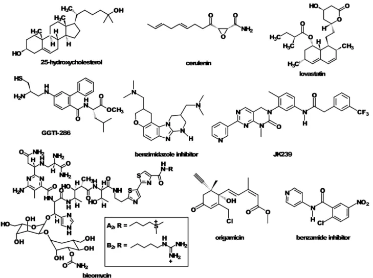
Transcriptional profiling of several systems including patient biopsies has suggested a link between HCV infection and a disruption of the metabolic state including pathways that govern lipid metabolism and cholesterol homeostasis in the infected livers.97In fact, both the peroxisome proliferator-activated receptor-alpha (PPARa) and sterol regulatory element binding protein (SREBP) pathways (Fig. 2) have been shown to be modulated during HCV replication and infection.54,56,98In addition, the importance of lipid metabolism and cholesterol homeostasis on HCV pathogenesis has been emphasized through the use of small molecule inhibitors. For example, inhibitors of HMG-CoA reductase, the rate-controlling enzyme of the mevalonate pathway, such as lovastatin and fluvastatin, have been demonstrated to inhibit HCV replication (Fig. 3).48,99Similarly, this effect is shared by 25-hydroxycholesterol and cerulenin,56 both of which act at the mevalonate pathway, and GGTI-286, an inhibitor of geranylgeranylation.99 Also, the geranylgeranylated host
protein FBL2 has been implicated as a host factor required for HCV RNA replication.48The lipidated form of FBL2 was shown to interact with NS5A via an immunoprecipitation experiment, and siRNA mediated knockdown of the protein
reduced viral replication by 65% (Fig. 3). Depletion of cholesterol using b-cyclodextrin disrupts the membranous web upon which HCV replication occurs.55Also, the PPAR receptor antagonist 2-chloro-5-nitro-N-(pyridyl)benzamide was shown to modulate HCV replication and rapidly disperses HCV RNA from replication complexes.98 Transcriptional profiling of Huh-7 cells treated with 25-hydroxycholesterol also revealed the changes in gene expression required to induce an anti-HCV state in the host cell. In addition to the modulation of many genes expressed in the mevalonate pathway, mRNA biomarkers of the outcome were identified. These included PROX1, INSIG-1, NK4, and UBD.78
miRNA profiling. MicroRNAs (miRNAs) are a class of small non-coding RNAs endogeneously expressed by metazoans with regulatory roles in a variety of cellular processes including proliferation, homeostasis, apoptosis, transcription, genome integrity, translation, and mRNA stability.100,101 The mature 21–25 nt miRNA forms are asymmetrically assembled into RNA-induced silencing complexes (RISC), where one strand of the mature miRNA is used to target homologous RNA inducing a block in translation or acceleration of deadenylation.102,103
Fig. 2 The effects of peroxisome proliferaor-activated receptor (PPAR) and sterol response element (SRE) signaling on lipid metabolism. The PPAR pathway controls genes that regulate fatty acid b-oxidation and the secretion of lipases involved in fatty acid breakdown. Upon ligand activation, these receptors heterodimerize with retinoic X receptor (RXR) to alter expression of lipid metabolism genes through binding response elements (PPREs). The two different SRE signaling pathways highlighted in blue and red represent sterol regulatory element binding protein (SREBP) isoforms 1c and 2 that control the response to synthesis of triglycerides and cholesterol esters, respectively. Both syntheses result in the formation of neutral lipids, which are stored in lipid droplets.
Downloaded by CISTI Archive Access on 08 March 2011
Each miRNA is estimated to potentially interact with over 100 mRNAs and regulate at least one third of all human genes.104–109The important roles miRNAs have in most cellular processes result in the aberrant expression of these genes correlating, in many incidences, with diseased states. Many miRNAs have now been linked to several pathologies including various genetic diseases,110–112types of cancers,113,114 including
hepatocellular carcinoma (HCC),4,80,115and viral infections.116–118 For this reason, miRNA profiling has become a very important tool in uncovering new biomarkers of disease and developing potential therapeutic targets. Probing the miRNA milieu has already facilitated the discovery of many oncogenes (reviewed in ref. 119–122).
Similar studies with regards to HCV have revealed valuable knowledge with regards to host–virus interactions. MicroRNA expression profiling study using small RNA cloning to compare human liver, human hepatoma Huh-7.5 cells, and Huh-7.5 cells harbouring a HCV replicon demonstrated miR-122 is the predominant microRNA in all samples and higher expression of miR-322, miR-197, miR-532-5p, and miR-374 miRNA families in the HCV infected cells.123
Varnholt et al. conducted quantitative RT-PCR to find aberrantly expressed miRNAs in HCV-related hepatic tumours when compared to normal liver parenchyma.124 The study yielded 10 upregulated and 19 down-regulated miRNAs including overexpression of miR-100, miR-122, and miR-10a and decreased expression of miR-198 and miR-145. A similar study on HCV-related HCC using qRT-PCR to study the miRNA expression in the progression of HCV disease to cancer in liver tissue samples revealed 17 miRNAs upregulated in HCC and 6 with decreased expression.125 A cDNA microarray facilitated examination of miRNA’s regulation of candidate genes and pathways. The 17 upregulated genes in HCC were associated with anti-tumour response, while the 17 downregulated targets were oncogenes relating to cell cycle, adhesion, proteolysis, transcription and translation.
Also, a parallel microarray and qRT-PCR study demonstrated that interferon beta (IFN-b), one of the current treatment regimens for HCV, modulates eight miRNAs which target highly conserved binding sites among all HCV genotypes.126 These eight miRNAs proved crucial to the anti-viral effects of the IFN-b treatment.
Fig. 3 The hypothesized effects of host protein FBL2 lipidation on HCV replication. The enlarged image is a cropped view of the membranous web where the replication complex resides adjacent to lipid droplet (LD)-associated membranes. The presence of FBL2 interacting with the HCV protein NS5A was shown to be necessary for the formation of a functional replication complex. FBL2 is post-translationally modified by geranylgeranyl transferase enzymes giving rise to a lipidated form that then becomes anchored on phospholipid bilayers of the ER membrane. Small molecule inhibitors that modulate host lipid metabolism can reduce the pool of geranylgeranyl diphosphate that is used to post-translationally modify FBL2 and anchor the FBL2 protein to the ER and associate with HCV replication complexes. The blue shading in the enlarged images represents the ER lumen.
Downloaded by CISTI Archive Access on 08 March 2011
While the majority of microRNAs are known to repress translation and transcription via interactions with the 30
non-coding regions (30NCRs) of target mRNAs, miRNA-122
was found to positively modulate HCV expression.49,50,127
Jopling et al. were the first to report this effect in Huh7 liver cells stably expressing a replicon.50The intracellular abundance of viral RNA was found to increase with ectopic expression of the microRNA while sequestration of miR-122 through the use of antisense oligonucleotides (ASOs) prevented accumulation of replication-competent HCV RNA in newly transfected cells. Jopling et al. identified two binding sites in the 50NCR
cooperatively mediating this effect through mutational analysis of the HCV RNA, with ectopic expression of miR-122 with corresponding mutations rescuing the viral load.49,50Henke et al. revealed a translational stimulation role of miR-122 through the use of replication-defective HCV mutant genome with ectopic expression of miR-122 causing an estimated increase in translation efficiency of 4-fold in liver cells over endogenous levels.127 Mutation of the 50UTR binding sites
revealed they were both necessary for translation regulation, and sequestration of miR-122 by ASOs resulted in a decrease of translation efficiency. The study also suggested miR-122 stimulates HCV translation through acceleration of the ribosomal 48S initiation complexes’ attachment to HCV RNA.
Transfection of miR-122 has also been found to stimulate viral translation and replication in non-hepatic cells.127,128
The therapeutic potential of ASOs revealed by the above miR-122 studies has led to additional studies using modified ASOs including locked nucleic acid (LNA) modified oligo-nucleotide probes129 and antagomirs (cholesterol conjugated ssRNA analogues).130Both probe types have yielded effective and specific silencing of miR-122 and represent potential therapeutic applications. A qPCR study revealed decreased levels of miRNA-122 in HCV patients with poor response to interferon therapy.131Also, it should be noted the in vivo study wasn’t able to reproduce the correlation between miR-122 levels and viral load. Therefore, one should proceed with caution when drawing conclusions from the in vitro studies. Nonetheless, a recent study in chimpanzees by Lanford et al. showed the promise of using LNA-modified ASO targeted against miR-122 as it resulted in long lasting suppression of viremia.132
Another miRNA that could potentially interact with the HCV genome was revealed through the use of miRNA target search algorithms: miR-199*, which was identified as having a binding site in the IRES of genotypes 1b and 2a.133 Overexpression of this microRNA was shown to cause an inhibitory effect on HCV replication.
Fig. 4 Examples of small molecules that were discovered to inhibit HCV–host cell interactions necessary for viral propagation.
Downloaded by CISTI Archive Access on 08 March 2011
These profiling studies have led to an increased understanding of the HCV aetiologies. Further investigations into miRNA profiles will help elucidate the mechanism of the disease’s progression from onset of infection to chronic hepatitis to HCC. Furthermore, miRNA signatures of disease states will be of diagnostic and prognostic relevance as they may reveal novel targets for antiviral therapy.
Proteomic approaches
Recently proteomic approaches have been applied to study the differential protein expression during HCV protein expression,61,134,135replication and infection136 as well as to discover biomarkers for HCC.137–140One of the first detailed proteomics study related to HCV biology was performed by Yan et al.141In this study, they used isotope-coded affinity tags (ICAT) to examine differentially expressed proteins during interferon treatment of Huh-7 cells. Their study identified novel IFN regulated proteins and pathways including metabolism and dynamics of microtubule function, thereby providing further insights to the possible mechanisms of antiviral effects of interferon at the protein level and a global view of the antiviral state that the host cell undertakes during interferon treatment.
Proteomics experiments have also been conducted with HCV core protein overexpression in cell lines. Using a combination of affinity-based pull down experiments and mass spectrometric analysis, Kang and co-workers were able to determine that HCV core protein interacts with 14 host cell factors.134 Some proteins that were identified include heat shock protein 60, implicated in modulation of apoptosis as well as fibrous cytoskeletal proteins, vimentin and cytokeratin 8. Sato et al. also examined the lipid droplet proteomes of cells expressing HCV core proteins and found a number of interesting lipid droplet proteins as well as differential expression of certain proteins such as adipocyte differentiation-related protein (ADRP) and tail-interacting protein of 47 kDa (TIP47).61
Proteomics experiments performed on the host cell lipid raft proteome, or the detergent-insoluble fractions, of cells replicating HCV identified 100 proteins in lipid rafts of which 39 were induced or repressed during HCV replication.136This study also used a modern technique called ‘stable isotope labeling by amino acids in cell culture’ (SILAC) where they discovered over 400 differentially expressed proteins based on two or more unique peptides from which 150 proteins showed at least 2.5 fold change in expression during HCV replication with many associated with vesicular and protein trafficking and in cell signaling. This represented the first evidence that there is a large scale change in protein subcellular localization that occurs at membrane rafts which facilitates HCV replication.
Our group has also been conducting functional proteomics during HCV replication and infection.142–145We have shown that activity-based protein profiling (ABPP) approaches, where covalent inhibitors label enzyme families according to their mechanism or substrate preferences, can be used to examine changes in enzyme activity during HCV replication.
Screening
Genome-wide siRNA knockdown experiments. Modern methods for high throughput biology have been applied successfully to discover interactions between HCV and its host. For example, siRNA-based screening has been successfully implemented to systematically knockdown genes within the host genome to discover the relative importance of host cell proteins and pathways.146–149 For example, Supekova et al. screened a panel of siRNAs that preferentially target human protein kinases against an HCV replicon expressing the firefly luciferase gene as a genetic reporter.147They identified three
human kinases, Csk, Jak1, and Vrk1, that reduce viral RNA and protein levels. They also demonstrated that a small molecule inhibitor of Csk, JK239 (Fig. 4), resulted in a significant reduction in HCV RNA and viral proteins, further supporting a role for Csk in HCV replication. Moreover, it was suggested that this role involved Csk-regulated kinase Fyn, which had lower levels of phosphorylation during JK239 treatment that led to lower levels of phosphorylation of NS5A. Two independent studies have been conducted using genome-wide RNA interference screens.148,149 Tai et al. identified 96 human genes that support HCV replication, with a significant number of them being involved in vesicle organization and biogenesis.148Phosphatidylinositol 4-kinase (PI4KA) and vesicle coat complex subunits were among genes that were further validated with small molecule inhibitors. Vaillancourt et al. screened an adenovirus-delivered shRNA library and 73 human genes that support HCV replication.149
They also identified PI4KA as being required for HCV propagation and demonstrated this to be true for HCV genotypes 1a, 1b and 2a. Li et al. performed similar genome-wide knockdown experiments in an infectious model for HCV and confirmed a large number of previously identified host–pathogen interactions.146The classification of the identified genes based on their molecular function and biological process showed a significant enrichment for kinases, factors involved in protein metabolism and modification, oncogenesis, nucleic acid binding, and nucleic acid metabolism.
The previously described miR-122 regulation of HCV is dependent on the RNA silencing pathway as siRNA-mediated knockdown of Drosha, DGCR8, Dicer, or one of the four Argonaute proteins results in a decreased intracellular accumulation of viral RNA.123
Small molecule screening. The use of small molecules to identify host–virus interactions, using techniques sometimes referred to as chemical genetics, has enabled identification of novel HCV–host cell interactions. For example, recently Parsons et al. showed that the internal ribosome entry site (IRES), a highly conserved structured element of the HCV RNA, can be disrupted with a benzimidazole inhibitor (Fig. 4).150 The benzimidazole was shown to induce a conformational change in the target RNA by interhelical widening of the IRES subdomain IIa. This causes a break-down of the critical interaction of the IRES with the host cell ribosomes that leads to an inhibition of translation of the HCV polyprotein. We also identified bleomycin as a novel probe for inhibiting HCV translation through targeted HCV
Downloaded by CISTI Archive Access on 08 March 2011
genomic RNA degradation in cell culture.151Chemical genetics approaches were also used to identify origamicin (Fig. 4), an analog of the plant hormone abscisic acid, as an inhibitor of HCV replication, and experiments suggested that its function was mediated through blocking protein folding thus preventing maturation of HCV proteins.152 The PPAR-a receptor antagonist 2-chloro-5-nitro-N-(pyridyl)benzamide (benzamide in Fig. 4) was also identified as an inhibitor of HCV replication using chemical genetic methods.54
Bioimaging approaches
Bioimaging studies allow us to understand unique interactions that are dynamic or transient in nature. Molecular imaging techniques can aid in measuring pathogen-induced alterations of host organelles, from the start of infection progressing to the production of new virion particles, thus enabling a better understanding of the complexity of dynamic processes. Traditional structure methods, such as atomic force microscopy and spectroscopic methods, have increased our understanding on the HCV lifecycle. By using chemical probes in molecular imaging to help visualize and quantify host–pathogen processes, we can spy on these interactions and measure the changes associated with the lifecycle of viral–host infection.153 Notably, techniques in microscopy have advanced in live cell imaging to a point where we can identify, with high contrast, proteins of choice, without any biochemical ambiguities that cell fixing methods may impose. By being able to probe both host and viral proteins that are specifically involved in the host dependent viral lifecycle, we have the unique capability to explore the kinetics of the HCV biological processes.
Atomic force microscopy.Atomic force microscopy (AFM) represents an interesting tool to study viral structure and biological complexes without fixing in aqueous environments. The advancements of non-contact-mode methods have permitted the probing of complexes in a mechanically non-invasive manner. The AFM analysis can elucidate detailed structures of viruses; for example, it has been used to show the long-range RNA interactions mediating cyclization of the dengue virus, another member of the Flaviviridae family. Davis et al. used AFM as a tool to validate predictions of genome-scale ordered RNA structure (GORS) in HCV. This tool, however, has yet to be exploited for the investigation of host and viral factors’ effect on different structural aspects of HCV as it has been for other viruses.154
Spectroscopy-based protein structure determination methods. In general, nuclear magnetic resonance (NMR) and circular dichroism (CD) spectroscopy are invaluable tools for providing protein structural analyses. With regards to HCV, NMR spectroscopy has proved useful in delineating structural determinants of both host and viral factors for essential interactions in complexes. For example, NMR spectroscopy was used to confirm structural determinants of NS3-4A for membrane association and the organization of functional replication complex155and, similarly, the structural determinants of the La protein156 and eIF3157 for facilitating IRES-mediated translation of HCV RNA. Hanoulle et al. used this
technique along with CD spectroscopy and gel filtration chromatography to confirm that NS5A-D2 domain is a substrate of both cyclophilins A and B.158 Both NMR and CD spectroscopy were also exploited by Lindenbach et al. to identify a C-terminal peptide of NS4A that regulates hyper-phosphorylation of NS5B and viral replication.159 These spectroscopy methods will continue to play crucial roles in identifying and validating novel HCV host–viral interactions.
Fusion proteins. The use of reporters to track HCV viral proteins in the progression of infection has played a fundamental role in understanding the connection between the host and virus. In particular, fluorescent proteins that are fused with a protein of interest provide a suitable tool for assessing both structure and function during an important physiological time period.160–163 For example, Wolk et al. has fused a green fluorescent protein to NS5A, which localizes on the HCV-induced membranous web-like matrix;164 this permitted live
cell imaging to track the fluorescent proteins localized on these continuous membranous webs and critically analysis of kinetics by following their movements. With the help of fusion proteins, the membranous web was identified to exist in two distinct populations and their physical movements pertain to small and large replication complexes, which display rapid saltatory movements in the former, and restricted motion for the latter. By using microtubule specific probes, the movements of the small replication complexes were further shown to travel along the microtubule networks of the cell.164 In contrast, the large replication complexes are continuously associated with the ER network and their restricted movements preclude any traveling along microtubule networks. The role of these small replication complexes was suggested to broaden the reach of HCV propagation throughout the entire cell, to ensure a better chance to escape deleterious mutations and establish highly adaptive infection. Time lapse microscopy has identified these two types of replication complexes perpetually involved in HCV propagation and has expanded the scope towards finally understanding the HCV–host pathways.164
Non-linear microscopy.Modern techniques using non-linear optics such as multiphoton fluorescence provides high contrast imaging with 3-D cross section capabilities. An emerging modern tool in microscopy is coherent anti-Stokes Raman scattering (CARS) microscopy, which delivers high specificity in spatial and lateral resolutions by using non-linear optics to detect inherent natural resonances such as that of C–H bond stretching.165–167 C–H bonds are most prevalent in the cell within lipid-rich bodies such as VLDL particles and lipid droplets. CARS microscopy, therefore, has the capacity to visualize these host organelles without the need for chemical staining or fluorescent dyes of fluorescent fusion proteins. Notably this system can also be coupled with fluorescence microscopy to simultaneously obtain information with fluor-escent probes and information about the changes associated with lipid metabolism by CARS microscopy.
As previously mentioned, lipid metabolism in the host plays an important role in the HCV lifecycle. Several aspects of host cell lipid metabolism and HCV have been examined by CARS
Downloaded by CISTI Archive Access on 08 March 2011
microscopy including the influence of copper,168induction of lipid storage,169 and the influence of metabolic inhibitors on
both the timing and mechanism of inhibition of HCV replication.54,98
Summary and outlook
New strategies are emerging for the rapid and genome-scale screening of HCV–host cell interactions that are helping to discover and define these interactions. The use of genomics, proteomics, RNA interference, and small molecule screening has aided in the discovery of host–HCV interactions that represent novel targets for therapeutic intervention. However, challenges still remain in understanding the dynamics and interrelations between the various molecular interactions as a function of time during infection. New techniques will undoubtedly need to be applied to further understand the spatial and temporal changes in the host cell and how they relate to HCV’s ability to propagate. Also, the clinical validation of these potential therapeutic targets remains an important challenge. While known HCV–host interactions are many in number, none-to-date represent clinically validated targets that can be used as alternate therapeutic strategies. Consequently, the identification of novel interactions and the development of new methods with which to facilitate their discovery may prove essential to future strategies for curing chronic HCV infection.
References
1 B. J. Thomson and R. G. Finch, Clin. Microbiol. Infect., 2005, 11, 86.
2 NIH Consensus and State-of-the-Science Statement, 2002, 19, 1. 3 N. N. Zein, Clin. Microbiol. Rev., 2000, 13, 223.
4 Y. Murakami, T. Yasuda, K. Saigo, T. Urashima, H. Toyoda, T. Okanoue and K. Shimotohno, Oncogene, 2006, 25, 2537. 5 D. Moradpour, F. Penin and C. M. Rice, Nat. Rev. Microbiol.,
2007, 5, 453.
6 B. D. Lindenbach and C. M. Rice, Nature, 2005, 436, 933. 7 K. E. Reed and C. M. Rice, Curr. Top. Microbiol. Immunol., 2000,
242, 55.
8 A. Grakoui, C. Wychowski, C. Lin, S. M. Feinstone and C. M. Rice, J. Virol., 1993, 67, 1385.
9 R. Bartenschlager, L. Ahlbornlaake, J. Mous and H. Jacobsen, J. Virol., 1994, 68, 5045.
10 T. Hugle, F. Fehrmann, E. Bieck, M. Kohara, H. G. Krausslich, C. M. Rice, H. E. Blum and D. Moradpour, Virology, 2001, 284, 70.
11 N. Ivashkina, B. Wolk, V. Lohmann, R. Bartenschlager, H. E. Blum, F. Penin and D. Moradpour, J. Virol., 2002, 76, 13088.
12 D. Moradpour, R. Gosert, D. Egger, F. Penin, H. E. Blum and K. Bienz, Antiviral Res., 2003, 60, 103.
13 R. Gosert, D. Egger, V. Lohmann, R. Bartenschlager, H. E. Blum, K. Bienz and D. Moradpour, J. Virol., 2003, 77, 5487. 14 D. Egger, B. Wolk, R. Gosert, L. Bianchi, H. E. Blum,
D. Moradpour and K. Bienz, J. Virol., 2002, 76, 5974.
15 T. Masaki, R. Suzuki, K. Murakami, H. Aizaki, K. Ishii, A. Murayama, T. Date, Y. Matsuura, T. Miyamura, T. Wakita and T. Suzuki, J. Virol., 2008, 82, 7964.
16 T. L. Tellinghuisen and K. L. Foss, PLoS Pathog., 2009, 4, e1000032.
17 M. Quintavalle, S. Sambucini, V. Summa, L. Orsatti, F. Talamo, R. De Francesco and P. Neddermann, J. Biol. Chem., 2007, 282, 5536.
18 M. Quintavalle, S. Sambucini, C. Di Pietro, R. De Francesco and P. Neddermann, J. Virol., 2006, 80, 11305.
19 K. E. Reed, J. Xu and C. M. Rice, J. Virol., 1997, 71, 7187. 20 N. Zitzmann, A. S. Mehta, S. Carrouee, T. D. Butters,
F. M. Platt, J. McCauley, B. S. Blumberg, R. A. Dwek and T. M. Block, Proc. Natl. Acad. Sci. U. S. A., 1999, 96, 11878. 21 B. Bartosch, J. Dubuisson and F. L. Cosset, J. Exp. Med., 2003,
197, 633.
22 N. Callens, Y. Ciczora, B. Bartosch, N. Vu-Dac, F. L. Cosset, J. M. Pawlotsky, F. Penin and J. Dubuisson, J. Virol., 2005, 79, 15331.
23 F. Penin, J. Dubuisson, F. A. Rey, D. Moradpour and J. M. Pawlotsky, Hepatology, 2004, 39, 5.
24 D. Moradpour and H. E. Blum, Liver Int., 2004, 24, 519. 25 M. E. Burlone and A. Budkowska, J. Gen. Virol., 2009, 90, 1055. 26 L. Cocquerel, C. Voisset and J. Dubuisson, J. Gen. Virol., 2006,
87, 1075.
27 L. Cukierman, L. Meertens, C. Bertaux, F. Kajumo and T. Dragic, J. Virol., 2009, 83, 5477.
28 J. Dubuisson, World J. Gastroenterol., 2007, 13, 2406.
29 J. Dubuisson, F. Helle and L. Cocquerel, Cell. Microbiol., 2008, 10, 821.
30 V. Agnello, G. Abel, M. Elfahal, G. B. Knight and Q. X. Zhang, Proc. Natl. Acad. Sci. U. S. A., 1999, 96, 12766.
31 E. G. Cormier, R. J. Durso, F. Tsamis, L. Boussemart, C. Manix, W. C. Olson, J. P. Gardner and T. Dragic, Proc. Natl. Acad. Sci. U. S. A., 2004, 101, 14067.
32 E. G. Cormier, F. Tsamis, F. Kajumo, R. J. Durso, J. P. Gardner and T. Dragic, Proc. Natl. Acad. Sci. U. S. A., 2004, 101, 7270. 33 M. J. Evans, T. von Hahn, D. M. Tscherne, A. J. Syder, M. Panis,
B. Wolk, T. Hatziioannou, J. A. McKeating, P. D. Bieniasz and C. M. Rice, Nature, 2007, 446, 801.
34 M. J. Farquhar, H. J. Harris, M. Diskar, S. Jones, C. J. Mee, S. U. Nielsen, C. L. Brimacombe, S. Molina, G. L. Toms, P. Maurel, J. Howl, F. W. Herberg, S. C. van Ijzendoorn, P. Balfe and J. A. McKeating, J. Virol., 2008, 82, 8797. 35 M. Flint, T. von Hahn, J. Zhang, M. Farquhar, C. T. Jones,
P. Balfe, C. M. Rice and J. A. McKeating, J. Virol., 2006, 80, 11331.
36 J. Grove, S. Nielsen, J. Zhong, M. F. Bassendine, H. E. Drummer, P. Balfe and J. A. McKeating, J. Virol., 2008, 82, 12020.
37 H. J. Harris, M. J. Farquhar, C. J. Mee, C. Davis, G. M. Reynolds, A. Jennings, K. Hu, F. Yuan, H. Deng, S. G. Hubscher, J. H. Han, P. Balfe and J. A. McKeating, J. Virol., 2008, 82, 5007.
38 Z. Y. Keck, S. H. Li, J. Xia, T. von Hahn, P. Balfe, J. A. McKeating, J. Witteveldt, A. H. Patel, H. Alter, C. M. Rice and S. K. Foung, J. Virol., 2009, 83, 6149.
39 V. Rocha-Perugini, M. Lavie, D. Delgrange, J. Canton, A. Pillez, J. Potel, C. Lecoeur, E. Rubinstein, J. Dubuisson, C. Wychowski and L. Cocquerel, BMC Microbiol., 2009, 9, 111.
40 Z. Stamataki, J. Grove, P. Balfe and J. A. McKeating, Clin. Liver Dis., 2008, 12, 693.
41 T. von Hahn and C. M. Rice, J. Biol. Chem., 2008, 283, 3689. 42 M. B. Zeisel, H. Barth, C. Schuster and T. F. Baumert, Front.
Biosci., 2009, 14, 3274.
43 Y. Miyanari, K. Atsuzawa, N. Usuda, K. Watashi, T. Hishiki, M. Zayas, R. Bartenschlager, T. Wakita, M. Hijikata and K. Shimotohno, Nat. Cell Biol., 2007, 9, 1089.
44 J. McLauchlan, Biochem. Soc. Trans., 2009, 37, 986.
45 J. McLauchlan, Biochim. Biophys. Acta, Mol. Cell Biol. Lipids, 2009, 1791, 552.
46 P. Gastaminza, S. B. Kapadia and F. V. Chisari, J. Virol., 2006, 80, 11074.
47 P. Gastaminza, G. F. Cheng, S. Wieland, J. Zhong, W. Liao and F. V. Chisari, J. Virol., 2008, 82, 2120.
48 C. F. Wang, M. Gale, B. C. Keller, H. Huang, M. S. Brown, J. L. Goldstein and J. Ye, Mol. Cell, 2005, 18, 425.
49 C. L. Jopling, S. Schuetz and P. Sarnow, Cell Host Microbe, 2008, 4, 77.
50 C. L. Jopling, M. K. Yi, A. M. Lancaster, S. M. Lemon and P. Sarnow, Science, 2005, 309, 1577.
51 K. D. Tardif, K. Mori, R. J. Kaufman and A. Siddiqui, J. Biol. Chem., 2004, 279, 17158.
52 S. T. Shi, S. J. Polyak, H. Tu, D. R. Taylor, D. R. Gretch and M. M. C. Lai, Virology, 2002, 292, 198.
Downloaded by CISTI Archive Access on 08 March 2011
53 S. B. Kapadia and F. V. Chisari, Proc. Natl. Acad. Sci. U. S. A., 2005, 102, 2561.
54 B. Rakic, S. M. Sagan, M. Noestheden, S. Belanger, X. L. Nan, C. L. Evans, X. S. Xie and J. P. Pezacki, Chem. Biol., 2006, 13, 23. 55 S. M. Sagan, R. Rouleau, C. Leggiadro, L. Supekova, P. G. Schultz, A. I. Su and J. P. Pezacki, Biochem. Cell Biol., 2006, 84, 67.
56 A. I. Su, J. P. Pezacki, L. Wodicka, A. D. Brideau, L. Supekova, R. Thimme, S. Wieland, J. Bukh, R. H. Purcell, P. G. Schultz and F. V. Chisari, Proc. Natl. Acad. Sci. U. S. A., 2002, 99, 15669. 57 A. Breiman, N. Grandvaux, R. T. Lin, C. Ottone, S. Akira,
M. Yoneyama, T. Fujita, J. Hiscott and E. F. Meurs, J. Virol., 2005, 79, 3969.
58 X. D. Li, L. J. Sun, R. B. Seth, G. Pineda and Z. J. J. Chen, Proc. Natl. Acad. Sci. U. S. A., 2005, 102, 17717.
59 T. Saito, R. Hirai, Y. M. Loo, D. Owen, C. L. Johnson, S. C. Sinha, S. Akira, T. Fujita and M. Gale, Proc. Natl. Acad. Sci. U. S. A., 2007, 104, 582.
60 T. Saito, D. M. Owen, F. G. Jiang, J. Marcotrigiano and M. Gale, Nature, 2008, 454, 523.
61 S. Sato, M. Fukasawa, Y. Yamakawa, T. Natsume, T. Suzuki, I. Shoji, H. Aizaki, T. Miyamura and M. Nishijima, J. Biochem., 2006, 139, 921.
62 M. Dimitrova, I. Imbert, M. P. Kieny and C. Schuster, J. Virol., 2003, 77, 5401.
63 M. Matsumoto, S. B. Hwang, K. S. Jeng, N. L. Zhu and M. M. C. Lai, Virology, 1996, 218, 43.
64 K. Nakai, T. Okamoto, T. Kimura-Someya, K. Ishii, C. K. Lim, H. Tani, E. Matsuo, T. Abe, Y. Mori, T. Suzuki, T. Miyamura, J. H. Nunberg, K. Moriishi and Y. Matsuura, J. Virol., 2006, 80, 11265.
65 T. L. Tellinghuisen and C. M. Rice, Curr. Opin. Microbiol., 2002, 5, 419.
66 I. Hamamoto, Y. Nishimura, T. Okamoto, H. Aizaki, M. Y. Liu, Y. Mori, T. Abe, T. Suzuki, M. M. C. Lai, T. Miyamura, K. Moriishi and Y. Matsuura, J. Virol., 2005, 79, 13473. 67 H. Tu, L. Gao, S. T. Shi, D. R. Taylor, T. Yang, A. K. Mircheff,
Y. M. Wen, A. E. Gorbalenya, S. B. Hwang and M. M. C. Lai, Virology, 1999, 263, 30.
68 K. Moriishi, T. Okabayashi, K. Nakai, K. Moriya, K. Koike, S. Murata, T. Chiba, K. Tanaka, R. Suzuki, T. Suzuki, T. Miyamura and Y. Matsuura, J. Virol., 2003, 77, 10237. 69 L. Gao, H. Tu, S. T. Shi, K. J. Lee, M. Asanaka, S. B. Hwang and
M. M. C. Lai, J. Virol., 2003, 77, 4149.
70 C. S. Kim, S. K. Seol, O. K. Song, J. H. Park and S. K. Jang, J. Virol., 2007, 81, 3852.
71 L. R. You, C. M. Chen, T. S. Yeh, T. Y. Tsai, R. T. Mai, C. H. Lin and Y. H. W. Lee, J. Virol., 1999, 73, 2841.
72 N. Mamiya and H. J. Worman, J. Biol. Chem., 1999, 274, 15751. 73 A. M. Owsianka and A. H. Patel, Virology, 1999, 257, 330. 74 C. M. Chen, L. R. You, L. H. Hwang and Y. H. W. Lee, J. Virol.,
1997, 71, 9417.
75 M. Matsumoto, T. Y. Hsieh, N. L. Zhu, T. VanArsdale, S. B. Hwang, K. S. Jeng, A. E. Gorbalenya, S. Y. Lo, J. H. Ou, C. F. Ware and M. M. C. Lai, J. Virol., 1997, 71, 1301. 76 B. de Chassey, V. Navratil, L. Tafforeau, M. S. Hiet, A.
Aublin-Gex, S. Agaugue, G. Meiffren, F. Pradezynski, B. F. Faria, T. Chantier, M. Le Breton, J. Pellet, N. Davoust, P. E. Mangeot, A. Chaboud, F. Penin, Y. Jacob, P. O. Vidalain, M. Vidal, P. Andre, C. Rabourdin-Combe and V. Lotteau, Mol. Sys. Biol., 2008, 4, 230.
77 H. Aizaki, T. Harada, M. Otsuka, N. Seki, M. Matsuda, Y. W. Li, H. Kawakami, Y. Matsuura, T. Miyamura and T. Suzuki, Hepatology, 2002, 36, 1431.
78 J. P. Pezacki, S. M. Sagan, A. M. Tonary, Y. Rouleau, L. Supekova and A. I. Su, BMC Chem. Biol., 2009, 9, 2. 79 A. Basu, K. Meyer, K. K. Lai, K. Saito, A. M. Di Bisceglie,
L. E. Grosso, R. B. Ray and R. Ray, Virology, 2006, 349, 347. 80 A. Budhu, Y. Chen, J. W. Kim, M. Forgues, K. Valerie,
C. C. Harris and X. W. Wang, Carcinogenesis, 2007, 28, 1552. 81 G. K. Geiss, V. S. Carter, Y. P. He, B. K. Kwieciszewski,
T. Holzman, M. J. Korth, C. A. Lazaro, N. Fausto, R. E. Bumgarner and M. G. Katze, J. Virol., 2003, 77, 6367. 82 S. Girard, P. Shalhoub, P. Lescure, A. Sabile, D. E. Misek,
S. Hanash, C. Brechot and L. Beretta, Virology, 2002, 295, 272.
83 M. A. Joyce, K. A. Walters, S. E. Lamb, M. M. Yeh, L. F. Zhu, N. Kneteman, J. S. Doyle, M. G. Katze and D. L. Tyrrell, PLoS Pathog., 2009, 5, e1000291.
84 K. A. Walters, M. A. Joyce, J. C. Thompson, M. W. Smith, M. M. Yeh, S. Proll, L. F. Zhu, T. J. Gao, N. M. Kneteman, D. L. Tyrrell and M. G. Katze, PLoS Pathog., 2006, 2, 591. 85 C. B. Bigger, K. M. Brasky and R. E. Lanford, J. Virol., 2001, 75,
7059.
86 S. L. Lederer, K. A. Walters, S. Proll, B. Paeper, S. Robinzon, L. Boix, N. Fausto, J. Bruix and M. G. Katze, Virol. J., 2006, 3, 98.
87 V. R. Mas, D. G. Maluf, K. J. Archer, K. Yanek, B. Williams and R. A. Fisher, Mol. Med., 2006, 12, 97.
88 M. Okamoto, T. Utsunomiya, S. Wakiyama, M. Hashimoto, K. Fukuzawa, T. Ezaki, T. Hanai, H. Inoue and M. Mori, Ann. Surg. Oncol., 2006, 13, 947.
89 I. Borozan, L. Chen, J. Sun, L. L. Tannis, M. Guindi, O. D. Rotstein, J. Heathcote, A. M. Edwards, D. Grant and I. D. McGilvray, Am. J. Transplant., 2006, 6, 806.
90 M. W. Smith, K. A. Walters, M. J. Korth, M. Fitzgibbon, S. Proll, J. C. Thompson, M. M. Yeh, M. C. Shuhart, J. C. Furlong, P. P. Cox, D. L. Thomas, J. D. Phillips, J. P. Kushner, N. Fausto, R. L. Carithers and M. G. Katze, Gastroenterology, 2006, 130, 179.
91 I. Bieche, T. Asselah, I. Laurendeau, D. Vidaud, C. Degot, V. Paradis, P. Bedossa, D. C. Valla, P. Marcellin and M. Vidaud, Virology, 2005, 332, 130.
92 Y. Kurokawa, R. Matoba, S. Nakamori, I. Takemasa, H. Nagano, K. Dono, K. Umeshita, M. Sakon, M. Monden and K. Kato, J. Exp. Clin. Cancer Res., 2004, 23, 135.
93 M. W. Smith, Z. X. N. Yue, M. J. Korth, H. A. Do, L. Boix, N. Fausto, J. Bruix, R. L. Carithers and M. G. Katze, Hepatology, 2003, 38, 1458.
94 K. A. Walters, M. W. Smith, S. Pal, J. C. Thompson, M. J. Thoinas, M. M. Yeh, D. L. Thomas, M. Fitzgibbon, S. Proll, N. Fausto, D. R. Gretch, R. L. Carithers, M. C. Shuhart and M. G. Katze, Virology, 2006, 350, 453. 95 N. Wong, K. Y. Y. Chan, P. F. Macgregor, P. B. S. Lai,
J. A. Squire, B. Beheshti, M. Albert and T. W. T. Leung, Clin. Cancer Res., 2005, 11, 1319.
96 T. Huang, K. Tu, Y. Shyr, C. C. Wei, L. Xie and Y. X. Li, J. Transl. Med., 2008, 6, 44.
97 S. Dharancy, M. Malapel, G. Perlemuter, T. Roskams, Y. Cheng, L. Dubuquoy, P. Podevin, F. Conti, V. Canva, D. Philippe, L. Gambiez, P. Mathurin, J. C. Paris, K. Schoonjans, Y. Calmus, S. Pol, J. Auwerx and P. Desreumaux, Gastroenterology, 2005, 128, 334.
98 R. K. Lyn, D. C. Kennedy, S. M. Sagan, D. R. Blais, Y. Rouleau, A. F. Pegoraro, X. S. Xie, A. Stolow and J. P. Pezacki, Virology, 2009, 394, 130.
99 J. Ye, C. F. Wang, R. Sumpter, M. S. Brown, J. L. Goldstein and M. Gale, Proc. Natl. Acad. Sci. U. S. A., 2003, 100, 15865. 100 V. Ambros, Nature, 2004, 431, 350.
101 P. D. Zamore and B. Haley, Science, 2005, 309, 1519.
102 L. G. Wu, J. H. Fan and J. G. Belasco, Proc. Natl. Acad. Sci. U. S. A., 2006, 103, 4034.
103 R. Yi, Y. Qin, I. G. Macara and B. R. Cullen, Genes Dev., 2003, 17, 3011.
104 E. Berezikov, V. Guryev, J. van de Belt, E. Wienholds, R. H. A. Plasterk and E. Cuppen, Cell (Cambridge, Mass.), 2005, 120, 21. 105 J. Brennecke, A. Stark, R. B. Russell and S. M. Cohen,
PLoS Biol., 2005, 3, 404.
106 A. Krek, D. Grun, M. N. Poy, R. Wolf, L. Rosenberg, E. J. Epstein, P. MacMenamin, I. da Piedade, K. C. Gunsalus, M. Stoffel and N. Rajewsky, Nat. Genet., 2005, 37, 495. 107 B. P. Lewis, C. B. Burge and D. P. Bartel, Cell (Cambridge,
Mass.), 2005, 120, 15.
108 P. Sood, A. Krek, M. Zavolan, G. Macino and N. Rajewsky, Proc. Natl. Acad. Sci. U. S. A., 2006, 103, 2746.
109 X. H. Xie, J. Lu, E. J. Kulbokas, T. R. Golub, V. Mootha, K. Lindblad-Toh, E. S. Lander and M. Kellis, Nature, 2005, 434, 338.
110 J. F. Abelson, K. Y. Kwan, B. J. O’Roak, D. Y. Baek, A. A. Stillman, T. M. Morgan, C. A. Mathews, D. A. Pauls, M. R. Rasin, M. Gunel, N. R. Davis, A. G. Ercan-Sencicek,
Downloaded by CISTI Archive Access on 08 March 2011
D. H. Guez, J. A. Spertus, J. F. Leckman, L. S. Dure, R. Kurlan, H. S. Singer, D. L. Gilbert, A. Farhi, A. Louvi, R. P. Lifton, N. Sestan and M. W. State, Science, 2005, 310, 317.
111 P. Jin, R. S. Alisch and S. T. Warren, Nat. Cell Biol., 2004, 6, 1048.
112 M. Landthaler, A. Yalcin and T. Tuschl, Curr. Biol., 2004, 14, 2162.
113 A. Hossain, M. T. Kuo and G. F. Saunders, Mol. Cell. Biol., 2006, 26, 8191.
114 E. J. Lee, Y. Gusev, J. M. Jiang, G. J. Nuovo, M. R. Lerner, W. L. Frankel, D. L. Morgan, R. G. Postier, D. J. Brackett and T. D. Schmittgen, Int. J. Cancer, 2007, 120, 1046.
115 J. Jiang, Y. Gusev, I. Aderca, T. A. Mettler, D. M. Nagorney, D. J. Brackett, L. R. Roberts and T. D. Schmittgen, Clin. Cancer Res., 2008, 14, 419.
116 C. H. Lecellier, P. Dunoyer, K. Arar, J. Lehmann-Che, S. Eyquem, C. Himber, A. Saib and O. Voinnet, Science, 2005, 308, 557.
117 R. Triboulet, B. Mari, Y. L. Lin, C. Chable-Bessia, Y. Bennasser, K. Lebrigand, B. Cardinaud, T. Maurin, P. Barbry, V. Baillat, J. Reynes, P. Corbeau, K. T. Jeang and M. Benkirane, Science, 2007, 315, 1579.
118 M. L. Yeung, Y. Bennasser, T. G. Myers, G. J. Jiang, M. Benkirane and K. T. Jeang, Retrovirology, 2005, 2, 81. 119 G. A. Calin and C. M. Croce, Nat. Rev. Cancer, 2006, 6, 857. 120 R. Garzon, M. Fabbri, A. Cimmino, G. A. Calin and
C. M. Croce, Trends Mol. Med., 2006, 12, 580.
121 V. Molnar, V. Tamasi, B. Bakos, Z. Wiener and A. Falus, Semin. Cancer Biol., 2008, 18, 111.
122 J. C. S. Pang, W. K. Kwok, Z. P. Chen and H. K. Ng, Acta Neuropathol., 2009, 117, 599.
123 G. Randall, M. Panis, J. D. Cooper, T. L. Tellinghuisen, K. E. Sukhodolets, S. Pfeffer, M. Landthaler, P. Landgraf, S. Kan, B. D. Lindenbach, M. Chien, D. B. Weir, J. J. Russo, J. Ju, M. J. Brownstein, R. Sheridan, C. Sander, M. Zavolan, T. Tuschl and C. M. Rice, Proc. Natl. Acad. Sci. U. S. A., 2007, 104, 12884.
124 H. Varnholt, U. Drebber, F. Schulze, I. Wedemeyer, P. Schirmacher, H. P. Dienes and M. Odenthal, Hepatology, 2008, 47, 1223.
125 S. Ura, M. Honda, T. Yamashita, T. Ueda, H. Takatori, R. Nishino, H. Sunakozaka, Y. Sakai, K. Horimoto and S. Kaneko, Hepatology, 2009, 49, 1098.
126 I. M. Pedersen, G. Cheng, S. Wieland, S. Volinia, C. M. Croce, F. V. Chisari and M. David, Nature, 2007, 449, 919.
127 J. I. Henke, D. Goergen, J. F. Zheng, Y. T. Song, C. G. Schuttler, C. Fehr, C. Junemann and M. Niepmann, EMBO J., 2008, 27, 3300.
128 J. H. Chang, J. T. Cruo, D. Jiang, H. T. Guo, J. M. Taylor and T. M. Block, J. Virol., 2008, 82, 8215.
129 J. Elmen, M. Lindow, A. Silahtaroglu, M. Bak, M. Christensen, A. Lind-Thomsen, M. Hedtjarn, J. B. Hansen, H. F. Hansen, E. M. Straarup, K. McCullagh, P. Kearney and S. Kauppinen, Nucleic Acids Res., 2008, 36, 1153.
130 J. Krutzfeldt, N. Rajewsky, R. Braich, K. G. Rajeev, T. Tuschl, M. Manoharan and M. Stoffel, Nature, 2005, 438, 685. 131 M. Sarasin-Filipowicz, J. Krol, I. Markiewicz, M. H. Heim and
W. Filipowicz, Nat. Med., 2009, 15, 31.
132 R. E. Lanford, E. S. Hildebrandt-Eriksen, A. Petri, R. Persson, M. Lindow, M. E. Munk, S. Kauppinen and H. Orum, Science, 2010, 327, 198.
133 Y. Murakami, H. H. Aly, A. Tajima, I. Inoue and K. Shimotohno, J. Hepatol., 2009, 50, 453.
134 S. M. Kang, M. J. Shin, J. H. Kim and J. W. Oh, Proteomics, 2005, 5, 2227.
135 W. Yang, B. L. Hood, S. L. Chadwick, S. F. Liu, S. C. Watkins, G. X. Luo, T. P. Conrads and T. Y. Wang, Hepatology, 2008, 48, 1396.
136 P. Mannova, R. H. Fang, H. Wang, B. Deng, M. W. McIntosh, S. M. Hanash and L. Beretta, Mol. Cell. Proteomics, 2006, 5, 2319.
137 R. Chaerkady, H. C. Harsha, A. Nalli, M. Gucek, P. Vivekanandan, J. Akhtar, R. N. Cole, J. Simmers,
R. D. Schulick, S. Singh, M. Torbenson, A. Pandey and P. J. Thuluvath, J. Proteome Res., 2008, 7, 4289.
138 D. L. Diamond, J. M. Jacobs, B. Paeper, S. C. Proll, M. A. Gritsenko, R. L. Carithers, A. M. Larson, M. M. Yeh, D. G. Camp, R. D. Smith and M. G. Katze, Hepatology, 2007, 46, 649.
139 I. N. Lee, C. H. Chen, J. C. Sheu, H. S. Lee, G. T. Huang, D. S. Chen, C. Y. Yu, C. L. Wen, F. J. Lu and L. P. Chow, Proteomics, 2006, 6, 2865.
140 H. W. Ressom, R. S. Varghese, L. Goldman, Y. An, C. A. Loffredo, M. Abdel-Hamid, Z. Kyselova, Y. Mechref, M. Novotny, S. K. Drake and R. Goldman, J. Proteome Res., 2008, 7, 603.
141 W. Yan, H. Lee, E. C. Yi, D. Reiss, P. Shannon, B. K. Kwieciszewski, C. Coito, X. J. Li, A. Keller, J. Eng, T. Galitski, D. R. Goodlett, R. Aebersold and M. G. Katze, Genome Biol., 2004, 5, R54.
142 D. R. Blais, M. Brulotte, Y. Qian, S. Be´langer, S. Q. Yao and J. P. Pezacki, J. Proteome Res., 2010, 9, 912.
143 J. Raez, D. R. Blais, Y. Zhang, R. A. Alvarez-Puebla, J. P. Bravo-Vasquez, J. P. Pezacki and H. Fenniri, Langmuir, 2007, 23, 6482. 144 R. Singaravelu, D. R. Blais, C. McKay and J. P. Pezacki,
Proteome Sci., 2010, 8, 5.
145 D. R. Blais, R. K. Lyn, M. Joyce, Y. Rouleau, A. Stolow, A. F. Pegoraro, D. L. Tyrrell and J. P. Pezacki, J. Biol. Chem., submitted.
146 Q. S. Li, A. L. Brass, A. Ng, Z. Y. Hu, R. J. Xavier, T. J. Liang and S. J. Elledge, Proc. Natl. Acad. Sci. U. S. A., 2009, 106, 16410. 147 L. Supekova, F. Supek, J. Lee, S. Chen, N. Gray, J. P. Pezacki, A. Schlapbach and P. G. Schultz, J. Biol. Chem., 2008, 283, 29. 148 A. W. Tai, Y. Benita, L. F. Peng, S. S. Kim, N. Sakamoto,
R. J. Xavier and R. T. Chung, Cell Host Microbe, 2009, 5, 298. 149 F. H. Vaillancourt, L. Pilote, M. Cartier, J. Lippens, M. Liuzzi,
R. C. Bethell, M. G. Cordingley and G. Kukolj, Virology, 2009, 387, 5.
150 J. Parsons, M. P. Castaldi, S. Dutta, S. M. Dibrov, D. L. Wyles and T. Hermann, Nat. Chem. Biol., 2009, 5, 823.
151 B. Rakic, M. Brulotte, Y. Rouleau, S. Belanger and J. P. Pezacki, ChemBioChem, 2006, 7, 1330.
152 B. Rakic, J. Clarke, T. L. Tremblay, J. Taylor, K. Schreiber, K. M. Nelson, S. R. Abrams and J. P. Pezacki, Chem. Biol., 2006, 13, 1051.
153 Y. Wang, J. Y.-J. Shyy and S. Chien, Annu. Rev. Biomed. Eng., 2008, 10, 1.
154 C. V. Filomatori, M. F. Lodeiro, D. E. Alvarez, M. M. Samsa, L. Pietrasanta and A. V. Gamarnik, Genes Dev., 2006, 20, 2238. 155 V. Brass, J. M. Berke, R. Montserret, H. E. Blum, F. Penin and D. Moradpour, Proc. Natl. Acad. Sci. U. S. A., 2008, 105, 14545. 156 T. Mondal, U. Ray, A. K. Manna, R. Gupta, S. Roy and S. Das,
J. Virol., 2008, 82, 11927.
157 J. Perard, R. Rasia, J. Medenbach, I. Ayala, J. Boisbouvier, E. Drouet and F. Baudin, FEBS Lett., 2009, 583, 70.
158 X. Hanoulle, A. Badillo, J. M. Wieruszeski, D. Verdegem, I. Landrieu, R. Bartenschlager, F. Penin and G. Lippens, J. Biol. Chem., 2009, 284, 13589.
159 B. D. Lindenbach, B. M. Pragai, R. Montserret, R. K. F. Beran, A. M. Pyle, F. Penin and C. M. Rice, J. Virol., 2007, 81, 8905. 160 A. Miyawaki, Dev. Cell, 2003, 4, 295.
161 B. A. Pollok and R. Heim, Trends Cell Biol., 1999, 9, 57. 162 R. Y. Tsien and A. Miyawaki, Science, 1998, 280, 1954. 163 J. Zhang, R. E. Campbell, A. Y. Ting and R. Y. Tsien, Nat. Rev.
Mol. Cell Biol., 2002, 3, 906.
164 B. Wolk, B. Buchele, D. Moradpour and C. M. Rice, J. Virol., 2008, 82, 10519.
165 C. L. Evans and X. S. Xie, Annu. Rev. Anal. Chem., 2008, 1, 883. 166 J. X. Cheng and X. S. Xie, J. Phys. Chem. B, 2004, 108, 827. 167 C. W. Freudiger, W. Min, B. G. Saar, S. Lu, G. R. Holtom,
C. W. He, J. C. Tsai, J. X. Kang and X. S. Xie, Science, 2008, 322, 1857.
168 D. C. Kennedy, R. K. Lyn and J. P. Pezacki, J. Am. Chem. Soc., 2009, 131, 2444.
169 X. L. Nan, A. M. Tonary, A. Stolow, X. S. Xie and J. P. Pezacki, ChemBioChem, 2006, 7, 1895.
Downloaded by CISTI Archive Access on 08 March 2011