Publisher’s version / Version de l'éditeur:
Journal of Clinical Microbiology, 53, 2, pp. 477-485, 2014-11-26
READ THESE TERMS AND CONDITIONS CAREFULLY BEFORE USING THIS WEBSITE. https://nrc-publications.canada.ca/eng/copyright
Vous avez des questions? Nous pouvons vous aider. Pour communiquer directement avec un auteur, consultez la
première page de la revue dans laquelle son article a été publié afin de trouver ses coordonnées. Si vous n’arrivez pas à les repérer, communiquez avec nous à PublicationsArchive-ArchivesPublications@nrc-cnrc.gc.ca.
Questions? Contact the NRC Publications Archive team at
PublicationsArchive-ArchivesPublications@nrc-cnrc.gc.ca. If you wish to email the authors directly, please see the first page of the publication for their contact information.
NRC Publications Archive
Archives des publications du CNRC
This publication could be one of several versions: author’s original, accepted manuscript or the publisher’s version. / La version de cette publication peut être l’une des suivantes : la version prépublication de l’auteur, la version acceptée du manuscrit ou la version de l’éditeur.
For the publisher’s version, please access the DOI link below./ Pour consulter la version de l’éditeur, utilisez le lien DOI ci-dessous.
https://doi.org/10.1128/JCM.02824-14
Access and use of this website and the material on it are subject to the Terms and Conditions set forth at
Development of a rapid multiplex PCR assay to genotype pasteurella
multocida strains by use of the lipopolysaccharide outer core
biosynthesis locus
Harper, Marina; John, Marietta; Turnic, Conny; Edmundsa, Mark; St.
Michael, Frank; Adler, Ben; Blackall, P. J.; Cox, Andrew D.; Boyce, John D.
https://publications-cnrc.canada.ca/fra/droits
L’accès à ce site Web et l’utilisation de son contenu sont assujettis aux conditions présentées dans le site LISEZ CES CONDITIONS ATTENTIVEMENT AVANT D’UTILISER CE SITE WEB.
NRC Publications Record / Notice d'Archives des publications de CNRC:
https://nrc-publications.canada.ca/eng/view/object/?id=12e0e9ba-b0d6-4739-9c04-1d41780309a1 https://publications-cnrc.canada.ca/fra/voir/objet/?id=12e0e9ba-b0d6-4739-9c04-1d41780309a1Pasteurella multocida Strains by Use of the Lipopolysaccharide Outer
Core Biosynthesis Locus
Marina Harper,a,b
Marietta John,a,b
Conny Turni,c
Mark Edmunds,a,b,d
Frank St. Michael,e
Ben Adler,b
P. J. Blackall,c
Andrew D. Cox,e John D. Boycea
Department of Microbiology, Monash University, Melbourne, Australiaa; Australian Research Council Centre of Excellence in Structural and Functional Microbial Genomics, Monash University, Melbourne, Australiab; Queensland Alliance for Agriculture and Food Innovation, The University of Queensland, Brisbane, Australiac; Poultry CRC, University of New England, Armidale, Australiad; Vaccine Program, Human Health Therapeutics Portfolio, National Research Council, Ottawa, Canadae
Pasteurella multocidais a Gram-negative bacterial pathogen that is the causative agent of a wide range of diseases in many
ani-mal species, including humans. A widely used method for differentiation of P. multocida strains involves the Heddleston sero-typing scheme. This scheme was developed in the early 1970s and classifies P. multocida strains into 16 somatic or lipopolysac-charide (LPS) serovars using an agar gel diffusion precipitin test. However, this gel diffusion assay is problematic, with difficulties reported in accuracy, reproducibility, and the sourcing of quality serovar-specific antisera. Using our knowledge of the genetics of LPS biosynthesis in P. multocida, we have developed a multiplex PCR (mPCR) that is able to differentiate strains based on the genetic organization of the LPS outer core biosynthesis loci. The accuracy of the LPS-mPCR was compared with classical Heddleston serotyping using LPS compositional data as the “gold standard.” The LPS-mPCR correctly typed 57 of 58 isolates; Heddleston serotyping was able to correctly and unambiguously type only 20 of the 58 isolates. We conclude that our LPS-mPCR is a highly accurate LPS genotyping method that should replace the Heddleston serotyping scheme for the classifica-tion of P. multocida strains.
P
asteurella multocida is the primary causative agent of a widerange of economically important diseases, including hemor-rhagic septicemia in ungulates, atrophic rhinitis in pigs, fowl chol-era in birds, snuffles in rabbits, and enzootic pneumonia and ship-ping fever in cattle, sheep, and pigs (1). P. multocida also causes opportunistic infections in humans, often following cat or dog bites, and plays a contributory role, together with other patho-gens, in a range of lower respiratory tract infections and sporadic septicemias in ungulates (1).
P. multocida strains have classically been differentiated using
serological techniques. Strains can be classified into five capsular serogroups (A, B, D, E, and F) using an indirect hemagglutination test (2) and into 16 somatic or lipopolysaccharide (LPS) serovars (serotypes) using the Heddleston gel diffusion precipitin test (3). Both of these schemes have been widely used. Isolates are com-monly assigned a combined designation, such as A:1 (capsular serogroup A and LPS serovar 1) or B:2 (capsular serogroup B and LPS serovar 2).
P. multocida LPS is an immunodominant antigen critical for
homologous protection stimulated by bacterin (killed-cell) vac-cines (4). Furthermore, in the P. multocida strain VP161, a full-length LPS molecule is essential for the ability to cause acute dis-ease (5,6). Heddleston serotyping is currently the only method used to differentiate P. multocida strains on the basis of LPS type. However, the accuracy of Heddleston serotyping has never been objectively tested, as the precise LPS structures produced by dif-ferent strains have not been known. Indeed, there have been many informal as well as formal reports that the Heddleston system fails to type many isolates and lacks accuracy and reproducibility (7,8). Furthermore, Heddleston serotyping is time-consuming and re-quires access to good-quality, serovar-specific antisera.
We have recently carried out a comprehensive analysis of the
LPS structures expressed by the 16 Heddleston type strains and identified the genes required for LPS assembly in each strain (9– 16). These combined analyses showed that the LPS produced by all strains consisted of a highly conserved inner core and a variable outer core and revealed that each of the 16 Heddleston type strains expresses structurally distinct LPS. Importantly, these analyses also showed that only eight unique LPS outer core biosynthesis loci are found in the 16 Heddleston type strains (Fig. 1). We have designated these genetic loci L1 through to L8. The type strains of Heddleston serovars 1, 2, 3, 5, 6, 8, 9, 12, and 16 express full-length or “parent” LPS structures, and the type strains of Heddleston serovars 4, 7, 10, 11, 13, 14, and 15 express truncated LPS—the result of mutations within the LPS outer core biosynthesis loci.
The partial differentiation of P. multocida strains on the basis of LPS biosynthesis genes has been reported previously (17). Us-ing PCR-restriction fragment length polymorphism (RFLP) anal-ysis, P. multocida strains were grouped into 5 PCR-RFLP types. However, only 11 of the 16 Heddleston serovars were included in
Received 30 September 2014 Returned for modification 5 November 2014 Accepted 16 November 2014
Accepted manuscript posted online 26 November 2014
Citation Harper M, John M, Turni C, Edmunds M, St. Michael F, Adler B, Blackall PJ, Cox AD, Boyce JD. 2015. Development of a rapid multiplex PCR assay to genotype Pasteurella multocidastrains by use of the lipopolysaccharide outer core biosynthesis locus. J Clin Microbiol 53:477–485.doi:10.1128/JCM.02824-14. Editor: B. W. Fenwick
Address correspondence to John D. Boyce, john.boyce@monash.edu. Copyright © 2015, American Society for Microbiology. All Rights Reserved.
doi:10.1128/JCM.02824-14
on October 12, 2016 by Canada Institute for STI, National Research Council
http://jcm.asm.org/
the study. Here we report the development and testing of a mul-tiplex PCR (mPCR) using the full set of Heddleston serovar type strains, which can accurately differentiate P. multocida strains into one of the eight distinct LPS genotypes. By comparing the results of Heddleston serotyping and the LPS-mPCR to the LPS struc-tures predicted from LPS compositional analysis by mass spec-trometry (MS), we have determined the accuracy of this mPCR and Heddleston serotyping for predicting LPS type. The LPS-mPCR gave a result that was indicative of LPS genotype ⬎98% of the time, and we propose that the LPS-mPCR assay should be used to differentiate strains into their appropriate LPS genotype and, together with the cap mPCR, form a new molecularly based typing system for accurate strain differentiation of P. multocida.
MATERIALS AND METHODS
Strains used. All P. multocida strains were grown at 37°C in heart infusion
(HI) (Oxoid, Basingstoke, United Kingdom) liquid broth with shaking or on solid HI medium containing 1.5% agar. The P. multocida strains used
in the study are described inTable 1. All field isolates were confirmed as P.
multocida by use of one of two P. multocida-specific PCR assays (18,19).
Serotyping. Each isolate of P. multocida was serotyped via the
Hed-dleston method as described previously (3).
Molecular biology techniques. Genomic DNA was purified from 1 ml
of P. multocida overnight culture using the RBC genomic DNA purifica-tion kit (RBC, Taiwan). Each of the final LPS-mPCRs (50-l final volume) was performed in 1⫻ Taq polymerase buffer (10 mM Tris-HCl, 1.5 mM
MgCl2, 50 mM KCl [Roche Diagnostic GmbH, Mannheim, Germany])
containing 0.4 M each primer (Table 2), 0.2 mM deoxynucleoside triphosphates (dNTPs), and 1.7 U Taq polymerase (Roche Diagnostic GmbH, Mannheim, Germany). For each colony PCR, material from 2 to 3 well-isolated P. multocida colonies (obtained from overnight growth of each isolate at 37°C on an HI–1.5% agar plate) was collected using a sterile tip on a 20-l micropipette (volume set at 20 l) inserted into the middle of each colony. The collected material was then added to a 50-l PCR mixture and mixed thoroughly by pipetting. For PCR using genomic DNA, approximately 50 ng of column-purified DNA was added to each PCR mixture. All reaction mixtures were mixed briefly then centrifuged
Serovar 14 Serovar 1 L1 Serovars 2/5 L2 Serovar 4 Serovar 3 L3 Serovar 6 Serovar 7 L4 Serovar 9 L5 Serovar 15 Serovar 11 Serovar 12 Serovar 10 L6 Serovar 8 L7 Serovar 13 Serovar 16 L8 hptE gatG natB gatF gctC natC 3Ac3Ac gatChptE gatE natA ppgB ppgA ppgC Heddleston serovar LPS genotype LPS outer core LPS outer core biosynthesis locus
glucose, N-acetyl-glucosamine, galactose, N-acetyl-galactosamine,
(1S)-2-acetamido-2-deoxy-D-galactose,
heptose, rhamnose
1-((4-aminobutyl)amino)-3-hydroxy-1-oxopropan-2-yl hydrogen phosphate, 3-acetamido-3,6-dideoxy-α-D-glucose, phosphoethanolamine phosphocholine 474bp V1:415bp 931bp 1kb gatD nctA hptF hptE
810bp gatA hptE pcgD pcgApcgBpcgC 1307bp
gatL latB gatK natD natE 550bp
hptG plbA rmlB plbB rhtA rhtB qdtB qdtD latA rmlA rmlD rmlC
1175bp
nat_ps hetA nctB gatH gctD gatB hptE gatJ 668bp hetB natG gatK gatM natF 255bp
FIG 1 LPS outer core structure produced by each of the Heddleston serovar type strains and the genes responsible for LPS outer core biosynthesis in each strain.
(Left) Schematic representation of the outer core LPS structures produced by each of the Heddleston serovar type strains. The last residue (glucose) of the conserved LPS inner core is shown on the far left as a reference point. Specific linkages between each of the residues are not shown. (Right) LPS genotype and genetic organization of each LPS outer core biosynthesis locus. The relative position and size of each genotype-specific PCR amplicon are shown above each LPS outer core biosynthesis locus. Each gene is color coded according to its known/predicted role in LPS biosynthesis; gctD and gatB (yellow and blue striped) in locus L6 differ by only a single nucleotide and are involved in the addition of glucose or galactose, respectively, to the outer core heptose. The rpL31_2 gene, encoding ribosomal protein L31, is not involved in LPS biosynthesis and is colored brown.
Harper et al.
on October 12, 2016 by Canada Institute for STI, National Research Council
http://jcm.asm.org/
(10 s, 13,000 ⫻ g). All PCRs were performed in an Eppendorf Mastercy-cler. For colony PCR, the cycling conditions were 96°C for 10 min, fol-lowed by 30 cycles of 96°C for 30 s, 52°C for 30 s, and 72°C for 2.5 min, with a final extension at 72°C for 5 min. For PCR using genomic DNA as the template, the cycling conditions were identical to those in the colony PCR, except that the initial denaturation step at 96°C was reduced to 5 min.
The PCR products generated from the LPS-mPCR were analyzed by gel electrophoresis using 2% agarose–Tris-acetate-EDTA (TAE) gel in 1⫻ TAE buffer for 90 min at constant voltage (70 V).
For the initial LPS-mPCR (LPS-mPCR version 1 [LPS-mPCRv1]), all primers were used at a concentration of 0.3 M, except for the L6 primers, which were used at 0.5 M. The cycling conditions for the LPS-mPCRv1 using bacterial colony material as the template were 95°C for 10 min, followed by 30 cycles of 95°C for 30 s, 54°C for 30 s, and 72°C for 2.5 min, with a final extension at 72°C for 2 min.
Nucleotide sequences were determined by direct sequencing from genomic DNA and/or by sequencing of amplified PCR fragments as de-scribed previously (10). Sequencing reactions were analyzed using the Applied Biosystems 3730S genetic analyzer, and sequencing chromato-grams were analyzed and the LPS loci assembled using Vector NTI Ad-vance 11 (Invitrogen). Bioinformatic analyses, including amino acid se-quence alignments, were conducted using BLAST and ClustalW2.
LPS sugar compositional analyses. For compositional analysis of LPS
produced by the Australian field isolates, small quantities of LPS were isolated from plate-grown cells as described previously (20). O-deacylated LPS (LPS-OH), core oligosaccharide (OS), and completely deacylated LPS were all isolated and purified from LPS as described previously (21). The sugar composition of the LPS from selected strains was determined by mass spectrometry as previously described (22). The predicted LPS struc-tures produced by the P. multocida isolates were determined using MS compositional analysis and comparison with the known compositions and structures of the 16 Heddleston serovar type strains (9–16).
RESULTS
Design of a first-generation mPCR capable of differentiating P.
multocidastrains based on the genetics of LPS biosynthesis. We
have shown previously that the 16 unique LPS outer core struc-tures produced by the P. multocida Heddleston type strains are
TABLE 1 Characteristics of the strains used in this studya Strain
no.
Heddleston serovar designation(s)b
Isolation
date Host species
X73 H1 type strain Prior to 1943 Chicken
M1404 H2 type strain Prior to 1943 Bison
P1059 H3 type strain Prior to 1943 Turkey
P1662 H4 type strain 1968 Turkey
P1702 H5 type strain 1971 Turkey
P2192 H6 type strain 1971 Chicken
P1997 H7 type strain 1971–1973 Herring gull
P1581 H8 type strain 1971–1973 Pine siskin
P2095 H9 type strain 1971–1973 Turkey
P2100 H10 type strain 1971–1973 Turkey
P903 H11 type strain 1971–1973 Pig
P1573 H12 type strain 1971–1973 Human
P1591 H13 type strain 1971–1973 Human
P2225 H14 type strain 1971–1973 Cattle
P2237 H15 type strain 1971–1973 Turkey
P2723 H16 type strain 1974 Turkey
PM1 H3 (H3, H4) 1993 Turkey PM3 H15 (H4, H10, H15) 1993 Turkey PM8 H10 (H10) 1993 Turkey PM18 NT (H3) 1986 Chicken PM19 H13 (H3) 1986 Chicken PM36 H14 (H14) 1985 Unknown PM37 H3 (H3) 1988 Chicken PM45 NT (H3, H4) 1986 Chicken PM46 H6 (H6) 1992 Chicken PM48 H3 (H3, H4) 1983 Chicken PM49 NT (H1, H15) 1984 Chicken PM51 H9 (H4, H12) 1984 Chicken PM64 NT (H3) 1979 Chicken PM67 H3 (H3, H12) 1969 Turkey PM72 NT (H3, H14) 1977 Chicken PM120 H12 (H12) 1993 Chicken PM135 H8, H13 (H13) Unknown Turkey PM140 NT (H13, H14, H15) 1994 Chicken PM147 H7 (H7) 1993 Chicken PM878 H1, H4 2001 Chicken PM993 H8 2002 Duck PM995 H3 2002 Chicken PM1075 H16 2004 Chicken PM1098 H15 2004 Unknown PM1099 H10 2004 Unknown PM1103 H10 2004 Unknown PM1113 NT 2004 Avian PM1120 NT 2005 Chicken PM1124 H1, H4, H12 2005 Unknown PM1128 H10 2005 Bovine PM1132 H1, H3, H4, H10, H14 2005 Pig PM1153 H1, H3, H7 2005 Avian PM1165 H1 2006 Duck PM1193 H3 2006 Duck PM1205 H1 2007 Emu PM1258 NT 2010 Chicken PM1268 NT 2010 Chicken PM1300 H4 2009 Turkey PM1304 H1 2009 Chicken PM1315 H1 2009 Chicken PM1316 H4 2009 Unknown PM1317 H3 2009 Unknown PM1320 H10, H13, H14 2010 Chicken PM1369 H1 2010 Chicken PM1396 H1, H3 2010 Unknown TABLE 1 (Continued) Strain no. Heddleston serovar designation(s)b Isolation
date Host species
PM1398 H1 2010 Chicken PM1405 NT 2010 Chicken PM1417 H4 2010 Chicken PM1434 NT 2010 Chicken PM1435 NT 2010 Chicken PM1439 NT 2010 Chicken PM1441 H2 2010 Turkey PM1455 H1 2011 Chicken PM1456 H14 2011 Chicken PM1457 NT 2011 Chicken PM1458 H14 2011 Chicken PM1470 H1 2011 Turkey PM1474 H12 2011 Duck
aIncluded are the Heddleston serovar type strains and details on the Australian P.
multocida field isolates, including Heddleston serotyping results, isolation date, and host species.
b
The format in which multiple numbers are separated by a comma indicates that a precipitin line was observed with more than one type of serum. The presence of results in parentheses indicates that two distinct and separate serotyping assays were performed: the result in the parentheses is the first result with this isolate. NT, nontypeable by Heddleston serotyping.
on October 12, 2016 by Canada Institute for STI, National Research Council
http://jcm.asm.org/
generated from only eight distinct genetic loci, which we have named L1 to L8 (Fig. 1) (9–15,20). We have therefore designated the following Heddleston serovar type strains as the LPS genotype type strains: X73 (L1), P1702 (L2), P1059 (L3), P2192 (L4), P2095 (L5), P1573 (L6), P1581 (L7), and P2723 (L8). Within each LPS genotype, strains displaying variation and/or truncation of the LPS structure can arise from random point mutations or dele-tions, in almost all cases, within the LPS outer core biosynthesis genes. These mutations can result in a change of function (sugar or donor specificity) or a total loss of function resulting in early ter-mination of LPS assembly (10,11). Given the random nature of LPS mutations, it was concluded that designing a PCR specific for each precise LPS structure identified was not possible. Thus, an mPCR assay was designed that was capable of differentiating the eight different LPS genotypes.
In all of the P. multocida strains so far examined, the genes required for synthesis of the LPS outer core are located between the conserved non-LPS genes priA and fpg (23). Each LPS outer core biosynthesis locus contains between 5 and 13 genes, includ-ing the highly conserved rpL31_2 gene, encodinclud-ing ribosomal pro-tein L31, which is not involved in LPS assembly (Fig. 1). To design an mPCR specific for the LPS outer core biosynthesis loci, a bioin-formatic comparison was first performed using all the predicted protein sequences from each of the eight LPS outer core biosyn-thesis loci. One unique (or least similar) protein sequence was selected, and the corresponding nucleotide sequence was com-pared with the entire nucleotide sequence of each of the eight LPS loci. Where some similarity was observed in the selected region with the nucleotide sequence from another locus, nucleotide alignments were generated and the alignments visually inspected to identify the most divergent DNA sections. This information
was used to design a set of eight primer pairs specific for each of the eight LPS biosynthetic loci (Table 2). Each primer pair was also designed to generate a distinct amplicon size for optimal electro-phoretic separation on 2% agarose gels.
Testing of the LPS-mPCRv1. All primer sets were initially
tested in separate PCRs using either genomic DNA or colony-derived cells from each of the Heddleston type strains as the tem-plate. All PCRs amplified a product of the correct size when the appropriate type strains were used as the template (e.g., when the L1 primers were used against the L1 strains X73 and P2225). The different primer pairs were then combined and used in a single mPCR using either genomic DNA or colony-derived cells from each of the Heddleston type strains as the template. Follow-ing PCR optimization, a reproducible mPCR (LPS-mPCR version 1 [LPS-mPCRv1]) was developed that generated a single product of the expected size for all strains (Fig. 2A).
Testing of the LPS-mPCRv1 against Australian P. multocida field isolates. To test the reproducibility and accuracy of
mPCRv1, 58 P. multocida field isolates were typed using both LPS-mPCRv1 and classical Heddleston serotyping. The 58 field isolates included strains obtained from a range of Australian poultry farms and other sources between 1977 and 2011. In total, 33 of the strains were recorded as being isolated from chickens, 8 from tur-keys, 4 from ducks, and 1 from an emu. The host species was not recorded for 10 of the isolates. One bovine isolate and one porcine isolate were also included (Table 1).
Historical strains were serologically typed using the Hed-dleston serotyping system when they were first received at the Australian reference laboratory (Agri-Science Queensland), and the typing was then repeated again for this study. For some strains, the serovar determined when the Heddleston serotyping was
re-TABLE 2 DNA sequence and genetic location of the primers used in the LPS-mPCR
Locus Primer Sequence Location
Product size (bp) Oligonucleotides used in final
LPS-mPCR typing assay
L1 BAP6119BAP6120 ACATTCCAGATAATACACCCGATTGGAGCACCTAGTAACCC Forward primer in pcgDReverse primer in pcgB 1,307
L2 BAP6121BAP6122 CTTAAAGTAACACTCGCTATTGCTTTGATTTCCCTTGGGATAGC Forward primer in nctAReverse primer in nctA 810
L3 BAP7213BAP7214 TGCAGGCGAGAGTTGATAAACCATCCAAAGATTGGTTCCAAATCTGAATGGA Forward primer in gatFReverse primer in gatF 474
L4 BAP6125BAP6126 TTTCCATAGATTAGCAATGCCGCTTTATTTGGTCTTTATATATACC Reverse primer in latBForward primer in latB 550
L5 BAP6129BAP6130 AGATTGCATGGCGAAATGGCCAATCCTCGTAAGACCCCC Forward primer in rmlAReverse primer in rmlC 1,175
L6 BAP7292BAP7293 TCTTTATAATTATACTCTCCCAAGGAATGAAGGTTTAAAAGAGATAGCTGGAG Forward primer in nctBReverse primer in nctB 668
L7 BAP6127BAP6128 CCTATATTTATATCTCCTCCCCCTAATATATAAACCATCCAACGC Forward primer in ppgBReverse primer in ppgB 931
L8 BAP6133BAP6134 GAGAGTTACAAAAATGATCGGCTCCTGGTTCATATATAGGTAGG Forward primer in natGReverse primer in natG 255
Oligonucleotides used only in LPS-mPCRv1a
L3 BAP6123BAP6124 TCCTTATCTGACATTGAAATCGCTAGACATCTGGTGGTTGCG Forward primer in gatGReverse primer in gatG 415
L6 BAP7039BAP6132 AATATCTTTATAATTATACTCTCCCAATGAAGGTTTAAAAGAGATAGC Forward primer in nctBReverse primer in nctB 668
a
These L3 and L6 primer sets were used in the initial LPS-mPCRv1 but were replaced in the final LPS-mPCR. Harper et al.
on October 12, 2016 by Canada Institute for STI, National Research Council
http://jcm.asm.org/
peated was not in agreement with the initial serovar typing result (Table 1). Of the 58 strains, 32 gave an unambiguous Heddleston serovar result (55%) (Table 1), 17 gave an ambiguous result of two or more possible serovars, and 9 were nontypeable (no precipitin line observed). The most common serovars identified unambigu-ously were serovars 1 and 3.
All strains were then LPS genotyped using the LPS-mPCRv1. An example of an LPS-mPCRv1 result is shown inFig. 2B. Of the 58 strains tested, the LPS-mPCRv1 gave an unambiguous LPS genotype for 48 of the strains (Table 3), but no PCR product could be generated for 10 strains (nontypeable). A comparison of the LPS-mPCRv1 genotype and Heddleston serovar designations of each strain (Table 3) revealed that there was complete agreement between the typing methods for only 16 of the 58 strains. Partial agreement was obtained for a further 11 strains (where serotyping gave an ambiguous result and the LPS-mPCRv1 result was in agreement with one of the serotyping results). For 15 strains, the LPS-mPCRv1 gave a locus designation that was incompatible with the serovar designation (Table 3). These data indicate that there were clear discrepancies between Heddleston serotyping and LPS genotype, as determined by the LPS-mPCRv1. Importantly, the LPS-mPCRv1 consistently assigned strains to a single genotype, whereas serotyping frequently assigned strains to multiple Hed-dleston serovars.
In order to determine whether Heddleston serotyping or the LPS-mPCRv1 gave a more accurate representation of the LPS pro-duced by each strain, the LPS composition from a set of selected strains was analyzed by mass spectrometry. The strains examined included five strains from the agreement group, all strains from the nonagreement group, 9 of the 11 strains from the partial agree-ment group (where Heddleston serotyping gave ambiguous re-sults), and all strains that remained nontypeable in one or both typing systems. As we have reported previously, these analyses identified a number of strains belonging to the L3 genotype, which expressed multiple LPS glycoforms (10). When interpreting the typing results, the LPS glycoform that contained the largest num-ber of sugars/residues was deemed to be representative of the most extended LPS structure produced by the strain, and any additional glycoforms observed that contained fewer sugars (but common to the largest glycoform) were considered truncated variants.
As expected, the five strains analyzed within the agreement group (Table 3) gave LPS-mPCRv1 designations that were in agreement with both LPS composition and with serotyping. The LPS composition of the nine strains examined within the partial agreement group correlated always with the genotype designation but correlated with one of the multiple Heddleston serovar desig-nations in only 6 of the 9 strains (Table 3). The LPS composition of two strains in the agreement group (PM120 and PM1193) and two
FIG 2 Gel electrophoresis separation of LPS-mPCRv1 products amplified using template of lysed colonies from each of the Heddleston type strains (H1 to H16)
(A) or template derived from lysed colonies from selected P. multocida field isolates (B). A 100-bp ladder marker was loaded in lane 1 of each gel. Pooled amplicons, generated from separate PCRs using each of the LPS genotype type strains as the template, are shown on either side of each gel for comparison. Each LPS genotype amplicon is shown on the right, and the products are labeled L1 to L8.
on October 12, 2016 by Canada Institute for STI, National Research Council
http://jcm.asm.org/
TABLE 3 Comparison of strain typing of 58 Australian field isolates by Heddleston serotyping and LPS-mPCR
Parameter and strain no. Serotypinga,b
LPS-mPCRv1
Final LPS-mPCR (Heddleston
serovars within each genotype)c LPS compositiond LPS-mPCR and serotyping in agreement PM36 H14 (H14) L1 L1 (H1, H14) No LPS analysis PM37 H3 (H3) L3 L3 (H3, H4) No LPS analysis PM45 NT (H3, H4) L3 L3 (H3, H4) No LPS analysis PM46 H6 (H6) L4 L4 (H6, H7) 3HexNAc, 2Hex (H6) PM120 H12 (H12) L6 L6 (H10, H11, H12, H15) 2Hex, 1Hep PM1165 H1 L1 L1 (H1, H14) No LPS analysis PM1193 H3 L3 L3 (H3, H4) 4Hex, Hep PM1300 H4 L3 L3 (H3, H4) No LPS analysis PM1304 H1 L1 L1 (H1, H14) No LPS analysis PM1315 H1 L1 L1 (H1, H14) No LPS analysis PM1316 H4 L3 L3 (H3, H4) No LPS analysis PM1317 H3 L3 L3 (H3, H4) No LPS analysis
PM1398 H1 L1 L1 (H1, H14) 2PCho, 2Hex, Hep (H1)
PM1417 H4 L3 L3 (H3, H4) No LPS analysis
PM1455 H1 L1 L1 (H1, H14) 2PCho, 2Hex, Hep (H1)
PM1458 H14 L1 L1 (H1, H14) No LPS analysis
LPS-mPCR and serotyping in partial agreement
PM3 H15 (H4, H10, H15) L3 L3 (H3, H4) 1Hex, Hep/2Hex, Hep
PM19 H13 (H3) L3 L3 (H3, H4) No LPS analysis
PM49 NT (H1, H15) L1 L1 (H1, H14) 2PCho, 2Hex, Hep (H1)
PM51 H9 (H4, H12) L6 L6 (H10, H11, H12, H15) 2Hex, Hep
PM67 H3 (H3, H12) L6 L6 (H10, H11, H12, H15) 1HexNAc, 3Hex, Hep (H12)
PM72 NT (H3, H14) L3 L3 (H3, H4) 3Hex, Hep (H4)/4Hex, Hep/1HexNAc, 4Hex, Hep (H3)/
2HexNAc, 4Hex, Hep
PM140 NT (H13, H14, H15) L1 L1 (H1, H14) 1 Hex, Hep (H14)
PM878 H1, H4 L1 L1 (H1, H14) No LPS analysis
PM1124 H1, H4, H12 L1 L1 (H1, H14) 2PCho, 2Hex, Hep (H1)
PM1132 H1, H3, H4, H10, H14 L6 L6 (H10, H11, H12, H15) 3Hex, Hep/HexNAc, 3Hex, Hep (H12)
PM1396 H1, H3 L1 L1 (H1, H14) 2PCho, 2Hex, Hep (H1)
LPS-mPCR and serotyping not in agreement
PM8 H10 L3 L3 (H3, H4) 2Hex, Hep/Hex, Hep/Hep
PM64 NT (H3) L6 L6 (H10, H11, H12, H15) 1HexNAc, 3Hex, Hep (H12)
PM147 H7 (H7) L3 L3 (H3, H4) 2Hex, Hep
PM993 H8 L6 L6 (H10, H11, H12, H15) No outer core (H10)
PM995 H3 L6 L6 (H10, H11, H12, H15) 1HexNAc, 3Hex, Hep (H12)
PM1098 H15 L3 L3 (H3, H4) 3Hex, Hep (H4)
PM1099 H10 L3 L3 (H3, H4) 3Hex, Hep (H4)/4Hex, Hep/1HexNAc, 4Hex, Hep (H3)
PM1103 H10 L3 L3 (H3, H4) 4Hex, Hep/1HexNAc, 4Hex, Hep (H3)
PM1128 H10 L3 L3 (H3, H4) 1HexNAc, 4Hex, Hep (H3)
PM1205 H1 L3 L3 (H3, H4) 3Hex, Hep (H4)
PM1320 H10, H13, H14 L3 L3 (H3, H4) Hex, Hep/2Hex, Hep (H4)
PM1441 H2 L3 L3 (H3, H4) 3Hex, Hep (H4)/4Hex, Hep/1HexNAc, 4Hex, Hep (H3)
PM1456 H14 L4 L4 (H6, H7) 1Hex (H7)
PM1470 H1 L3 L3 (H3, H4) 1HexNAc, 4Hex, Hep (H3)/2HexNAc, 4Hex, Hep
PM1474 H12 L3 L3 (H3, H4) 3Hex, Hep (H4), 4Hex, Hep
Nontypeable using Heddleston serotyping
PM1113 NT L4 L4 (H6, H7) 1Hex (H7)
PM1268 NT L3 L3 (H3, H4) 2Hex, Hep, 3Hex, Hep (H4)
PM1405 NT L1 L1 (H1, H14) 2PCho, 2Hex, Hep (H1)
PM1435 NT L1 L1 (H1, H14) 2PCho, 2Hex, Hep (H1)
PM1439 NT L3 L3 (H3, H4) 3Hex, Hep (H4)
PM1457 NT L4 L4 (H6, H7) 3HexNAc, 1Hex/3HexNAc, 2Hex (H6)
(Continued on following page) Harper et al.
on October 12, 2016 by Canada Institute for STI, National Research Council
http://jcm.asm.org/
in the partial agreement group (PM3 and PM51) did not correlate precisely with the serovar-specific LPS structures within the as-signed LPS genotype. However, in each case the LPS composition did correlate with a truncated version of the LPS structure specific to the assigned LPS genotype (10,11).
For the strains where serotyping and PCR were in nonagree-ment, the LPS compositional analysis was always compatible with the LPS genotype assigned using the LPS-mPCRv1. In contrast, the Heddleston serotyping designation did not correlate with the predicted LPS composition for any strain in this group, clearly showing that Heddleston serotyping is unreliable for prediction of LPS composition. Importantly, the LPS-mPCRv1 gave unambig-uous LPS genotyping results (producing only a single amplicon) that always correlated with LPS composition (Table 3).
Redesign of the mPCR to increase coverage. The
LPS-mPCRv1 gave an unambiguous LPS genotype for 48 of 58 field strains (Table 3) but failed to amplify a product from 10 isolates. PCR and nucleotide sequence analyses of the LPS outer core bio-synthesis locus in each strain revealed that nine of the untypeable strains contained an L3 LPS locus but with significant nucleotide differences within gatG, where the L3 primers were located. The tenth strain, PM135, contained an L7 LPS locus but with a major deletion of 2,210 nucleotides (14) that included ppgB, where the L7 primers for the LPS-mPCRv1 were located.
To improve the strain coverage of the LPS-mPCRv1, the nu-cleotide sequence of the L3 type strain (P1059) was used to design new primers in a region within gatF that shared 100% identity with the nine L3 strains that were nontypeable using the LPS-mPCRv1 (Fig. 1). Substitution of the gatF L3 primers in the mul-tiplex PCR resulted in amplification of all locus-specific products from the appropriate templates, except for locus 6, where only weak amplification of the product was observed for some strains
(data not shown). To improve the L6 amplicon yield, the L6 prim-ers were slightly modified. This final typing PCR was designated the LPS-mPCR. The full set of the final LPS-mPCR primers and amplicon sizes is shown inTable 2.
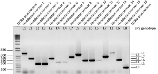
The final LPS-mPCR was used to genotype the 16 Heddleston type strains and was able to accurately differentiate all of these strains into the eight LPS genotypes (Fig. 3). The LPS-mPCR was then used to genotype the 58 field isolates. This final LPS-mPCR gave a single reproducible amplification product for 57 of 58 strains (strain PM135 was nontypeable), including the nine L3 strains that were nontypeable using the LPS-mPCRv1 (data not shown). All positive LPS-mPCR results were compatible with the LPS compositions that were determined (Table 3).
DISCUSSION
In this study, a multiplex PCR was designed to differentiate P.
multocida strains on the basis of the LPS genotype. The final
LPS-mPCR was able to unambiguously type the 16 Heddleston type strains and 57 of the 58 field isolates. However, strain PM135 remained untypeable, due to a large deletion in the region where the L7 LPS-mPCR primers were located (14). The failure of the LPS-mPCR due to large deletions within the LPS loci where prim-ers are located cannot be avoided. However, our previous analyses of the Heddleston type strains and field isolates containing LPS gene mutations indicate that such large deletions are rare; most mutations within the P. multocida LPS outer core biosynthesis loci involve single point mutations (9–11) and would be unlikely to compromise PCR amplification.
During the testing of the LPS-mPCR for differentiation of P.
multocida isolates, mass spectrometry analysis was used as the
“gold standard” to assess the composition of the LPS produced by individual strains. This method of LPS analysis identifies sugar
TABLE 3 (Continued)
Parameter and strain no. Serotypinga,b
LPS-mPCRv1
Final LPS-mPCR (Heddleston
serovars within each genotype)c LPS compositiond Nontypeable using initial
LPS-mPCRv1e
PM1 H3 (H3, H4) NT L3 (H3, H4) 3Hex, Hep (H4)/4Hex, Hep/1HexNAc, 4Hex, Hep (H3)
PM18 NT (H3) NT L3 (H3, H4) 2Hex, Hep
PM48 H3 (H3, H4) NT L3 (H3, H4) 3Hex, Hep (H4)/4Hex, Hep/1HexNAc, 4Hex, Hep (H3)
PM135 H8, H13 (H13) NT NT, L7 sequencef
1HexNAc, 2Hex, Hep (H13)
PM1075 H16 NT L3 (H3, H4) No outer core
PM1120 NT NT L3 (H3, H4) No outer core
PM1153 H1, H3, H7 NT L3 (H3, H4) 3Hex, Hep (H4)/4Hex, Hep/1HexNAc, 4Hex, Hep (H3)/
2HexNAc, 4Hex, Hep
PM1258 NT NT L3 (H3, H4) No outer core
PM1369 H1 NT L3 (H3, H4) 3Hex, Hep (H4)/4Hex, Hep
PM1434 NT NT L3 (H3, H4) 1HexNAc, 4Hex, Hep (H3)
aThe format in which multiple numbers are separated by a comma indicates that a precipitin line was observed with more than one type serum. Results in parentheses indicate that
two distinct and separate serotyping assays were performed: the result in parentheses is the first result with this isolate. NT, not able to be typed by this method (i.e., no precipitin line was observed using serotyping, or no amplicon was produced by mPCR).
b
A Heddleston serovar shown in boldface correlates with both LPS composition and LPS genotype.
cAn LPS genotype shown in boldface correlates with LPS composition. d
Outer core LPS sugar composition as predicted by MS/MS compositional analysis. The Heddleston serovar within the designated genotype that matches the LPS outer core composition is shown in boldface and in parentheses. Those compositions without a Heddleston serovar designation shown in parentheses do not precisely match any of the Heddleston type strain LPS structures. Multiple LPS glycoforms when detected have been separated by “/.” Hex, hexose (glucose or galactose); HexNAc, N-acetyl hexosamine [N-acetylglucosamine, N-acetyl galactosamine, or (1S)-2-acetamido-2-deoxy-D-galactose]; Hep, heptose; PCho, phosphocholine. Nonstoichiometric phosphoethanolamine additions to the outer core have not been determined.
eSequencing of the LPS outer core biosynthesis locus revealed significant nucleotide differences where the LPS-mPCRv1 primers were located. f
Sequencing of the PM135 LPS outer core biosynthesis locus revealed a large deletion in the region where the L7 LPS-mPCR primers were located.
on October 12, 2016 by Canada Institute for STI, National Research Council
http://jcm.asm.org/
type and overall sugar content, which are then compared to the LPS compositions of the fully elucidated Heddleston LPS struc-tures to predict the LPS glycoforms expressed by any particular strain.
Our previous analyses of the LPS genetics and structure in P.
multocida showed that strains with the same serological
designa-tion can produce structurally distinct LPS (9–16). These analyses also showed that the LPS structures produced by many P.
multo-cida strains are truncated variants of the full-length or “parent”
LPS structure and that there is significantly more LPS diversity in the field than is represented by the current 16 type strains (9–12, 14). Interestingly, many P. multocida field isolates belonging to the L3, L4, and L6 genotypes produce a wide range of LPS glycoforms, including some which have a significantly truncated LPS outer core or no outer core at all. Many of these strains were isolated from poultry exhibiting clear signs of fowl cholera (data not shown), indicating that strains belonging to these genotypes do not require a full-length LPS molecule to cause disease. Our stud-ies on the L1/serovar 1 strain VP161 have shown that any short-ening of the LPS outer core structure in this strain results in atten-uation of virulence in chickens (24), but it is possible that isolates expressing truncated LPS may be able to persist in some host niches but are not as virulent as parent strains expressing full-length LPS. Indeed, infection of chickens with a VP161 hptE LPS mutant (which produces a highly truncated outer core) showed that this mutant could persist at the site of muscle injection but could not be recovered from the blood, thus supporting this hy-pothesis (20). Importantly, many of the structures expressed by L3 and L6 genotype strains mimic host glycosphingolipids, and this may allow the bacteria to avoid recognition by the components of the innate immune system (10,11).
Our comparison of LPS composition, Heddleston serotyping, and LPS-mPCR typing showed clearly that the Heddleston serovar designation frequently failed to correlate with the composition of the LPS produced by each strain. In contrast, the LPS-mPCR as-say, specific for the identification of the LPS outer core biosynthe-sis loci, always correlated with LPS composition. Knowledge of the LPS genotype allows for the identification of the LPS type and possible range of LPS structures that strains can produce.
How-ever, the LPS-mPCR cannot predict the precise LPS structures produced by individual strains as random mutations within the LPS locus often lead to changes in LPS structure. We propose that the high diversity in numbers and types of LPS molecules pro-duced by P. multocida strains indicates that a typing system exactly predictive of LPS structure is not feasible. If knowledge of the precise LPS structure is important for diagnosis and the control of outbreaks, then following mPCR analysis, further experiments would need to be conducted, such as carbohydrate-specific silver staining of cell lysates to assess the relative size of the LPS pro-duced. Alternatively, for more detailed analysis, nucleotide se-quencing of the LPS biosynthesis locus to identify the specific LPS gene mutations combined with carbohydrate mass spectrometry could be employed. However, these detailed analyses are beyond the scope of diagnostic laboratories.
The LPS-mPCR developed here is a highly reproducible typ-ing system for differentiattyp-ing P. multocida strains. We have also shown that many field isolates produce multiple LPS glycoforms simultaneously; these naturally occurring “multivalent” strains could be excellent candidates for killed-cell vaccines as they may show broader protective efficacy than strains expressing single LPS molecules. We are currently assessing this possibility.
ACKNOWLEDGMENTS
This research was partly conducted within the Poultry CRC, established and supported under the Australian Government’s Cooperative Research Centres, and was also funded in part by the Australian Research Council, Canberra, Australia.
We thank Perry Fleming for bacterial growths and Jacek Stupak for capillary electrophoresis (CE)-MS.
REFERENCES
1. Wilkie IW, Harper M, Boyce JD, Adler B. 2012. Pasteurella multocida: diseases and pathogenesis. Curr Top Microbiol Immunol 361:1–22.http: //dx.doi.org/10.1007/82_2012_216.
2. Carter GR. 1952. The type specific capsular antigen of Pasteurella
multo-cida. Can J Med Sci 30:48 –53.
3. Heddleston KL, Gallagher JE, Rebers PA. 1972. Fowl cholera: gel diffu-sion precipitin test for serotyping Pasteurella multocida from avian spe-cies. Avian Dis 16:925–936.http://dx.doi.org/10.2307/1588773. 4. Glisson JR, Hofacre CL, Christensen JP. 2008. Fowl cholera, p 739 –758.
FIG 3 Gel electrophoresis separation of products generated using the final LPS-mPCR with the template derived from lysed colonies from each of the Heddleston
type strains (H1 to H16). The relative size of each LPS genotype PCR amplicon remains unchanged from that of the LPS-mPCRv1, with the exception of the L4 amplicon, which is now 474 bp.
Harper et al.
on October 12, 2016 by Canada Institute for STI, National Research Council
http://jcm.asm.org/
In Swayne DE (ed), Diseases of poultry, 12th ed. Blackwell Publishing,
Ames, IA.
5. Harper M, Boyce JD, Adler B. 2012. The key surface components of
Pasteurella multocida: capsule and lipopolysaccharide. Curr Top
Micro-biol Immunol 361:39 –51.http://dx.doi.org/10.1007/82_2012_202. 6. Harper M, Cox AD, St. Michael F, Wilkie IW, Boyce JD, Adler B. 2004. A
heptosyltransferase mutant of Pasteurella multocida produces a truncated lipopolysaccharide structure and is attenuated in virulence. Infect Immun
72:3436 –3443.http://dx.doi.org/10.1128/IAI.72.6.3436-3443.2004. 7. Singh R, Blackall PJ, Remington B, Turni C. 2013. Studies on the presence
and persistence of Pasteurella multocida serovars and genotypes in fowl cholera outbreaks. Avian Pathol 42:581–585.http://dx.doi.org/10.1080 /03079457.2013.854861.
8. Wilson MA, Morgan MJ, Barger GE. 1993. Comparison of DNA finger-printing and serotyping for identification of avian Pasteurella multocida isolates. J Clin Microbiol 31:255–259.
9. Harper M, St. Michael F, John M, Vinogradov E, Adler B, Boyce JD,
Cox AD. 2011. Pasteurella multocida Heddleston serovars 1 and 14 express
different lipopolysaccharide structures but share the same lipopolysaccha-ride biosynthesis outer core locus. Vet Microbiol 150:289 –296.http://dx .doi.org/10.1016/j.vetmic.2011.01.028.
10. Harper M, St. Michael F, John M, Vinogradov E, Steen JA, van Dorsten
L, Steen JA, Turni C, Blackall PJ, Adler B, Cox AD, Boyce JD. 2013.
Pasteurella multocida Heddleston serovar 3 and 4 strains share a common
lipopolysaccharide biosynthesis locus but display both inter- and intras-train lipopolysaccharide heterogeneity. J Bacteriol 195:4854 – 4864.http: //dx.doi.org/10.1128/JB.00779-13.
11. Harper M, St. Michael F, Steen JA, John M, Van Dorsten L, Parnas H,
Vinogradov E, Adler B, Cox A, Boyce JD. 2014. Structural analysis of
lipopolysaccharide produced by Heddleston serovars 10, 11, 12 and 15 and the identification of a new Pasteurella multocida LPS outer core bio-synthesis locus, L6. Glycobiology 24:649 – 659.http://dx.doi.org/10.1093 /glycob/cwu030.
12. Harper M, St. Michael F, Steen JA, John M, Wright CL, Van Dorsten L,
Vinogradov E, Adler B, Cox A, Boyce JD. 7 October 2014.
Characteri-sation of the lipopolysaccharide produced by Pasteurella multocida sero-vars 6, 7 and 16; identification of lipopolysaccharide genotypes L4 and L8. Glycobiologyhttp://dx.doi.org/10.1093/glycob/cwu110.
13. Harper M, St. Michael F, Vinogradov E, John M, Boyce JD, Adler B, Cox
AD. 2012. Characterization of the lipopolysaccharide from Pasteurella
mul-tocida Heddleston serovar 9; identification of a proposed bi-functional
dTDP-3-acetamido-3,6-dideoxy-␣-D-glucose biosynthesis enzyme.
Gly-cobiology 22:332–344.http://dx.doi.org/10.1093/glycob/cwr147. 14. Harper M, St. Michael F, Vinogradov E, John M, Steen JA, van Dorsten
L, Boyce JD, Adler B, Cox AD. 2013. Structure and biosynthetic locus of
the lipopolysaccharide produced by Pasteurella multocida serovars 8 and 13 and the identification of a novel phospho-glycero moiety. Glycobiology
23:286 –294.http://dx.doi.org/10.1093/glycob/cws154.
15. St. Michael F, Harper M, Parnas H, John M, Stupak J, Vinogradov E,
Adler B, Boyce JD, Cox AD. 2009. Structural and genetic basis for the
serological differentiation of Pasteurella multocida Heddleston serotypes 2 and 5. J Bacteriol 191:6950 – 6959.http://dx.doi.org/10.1128/JB.00787-09. 16. St. Michael F, Li J, Cox AD. 2005. Structural analysis of the core oligo-saccharide from Pasteurella multocida strain X73. Carbohydr Res 340: 1253–1257.http://dx.doi.org/10.1016/j.carres.2005.02.014.
17. Tsai YC, Shien JH, Wu JR, Shieh HK, Chang PC. 2011. Polymerase chain reaction-restriction fragment length polymorphism analysis of the genes involved in the biosynthesis of the lipopolysaccharide of Pasteurella
multocida. J Vet Diagn Invest 23:543–546.http://dx.doi.org/10.1177 /1040638711404145.
18. Miflin JK, Blackall PJ. 2001. Development of a 23S rRNA-based PCR assay for the identification of Pasteurella multocida. Lett Appl Microbiol
33:216 –221.http://dx.doi.org/10.1046/j.1472-765x.2001.00985.x. 19. Townsend KM, Frost AJ, Lee CW, Papadimitriou JM, Dawkins HJ.
1998. Development of PCR assays for species- and type-specific identifi-cation of Pasteurella multocida isolates. J Clin Microbiol 36:1096 –1100. 20. Boyce JD, Harper M, St. Michael F, John M, Aubry A, Parnas H, Logan
SM, Wilkie IW, Ford M, Cox AD, Adler B. 2009. Identification of novel
glycosyltransferases required for assembly of the Pasteurella multocida A:1 lipopolysaccharide and their involvement in virulence. Infect Immun 77: 1532–1542.http://dx.doi.org/10.1128/IAI.01144-08.
21. St. Michael F, Li J, Vinogradov E, Larocque S, Harper M, Cox AD. 2005. Structural analysis of the lipopolysaccharide of Pasteurella multocida strain VP161: identification of both Kdo-P and Kdo-Kdo species in the lipopolysaccharide. Carbohydr Res 340:59 – 68.http://dx.doi.org/10.1016 /j.carres.2004.10.017.
22. Harper M, Boyce JD, Cox AD, St. Michael F, Wilkie I, Blackall P, Adler
B. 2007. Pasteurella multocida expresses two LPS glycoforms
simultane-ously both in vitro and in vivo but only a single form is required for virulence: identification of two acceptor specific heptosyl I transferases. Infect Immun 75:3885–3893.http://dx.doi.org/10.1128/IAI.00212-07. 23. Harper M, Cox AD, Adler B, Boyce JD. 2011. Pasteurella multocida
lipopolysaccharide: the long and the short of it. Vet Microbiol 153:109 – 115.http://dx.doi.org/10.1016/j.vetmic.2011.05.022.
24. Harper M, Cox AD, St. Michael F, Parnas H, Wilkie I, Blackall PJ, Adler
B, Boyce JD. 2007. Decoration of Pasteurella multocida LPS with
phos-phocholine is important for virulence. J Bacteriol 189:7384 –7391.http: //dx.doi.org/10.1128/JB.00948-07.