J Neurol (2008) 255:1275–1277
DOI 10.1007/s00415-008-0894-7
LETTER TO THE EDITORS
JO
N 289
4
in the right arm as well as self-in-flicted pressure-induced pain in the right upper arm. The severity of the jerky movements was strongly associated with the inten-sity of the pain. The movements could be absent for days or appear once a day for about a second up to several times within several hours. They were unaffected by voluntary contractions and not associated with amnesia or additional sen-sory-motor disturbances. Examina-tion revealed a right-sided domi-nant hyporeflexia of the upper extremities and a paramedian right-sided dissociated sensory loss below the third cervical derma-tome with sparing of the right hand. In addition there was the known complete transverse spinal cord syndrome at the 6th thoracic level with sensory loss of all quali-ties, paresis of abdominal – and lower back musculature, paraplegia and bilateral extensor plantar re-flexes. At the upper extremities there was no atrophy, no weakness and no impairment of coordina-tion. Cranial nerves were intact. MRI of the cervical spine, per-formed 20 months before sensory symptoms started, was normal. MRI of the brain was normal. EEG showed an intermittent bifrontal slowing but no epileptiform poten-tials. Together with the intermittent nature of the jerks and the fluctuat-ing sensory symptoms a psycho-genic disorder or a relapse of drug-dependent behavior was suspected. Increasing the dosage of metha-done diminished pain and involun-tary movements somewhat. In the following months the involuntary clonic flexor movement of the arm changed to a more dystonic flexor type without any associated changes in motor-sensory symp-toms. Persistence of symptoms and increased disability of the patient led to a revaluation of the case. Me-dian nerve sensory evoked poten-tials were normal, but cutaneous M. Sollberger
P. Fuhr
Flexor myoclonus of the
arm due to posttraumatic
cervico-thoracic
syringomyelia
Received: 13 September 2007Received in revised form: 28 December 2007 Accepted: 6 February 2008
Published online: 4 July 2008
Sirs: Syringomyelia rarely causes involuntary movements such as re-spiratory synkinesis, myoclonus, dystonia and tremor, probably due to an increased excitability of the spinal motor neurons [1]. We re-port the case of a pure pain-sensi-tive segmental myoclonus of the right arm in posttraumatic cervico-thoracic syringomyelia, initially suspected to be a relapse of drug-dependant behavior.
A 33-year-old man developed painful paresthesias and hypesthe-sia over the shoulder-girdle and in both arms 3 years after a complete transverse spinal cord injury at the fourth thoracic level. His past med-ical history was known for abuse of heroin until the age of 16, chronic intake of methadone and an idio-pathic hypogonadism substituted with testosterone. Within few months the sensory symptoms dis-appeared on the left side, whereas they increased on the right side. Moreover, repetitive involuntary jerky flexion movements of the right arm developed, involving pre-dominantly the proximal arm mus-cles. They were exclusively trig-gered by spontaneous intense pain
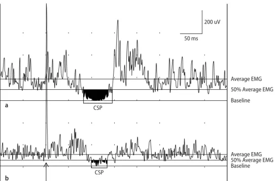
silent period (CSP) in the voluntary contracted right abductor pollicis brevis (APB) muscle was attenu-ated (Fig.1). The patient refused any additional neurophysiological exams. Repeated MRI of the cervi-cal spine now revealed an extensive syringomyelia from the second cer-vical segment down to the level of the spinal cord lesion at Th2/Th3 (Fig.2). After the syringo-perito-neal drainage, pain and involuntary movements decreased clearly within the first 2 weeks, followed by a stable clinical course ever since (9 months).
This case shows exclusively pain-sensitive segmental flexor myoclonus in an upper extremity, most likely caused by disinhibition of the flexor reflex due to posttrau-matic cervico-thoracic syringomy-elia. The marked decrease of the in-voluntary movements after surgical intervention together with the neu-rophysiological and imaging analy-ses point to the spinal origin of the myoclonus. Unfortunately no time-locked back-averaged EEG, no EMG polygraphs and no reflex studies other than CSP could be performed to better characterize the mechanism of the myoclonus. The case highlights the wide spec-trum of involuntary movements re-ported in patients with syringomy-elia, i.e. those which are
spontaneous and stimulus-depen-dant [1, 2, 4], spontaneous and stimulus-independent [3] or, as in our case, exclusively stimulus-de-pendent [2]. Interestingly, in spite the clinical heterogeneity a similar pathogenic mechanism is postu-lated, which is increased excitabil-ity of spinal motor neurons of probably different intensity due to altered processing at the inhibitory spinal interneuron circuits [1]. Of note in our case is the change of the clonic flexor movement to a more dystonic flexor type. That change in movement implicates longer lasting excitatory periods of
M. Sollberger, MD (,) · P. Fuhr, MD Dept. of Neurology
University Hospital of Basel Petersgraben 4
4031 Basel, Switzerland Tel.: +41 61 265 89 30 Fax: +41 61 265 37 88 E-Mail: [email protected]
1276
spinal motor neurons [1] probably as consequence of progression of the syrinx.
In the present case, posterior columns as assessed by median nerve sensory evoked potentials were normal, while CSP, an inhibi-tory part of the flexor reflex, in the right hand was attenuated. Afferent impulses that generate the CSP are carried by A-delta fibers, which conduct nociceptive impulses on spinal interneurons, located in close proximity to the central canal [5, 6, 12]. These findings, together with the phenomenology of the myoclonus and its response to methadone, suggest a mechanism involving an intraspinal pathway, susceptible to nociceptive input and endorphin receptor activation. There is evidence from human and animal literature that endogenous and exogenous opioids suppress spinal nociceptive flexion reflexes mainly by acting on spinal mu-opi-oid receptors [7–9]. However
,
ex-cept in patients with restless legs syndrome, respectively periodic limb movements during sleep [10] opioids are not known to decrease involuntary movements of spinal origin. In contrary, myoclonus has been described as a neuroexcit-atory side effect of opioids [11]. In this regard we report an unusualpharmacological effect of metha-done on spinal myoclonus, which, however, goes with the hypothesis of a disinhibited spinal flexor re-flex.
It is of note that CSP is also al-tered in patients with syringomy-elia without involuntary move-ments [12, 13], which implies an additional mechanism for the pain-associated motor release phenome-non in the reported case.
References
1. Nogués MA, Leiguarda RC, Rivero AD, et al. (1999) Involuntary movements and abnormal spontaneous EMG ac-tivity in syringomyelia and syringo-bulbia. Neurology 52:823–834 2. Hill MD, Kumar R, Lozano A, et al.
(1999) Syringomyelic dystonia and athetosis. Movement Disorders 14: 684–688
3. Bagnato S, Rizzo V, Quartarone A, et al. (2001) Segmental myoclonus in a pa-tient affected by syringomyelia. Neurol Sci 22:27–29
4. Gregoire SM, Laloux P, Hanson P, et al. (2006) Segmental spinal myoclonus and syringomyelia: A case report. Acta Neurol Belg 106:37–40
5. Floeter MK (2003) Cutaneous silent periods. Muscle Nerve 28:391–401
200 uV 50 ms Average EMG 50% Average EMG Baseline Average EMG 50% Average EMG Baseline CSP a b CSP
Fig. 1 CSP in the right APB of a healthy control (a) and the patient (b). Stimula-tion of second digit at 13× sensory threshold (ST) in (a) and at 16× ST in (b) (arrow). Stimulus duration 0.2 ms. Average EMG bases on the period of EMG activity prior to the stimulus
Fig. 2 Sagittal T2-weighted MRI of the cervical spine. Note the extensive syringomyelia from C2 down to the level of the spinal cord lesion at Th2/Th3
1277
6. Kofler M, Fuhr P, Leis AA, et al. (2001) Modulation of upper extremity motor evoked potentials by cutaneous affer-ents in humans. Clin Neurophysiol 112: 1053–1063
7. Willer JC, Dehen H, Cambier J (1981) Stress-induced analgesia in humans: endogenous opioids and naloxone-re-versible depression of pain reflexes. Science 212:689–691
8. Willer JC (1985) Studies on pain. Ef-ff fects of morphine on a spinal nocicep-tive flexion reflex and related pain sen-sation in man. Brain Res 331:105–114
9. Tao PL, Lai YS, Chow LH, et al. (2005) Effects of morphine and endomor-phins on the polysynaptic reflex in the isolated rat spinal cord. Naunyn Schmiedebergs Arch Pharmacol 371: 72–80
10. Karatas M (2007) Restless legs syn-drome and periodic limb movements during sleep: diagnosis and treatment. Neurologist 13:294–301
11. Glare P, Walsh D, Sheehan D (2006) The adverse effects of morphine: a prospective survey of common symp-toms during repeated dosing for chronic cancer pain. Am J Hosp Palliat Care 23:229–235
12. Stetkarova I, Kofler M, Leis AA (2001) Cutaneous and mixed nerve silent pe-riods in syringomyelia. Clin Neuro-physiol 112:78–85
13. Kaneko K, Kawai S, et al. (1997) Cuta-neous silent period in syringomyelia. Muscle Nerve 20:884–886