Received: 3 January 2002 Revised: 22 July 2002 Accepted: 6 August 2002 Published online: 17 October 2002 © Springer-Verlag 2002
Abstract 2-[F-18]-fluoro-2-deoxy-D-glucose (FDG) positron emission tomography (PET) has become an important staging modality for many tumors, including bronchial carcino-ma; however it is important to know that there are several pitfalls in PET image interpretation. In this report we demonstrate three cases in which focal intrapulmonary FDG uptake could possibly represent iatrogenic microembolism. These FDG accumu-lations would have been interpreted
as malignant tumor mass in the lung if no anatomic correlation would have been performed. For this rea-son, we further present an integrated PET/CT scanner, which recently has been introduced. This correlation of molecular and morphological infor-mation enables the specification of the FDG-PET findings.
Keywords Positron emission tomography · CT FDG · PET/CT scanners · Tumors
Thomas F. Hany Juerg Heuberger
Gustav K. von Schulthess
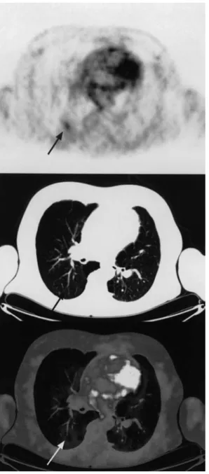
Iatrogenic FDG foci in the lungs:
a pitfall of PET image interpretation
Introduction
The 2-[F-18]-fluoro-2-deoxy-D-glucose positron emission tomography (FDG-PET) has become an important staging modality for many tumors including bronchial carcinoma. It has been demonstrated that FDG-PET is more sensitive and more specific than CT in the staging of many tumors [1]. It identifies lesions not seen in CT and lesions which cannot be specified as being malignant or benign based on morphological criteria only. A major drawback of FDG-PET is the almost complete absence of an anatomic refer-ence frame, which often prevents the diagnostician from being able to precisely localize disease foci with the FDG-PET examination alone. For this reason, integrated FDG-PET/ CT scanners have been recently introduced [2, 3]. The great advantage of these combined systems providing “hardware” co-registered images is that lesions seen in FDG-PET can be correlated to the morphological findings without the additional use of complex software packages. This correlation of molecular and morphological informa-tion helps to specify FDG-PET findings. This is important because it is well known that FDG not only accumulates in many tumors but also in inflammatory tissue and a variety of physiological and artificial settings [4].
Herein we report on three cases where patients dem-onstrated lung lesions on FDG-PET. It is hypothesized that these lesions were microembolisms formed during injection, with a size to make them lodge in the distal capillary lung bed very much like macroaggregates of albumin lodge in the lung parenchyma.
Case reports
The FDG-PET/CT examinations in three patients with focal FDG uptake in the lung were identified among 750 PET/CT scans performed from March until November 2001. The patients were examined for the diagnoses discussed below.
Case 1
A 58-year-old patient had a PET/CT scan for preoperative staging of a histologically proven non-small-cell lung cancer. This patient underwent a second PET/CT scan 5 days after the initial scan due to the unclear findings in the right upper lobe.
Case 2
An 11-year-old child had a PET/CT scan for re-staging after chemotherapy and radiotherapy of Hodgkin’s lymphoma.
T.F. Hany · J. Heuberger G.K. von Schulthess (
✉
) Department of Nuclear Medicine, University Hospital, Rämistrasse 100, 8091 Zurich, Switzerlande-mail: vonschulthess@dmr.usz.ch Tel.: +41-1-2552944
Results
The first patient showed a pathological FDG accumula-tion in the right apical lung segment (Fig. 1) in addiaccumula-tion to the FDG uptake in the known tumor mass in the left upper lobe adjacent to the aorta (Fig. 2). The co-regis-tered unenhanced CT image (Fig. 3) corresponding to the PET image of Fig. 1 showed no anatomical correlate to the FDG focus, which would have suggested the pres-ence of a pulmonary nodule. The focus projected onto a branching point of a peripheral lung vessel. The patient received a partially paravenous injection at the right arm (Fig. 4). This discrepancy of findings with potentially important clinical consequences prompted us to order a repeat scan, which was performed 5 days after the initial scan. While the known tumor lesion was identified again, the focal FDG accumulation in the right apical lung segment was not present in the second scan (Fig. 5). The other two patients showed similar findings than the first patient with FDG accumulation at a lung site where no anatomic correlate could be identified (Figs. 6, 7). In these patients no repeat scans were ordered because of the lack of clinical consequence of these findings.
Discussion
On the basis of the initial findings in the first patient, the hypothesis was made that the FDG focus was due to a tiny blood clot possibly arising from some aspiration/ paravenous injection, which lodged in the peripheral lung.
Inflammatory changes also show increased FDG uptake. Therefore, these lesions could have represented acute inflammation; however, no morphological changes were seen in CT used for co-registration. Also, such a high increase in FDG uptake is rarely seen in PET imag-ing due to inflammation and a certain degree of uptake would have been expected in the follow-up exam after 5 days; therefore, the absence of the prominent lesion on the second FDG PET/CT can be explained by iatrogenic microembolism. Circumstantial evidence to this hypoth-esis could be the high FDG concentration within the intrapulmonary lesion. This observation can only be ex-plained when the thrombus formation happened within the syringe or the site of injection as demonstrated in Fig. 4. Iatrogenic embolization is well known as a com-plication of permanent venous access devices [5]; how-ever, no data are available regarding microembolism due to peripheral venous access as used in our study. The findings suggest that occasionally, and particularly when FDG injection does not work flawlessly, minor blood clots can form. The two other cases, again exhibiting a mismatch between molecular imaging and morphologi-cal imaging findings, could serve as corroborating
2123
Case 3
A 56-year-old patient had a follow up PET/CT scan after surgical resection of a non-small-cell lung cancer.
Four PET/CT scans were performed on the three patients using a novel combined PET/CT system (Discovery LS, GE Medical Systems, Waukesha, Wis.), combining the ability to acquire CT im-ages and PET data of the same patient in “hardware” co-registered fashion in one session. The Discovery LS system consists of a GE Advance NXi PET and a multislice helical CT (LightSpeed plus) scanner. The axes of both systems are mechanically aligned to co-incide perfectly. Initially, 1000 ml of oral contrast (Micropaque scanner, Guerbet, Aulnay-sous-Bois, France) was given for delin-eation of intestinal structures. Sixty to 120 min after intravenous injection of 10 mCi of FDG, the PET/CT exam was started. First, a non-contrast-enhanced CT data acquisition with the following pa-rameters was acquired: tube-rotation time 0.5 s/evolution; 140 kV; 80 mA; 22.5 mm/rotation; slice pitch 6 (high-speed mode); recon-structed slice thickness 5 mm; scan length 867 mm; and acquisition time 22.5 s per CT scan. The CT data were acquired during shal-low breathing. After table transposition into the PET-sensitive field of view, emission data were acquired at six incremental table posi-tions, each 146 mm wide, thereby covering 867 mm of table travel. For each position, 35 2D non-attenuation-corrected scans were obtained simultaneously over a 4-min period. The CT data was used for attenuation correction [2]; therefore, data acquisition was performed within 25 min covering the patient from the head to the pelvic floor. Viewing of co-registered images was performed using a dedicated software (eNTEGRA, Elgems, Haifa, Israel).
Fig. 1 Coronal positron emission tomography (PET) image in
case 1: pathological 2-[F-18]-fluoro-2-deoxy-D-glucose (FDG) accumulation in the right apical lung is seen
2125
Fig. 3 Case 1. The co-registered CT image corresponding to the PET
image showed no anatomical correlate to the FDG focus. The focus projected onto a branching point of a peripheral lung vessel (arrow)
Fig. 4 Partially paravenous injection in the right arm in case 1
evidence. The advantage of FDG PET/CT in this setting is that anatomy helps to specify the findings.
In conclusion, our data suggest that extra care with FDG injection should be taken and aspiration of blood should be avoided. When intrpreting the presence of focal lesions in the lung field, a differential diagnosis of a microembolism containing FDG has to be considered, and if no corroborating anatomic evidence of the lesion is found, this diagnosis is likely.
Fig. 5a, b Case 1. a Coronal view of CT, PET, and PET/CT (left to right) demonstrating intrapulmonary FDG uptake in the right apical
2127
Fig. 6 Case 2 with FDG accumulation in the right lung without
anatomic correlate (arrows)
Fig. 7 Case 3 with FDG accumulation in the right lung without
anatomic correlate (arrows)
References
1. Pieterman RM, van Putten JW, Meuzelaar JJ, Mooyaart EL, Vaal-burg W, Koeter GH et al. (2000) Preoperative staging of non-small-cell lung cancer with positron-emission tomography. N Engl J Med 343:254– 261
2. Kamel E, Hany TF, Burger C, Treyer V, Lonn AH, Schulthess GK von et al. (2002) CT vs 68Ge attenuation correc-tion in a combined PET/CT system: evaluation of the effect of lowering the CT tube current. Eur J Nucl Med Mol Imaging 29:346–350
3. Townsend DW, Cherry SR (2001) Combining anatomy and function: the path to true image fusion. Eur Radiol 11:1968–1974
4. Engel H, Steinert H, Buck A, Berthold T, Huch Boni RA, Schulthess GK von (1996) Whole-body PET: physiological and artifactual fluorode-oxyglucose accumulations. J Nucl Med 37:441–446
5. Hoch JR (1997) Management of the complications of long-term venous access. Semin Vasc Surg 10:135–143
![Fig. 1 Coronal positron emission tomography (PET) image in case 1: pathological 2-[F-18]-fluoro-2-deoxy-D-glucose (FDG) accumulation in the right apical lung is seen](https://thumb-eu.123doks.com/thumbv2/123doknet/14865057.637013/2.892.92.419.99.523/coronal-positron-emission-tomography-pathological-fluoro-glucose-accumulation.webp)


