RESEARCH ARTICLE
18
F-FET PET/CT in Advanced Head and Neck
Squamous Cell Carcinoma: an Intra-individual
Comparison with
18
F-FDG PET/CT
Stephan Kurt Haerle,
1Dorothee R. Fischer,
2Daniel T. Schmid,
2Nader Ahmad,
1Gerhard F. Huber,
1Alfred Buck
21
Department of Otorhinolaryngology, Head and Neck Surgery, University Hospital Zurich, Frauenklinikstrasse 24, CH-8091 Zurich, Switzerland
2Department of Nuclear Medicine, University Hospital Zurich, Raemistrasse 100, CH-8091 Zurich, Switzerland
Abstract
Purpose: To assess the diagnostic value of O-2-fluoro-18(F)-ethyl-L-tyrosine (18F-FET) positron emission tomography/computed tomography (PET/CT) for patients with advanced head and neck squamous cell carcinoma compared with 18F-fluoro-2-deoxy-D-glucose (18F-FDG) PET/CT at initial staging and following radiochemotherapy.
Procedures: Thirteen patients were prospectively enrolled; each of them underwent an18F-FDG PET/CT and18F-FET PET/CT before treatment. Ten of those were scanned 10 weeks after treatment.
Results: Sensitivity, specificity, and accuracy for 18F-FDG PET/CT (primary and lymph node
metastases) at initial staging were 89%, 50%, and 81%. For18F-FET PET/CT the numbers were
70%, 90%, and 74%. Sensitivity, specificity, and accuracy for 18F-FDG PET/CT at follow-up
were 71%, 65%, and 67%. For 18F-FET PET/CT the numbers were 29%, 100%, and 83%.
Additionally, 18F-FDG PET/CT detected a higher number of second malignancies or distant metastases.
Conclusions: 18F-FET is no substitute for 18F-FDG. Although it is more specific, too many malignant lesions are missed due to its lower sensitivity.
Key words:18F-FET PET/CT,18F-FDG PET/CT, Tracer, Head and neck cancer, Staging
Introduction
T
he clinical value of 2-deoxy-2-[18F]fluoro-D-glucosepositron emission tomography/computed tomography (18F-FDG PET/CT) in primary staging of head and neck squamous cell carcinoma (HNSCC) is well documented [1]. Unfortunately, 18F-FDG also accumulates in inflammatory tissue, which may cause false-positive results and therefore the specificity for malignancies is decreased, in particular after treatment by radiotherapy [2]. As a result of this, radiolabelled amino acids were attracting increasing interest
in nuclear medicine over the past years. Amino acids are specifically accumulated in malignant cells owing to increased expression of amino acid transporters. In contrast to glucose derivates, the uptake of amino acids in macro-phages and other inflammatory cells is absent or at least reduced [3,4], therefore the amino acid tracers appear to be more specific for tumor than FDG. Promising results for the detection of HNSCC have been reported using L
-methyl-[11C]-methionine [5] and L-[1-(11) C]-tyrosine [3] as a
tracer. The drawback is the short physical half-life (20 min) of the 11C label. On the contrary, 18F-labelled amino acid analogues, such as O-(2-[18F]fluoroethyl)-L-tyrosine (18
F-FET), are logistically advantageous. Regarding the fact that inflammatory tissue and lymph nodes are often present in
patients with HNSCC the rate of false-positive results by
18
F-FDG PET is considerable. Previous studies describing
18
F-FET as a potential tracer in HNSCC were first evaluat-ing its value for initial stagevaluat-ing [6]. According to the disappointing results compared with 18F-FDG, later studies investigated18F-FET in the follow-up setting [7,8]. The aim of this prospective study was, for the first time, to assess the diagnostic value of18F-FET PET/CT in a cohort of patients with advanced HNSCC compared with18F-FDG PET/CT at the time of initial staging and following radiochemotherapy in the same patients, resulting in an intra-individual comparison of the two modalities.
Materials and Methods
Patients
A total of 13 patients from the Department of Otorhinolaryngology, Head and Neck Surgery at a tertiary referral center have been enrolled in this prospective study between June 2008 and June 2009. This patient cohort does not represent a consecutive group of all patients diagnosed with a HNSCC within the given time frame, because mainly patients with advanced disease (T3/4, N2/3) were referred to 18F-FDG PET/CT for search for potential distant metastases. All of the patients were suffering from a previously untreated HNSCC and all of the patients were staged by undergoing panendoscopy, ultrasound-guided fine needle aspiration cytology (USgFNAC) of the neck and biopsy of the primary. It is the institutional policy that all the patients suffering from HNSCC are primarly staged by USgFNAC as a first line examination. In our experience, it is one of the most accurate methods for staging the neck. In ten patients, the primary was located in the oropharynx, whereas two patients were suffering from SCC of the oral cavity, and one patient presented with a poorly differentiated SCC of the nasopharynx. Further tumor characteristics are listed in Table1.
Each of the 13 patients was examined with18F-FDG PET/CT and
18
F-FET PET/CT, before treatment. The order of the PET/CTs was arbitrary. The interval between the two imaging modalities was not longer than 2 weeks. Ten of those were again scanned 10 weeks after treatment. The initial PET imaging was done before panendoscopy. No therapy between the two imaging modalities was done. In the patients which had a contrast-enhanced CT or MRI before the PET-CT scan, the CT part of the PET-CT scan was done without contrast. Radiochemotherapy consisted of intensity-modulated radiotherapy (IMRT) and simultaneous systemic treatment was performed with cisplatin (40 mg/m2per week) or cetuximab (standard dosage). This
study was approved by university ethics committee. All subjects gave written informed consent for participation in this prospective study.
Positron Emission Tomography Imaging
For this study, we used a combined PET/CT system (Discovery LS, RX or Discovery STE, GE Health Systems, Milwaukee, WI). This device integrates a PET scanner with a multi-slice helical CT and permits the acquisition of coregistered CT and PET images in the same session. Patients were examined in supine position.
Immediately following the low-dose CT acquisition (FOV 50 cm, auto mA, 120 kV, slice-thickness 3.75 mm, 0.5 s rotation time, standard reconstruction type), 3D-PET emission data were acquired for 1.5 and 2 min/bed position, respectively. The CT data
were used for attenuation correction and images were reconstructed using a fully 3D iterative algorithm (VUE Point HD). The CT scan was acquired during breath holding in the normal expiratory position. The PET/CT scans were started 45 min following injection of 350 MBq18F-FDG, and, in accordance to a previous
report by Pauleit D et al. [7], 60 min after the application of 250 MBq18F-FET.
The acquired images were post-processed with a dedicated software (Volume Viewer PET/CT, AW 4.4 workstation, GE Healthcare) providing multiplanar reformatted images of PET alone, CT alone and fused PET/CT with linked cursors. At our institution, blood glucose is checked in every patient before the 18F-FDG injection. In this study, no patient suffered from hyperglycemia.
Standard of Reference and Data Analysis
The18F-FDG- and18F-FET PET/CT scans were evaluated by two physicians, board certified in radiology and nuclear medicine and both with years of PET/CT experience. Each read either the18 F-FET or the18F-FDG scan without access to the other scan. The results from panendoscopy and cytological/histological work-up were not available for the nuclear medicine specialists. The primary tumor and suspicious lymph nodes were identified on CT or on the PET scan. The standard of reference was the biopsy of the primary, and the fine needle aspiration cytology of all the suspicious cervical lymph nodes. As all the patients underwent USgFNAC before and after treatment, a false-negative finding in the PET/CT scans is defined as missed lymphatic disease in18F-FET or18F-FDG in a lesion identified either in only one of them or on the CT and verified as tumor in the USgFNAC. In addition, a clinical follow-up for at least 6 months served as the standard of reference (12/13, 92%). A verified positive lymph node during follow-up, which was not identified in the first session by either modality was labelled false negative. In one case, the patient died 1 month after having started an adequate therapy and therefore a follow-up for 6 months was not possible. The ultrasound-guided fine needle aspiration was always performed by the same specialist with a vast experience regarding this staging method.
In summary, all numbers for sensitivity, specificity, and accuracy are lesion based. Three lesions (two lesions suspicious for a second primary and one lesion suspicious for a distant metastasis) were initially not verified by cytology or histology. In two of these cases, a clinical referral was able to exclude a second primary, and in one patient a follow-up imaging examination 6 months later served as reference. All other patients with suspected distant metastasis or secondary primaries distant from the neck were referred for biopsy/cytology.
The tracer uptake in the lesions for both, the18F-FET and the 18
F-FDG scan, was visually graded as none, mild, moderate and strong compared with background tissue. Mild, moderate, or strong uptake was considered suspicious for tumor.
In addition to visual analysis, the uptake of18F-FDG and18 F-FET in the primary tumor was expressed as standardized uptake values (SUV). For this purpose a region of interest (ROI) was placed over the tumor using the image analysis software PMOD (PMOD Technologies Ltd., Zurich, Switzerland). The ROI was placed on the18F-FDG or the18F-FET scan, depending on where it
was best visible. It was then transferred to the other modality after co-registration. To avoid any statistical noise, the mean uptake in the ROI was chosen for reporting.
Results
Between 2008 and 2009, the total number of 13 patients (11 males, two females) was evaluated with 18F-FDG PET/CT and 18F-FET PET/CT for a previously untreated advanced HNSCC. The mean age of the patients was 63.3 years (range, 53–80 years), and the mean follow-up period of all patients was 12.7 months (range, 1–22 months). One patient could only be followed by a month because of death. All other patients were followed for at least 6 months. Patient information and tumor characteristics are shown in Table1. All patients presented in the advanced stages III and IV (International Union against Cancer (UICC) 1997).
Positron Emission Tomography Imaging
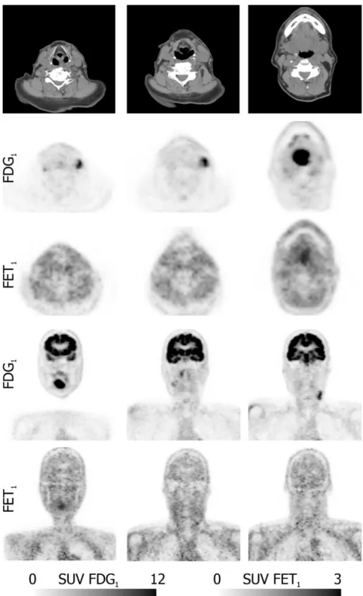
Typical examples of 18F-FDG and 18F-FET PET scans are illustrated in Figs.1 and 2. Figure1 demonstrates a patient with moderate to strong18F-FDG and18F-FET uptake in the primary tumor and the lymph node metastases with higher tumor-to-background ratio in the18F-FDG scan. In Fig. 2, the primary tumor and the nodal disease are clearly visible on the18F-FDG scan, whereas all malignant lesions, except the primary, are false negative on the 18F-FET scan. At initial staging a total of 37 lesions were detected in the 13 patients (13 primaries and 24 lymph nodes). The primary tumor was detected in all patients by18F-FDG- and18F-FET PET/CT. The 18F-FDG uptake was rated “strong” in all cases. The rating of the 18F-FET uptake was “strong” in three patients, “moderate” in two cases and mild in eight patients. At follow-up, 11 patients were available for 18 F-FDG and ten for 18F-FET imaging. The primary tumor site uptake was rated “none” in all patients by using 18F-FET, whereas with18F-FDG, four primary tumor sites were rated “mild”, one “moderate” and six were rated “none”.
With regard to lymph nodes suspicious findings on 18 F-FDG PET were found in 12 of the 13 patients (92%). The node uptake was rated “moderate” or “strong”. In contrast, only seven (54%) patients had suspicious nodes on18F-FET PET,
the uptake in which was rated “mild” in six patients and “strong” only in one patient. In summary, the lesion based rate at initial staging for the primaries using18F-FDG PET and18 F-FET PET was 13 out of 13 (100%). With regard to the lymph node metastases, the lesion based rate for18F-FDG PET was 20 out of 24 (83%), and 13 out of 24 (54%) for18F-FET PET. During follow-up,18F-FDG PET was able to detect all three histological confirmed local tumor persistences whereas18 F-FET PET only detected two out of three and therefore missed one true-positive lesion. With regard to the lymph node metastases, the lesion based rate for 18F-FDG PET and for
18
F-FET PET was two out of four (50%), respectively. The calculated sensitivity and specificity for18F-FDG and18F-FET are summarized in Table 2. The sensitivity to detect malig-nancy was markedly higher for18F-FDG than18F-FET. The specificity shows the reverse trend, at initial staging the specificity of 18F-FET in suspicious lymph nodes was 90% compared with only 50% for 18F-FDG. At follow-up, the pattern is similar. The sensitivity data at follow-up are less reliable due to the low number of true-positive lymph nodes. There were five false-positive lymph nodes on18F-FDG PET/ CT. All of these were cytologically confirmed as lymphadeni-tis. On the contrary,18F-FET PET/CT only detected one false-positive finding that turned out to be an inflammatory enlarge-ment of the lymph node. A different pattern was found in the follow-up setting:18F-FDG PET/CT revealed a total of two false-positive primaries and a total of six false-positive lymph nodes. The false-positive primaries histologically turned out to be chronic inflammatory and fibrotic tissue. Regarding false-positive lymph node metastases during follow-up, 18F-FDG PET/CT revealed six lymph nodes which in the pathological report turned out to be inflammatory and necrotic tissue.18 F-FET PET/CT, on the contrary, did not reveal any false-positive finding during follow-up resulting in a specificity of 100%.
Second Primaries and Distant Metastases
At initial staging,18F-FDG PET/CT revealed a total of four lesions suspicious for a second primary (one caecal, one
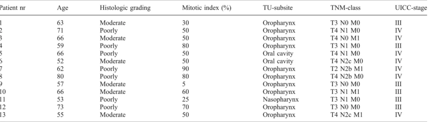
Table 1. Patient and tumor data
Patient nr Age Histologic grading Mitotic index (%) TU-subsite TNM-class UICC-stage 1 63 Moderate 30 Oropharynx T3 N0 M0 III 2 71 Poorly 50 Oropharynx T4 N1 M0 IV 3 66 Moderate 50 Oropharynx T4 N0 M1 IV 4 59 Poorly 80 Oropharynx T3 N1 M0 III 5 66 Poorly 50 Oral cavity T4 N1 M0 IV 6 52 Moderate 50 Oral cavity T4 N2c M0 IV 7 62 Poorly 90 Oropharynx T2 N2b M1 IV 8 80 Poorly 80 Oropharynx T4 N2b M0 IV 9 57 Moderate 5 Oropharynx T3 N0 M0 III 10 66 Moderate 60 Oropharynx T3 N1 M1 III 11 53 Poorly 25 Nasopharynx T3 N1 M0 III 12 73 Poorly 70 Oropharynx T3 N0 M0 III 13 55 Moderate 50 Oropharynx T4 N2c M1 IV nr number, TU tumor, class classification, UICC international union against cancer
gastric, one cutaneous, one prostate), and a total of three lesions suspicious for distant metastases (all situated in the lungs). The caecal lesion was confirmed as high-grade dysplasia, and two pulmonary lesions were confirmed as distant disease. In contrast,18F-FET PET/CT detected only two additional lesions suspicious for a lung metastasis, of which one was confirmed as distant disease. The other lesions detected by18F-FDG PET/CT were not seen on the
18
F-FET PET/CT scan.
At follow-up,18F-FDG PET/CT, revealed two additional findings suspicious for lung metastases. One was confirmed histologically, and the second was confirmed radiologically with follow-up scans.18F-FET PET/CT detected one
false-positive caecal lesion (polypoid lesion with fibrotic tissue). The work-up consisted of a biopsy. The two lung metastases were not seen on18F-FET PET/CT.
SUV Values at Initial Staging and after Therapy
in the Primary Tumor Site
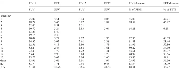
The results of the ROI analysis are demonstrated in Table 3. The mean SUV and the percentage reduction after therapy were markedly higher for 18F-FDG than for
18
F-FET. The average ROI SUV was also correlated with the mitotic index and the grade of differentiation. The
18
F-FDG SUV for the moderately differentiated tumors
Fig. 1. CT and PET examination at initial staging of a 55 years old male suffering from an oropharyngeal tumor, cT4 N2c cM1. Transaxial contrast-enhanced CT and corresponding matched PET slices are presented in thetop three rows, coronal PET slices are demonstrated in thebottom two rows. The contrast-enhanced CT was taken at the same time as the FDG PET. Note the higher tumor-to-background ratio in the FDG PET.
was 16.8 ± 6.4; whereas, for the poorly differentiated tumors it was 11.5 ± 4.1. However, the difference did not reach statistical significance (student’s t test). For 18
F-FET, the corresponding numbers were 4.1 ± 2.3 and 3.3 ±
1.1, respectively, not reaching statistical significance, neither. Furthermore, no significant correlation was found between the mitotic index and the 18F-FDG or 18F-FET uptake.
Fig. 2. CT and PET examination at initial staging of a 80 years old patient suffering from a oropharyngeal tumor, cT4 cN2b cM0. Transaxial low-dose CT and corresponding matched PET slices are presented in the top three rows, coronal PET slices are demonstrated in thebottom two rows. The low-dose CT was acquired at the same time as the FDG PET. While the primary tumor and the lymph node metastases are clearly visible on the FDG scan, all malignant lesions, except the primary, were negative on the FET scan.
Discussion
18F-FDG PET/CT has been shown to be a valuable tool in the evaluation of patients with head and neck cancer. Regarding the staging of nodal disease, recently published data empha-sizes the value of18F-FDG PET/CT at initial staging [9]. There is also a role for 18F-FDG PET/CT in detecting second primaries or distant metastases in patients with advanced HNSCC [10,11]. A major drawback of18F-FDG PET/CT is the relatively low specificity, which leads to a high number of false-positive findings, especially after radiochemotherapy due to the high18F-FDG uptake in inflammatory cells, which may be present after radiotherapy. This is reflected in the reduced specificity for18F-FDG in our study. Therefore, there is a need for a tracer with higher specificity for tumor tissue. Based on earlier studies18F-FET seemed to be a good candidate. Kaim et al. demonstrated in an abscess model in rats that18F-FET does not accumulate in inflammatory cells [4]. Other authors tested the suitability of18F-FET in human tumors: in their first study of18F-FET, Pauleit et al. looked at different tumor types (e.g., lymphoma, adenocarcinoma) originating at different sites of the body (colorectal, pancreatic, lung, ovarian, breast, head, and neck) [6]. In line with our study, they found a superior
specificity but a lower sensitivity of18F-FET for the primary tumor compared with 18F-FDG. They also reported that the tumor-to-background contrast was significantly lower in18 F-FET PET compared with18F-FDG PET. This is in line with our study (Figs.1and2).
After concluding that the use of 18F-FET allows a better distinction between tumors and inflammatory tissues in most cases of SCC [6], Pauleit and his colleagues looked at HNSCC only [7]: the sensitivity of 18F-FDG PET was 93%, the specificity was 79%, and the accuracy was 83% compared with18F-FET PET with a sensitivity of 75%, a specificity of 95%, and an accuracy of 90%. In our study, we found the same pattern, with18F-FDG being more sensitive but less specific than18F-FET in the investigated tumors. As Pauleit stated a potential benefit for18F-FET PET in the follow-up of irradiated patients [7], Balogova et al. looked also at 12 follow-up patients [8]. As in our study, the good specificity of18F-FET was confirmed, but the sensitivity was insufficient. However, they suggested, that in certain cases a wait and see strategy may be chosen for an18F-FDG-positive lesion if it is negative with
18
F-FET. Compared with the previous studies by Pauleit et al. [7] and Balogova et al. [8], it is the strength of this study to confirm the previous findings in a more powerful way by
Table 2. Sensitivities, specificities, and accuracies of both modalities Lesions True positive False negative True negative False positive
Sensitivity (%) Specificity (%) PPV (%) NPV (%) Accuracy (%)
Initial staging FDG primary+lymph nodes 33 4 5 5 89 50 87 56 81 Initial staging FET primary+lymph nodes 26 11 9 1 70 90 96 45 74 Initial staging FDG lymph nodes 20 4 5 5 83 50 80 56 74 Initial staging FET lymph nodes 13 11 9 1 54 90 93 45 65 Follow-up FDG primary+lymph nodes 5 2 15 8 71 65 38 88 67 Follow-up FET primary+lymph nodes 2 5 23 0 29 100 100 82 83 Follow-up FDG lymph nodes 2 2 10 6 50 63 25 83 60 Follow-up FET lymph nodes 2 2 16 0 50 100 100 89 90 pos positive, neg negative
Table 3. SUV values and percentage decrease after therapy in the primary lesion
FDG1 FET1 FDG2 FET2 FDG decrease FET decrease SUV SUV SUV SUV % of FDG1 % of FET1 Patient nr 1 25.07 3.51 3.74 2.03 85.09 42.21 2 18.24 3.45 3.92 1.87 78.52 45.82 3 22.46 8.51 3.51 4 10.70 3.24 3.83 3.04 64.21 6.29 5 13.23 5.40 6 19.16 2.30 7 10.04 3.27 2.77 1.95 72.35 40.39 8 14.35 3.03 2.61 2.38 81.81 21.44 9 12.56 4.15 3.90 1.75 68.95 57.80 10 8.52 2.46 1.68 1.61 80.22 34.30 11 7.44 1.72 1.22 1.31 83.61 23.37 12 6.44 2.90 3.80 1.78 40.92 38.56 13 13.23 3.67 2.14 1.66 83.83 54.81 Mean 13.96 3.66 3.01 1.94 73.95 36.50 SD 5.77 1.71 0.98 0.48 13.54 15.79 COV 41.31 46.75 32.59 24.63 18.31 43.27 SUV standardized uptake value, nr number, SD standard deviation, COV coefficient of variation (SD/mean*100)
intra-individual comparison. The loss of information was therefore kept to a minimum by analysing the data of the same patients undergoing both imaging modalities at initial staging and during follow-up. Another advantage of this study design is the comparison of the variation of the SUVs of18F-FDG and
18
F-FET before/after treatment and therefore the response to treatment.
After all, in daily practice of managing patients with HNSCC it is worse missing a true-positive lesion than detecting a false-positive one since suspicious lesions can always be further evaluated by fine needle puncture or biopsy, whereas missed tumors may grow into incurable disease. After having looked at the size of the false negatives, the range of the lesion size was between 1.2 and 3.5 cm. Given the 0.5 cm resolution of the used PET/CT system, the size of the lesion was therefore not a reason for the false-negative lesions revealed by18F-FET. In addition,
18
F-FDG PET/CT detected a higher number of second malignancies or distant metastases compared with18F-FET PET/CT. In our practice, we will continue to use 18F-FDG PET/CT for advanced disease routinely, as these additional findings heavily affect the management of the patient. Some question may arise regarding the reliability of sensitivities and specificities presented in this paper, due to the relatively low number of patients. However, the number of lesions used to calculate the figures should be sufficient yield a useful estimate. Furthermore, the numbers are in good agreement with earlier published data [8].
As mentioned above, there is another argument favouring
18
F-FDG based on our intra-individual analysis: the SUV measurements in the primary tumor were considerably higher for18F-FDG leading to a higher tumor-to-background ratio (the SUV values at follow-up represent more or less background), which may partially be responsible for the higher sensitivity. Furthermore, 18F-FET demonstrated a much smaller post-therapeutic percentage reduction com-pared with 18F-FDG, reducing its suitability for the evalua-tion of a treatment. A limitaevalua-tion of this study is that not all patients received a contrast-enhanced CT at the time of PET scanning. It is therefore possible, that some tumorous lesions may have been missed which would have increased the false-negative number for18F-FET and18F-FDG. However, given the high sensitivity of18F-FDG and the fact that most patients were scanned twice, the number of such missed tumorous lesions can be expected to be small. The18F-FDG scan was performed 45 min following injection. Other centers use a 60 min uptake time, which potentially increases the tumor-to-background ratio. However, since
the 18F-FDG uptake in the positive lesions was clearly visible at 45 min, it is unlikely that our results would have changed with a 60 min uptake time.
Conclusion
This study delivers further evidence that 18F-FET is no substitute for18F-FDG in the initial staging or follow-up of patients with HNSCC. Although it is more specific, too many malignant lesions are missed due to its lower sensitivity.
Acknowledgments. We thank Dr. R. Hesselmann, Radiopharmacy, USZ for his contribution and Sabine Knöfel for all technical support.
Conflict of Interest Disclosure. The authors declare that they have no conflict of interest.
References
1. Quon A, Fischbein NJ, McDougall IR et al (2007) clinical role of 18F-FDG PET/CT in the management of squamous cell carcinoma of the head and neck and thyroid carcinoma. J Nucl Med 48(Suppl 1):58–67 2. Kao J, Vu HL, Genden EM et al (2009) The diagnostic and prognostic
utility of positron emission tomography/computed tomography- based follow-up after radiotherapy for head and neck cancer. Cancer 115:4586–4594
3. De Boer JR, van der Laan BF, Pruim J et al (2002) Carbon-11 tyrosine PET for visualization and protein synthesis rate assessment of laryngeal and hypopharyngeal carcinomas. Eur J Nucl Med Imaging 29:1182– 1187
4. Kaim AH, Weber B, Kurrer MO et al (2002) (18)F-FDG and (18)F-FET uptake in experimental soft tissue infection. Eur J Nucl Med Mol Imaging 29:648–654
5. Lindholm P, Leskinen S, Lapela M (1998) Carbon-11-methionine uptake in squamous cell head and neck cancer. J Nucl Med 39:1393– 1397
6. Pauleit D, Stoffels G, Schaden W et al (2005) PET with O-(2-18F-Fluoroethyl)-L-Tyrosine in peripheral tumors: first clinical results. J Nucl Med 46:411–416
7. Pauleit D, Zimmermann A, Stoffels G et al (2006) 18F-FET PET compared with 18F-FDG PET and CT in patients with head and neck cancer. J Nucl Med 47:256–261
8. Balogova S, Périé S, Kerrou K et al (2008) Prospective comparison of FDG and FET PET/CT in patients with head and neck squamous cell carcinoma. Mol Imaging Biol 10:364–373
9. Rodrigues RS, Bozza FA, Christian PE et al (2009) Comparison of whole-body PET/CT, dedicated high-resolution head and neck PET/CT, and contrast-enhanced CT in preoperative staging of clinically M0 squamous cell carcinoma of the head and neck. J Nucl Med 50:1205– 1213
10. Strobel K, Haerle SK, Stoeckli SJ et al (2009) Head and neck squamous cell carcinoma (HNSCC)- detection of synchronous primaries with (18) F-FDG-PET/CT. Eur J Nucl Med Mol Imaging 36:919–927
11. Brouwer J, Senft A, de Bree R et al (2006) Screening for distant metastases in patients with head and neck cancer: is there a role for (18) FDG-PET? Oral Oncol 42:275–280