Frank J. Riihli
Clinical perspectives on secular trends
Maciej Henneberg
of intervertebral foramen diameters
in an industrialized European society
Received: 15 July 2003 Revised: 21 November 2003 Accepted: 17 January 2004 Published online: 1 April 2004 0 Springer-Verlag 2004
F. J. Ruhli
Clinical Paleopathology Unit, Orthopedic University Clinic Balgrist and Institute for the History of Medicine, University of Zurich,
Hirschengraben 82, 8001 Zurich, Switzerland F. J. Riihli • M. Henneberg
Department of Anatomical Sciences, The University of Adelaide, Adelaide, Australia F. J. Riihli (®) Institute of Anatomy, University of Zurich, Winterthurerstrasse 190, 8057 Zurich, Switzerland Tel.: +41-1-6355315, Fax: +41-1-6355702, e-mail: frank.ruhli@anatom.unizh.ch
Abstract
Low back pain origins
have been a matter of great
contro-versy. While spinal stenosis is now
radiologically traceable, the
alter-ation of intervertebral foramen is less
clear. The aim of this study was to
assess "secular trends" — alterations
occurring from one generation to the
next — in osseous intervertebral
foram-ina of the major vertebral segments
in an industrialized society, and to
discuss their possible clinical
impli-cation. The macerated "maximum
intervertebral foramen width" and
"intervertebral foramen height" of
all major vertebral levels in 71
non-pathologic Swiss adult skeletons
from the nineteenth and early
twen-tieth century, with known individual
age and sex and similar geographic
and socio-economic background,
were measured by sliding caliper at
validated landmarks. A secular trend
of the increase in "maximum
inter-vertebral foramen width" is found
for most levels, with females
show-ing a more prominent alteration.
Ad-ditionally, the non-pathologic
"maxi-mum intervertebral foramen width"
does not change with respect to
indi-vidual age, nor is a significant side
difference detectable. "Intervertebral
foramen height," hereby defined as
the difference of the dorsal vertebral
body height minus pedicle height,
demonstrates for most levels, and
ei-ther sex, an insignificant negative
secular trend. Neither stature nor
skeletal robustness vary significantly
through time within this particular
sample. The results of this study,
de-spite obvious inadequacies of
meth-ods used, exclude secular narrowing
of the "maximum intervertebral
fora-men width" as the only cause of
radiculopathy or spinal stenosis.
Fur-thermore, we found a mild
insignifi-cant decrease of the clinically more
relevant "intervertebral foramen
height." Nevertheless, the detected
short-time variability of the bony
in-tervertebral foramen, independent of
individual stature, skeletal robustness
or age, argues for an enhanced focus
on the understanding of clinically
relevant changes of spinal
morphol-ogy from generation to generation.
Keywords
Backache • Bone • Pain
Paleopathology • Spinal cord
The alteration of the intervertebral foramen plays a
signif-icant role in the pathophysiology of main back pain
eti-ologies [2, 9, 12, 19, 47, 58], such as radiculopathy or
spi-nal stenosis; therefore, the assessment of the intervertebral
foramen size and shape is worth surveying, especially as
back pain disabilities have risen more quickly than other
pathologies and cause enormous health care costs [32, 38].
Surprisingly, no study exploring a possible secular
alter-ation of the intervertebral foramen in industrialized
soci-eties exists. Since the teardrop-like shape of the superior
and inferior soft tissue parts of the foramen space are
dif-ferent from its osseous outline, clinical or cadaver
mea-surements [7, 25, 40, 52] cannot be easily reproduced on
dry bone specimens, which would form the basis of
com-parative historic studies. Whereas microevolutionary
sur-veys of human spinal pathologies [22, 24], vertebral body
[28], or neural canal size [44, 54] have been published,
es-tablished osteometric schemes [20, 39] do not include
in-tervertebral foramen diameters. Hitherto, the assessment
of the macerated intervertebral foramen was surprisingly
done for just one or two of the three main spinal regions
[1, 6, 10, 13] and explored in one prehistoric sample
(950-1300 A.D.) only [10].
The aim of this article is to present for the first time
"secular" (which derives linguistically from saeculum
meaning century or generation and is commonly used in
biological/anthropological literature to describe short-time
alterations occurring within a few succeeding generations
such as, e.g., the widely found increase in adult body height
in the twentieth century A.D.) trend data of the upper part
of the osseous intervertebral foramen at all major spinal
levels, and to discuss its possible clinical implications.
Materials and methods
Well-preserved adult skeletons were selected due to their excellent historic documentation with known sex and age at death (Table 1). No gross morphologic abnormalities were visible on the speci-mens, which represent in historic terms early-industrialization and current life style ("St. Johann" graveyard, Basel: n=37; born 1772-1837 A.D.; mean age at death 42.4 years; Museum of Nat-ural History, Basel; and rNat-ural cemeteries in Apples, Bex, La Sarraz, St. Prex — Western Switzerland: n=34; 1865-1934 A.D.; mean age at death 57.0 years; Department of Anthropology, University of Geneva). The samples show similar geographic origin — located within approximately 150km in comparable climate at Swiss low plains — and, according to individual death records, similar socio-economic background of mainly low- and middle class occupa-tions, e.g., peasants or industrial workers [15, 16].
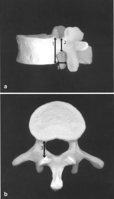
Intervertebral foramen were assessed bilaterally at vertebral level C3, C7, Thl, Th6, Th10, LI, and L5. Osteometric definitions were as follows (Fig. 1):
Maximum intervertebral foramen width: shortest horizontal dis-tance between the inferior posterior edges of the vertebral body and the corresponding anterior aspect of the inferior articular process
Intervertebral foramen height: dorsal vertebral body height [39] minus pedicle height [511
Femur maximum lengths, as a representation of individual stature [56], and femur mid-shaft circumferences [39], both from the right
Table 1 Sample composition (n=71; mean age 49.4 years, stan-dard deviation SD 18.4 years)
Age group No. of No. of
(years) malesa femalesb
20-39 13 15
40-59 14 8
>60 14 7
an,o 41; mean age 51.9 years, SD 18.6 years
bn,.w=30; mean age 45.9 years, SD 18.3 years
Fig. la, b Lateral and caudal view of a vertebra with definition of macerated intervertebral foramen height (1 minus 2) and maxi-mum intervertebral foramen width (3)
side, if preserved, were assessed, too. Femur robustness was de-fined as mid-shaft circumference divided by maximum length [39]. One observer (F.J.R.) performed all vertebral measurements twice with a sliding caliper to the nearest 0.1 mm. Statistical analy-ses were done by using primarily SPSS 11.0 (SPSS, Chicago, Ill.) or Microsoft Excel 2000 (Microsoft, Redmond, Wash.). Linear re-gression was used for data analysis and level of significance was defined at p<0.05, with Bonferroni's correction applied for multi-ple comparisons. When a significant correlation with birth years was found, a secular trend was detected. Paired t test was used for analysis of side differences. The comparison of the measurements of the intervertebral foramen with previous reports [1, 6, 13, 31, 37, 46] as well as their correlation with various osseous spinal land-marks will be addressed elsewhere.
Results
A, C3 left, r=0.77, N=27;Neither stature (Fig. 2A) nor femoral robustness (Fig. 2B)
show a significant secular trend and both sexes show no
significant difference in age at death between the samples
(r for females=0.05, r for males=0.03).
We found a positive secular trend (an increase) for nearly
all selected levels of the maximum intervertebral foramen
width, with females demonstrating mostly a stronger
ten-dency. For females, at level C3 left side (Fig. 3A), and
bi-lateral on Li (rri
ght=0
.
60,
rleft=
0
.
61
) the increase was
sig-nificant. Other secular trends, significant only before
ap-plication of Bonferroni's correction, were in females
bi-lateral at C7 (rri
ght=O.48,
cleft=
0
.
45
), bilateral at Thl (r,;g
ht=
0.39, r1eft=0.52), at Th6 right side (Fig. 3B), and in males at
C7 right side (r=0.37), at Thl bilateral (right side: Fig. 4A;
rleft=
0
.
33
), and at L5 left side (Fig. 4B).
A, femur length, r=0.02, N=62; 520 ¤ 500
¤
•
480 ¤• :¤ • ¤ 460 • •¤
Z•
• • ¤ y440 E • ¤ E¤
¤ ¤ • 420 • 400 ¤ 380•
360 1770 1790 1810 1830 1850 1870 1890 1910 1930 1950 birth year B, femur robusticity, r=0.21, N=68; 0.24 ••
0.23 ¤ . • 0.22 • • • • 021• •
: •
•••
.
•
i•4
•¤
0.19•• •
• • ¤ N 0.18s
0.17¤
0.16177n 179n 1R1n iRnn 1R5n 1R7n 1A9n 19m lsan 1950
11 ¤ ¤ 10 ¤ 9
•
Z
8E
•
• 7¤¤
6•
5 1770 1790 1810 1830 1850 1870 1890 1910 1930 1950 birth year B, Th6 right, r=0.46, N=25; • 15 14 ¤ • • ¤ 13 N • 12 • • E ¤ E ¤ 11 ¤ 10•
9•
8 1770 1790 1810 1830 1850 1870 1890 1910 1930 1950 birth yearFig. 3a, b Female maximum intervertebral foramen widths at se-lected vertebral levels and sides showing significant secular trends
Furthermore, the non-pathologic maximum
interverte-bral foramen width does not change with respect to
indi-vidual age and there is also no significant side difference
(Tables 2, 3).
Intervertebral foramen height shows mostly a negative
insignificant secular trend in both sexes, in females for
level C7 bilateral (r,
;gtt=-0.40, r
lefr=-0.42; Fig. 5A) and in
males for level C7 on the right side (r=—O.35; Fig. 5B).
In-tervertebral foramen height at level Th10 in females and
at ThiO, L1, and L5 in males, all bilateral, demonstrate an
insignificant, positive secular trend.
Discussion
The osteometric assessment of the intervertebral foramen
birth year
is just an approximation of its in vivo size, which
cru-Fig. 2a, b Femur length and femur robustness showing no secularcially depends on dynamic soft tissue components [3, 5, 7,
trend (sexes combined)8, 11, 14, 18, 21, 23, 26, 29, 33, 34, 36, 37, 40, 41, 43, 50,
A, Thl right, r=0.46, N=39; Table 2 Means and t values of maximum intervertebral foramen widths showing no significant side difference
14 Level/side Males Females
¤
¤
F 13
• $ • ¤ • Lj2 ¤ ¤ 1r 11 • •^ E • 10 S • • • 9• •
•
L8
• T7 1770 1790 1810 1830 1850 1870 1890 1910 1930 1950birth year
B, L5 left, r=0.42, N=37; • • 15 • 14 • 13 • 12 11¤ •
•••
•
•
10 E
•
¤
•
•
9
¤ • •
•
•
8
• • 7 6 •5
1770 1790 1810 1830 1850 1870 1890 1910 1930 1950birth year
Fig. 4a, b Male maximum intervertebral foramen widths at se-lected vertebral levels and sides showing significant secular trends
52, 53, 57, 59, 61, 62] such as, for example, the
interver-tebral disc. The interverinterver-tebral foramen is delimited by the
postero-lateral region of the vertebral body at level "X"
and the anterior aspect of the superior articular process at
level "X+1". The thickness of the intervertebral disc
be-tween level "X" and "X+1" contributes substantially, too,
as well as does the angle of the articular process at level
"X+1". Unfortunately, the latter one was beyond the scope
of this study, since no angles of any spinal structures were
assessed. In future projects this should be an essential part
of analysis and could be done with the use of the
geomet-ric morphometgeomet-ric software package "Morphologica"
(de-veloped by O'Higgins et al. at University College London,
UK) [42]. Average intervertebral disc sizes are known
[17, 27, 30, 55] and could, theoretically, be added to the
osseous data. Nevertheless, even this may not represent
the real living dimensions; however, our approach allows
at least reliable comparative studies of the non-pathologic
Mean t n Mean t n C3 left 8.2 - 35 8.3 - 27 C3 right 8.2 0.04 35 8.1 0.40 26 C7 left 10.1 - 38 10.0 - 26 C7 right 10.1 0.15 37 9.8 0.45 24 ThIleft 10.9 - 39 10.3 - 27 THl right 10.6 0.93 39 10.4 0.22 27 Th6 left 13.3 - 32 12.4 - 24 Th6 right 12.7 1.79 32 11.5 1.85 25 Th10 left 13.1 - 38 12.7 - 28 Th10 right 12.9 0.28 37 12.6 0.15 27 Ll left 13.1 - 34 13.6 - 27 L1 right 13.0 0.12 34 13.4 0.33 27 L5 left 10.1 - 37 11.3 - 27 L5 right 9.8 0.64 36 11.3 0.07 27
Table 3 Correlation coefficients of maximum intervertebral fora men widths with individual age showing no significant depen
dency
Level/side Males Females
r
n
r
n
C3 left -0.04 35 -0.05 27 C3 right 0.01 35 0.02 26 C7 left 0.16 38 0.30 26 C7 right 0.14 37 0.24 24 ThIleft 0.12 39 0.19 27 THl right 0.16 39 0.31 27 Th6 left 0.16 32 0.11 24 Th6 right 0.11 32 0.15 25 Th1Oleft 0.03 38 0.12 28 Th10 right -0.01 37 0.19 27 Ll left 0.07 34 -0.05 27 L1 right 0.22 32 0.13 27 L5 left 0.09 37 0.21 27 L5 right -0.15 36 0.14 27macerated intervertebral foramen. Unfortunately, the
os-seous sagittal intervertebral foramen width seems to be
in-dependent from intervertebral disc alterations [8].
In the present study the so-called intervertebral
fora-men height reflects
de factoonly the difference between
the height of the vertebral body at level "X" minus the
height of the pedicle at level "X" and does not represent in
any form the influence of the intervertebral disc, which may
actually undergo secular trends of any sort, too. Finally,
for the spines studied herein at least major taphonomic
al-terations seem to be negligible.
Although previous reports detected no secular trend of
the foramen size by focusing on influences of prehistoric
A, Females, r=-0.40, N=28; • 9.5 ' 9 • ' 8.5 • • 8 • . 7.5 • 7 ' • ' 6.5
•
• . 6 • • 5.5 • • 5 4.5 1770 1790 1810 1830 1850 1870 1890 1910 1930 1950 birth year B, Males, r=-0.35, N=38 • 10 • 9.5 • • • • 9 8.5 • 8 ¤ ¤• ¤ ¤• 7.5E
'•
. 7 • • • 6.5 • 6•
• 5.5 ' 5 1770 1790 1810 1830 1850 1870 1890 1910 1930 1950 birth yearFig. 5a, b Intervertebral foramen height at vertebral level C7, right side, showing insignificant negative secular trends
lifestyle changes [10], the samples from industrialized
so-cieties presented herein demonstrate a mild secular
alter-ation of the intervertebral foramen even without an
appar-ent major shift in culture. Possible causes of these
find-ings may be, as pointed out in previous microevolutionary
spinal studies [22, 24, 48], of genetic or environmental
nature, e.g., nutrition. Additionally, we found no
correla-tion between the upper part of the osseous maximum
in-tervertebral foramen width and individual age at death,
unlike previous clinical reports [25]. Changes in general
bony robustness, as expressed by femoral robustness rather
than stature, could partially explain secular alterations of
the intervertebral foramen size. This is not the case in this
particular sample showing an insignificant positive
in-crease in robustness, which would likely oppose a secular
enlargement of the mostly bone-enclosed foramen.
The mild secular trend of the intervertebral foramen
di-ameters may not correlate with clinical presentation, since
previous studies focusing on possible links between
al-tered spinal neural pathways and symptoms report
equiv-ocally [4, 21, 25, 60] and dynamic and day-time-related
alterations of its size are known, too [18]. Furthermore,
this study found that stronger secular trends in females
lack an evident interpretation and need further exploration,
especially given that in a recent sample intervertebral
foramen and spinal canal size showed mostly no
signifi-cant sex difference [13, 35]. Apparently, the upper part of
the osseous intervertebral foramen becomes wider and
lower in cranio-caudal direction towards most modem times,
independent of factors such as stature, skeletal robustness,
or age. The question as to whether this reshaping was
compensated by
an
adaptation of, for example, the
om-nipresent fat tissue and blood vessels [23, 53] remains
unanswered. If not, this reforming could prompt clinical
symptoms, since it is known that the dorsal root ganglion
and the spinal nerve occupy supero-lateral and inferior
sections, respectively, of the root canal [34, 53]; however,
the sagittal and vertical intervertebral foramen diameters
at least reflect well its overall dimension [8]. Furthermore,
the assessment of the intervertebral foramen dimensions
by caliper and on dried vertebrae produces less
inter-ob-server variability and lower standard errors than other
methods using fresh cadaveric samples with soft tissues
still present [8].
Conclusion
Our results contradict a secular narrowing of the
maxi-mum intervertebral foramen widths as a possible
micro-evolutionary precondition of increasing incidence of
ra-diculopathy or spinal stenosis. Nevertheless, the presented
data, though collected using a method that has serious
lim-itations, reveal a remarkable secular variability of the
up-per part of the osseous outline of the intervertebral
fora-men, independent of individual age, stature, and
robust-ness. Our challenging preliminary results will hopefully
stimulate the debate, which assesses spinal morphology
changes by using a broad historic perspective [6, 10, 22,
24, 28, 45, 49, 54] and may represent a pilot approach for
further investigations on changing clinical entities in
mod-em societies.
Acknowledgement This study was supported by an International Postgraduate Research Scholarship of the University of Adelaide awarded to F.J.R.
References
1. Amonoo-Kuofi HS (1985) The sagittal diameter of the lumbar vertebral canal in normal adult Nigerians. J Anat 140: 69-78
2. Arnoldi CC, Brodsky AE, Cauchoix J et al. (1976) Lumbar spinal stenosis and nerve root entrapment syndromes. Definition and classification. Clin Or-thop 115:4-5
3. Bailey P, Casamajor L (1911) Osteo-arthritis of the spine as a cause of com-pression of the spinal cord and its roots. J Nery Ment Dis 38:588-609 4. Boden SD, Davis DO, Dina TS,
Patronas NJ, Wiesel SW (1990) Ab-normal magnetic-resonance scans of the lumbar spine in asymptomatic subjects. A prospective investigation. J Bone Joint Surg Am 72:403-408 5. Bose K, Balasubramaniam P (1984)
Nerve root canals of the lumbar spine. Spine 9:16-18
6. Boszczyk BM, Boszczyk AA, Putz R (2001) Comparative and functional anatomy of the mammalian lumbar spine. Anat Rec 264:157-168 7. Chung SS, Lee CS, Kim SH, Chung
MW, Alm JM (2000) Effect of low back posture on the morphology of the spinal canal. Skeletal Radiol 29:217-223
8. Cinotti G, Santis P de, Nofroni I, Postacchini F (2002) Stenosis of lum-bar intervertebral foramen. Spine 27: 223-229
9. Ciric I, Mikhael MA, Tarkington JA, Vick NA (1980) The lateral recess syn-drome. A variant of spinal stenosis. J Neurosurg 53:433-443
10. Clark GA, Panjabi MM, Wetzel FT (1985) Can infant malnutrition cause adult vertebral stenosis? Spine 10:
165-170
11. Crock HV (1981) Normal and patho-logical anatomy of the lumbar spinal nerve root canals. J Bone Joint Surg Br 63B:487-490
12. Dorwart RH, Vogler JB, Helms CA (1983) Spinal stenosis. Radiol Clin North Am 21:301-325
13. Ebraheim NA, An HS, Xu R, Ahmad M, Yeasting RA (1996) The quantita-tive anatomy of the cervical nerve root groove and the intervertebral foramen. Spine 21:1619-1623
14. Epstein JA, Epstein BS, Lavine L (1962) Nerve root compression associated with narrowing of the lumbar spinal canal. J Neurol Neurosurg Psychiaty 25:165-176
15. Etter HF (1988) Der aussere St. Johann Gottesacker in Basel. Was ein Spital-friedhof des 19. Jahrhunderts verrat. CH-Forschung 11:23-28
16. Etter HF, Lorcher M (1993) Armut, Krankheit, Tod im fruhindustriellen Basel. Cratander, Basel
17. Frobin W, Brinckmann P, Biggemann M (1997) Objektive Messung der Hohe lumbaler Bandscheiben aus seitlichen Rontgen-Ubersichtsaufnahmen. Z Or-thop Grenzgeb 135:394-402
18. Fujiwara A, An HS, Lim TH, Haughton VM (2001) Morphologic changes in the lumbar intervertebral foramen due to flexion-extension, lat-eral bending, and axial rotation: an in vitro anatomic and biomechanical study. Spine 26:876-882
19. Gaskill MF, Lukin R, Wiot JG (1991) Lumbar disc disease and stenosis. Radiol Clin North Am 29:753-764 20. Hasebe K (1913) Die Wirbelsaule der
Japaner. Z Morphol Anthropol 15:259-380
21. Hasegawa T, An HS, Haughton VM, Nowicki BH (1995) Lumbar foraminal stenosis: critical heights of the inter-vertebral discs and foramina. A cryo-microtome study in cadavera. J Bone Joint Surg Am 77:32-38
22. Henneberg RJ, Henneberg M (1999) Variation in the closure of the sacral canal in the skeletal sample from Pom-peii, Italy, 79 A.D. Persp Hum Biol 4:177-188
23. Hoyland JA, Freemont AJ, Jayson MI (1989) Intervertebral foramen venous obstruction. A cause of periradicular fibrosis? Spine 14:558-568
24. Hukuda S, Inoue K, Nakai M,
Katayama K (2000) Did ossification of the posterior longitudinal ligament of the spine evolve in the modern period? A paleopathologic study of ancient hu-man skeletons in Japan. J Rheumatol 27:2647-2657
25. Humphreys SC, Hodges SD,
Patwardhan A, Eck JC, Covington LA, Sartori M (1998) The natural history of the cervical foramen in symptomatic and asymptomatic individuals aged 20-60 years as measured by magnetic resonance imaging. A descriptive ap-proach. Spine 23:2180-2184
26. Inufusa A, An HS, Lim TH, Hasegawa T, Haughton VM, Nowicki BH (1996) Anatomic changes of the spinal canal and intervertebral foramen associated with flexion-extension movement. Spine 21:2412-2420
27. Jacobi H (1927) Messungen der Brust-und oberen Lendenwirbelsaule unter Beriicksichtigung der Veranderungen an Bandscheiben and Wirbelkorpern. Beitr Pathol Anat 78:303-314 28. Jankauskas R (1994) Variability of
vertebral column measurements in Lithuanian paleopopulation. Int J An-thropol 9:137-151
29. Jones RA, Thomson JL (1968) The narrow lumbar canal. A clinical and ra-diological review. J Bone Joint Surg Br 50:595-605
30. Kandziora F, Pflugmacher R, Scholz M et al. (2001) Comparison between sheep and human cervical spines: an anatomic, radiographic, bone mineral density, and biomechanical study. Spine 26:1028-1037
31. Karaikovic EE, Daubs MD, Madsen RW, Gaines RW Jr. (1997) Morpho-logic characteristics of human cervical pedicles. Spine 22:493-500
32. Kelsey JL, White AA (1980) Epidemi-ology and impact of low-back pain. Spine 5:133-142
33. Larmon WA (1944) An anatomic study of the lumbosacral region in relation to low back pain and sciatica. Ann Surg
119:892-896
34. Lee CK, Rauschning W, Glenn W (1988) Lateral lumbar spinal canal stenosis: classification, pathologic anatomy and surgical decompression. Spine 13:313-320
35. Lee HM, Kim NH, Kim HJ, Chung IH (1995) Morphometric study of the lum-bar spinal canal in the Korean popula-tion. Spine 20:1679-1684
36. Lu J, Ebraheim NA, Huntoon M, Haman SP (2000) Cervical interverte-bral disc space narrowing and size of intervertebral foramina. Clin Orthop 370:259-264
37. Magnuson PB (1944) Differential diag-nosis of causes of pain in the lower back accompanied by sciatic Pain. Ann Surg 119:878-891
38. Maniadakis N, Gray A (2000) The eco-nomic burden of back pain in the UK. Pain 84:95-103
39. Martin R (1928) Lehrbuch der Anthro-pologie. Gustav Fischer, Jena
40. Mayoux-Benhamou MA, Revel M, Aaron C, Chomette G, Amor B (1989) A morphometric study of the lumbar foramen. Influence of flexion-exten-sion movements and of isolated disc collapse. Surg Radiol Anat 11:97-102 41. Nowicki BH, Haughton VM, Schmidt T et al. (1996) Occult lumbar lateral spinal stenosis in neural foramina sub-jected to physiologic loading. Am J Neuroradiol 17:1605-1614
42. Pan R, Wei F, Li M (2003) Craniofa-cial variation of the Chinese macaques explored with morphologica. J Mor-phol 256:342-348
43. Panjabi MM, Takata K, Goel VK (1983) Kinematics of lumbar interver-tebral foramen. Spine 8:348-357 44. Piontek J, Budzynska J (1972)
Zmien-nosc Cech Metrycznych Kregoslupa. Przeglad Antropol 38:17-26
45. Porter RW, Pavitt D (1987) The verte-bral canal: I. Nutrition and develop-ment, an archaeological study. Spine
12:901-906
46. Postacchini F, Ripani M, Carpano S (1983) Morphometry of the lumbar vertebrae. An anatomic study in two caucasoid ethnic groups. Clin Orthop
172:296-303
47. Resnick D (1985) Degenerative dis-eases of the vertebral column. Radiol-ogy 156:3-14
48. Rothschild BM, Rothschild C (1996) Is there an epidemic/epizootic of spondyloarthropathy in baboons? J Med Primatol 25:69-70
49. Riihli FJ, Schultz M, Henneberg M (2002) Microevolution of the central European human vertebral column since the Neolithic: preliminary osteo-metric assessment and interpretations. Am J Phys Anthropol (Suppl) 34:134-135
50. Schmid MR, Stucki G, Duewell S. Wildermuth S, Romanowski B, Hodler J (1999) Changes in cross-sectional measurements of the spinal canal and intervertebral foramina as a function of body position: in vivo studies on an open-configuration MR system. AJR 172:1095-1102
51. Shapiro L (1993) Evaluation of "unique" aspects of human vertebral bodies and pedicles with a considera-tion of Australopithecus africanus. J Hum Evol 25:433-470
52. Stephens MM, Evans JH, O'Brien JP (1991) Lumbar intervertebral fora-mens. An in vitro study of their shape in relation to intervertebral disc pathol-ogy. Spine 16:525-529
53. Swanberg H (1915) The intervertebral foramina in man. Med Rec 87:176-180 54. Tatarek NE (2001) Variation in the
lumbar neural canal. Am J Phys An-thropol (Suppl) 32:147
55. Tribus CB, Belanger T (2001) The vas-cular anatomy anterior to the L5—S 1 disk space. Spine 26:1205-1208 56. Trotter M, Gleser GC (1952)
Estima-tion of stature from long bones of American whites and negroes. Am J Phys Anthropol 10:463-514
57. Vanderlinden RG (1984) Subarticular entrapment of the dorsal root ganglion as a cause of sciatic pain. Spine 9:19-22
58. Verbiest H (1954) A radicular syn-drome from developmental narrowing of the lumbar vertebral canal. J Bone Joint Surg Br 36:230-237
59. Vital JM, Lavignolle B, Grenier N, Rouais F, Malgat R, Senegas J (1983) Anatomy of the lumbar radicular canal. Anat Clin 5:141-151
60. Wiesel SW, Tsourmas N, Feffer HL, Citrin CM, Patronas N (1984) A study of computer-assisted tomography. I. The incidence of positive CAT scans in an asymptomatic group of patients. Spine 9:549-551
61. Yoo JU, Zou D, Edwards WT, Bayley J, Yuan HA (1992) Effect of cervical spine motion on the neuroforaminal dimensions of human cervical spine. Spine 17:1131-1136
62. Yoshida M, Shima K, Taniguchi Y, Tamaki T, Tanaka T (1992) Hypertro-phied ligamentum flavum in lumbar spinal canal stenosis. Pathogenesis and morphologic and immunohistochemi-cal observation. Spine 17:1353-1360