D
E
V
E
LO
P
M
E
N
T
INTRODUCTIONThe adrenal cortex and gonad are the main steroid-producing tissues in mammals. Although they produce different steroid hormones in the adult, histological and molecular studies have shown that they share a common precursor during development. Expression analysis of the nuclear hormone receptor Sf-1 (steroidogenic factor 1; also known as Nr5a1), an essential factor in the development of both gonads and adrenals (Luo et al., 1994; Val et al., 2003), suggests that these two tissues originate from a so-called adrenogonadal primordium (AGP) present at embryonic day 9 (E9.0) in mouse (Hatano et al., 1996; Ikeda et al., 1994). This is consistent with the absence of both adrenal glands and gonads in mice mutant for the transcription factor Wt1, although Wt1 was not found in the adrenal glands but was expressed in the coelomic epithelium of the urogenital region at E9.0 (Moore et al., 1999; Moore et al., 1998; Vidal and Schedl, 2000). As development proceeds, the adrenal cortical primordium separates from the gonadal primordium in the rostral region of the AGP and undergoes an adrenal cortical-specific differentiation programme. This is characterised by expression of adrenal steroidogenic enzymes, zonation of their expression and establishment of hormonal sensitivity (Morohashi, 1997). The innermost region of the adrenal gland, the medulla, is composed of chromaffin cells that derive from neural crest cells that invade the adrenal cortical primordium during development.
Most mutant mouse models in which a defect in adrenal development is observed also show impaired gonad development (Else and Hammer, 2005). This is the case for
genetic ablation of factors such as Sf-1 (Luo et al., 1994), Pbx1 (Schnabel et al., 2003), M33 (Cbx2 – Mouse Genome Informatics) (Katoh-Fukui et al., 2005) and Odd1 (Osr1 – Mouse Genome Informatics) (Wang et al., 2005). Although these observations conform to the idea of a common developmental origin of these two tissues, these models do not provide insight into the developmental and molecular mechanisms that underlie the specification of one or other primordium from the AGP.
Interestingly, mice mutant for the transcription co-factor Cited2 were reported to have no adrenal gland but apparently normal gonads at E17.5 (Bamforth et al., 2001), suggesting that Cited2 might participate in adrenal development. Studies have shown that Cited2 is also involved in the development of the heart and neural tube and is implicated in left-right patterning through interaction with the transcription factor Tfap2 (Tcfap2a – Mouse Genome Informatics) on the Pitx2 promoter (Bamforth et al., 2001; Bamforth et al., 2004; Bragança et al., 2003; Barbera et al., 2002; Weninger et al., 2005; Yin et al., 2002). The Cited2 protein lacks DNA-binding properties and has been shown to function as a transcription coactivator for the nuclear hormone receptors Ppar␣ and Ppar␥ in vitro, although the physiological significance of such an interaction is still unclear (Tien et al., 2004). Cited2 has also been proposed to repress Hif1␣ transcription activity by competing for the CH1-binding domain on CBP/p300 (Crebbp – Mouse Genome Informatics) (Bhattacharya et al., 1999; Freedman et al., 2003).
Here, we have investigated the role of Cited2 in adrenal development. We provide molecular and genetic evidence that Cited2 acts in the AGP, interacting with the transcription factor Wt1 to promote adrenal cortical primordium development from the AGP. Analysis of Sf-1 expression in Cited2–/– and Cited+/– Wt1+/– embryos by quantitative real-time PCR indicates a model in which the interaction between Cited2 and Wt1 results in an increase in the expression of Sf-1 above a threshold required for adrenal cortical development.
Adrenal development is initiated by Cited2 and Wt1 through
modulation of Sf-1 dosage
Pierre Val1, Juan-Pedro Martinez-Barbera2and Amanda Swain1,*
It has been proposed that the mammalian adrenal cortex and gonad are derived from the same primordium present during early urogenital development. Molecular pathways involved in the differentiation of the adrenal cortex from the adrenogonadal primordium (AGP) have yet to be determined. Here we show in mice that the transcription co-factor Cited2 is required for the specification of the adrenal cortex from the AGP. We present genetic and molecular evidence demonstrating that Cited2 interacts with the transcription factor Wt1 to stimulate expression of the nuclear hormone receptor Sf-1 (Nr5a1) in the AGP prior to the separation between gonad and adrenal cortex. We show a direct correlation between the expression levels of Sf-1 in the AGP and the defects in adrenal development found in mice with different Cited2 and Wt1 mutant backgrounds. Analysis of embryos heterozygous for mutations in both Sf-1 and Cited2 confirmed that these genes act in the same pathway during adrenal development. Our studies reveal a regulatory mechanism in which Cited2 acts as a Wt1 co-factor to increase, at a critical time in embryogenesis, the levels of the essential transcription factor Sf-1 in the AGP above the threshold required to determine adrenal development. These results highlight the importance of transcription factor dosage in organogenesis and the role of transcription co-factors such as Cited2 in determining the levels of these factors.
KEY WORDS: Adrenal development, Gene dosage, Organogenesis, Mouse
Development 134, 2349-2358 (2007) doi:10.1242/dev.004390
1Section of Gene Function and Regulation, Institute of Cancer Research, 237 Fulham
Road, London SW3 6JB, UK. 2Neural Development Unit, Institute of Child Health,
University College London, London, UK.
*Author for correspondence (e-mail: amanda.swain@icr.ac.uk)
D
E
V
E
LO
P
M
E
N
T
MATERIALS AND METHODS
Whole-mount in situ hybridisation (WISH)
Single and double WISH experiments were carried out on an in situ processor (Intavis In Situ Pro) as described by Val et al. (Val et al., 2006). Probes for Cyp11a1 (P450scc), Sf-1, Sox9, Wnt4 and Dax1 have been described previously (Ikeda et al., 1996; Kent et al., 1996; Swain et al., 1996; Vainio et al., 1999). The Cited2 probe was synthesized from a fragment of Cited2 cDNA (positions 4 to 561) subcloned from AA023157 (Image clone 456049) into pT7T3-PacI. Probes for Lim1, Wt1, Hoxb9 and tyrosine hydroxylase (Th) were synthesized directly from PCR templates, which included the sequences encoding the T7 RNA polymerase promoter. PCR fragments were made from MF1 mouse genomic DNA (Lim1, Wt1, Hoxb9) or E18.5 brain cDNAs (Th) using the following primers (F, forward; R, reverse): Lim1, 5⬘-CACTGCAAGAAACCGGCAATG-3⬘ (F) and
5⬘-GTAATACGACTCACTATAGGGTTGCAGCCCGCACAGTGC-3⬘ (R);
Wt1, 5⬘TTTGAGGGGTCCTGACACGG3⬘ (F) and 5⬘GTAATAC -GACTCACTATAGGGCTGGCCTTCCAGGTTTCCAG-3⬘ (R); Hoxb9,
5⬘-CCACTCTCCTGGTGTTAAGA-3⬘ (F) and 5⬘GTAATACGAC
-TCACTATAGGGAGACTTGTCTTGCTGGTG-3⬘ (R); and for Th, 5⬘CCGTGCAGCCCTACCAAG3⬘ (F) and 5⬘GTAATACGACT CA
-CTATAGGGGTAGAATACAGCATGAAGGGC-3⬘ (R). These PCR
fragments were isolated and used as templates in a DIG labelling reaction using T7 RNA polymerase (Val et al., 2006).
Mice
Cited2 mutant mice have been described previously and were bred on a C57/Bl6 strain background (Barbera et al., 2002). Unless otherwise stated, the phenotype of Cited2+/–mice was identical to Cited2+/+mice and
therefore these mice were sometimes used as wild-type control samples. Wt1 mutant mice were kindly provided by Dr Andreas Schedl (INSERM U470, Nice, France) and were initially created by Kreidberg et al. (Kreidberg et al., 1993). Sf-1 mutant mice were kindly provided by Dr Lovell-Badge (NIMR, London, UK) and were initially created by Luo et al. (Luo et al., 1994). Both lines were bred on a mixed strain background. The expression studies were performed on embryonic tissue from MF1 mice. For early development, staging of the embryos was determined by counting the number of tail somites (ts). E10, E10.5, E11, E11.5 and E12 were considered equivalent to approximately 5ts, 10ts, 14ts, 18ts and 21ts, respectively. Older embryos were staged by limb morphology.
Immunohistochemistry
Laminin was detected on cryosections (10 m) or on whole-mount urogenital regions using anti-laminin antibody (1/400, L9393, Sigma). Secondary fluorescent antibodies were obtained from Molecular Probes and used at 1/1000 dilution.
Apoptosis and proliferation
Apoptosis was detected by incubating dissected embryos in the presence of Lysotracker Red (Molecular Probes) (Zucker et al., 1999) according to the protocol of Schmahl and Capel (Schmahl and Capel, 2003). For proliferation analysis, pregnant mice were injected with Bromodeoxyuridine (BrdU) (50 mg/kg) 2 hours before culling. Incorporated BrdU was detected on cryosections after acid treatment using a 1/50 dilution of a monoclonal anti-BrdU antibody (clone BMC 9318, Roche). For double immunohistochemistry with Sf-1 and BrdU antibodies, sections were incubated with Sf-1 antibody (rabbit anti-Sf-1, 1/4000, a kind gift from K. Morohashi, NIBB, Okasaki, Japan) prior to acid treatment. After washes, the primary antibody was detected with the Vectastain ABC Amplification Kit (Vector Labs) according to the manufacturer’s instructions. Peroxidase activity was detected with the fluorescent substrate TSA (Pierce) and sections were then processed through acid treatment and BrdU detection. Immunoprecipitation
Total proteins (500 g) from transfected 293A cells or nuclear proteins from M15 cells were immunoprecipitated overnight with anti-Wt1 antibody (1.6 g, Wt1 C-19, SC-192, Santa Cruz), anti--galactosidase antibody (1.6 g, AB1211, Chemicon), non-immune rabbit IgG (1.6 g, SC-2027, Santa Cruz), anti-Sf-1 antibody (1.6 g, 07-618, Upstate/Millipore) or anti-Cited2 antibody (10 g, clone JA22, Abcam). Immunoprecipitates were collected
on protein A- or protein G-sepharose. After four washes in PBS in the presence of 0.1% Triton X-100, protease inhibitors (Complete Mix, Roche) and 150 mM NaCl, immunocomplexes were eluted in Laemmli buffer and analysed by western blotting.
GST pull-down
GST fusions of the mouse Wt1(–/–) protein were provided by Dr Jonathan Licht and are described in Johnstone et al. (Johnstone et al., 1996). Recombinant proteins were prepared and attached to glutathione-sepharose 4B beads (GE Healthcare) according to a standard protocol. Mouse Cited2 protein (AA023157) was translated in vitro in the presence of 35
S-methionine using the TNT T7/T3-Coupled Reticulocyte Lysate Kit (Promega). 35S-labelled Cited2 protein was incubated with the beads in
binding buffer (20 mM Hepes pH 7.4, 100 mM NaCl, 0.1% Triton X-100, 0.2 mg/ml BSA, 2 mM dithiothreitol, 10 M ZnCl2, 2 mM EDTA, 1 mM
PMSF, 1⫻complete protease inhibitor mix) for 2 hours at 4°C. Glutathione-sepharose beads were then washed six times in PBS in the presence of 0.2% Triton X-100 and 150 mM NaCl, and resuspended in Laemmli buffer. Bound proteins were recovered by boiling for 5 minutes and subjected to SDS-PAGE analysis. After drying the gel, proteins were visualised by Coomassie Blue staining and Cited2 was detected by autoradiography.
Adrenal surface measurement
After WISH for Sf-1 (Fig. 5) or Hoxb9 (Fig. 7), images of all the samples were taken at the same magnification, processed into ImageJ software and the surface of the adrenal gland was measured using the ‘measure’ tool. Experiments were performed on at least three independent samples and results are expressed as a percentage of the average surface of the wild-type adrenal±s.d.
Real-time PCR
For real-time RT-PCR, E10.5 urogenital regions (8-11ts) or E11.5 gonads were microdissected, based on the in situ expression pattern of the genes analysed: at E10.5, the urogenital region was dissected away from the rest of the embryo and at E11.5 the gonad and mesonephroi were dissected away from the dorsal aorta and adrenal region. Total mRNAs were extracted using the RNAqueous-Micro Kit (Ambion). cDNA synthesis was primed with oligo(dT) and performed with Superscript II reverse transcriptase (Invitrogen). Sf-1, Wt1, Sox9, Cyp11a1 and Hprt1 mRNA accumulation was quantified by real-time PCR using the Taqman system (Applied Biosystems). The premade and pretested primer/probe sets that were used were: Sf-1, Mm00446826_m1; Wt1, Mm00460570_m1; Sox9,
Mm00448840_m1; Cyp11a1, Mm00490735_m1; Hprt1,
Mm00446968_m1. Relative mRNA accumulation was determined by the ⌬⌬Ct method in which Hprt1 was used as the normaliser and one of the wild-type samples was used as the baseline value. Results are expressed as a percentage of the mRNA accumulation in wild-type embryos and represent the mean values obtained with at least four urogenital regions or four gonads for each genotype±s.d.
Cell transfections
C2C12 cells were maintained in DMEM with 10% foetal calf serum and passaged at 50-60% confluence to prevent differentiation. Cells were transfected with JetPEI (Autogen Bioclear) 20 hours after seeding in six-well plates (Falcon) at a density of 2⫻105cells per well. Transfections were
performed with 1 g of pGL3-Sf1P, or the Wt1-responsive element mutant pGL3-Sf1Pm (Wilhelm and Englert, 2002), or pGL3-amphiregulin (Lee et al., 1999) reporter genes and 50 ng of RcCMV-Wt1(–/–) (Kim et al., 1999) alone or in combination with 50 ng of pCMV5-Cited2 (Cited2 sequence derived from IMAGE clone AA023157 and cloned into the EcoRI/HindIII sites of pCMV5). Amounts of DNA and constitutive promoter were kept constant by addition of pCMV5 empty vector. Luciferase activity was assayed 24 hours after transfection using Genofax A reagent (Yelen, France) and was normalised to the activity of 2 ng of co-transfected pRLSV40 vector coding for renilla. Renilla activity was assayed with Genofax C reagent (Yelen, France). Each experiment was performed in triplicate and repeated at least three times. All data are expressed as mean±s.d. Western blotting confirmed that Wt1 expression levels were not affected by Cited2 co-transfection (data not shown).
D
E
V
E
LO
P
M
E
N
T
RESULTSCited2 is required for adrenal cortical cell specification
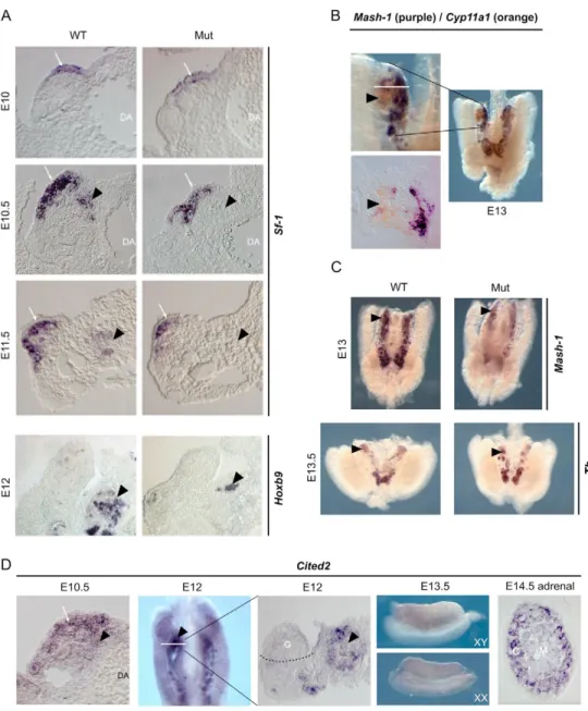
In their study of Cited2–/–embryos, Bamforth et al. showed an absence of adrenal gland at E17.5 (Bamforth et al., 2001). To study the early stages of adrenal development in these mice, we analysed the expression of Sf-1, which marks adrenal cortical cells at all stages of development, by whole-mount in situ hybridisation (WISH) followed by sections of the urogenital region (Fig. 1A). At E10.5, when the adrenal primordium is first apparent in wild-type embryos, we observed a lack of Sf-1 expression in this region. This lack of expression was also seen at later stages and for other adrenal cortical markers including Cyp11a1, Dax1 (Nr0b1 – Mouse Genome Informatics) and Wnt4 (Fig. 2 and data not shown). We also investigated adrenal development by WISH for Hoxb9, a recently identified marker of adrenal steroidogenic cells that is not known to be regulated by Sf-1 (Zubair et al., 2006). At E12, the adrenal primordium appeared as a distinct and organised Hoxb9 expression-positive structure in the wild-type embryo. By contrast, such organisation was not apparent in the Cited2–/–mutant embryo, although a few Hoxb9 expression-positive cells were found in the
presumptive adrenal area. The developing gonad of the mutant embryo showed Sf-1 expression, although the levels appeared to be decreased at E10-11 (Fig. 1A). These studies show that development of the adrenal cortical primordium is markedly impaired in Cited2–/–
embryos.
Whereas the outer cortical layers of the adrenal gland are thought to be derived from the AGP, the inner medulla is derived from neural crest cells that invade the cortical primordium during development and differentiate into chromaffin cells. Interestingly, Bamforth et al. showed that Cited2 mutant embryos had a defect in neural crest cell development. Because of the known interactions between the adrenal cortex and medulla (Ehrhart-Bornstein et al., 1998), they hypothesised that the neural crest phenotype could account for impaired adrenal cortical development (Bamforth et al., 2001). However, previous reports (Bland et al., 2004; Gut et al., 2005) and our observations showed that although neural crest cells were found in the urogenital region at E11.5 and E12.5, it was not until E13 that these cells were found near the presumptive adrenal area (Fig. 1B). This late stage of neural crest cell migration indicated that the adrenal cortical phenotype in the mutant mice was unlikely to result from a neural crest developmental defect. Analysis of expression of
Fig. 1. Cited2 acts in the
adrenogonadal primordium (AGP) to promote adrenal cortex
development. (A) Adrenal cortex differentiation is markedly impaired in Cited2 mutant mouse embryos. Urogenital regions from wild-type (WT) or mutant embryos (Mut) were subjected to whole-mount in situ hybridisation (WISH) for Sf-1 or Hoxb9 expression and sectioned. Arrowhead, position of the adrenal anlagen; arrow, Sf-1 expression in the developing gonad; DA, dorsal aorta. (B) Neural crest migration to the adrenal anlagen occurs at E13. Neural crest migration to the adrenal anlagen (arrowhead) was evaluated by double WISH for Cyp11a1 (orange) and Mash1 (purple). The lower left panel shows a section (taken at the line in the top panel) through the adrenal anlagen. (C) Neural crest migration is only mildly affected in Cited2 mutant embryos. WISH for Mash1 or tyrosine hydroxylase (Th) at E13.5 shows that neural crest cells still migrate to the presumptive adrenal area (arrowhead) of mutant embryos, albeit in reduced numbers. (D) Cited2 is expressed in the AGP and the adrenal cortex anlagen. Cited2 mRNA is expressed in the genital ridge (arrow) and the presumptive adrenal area (arrowhead) at E10.5. At E12 (left panel shows WISH; right panel shows section of WISH sample at the white line), expression in the gonad (G) is barely detectable but Cited2 is present in the adrenal anlagen (arrowhead). At E13.5, Cited2 is faintly expressed in the gonads of both sexes. At E14.5, Cited2 is strongly expressed in the cells of the adrenal cortex (C) and weakly expressed in the medulla (M).
D
E
V
E
LO
P
M
E
N
T
the neural crest marker Mash1 (Ascl1 – Mouse Genome Informatics) in the E13 urogenital region of Cited2–/–embryos showed that neural crest cells were still present in the urogenital region, although the numbers were reduced. This observation was confirmed by WISH for the chromaffin cell marker tyrosine hydroxylase (Th) at E13.5 (Fig. 1C). Altogether, this indicated that the adrenal cortical development failure was independent of the neural crest phenotype. Consistent with this, Erbb3–/–mice, which are completely devoid of neural crest cells in the urogenital region, show development of an adrenal cortex, although it becomes disorganised at later stages (Britsch et al., 1998).
Expression of Cited2 in the urogenital region To investigate whether Cited2 could play a direct role in the differentiation of the AGP, we analysed its expression in the urogenital region by WISH at various developmental stages. At E10, Cited2 was expressed in the coelomic epithelium and in cells in the underlying nephrogenic mesenchyme. Cited2 was also expressed in mesonephric tubules (data not shown). At E10.5, just before the adrenal primordium has separated from the AGP, Cited2 expression was decreased in the coelomic epithelium and increased in the
underlying nephrogenic mesenchyme (Fig. 1D). At E12, gonads and adrenals were clearly separated and Cited2 was highly expressed in the adrenal primordium and barely detectable in the coelomic epithelium or the gonad. At E13.5 and later, Cited2 remained highly expressed in the adrenal cortical cells and showed low-level expression in the adrenal medulla and gonads of both sexes (Fig. 1D).
Gonad phenotype in Cited2–/–embryos
Because of the early adrenal phenotype in Cited2–/–embryos and the common developmental origin of the gonad and adrenal cortex, we analysed the differentiation of the gonad in these mutant embryos by WISH for expression of gonadal markers (Fig. 2A). Expression levels of Sox9, a Sertoli cell marker and of Cyp11a1, a steroidogenic cell marker, were decreased in Cited2–/–XY gonads at E11.5, but their expression levels were normal at E13.5. Sf-1 expression levels were mildly decreased in Cited2–/–gonads at E11.5 and appeared normal at E13.5. These in situ observations were confirmed by real-time RT-PCR on E11.5 gonads (Fig. 2B). Expression of Sox9 and Cyp11a1 was decreased in the mutant to 37% and 54% of wild-type levels, respectively. Sf-1 expression was also decreased in the mutant, albeit to a lesser extent (64% of wild-type levels). A similar decrease in Cyp11a1 and Sf-1 was observed in the XX mutant E11.5 gonad (data not shown). These data show that early differentiation of the gonads in Cited2–/–embryos is impaired, but that at later stages the gonads recover to develop normally. This is consistent with the higher levels of Cited2 expression observed at the early stages of AGP development, which decreased as gonad development occurred (Fig. 1D).
Proliferation and apoptosis are not altered at the
early stages of AGP development in Cited2–/–
embryos
Previous studies had implicated Cited2 in the control of apoptosis and cell proliferation during embryonic development (Kranc et al., 2003; Barbera et al., 2002; Yin et al., 2002). Therefore, we investigated whether an increase in apoptosis or a decrease in cell proliferation could account for impaired adrenal cortex development in Cited2–/–embryos. Apoptosis was analysed by Lysotracker Red incorporation (Fig. 3A) or TUNEL assays (data not shown). There was no difference in apoptosis rates at E10.5 between Cited2–/–and the wild type. However, apoptotic cells were detected in the presumptive adrenal area of Cited2–/–embryos at E12.5 (Fig. 3A). Analysis of cell proliferation by in vivo BrdU incorporation and double immunohistochemistry with BrdU and laminin or anti-Sf-1 antibodies in urogenital regions at E10.5 (Fig. 3B) and E11.5 (data not shown) showed no difference between wild-type and mutant embryos at these stages.
Genetic interaction between Cited2 and Wt1 in the urogenital region
Detailed analysis of the urogenital region in Cited2–/–embryos uncovered an additional phenotype characterised by abnormal development of the mesonephric tubules. Analysis of Lim1 (Lhx1 – Mouse Genome Informatics) and laminin expression, to highlight the tubules within the mesonephros, showed that the caudal tubules, which are not connected to the Wolffian duct, failed to develop in the mutant embryos (Fig. 4A). Interestingly, this phenotype was also observed in Wt1 mutant embryos, which also lack adrenal glands and gonads (Sainio et al., 1997). This similarity in phenotypes within the adrenal and mesonephric tubules in Cited2 and Wt1 mutant embryos suggested that these factors are part of a common
Fig. 2. Early gonad development is affected in Cited2 mutant mouse embryos. (A) Gonad development was analysed in wild-type (WT, left panels) and mutant (Mut, right panels) embryos by WISH for Sox9, Cyp11a1 and Sf-1 mRNAs at E11.5 and E13.5. Arrow, gonad; arrowhead, adrenal primordium. (B) Expression of Sox9, Cyp11a1 and Sf-1 was analysed by quantitative real-time RT-PCR on E11.5 microdissected XY gonads from wild-type (black bars) and Cited2 mutant (grey bars) embryos. Results are shown as a percentage of mRNA accumulation in wild-type embryos and represent the mean values obtained with at least four urogenital regions for each genotype (±s.d.); *, P<0.05 in Student’s t-test.
D
E
V
E
LO
P
M
E
N
T
molecular pathway within the AGP. WISH analysis of Cited2 and Wt1 expression showed that these genes are coexpressed in the coelomic epithelium of E10 embryos and within some mesonephric tubules, although their general expression patterns in the urogenital region were not identical. Once the adrenal primordium had formed, however, their expression patterns differed in that Cited2, but not Wt1, was expressed in adrenal cortical cells (Fig. 4B).
To investigate whether there was a genetic interaction between Cited2 and Wt1, we bred Cited2+/–mice with Wt1+/–mice and analysed the adrenal phenotype of the progeny by WISH for Sf-1 expression at E11.5. As shown in Fig. 5, the presence of one mutant allele for either Cited2 or Wt1 had no significant effect on adrenal primordium development. However, double heterozygous embryos
(Cited2+/–Wt1+/–) showed a 60% decrease in adrenal size at E11.5, which consistently developed in an abnormal site closer to the dorsal aorta as compared with adrenals from wild-type embryos (Fig. 5). This decrease was also observed when we analysed the expression of Cyp21a1, an adrenal differentiation marker (data not shown). Gonad development appeared normal for all genotypes. These data show that Cited2 and Wt1 are part of the same genetic pathway that promotes adrenal development from the AGP.
Physical interaction between Cited2 and Wt1 Our data demonstrated a genetic interaction between Cited2 and Wt1. To establish whether there was a physical interaction between these two proteins we performed immunoprecipitation and GST pull-down experiments (Fig. 6). Endogenous Wt1 protein from M15 cells, which are derived from mouse embryonic mesonephros, was immunoprecipitated with an anti-Wt1 antibody raised against the C-terminus of the protein. Western blot analysis showed that endogenous Cited2 protein was present in the immunoprecipitate isolated with Wt1 antibody, but not in an immunoprecipitate isolated with a control anti--galactosidase antibody (Fig. 6A). This indicated that endogenous Wt1 and Cited2 proteins physically interact in M15 cells. The transcripts of
Fig. 3. Apoptosis and proliferation in Cited2 mutant mouse embryos. (A) Analysis of apoptosis in E10.5 and E12.5 Cited2 mutant embryos. Apoptosis was analysed by incubation of E10.5 and E12.5 wild-type (WT) and mutant (Mut) embryos with Lysotracker Red (red staining). Urogenital regions were then sectioned and incubated with anti-laminin antibody (green) and stained with DAPI (blue). Arrowhead, mesonephric tubules; arrow, coelomic epithelium; dashed line, adrenal anlage at E12.5. (B) Proliferation is not decreased in E10.5 Cited2 mutant embryos. Proliferation was detected by BrdU incorporation at E10.5. Sections of the urogenital region were stained with anti-BrdU (red) and anti-laminin or anti-Sf-1 (green) antibodies. Arrow, coelomic epithelium. Note the decrease in Sf-1 protein levels in the Cited2 mutant.
Fig. 4. Similar mesonephric tubule phenotype in Cited2 and Wt1 mutant mice. (A) Cited2 mutant mouse embryos lack caudal mesonephric tubules. Development of mesonephric tubules was analysed by WISH for Lim1 at E10 (upper panel) and by
immunohistochemistry for laminin at E11.5 (lower panel). Caudal mesonephric tubules (arrowheads) are absent from Cited2 mutant (Mut) embryos. (B) Cited2, Wt1 and Sf-1 are coexpressed in the early AGP. Expression of Cited2, Wt1 and Sf-1 was analysed by WISH during urogenital development. The three factors are colocalised in the coelomic epithelium (arrow) and underlying cell layers at E10. Cited2 and Sf-1, but not Wt1, are coexpressed in the adrenal primordium (arrowhead) at E12.5. G, gonad.
D
E
V
E
LO
P
M
E
N
T
Wt1 undergo two main alternative splicing events. The first introduces an extra exon 5 encoding 17 amino acids that can alter co-factor recruitment. The second introduces a KTS motif between the third and fourth zinc-fingers of the DNA-binding domain; this is thought to prevent transcriptional activity and to promote interaction with the RNA-splicing machinery (Hastie, 2001). To determine whether Cited2 interacted with these Wt1 isoforms, we performed immunoprecipitation assays on human embryonic kidney 293A cell lines, transfected with expression plasmids for Cited2 and either the Wt1(–/–) (lacking the 17 aa and KTS motifs) or Wt1(+/+) (harbouring both the 17 aa and KTS motifs) isoforms. In these experiments, Cited2 was shown to co-precipitate with the two isoforms of Wt1 (Fig. 6B, top left panel). Specificity of the interaction was confirmed with non-immune isotype-matched rabbit IgG and Sf-1 antibody (Fig. 6B, bottom left panel). In reciprocal experiments, both Wt1 isoforms were found in the immunoprecipitate formed with a monoclonal antibody raised against Cited2 (Fig. 6B, right panel). This showed that Cited2 interacted with Wt1(–/–) and Wt1(+/+) proteins, although the level of interaction with the non-DNA-binding Wt1(+/+) isoform was
consistently lower than that with the DNA-binding Wt1(–/–) isoform (Fig. 6B). These observations were extended by GST pull-down experiments in which GST fusions of the full-length Wt1(–/–) protein, its N-terminal transactivation domain or its zinc-finger DNA-binding domain were incubated with in vitro translated, radioactively labelled Cited2 protein. In these experiments, Cited2 interacted with full-length protein or the DNA-binding domain but not with the N-terminal domain of Wt1 (Fig. 6C).
Levels of Sf-1 expression are decreased in the AGP of Cited2–/–embryos
Previous in vitro and in vivo studies showed that Sf-1 expression in the urogenital ridge is controlled by Wt1 (Wilhelm and Englert, 2002). As Sf-1 is required for the development of adrenals and gonads (Luo et al., 1994; Val et al., 2003), we analysed Sf-1 expression levels in the AGP of Cited2–/– embryos. Careful examination of WISH experiments suggested that Sf-1 expression was markedly reduced in the urogenital ridge of E10.5 mutant embryos (Fig. 7A and Fig. 1A). To confirm these observations,
Fig. 5. Cited2 and Wt1 are in the same genetic pathway. Genetic interaction between Cited2 and
Wt1 was analysed by interbreeding Cited2+/–and Wt1+/–mice. Urogenital regions from embryos of
the four resulting genotypes were analysed by WISH for Sf-1 at E11.5 (left panel). Adrenal surface was measured as the non-gonadal area staining positive for Sf-1 (arrowhead) and is expressed as a percentage of wild-type surface (right panel). Results are the mean of three independent experiments; *, P<0.05 in Student’s t-test.
Fig. 6. Mouse Cited2 and Wt1 interact physically. (A) Wt1 and Cited2 endogenous proteins interact physically in M15 cells. Nuclear proteins from M15 cells were immunoprecipitated with antibodies for Wt1 (IP Wt1) or -galactosidase (IP Gal, specificity control). Cited2 (␣C) was only detected in the immunoprecipitate formed with Wt1 antibody. ‘5%’ means 5% of the amount of protein used for immunoprecipitation. (B) Wt1 and Cited2 interact physically in transfected cells. 293A cells were transfected with expression vectors for Wt1(–/–) or Wt1(+/+) and Cited2. Proteins were immunoprecipitated with antibodies raised against Wt1 (left panel) or Cited2 (right panel). Non-immune rabbit IgGs or rabbit anti-Sf-1 antibody did not immunoprecipitate Cited2 in control experiments. EV, empty vector; ␣C, western blot with Cited2 antibody; ␣W, western blot with Wt1 antibody. (C) Cited2 interacts directly with the Wt1 DNA-binding domain. In vitro translated 35S-labelled Cited2 protein (IVT Cited) was
incubated with GST alone (GST, negative control) or immobilised GST-tagged full-length (FL), N-terminal (N-ter) or zinc-finger DNA-binding domain (ZF) fragments of Wt1(–/–) protein. After washes, bound proteins were eluted and separated on a SDS-PAGE gel and detected by autoradiography (upper panel). A Coomassie staining of the gel is provided as a loading control for the amounts of the GST fusion proteins (lower panel).
D
E
V
E
LO
P
M
E
N
T
urogenital regions from E10.5 wild-type and mutant embryos were microdissected, mRNAs were extracted and Sf-1 expression levels were determined by quantitative real-time RT-PCR (Fig. 7B). Strikingly, Sf-1 mRNA accumulation in Cited2–/–embryos was decreased to 36% (±6%) of wild-type levels. Wt1 mRNA levels were not affected in Cited2–/–mutant embryos, confirming that there was no general defect in urogenital development. To confirm that the levels of Sf-1 expression in the urogenital region in early embryos were coordinately controlled by Wt1 and Cited2, we measured Sf-1 mRNA accumulation in E10.5 Cited2+/–Wt1+/–embryos (Fig. 7C). Sf-1 levels were higher than in Cited2–/– embryos, but were decreased to 45% of wild-type levels. Heterozygosity for either Cited2 or Wt1 led to a mild decrease in Sf-1 mRNA accumulation, which was only statistically significant in Wt1+/–embryos. We thus concluded that Sf-1 levels were correlated to the severity of the adrenal phenotype. This strongly suggested that the marked decrease in Sf-1 expression in Cited2–/–embryos could account for the adrenal phenotype.
In order to confirm that Cited2 and Sf-1 were part of the same pathway promoting adrenal development, we mated Cited2+/–mice with Sf-1+/–mice and evaluated adrenal development in the progeny of these crosses (Fig. 7D). Whereas heterozygosity for Cited2 did not affect adrenal development significantly, heterozygosity for Sf-1 induced a marked reduction in adrenal size at ESf-13.5, which is consistent with previously published observations (Bland et al., 2004; Bland et al., 2000). Strikingly, Cited2+/–Sf-1+/–embryos displayed drastically reduced adrenal development at this stage. These results show that these two factors act in the same pathway. Cited2 stimulates Wt1 transcriptional activity at the Sf-1 promoter
To establish a functional interaction between Cited2 and Wt1 at Sf-1 regulatory regions, we performed co-transfection experiments in C2C12 cells that express low endogenous levels of Wt1, Cited2 and Sf-1 (data not shown). We elected to use the 674 bp Sf-1 promoter fragment previously shown to be active in the urogenital ridge of
Fig. 7. Cited2 and Wt1 coordinately control Sf-1 mRNA accumulation in the AGP. (A) Sf-1 accumulation is decreased in the urogenital region of Cited2–/–mouse embryos. In the upper panel, Sf-1 expression was analysed by WISH in Cited2+/+and Cited2–/–embryos at E10.5. In the lower
panels, urogenital regions were sectioned at the line shown in the upper panels. (B) Quantitative real-time RT-PCR analysis of Sf-1 and Wt1 mRNA accumulation at E10.5. Urogenital regions from Cited2+/+and Cited2–/–embryos were microdissected and relative Sf-1 and Wt1 mRNA
accumulation was analysed by quantitative real-time RT-PCR. Results are shown as a percentage of mRNA accumulation in wild-type embryos and represent the mean values obtained with at least four urogenital regions for each genotype (±s.d.). (C) Sf-1 mRNA accumulation in the offspring of Cited2+/–⫻Wt1+/–matings at E10.5. Experiments were performed as in B (*, P<0.05 in Student’s t-test). (D) Cited2 and Sf-1 are in the same genetic
pathway. Genetic interaction between Cited2 and Sf-1 was analysed by interbreeding Cited2+/–and Sf1+/–mice. E13.5 adrenals and kidneys were
then subjected to WISH for Hoxb9. The adrenal gland was identified as the area outside the kidney staining positive for Hoxb9 (arrowhead). Its surface was measured using ImageJ software and is expressed as a percentage of the wild-type surface (values included in the appropriate panels). Results are the mean of four independent experiments (±s.d.). (E) Cited2 stimulates Wt1 transcriptional activity at the Sf-1 promoter. C2C12 cells were transfected with luciferase reporter genes driven by either the Sf-1 (Sf1P, Sf1Pm, left panel) or amphiregulin (labelled as pAmphiregulin, right panel) promoters. The effect of 50 ng RcCMV-Wt1(–/–), 50 ng pCMV5-Cited2, or both, was evaluated in co-transfection experiments as indicated. The version of the Sf-1 promoter harbouring four mutated Wt1-responsive elements (Sf1Pm) was included as a control. Results are expressed as fold induction over the activity of Sf1P or amphiregulin in the absence of Wt1 and Cited2, and represent the mean of at least three experiments performed in triplicate (±s.d.).
D
E
V
E
LO
P
M
E
N
T
transgenic mice (Wilhelm and Englert, 2002). As expected, Wt1 transfection caused a mild increase in Sf-1 promoter activity (1.7-fold), consistent with previously published observations (Fig. 7E). Cited2 alone was also able to induce Sf-1 promoter activity, presumably through endogenous Wt1 protein in C2C12 cells. Consistent with our in vivo observations, co-transfection of Cited2 and Wt1 led to a further increase in Sf-1 promoter activity (3.0-fold), with Cited2 causing a 1.7-fold increase in Wt1 transcriptional activity. This effect of Cited2, although relatively mild, is consistent with previous reports on its activity on Tfap2-mediated transcription and with the effect of Cited2 on Sf-1 levels in vivo (Bamforth et al., 2001; Bamforth et al., 2004). Mutation of the four Wt1-responsive elements within the Sf-1 promoter induced a drastic decrease in its activity, in line with endogenous Wt1 expression in C2C12 cells. Importantly, this also abrogated the effect of Cited2 on Wt1-mediated transcription, indicating that Cited2 function at the Sf-1 promoter is dependent on Wt1.
We then evaluated the outcome of the functional interaction between Wt1 and Cited2 at the amphiregulin promoter, a known Wt1 target gene (Lee et al., 1999). Activity of the amphiregulin promoter was markedly induced by Wt1 transfection in C2C12 cells (38 fold). In contrast to the Sf-1 promoter, co-transfection of Cited2 induced a decrease in Wt1-mediated amphiregulin promoter activity (Fig. 7E). This indicated that the effect of Cited2 on Wt1 activity was dependent on the promoter context.
DISCUSSION
Genetic ablation of the transcription co-factor Cited2 results in multiple developmental abnormalities, including cardiovascular malformations, neural crest defects and exencephaly (Bamforth et al., 2001; Barbera et al., 2002; Yin et al., 2002). Although a previous report hypothesised that the absence of the adrenal gland in Cited2–/– embryos was dependent on impaired neural crest development (Bamforth et al., 2001), we provide compelling evidence for a new physiological role for Cited2 in the development of the adrenal cortical anlagen from the AGP that is independent of the neural crest phenotype. The following aspects of our data strongly suggest that Cited2 plays a direct role in the AGP to promote adrenal cortical development by upregulating Sf-1 expression. (1) Cited2 and Sf-1 were colocalised in the AGP prior to adrenal development. (2) As observed in Sf-1–/–mice, there was no overt adrenal differentiation in Cited2–/– embryos, but apoptotic cells were found in the presumptive adrenal area at E12.5 (Bland et al., 2004; Luo et al., 1994). (3) Sf-1 expression was downregulated to 36% of wild-type levels in the Cited2 mutant AGP at E10.5, prior to adrenal differentiation. (4) Cited2+/–Sf-1+/–embryos displayed reduced adrenal development as compared with either Sf-1+/–or Cited2+/– embryos. (5) We showed a genetic, physical and functional interaction between Cited2 and Wt1 that resulted in a direct stimulation of Sf-1 promoter activity and an increase in Sf-1 dosage in the AGP.
Sf-1 dosage has been associated with different adrenal and gonadal phenotypes in mice and humans. Sf-1 mutant mice show no gonad or adrenal development, whereas Sf-1+/–mice show a marked impairment in early adrenal development, which gives rise to a smaller adrenal gland at early stages of development (Bland et al., 2004; Bland et al., 2000). Sf-1 haploinsufficiency also affects early mouse gonadal differentiation, but sex determination occurs normally and both male and female mice are fertile (Park et al., 2005). In this study, we show a direct correlation between levels of Sf-1 expression in the AGP and induction of adrenal and gonad development. With doses of Sf-1 expression in the AGP at 36% of
wild-type levels, as seen in the Cited2–/– embryos, adrenal development does not occur but gonad development is initiated, although differentiation is impaired. Increasing the dose of Sf-1 expression, as seen in Cited2+/–Wt1+/–mice, is enough to ensure
adrenal specification, although the organ is smaller. This indicates that adrenal development is more sensitive to Sf-1 dosage than gonadal development in mouse. Consistent with this, transgenic overexpression of Sf-1 rescued the gonad but not the adrenal defect in Sf-1–/–mice, although the transgenic constructs were expressed in both tissues in wild-type animals (Fatchiyah et al., 2006). Therefore, our data strongly suggest that Sf-1 dosage has to reach a critical threshold in the AGP to trigger adrenal development and that Cited2 is required to raise Sf-1 expression above this threshold. Strain background has been shown to affect gene levels in the gonad during sex determination (Albrecht et al., 2003). Consistent with this, in our real-time RT-PCR experiments we observed a higher level (144±15%) of Sf-1 expression in the AGP in wild-type embryos on the mixed background derived from our breeding of Wt1 and Cited2 heterozygous mice than on the C57/Bl6 background. Although this will affect our relative values of Sf-1 expression in mutant versus wild-type alleles (Sf-1 levels in Cited2+/–Wt1+/–embryos were 65%, instead of 45%, of C57/Bl6 wild-type levels), the reduction observed in Sf-1 levels always correlated with the Cited2 and Wt1 mutant alleles. Altogether, these studies highlight the importance of gene dosage and the factors that modulate gene dosage (co-factors and/or strain background) in specific tissues and at critical times in embryonic development, in order to ensure induction of organ formation.
Consistent with the phenotype of Sf-1–/–embryos (Bland et al., 2004; Luo et al., 1994), our data show the late appearance of apoptotic cells in the presumptive adrenal area. Although genetic ablation of Cited2 has been shown to induce apoptosis in the neuroectoderm (Bamforth et al., 2001; Martinez Barbera et al., 2002), it is unclear whether apoptosis in the presumptive adrenal region is a primary effect of the lack of Cited2, or whether cells in this area require adrenal cortical cells for their survival.
Wt1 is a transcription regulator that can either activate or repress transcription depending on the cellular context (Roberts, 2005). Wt1 was previously shown to positively regulate Sf-1 expression in vitro and in the AGP of transgenic mice (Wilhelm and Englert, 2002). In this paper we provide genetic and molecular evidence that Cited2 interacts with Wt1 in order to stimulate Sf-1 gene transcription through its 5⬘ regulatory sequences. This indicates that Cited2 can be considered a bona fide transcriptional co-factor for Wt1. Although Cited2 exerted a positive effect on Wt1-mediated transcription from the Sf-1 promoter in C2C12 cells, it had no effect in other cell types (data not shown). Furthermore, we show that Cited2 repressed Wt1 activation of the amphiregulin promoter, another Wt1 target gene (Lee et al., 1999). This shows that Cited2 can function either as a coactivator or a co-repressor of Wt1 transcriptional activity, depending on the promoter and cellular context. A similar effect has been observed for the co-factor Par-4 (Pawr – Mouse Genome Informatics), in that it acts as a coactivator of the Wt1 isoforms containing the alternatively spliced 17 amino acid motif, and as a co-repressor of the isoforms lacking this motif (Johnstone et al., 1996; Richard et al., 2001). However, in our studies, we did not observe any difference in the interaction of Cited2 and the different isoforms of Wt1 that contained or lacked the 17 amino acid motif (data not shown). Future work will focus on identifying the factors that cooperate with Cited2 and Wt1 to achieve correct spatiotemporal expression of Sf-1 in the AGP.
D
E
V
E
LO
P
M
E
N
T
The hypothesis that the adrenal cortex and gonads share a common precursor cell population initially stemmed from the analysis of Sf-1 expression at early stages of urogenital development, and was further reinforced by the presence of both adrenal and gonadal developmental defects in a number of mouse mutant models or human pathologies (Else and Hammer, 2005). Although Wt1 is never found in the overtly differentiated adrenal cortex, we demonstrate that Cited2 and Wt1 genetically interact to promote adrenal development. This indicates that the interaction between Cited2 and Wt1 has to occur at a stage prior to overt adrenal differentiation, when the two factors are coexpressed in the AGP. Consistent with this idea, we also show an early defect in gonad development. Therefore, our data further substantiate the idea that adrenal cortex and gonads originate from the AGP and show that Cited2 and Wt1 act at the earliest stages of adrenal differentiation, inside the AGP. Whereas the promoter fragment used in this study was shown to drive Sf-1 expression in the AGP (Wilhelm and Englert, 2002), Zubair et al. recently identified a fetal adrenal-specific enhancer (FAdE) in the fourth intron of Sf-1. This enhancer was inactive in the adrenogonadal primordium, but its activity became evident in the adrenal anlagen as soon as it separated from the AGP (Zubair et al., 2006). Consistent with the absence of Wt1 expression in the overtly differentiated adrenal anlagen, our computer analysis of the FAdE sequence using the Genomatix suite (www.genomatix.de) did not show any Wt1-responsive elements. Based on our data, we suggest that expression of Sf-1 in the precursors of the adrenal cortex is first induced in the AGP at the level of its 5⬘ regulatory sequences through the interaction between Cited2 and Wt1, and that the FAdE enhancer element then takes over after overt adrenal cortex differentiation. Whether Cited2 also cooperates with transcription factors bound to the FAdE element remains open to investigation.
Our studies demonstrate the essential role of Cited2 and Wt1 in modulating Sf-1 expression in the AGP and promoting adrenocortical development in mouse. It would therefore be interesting to determine if mutations in CITED2 can be linked to adrenal and/or gonadal deficiencies in humans.
We thank Andreas Schedl and Robin Lovell-Badge for providing Wt1 and Sf-1 mutant mice, respectively; Stefan Roberts, Jonathan Licht and Christoph Englert for providing numerous constructs; and Robin Lovell-Badge, Blanche Capel and Alan Ashworth for critical reading of the manuscript. P.V. is a recipient of grants from Fondation Recherche Médicale, Fondation Singer Pollignac and the Medical Research Council.
References
Albrecht, K. H., Young, M., Washburn, L. L. and Eicher, E. M. (2003). Sry
expression level and protein isoform differences play a role in abnormal testis development in C57BL/6J mice carrying certain Sry alleles. Genetics 164, 277-288.
Bamforth, S. D., Bragança, J., Eloranta, J. J., Murdoch, J. N., Marques, F. I. R., Kranc, K. R., Farza, H., Henderson, D. J., Hurst, H. C. and Bhattacharya, S.
(2001). Cardiac malformations, adrenal agenesis, neural crest defects and exencephaly in mice lacking Cited2, a new Tfap2 co-activator. Nat. Genet. 29, 469-474.
Bamforth, S. D., Bragança, J., Farthing, C. R., Schneider, J. E., Broadbent, C., Michell, A. C., Clarke, K., Neubauer, S., Norris, D., Brown, N. A. et al.
(2004). Cited2 controls left-right patterning and heart development through a Nodal-Pitx2c pathway. Nat. Genet. 36, 1189-1196.
Barbera, J. P. M., Rodriguez, T. A., Greene, N. D. E., Weninger, W. J., Simeone, A., Copp, A. J., Beddington, R. S. P. and Dunwoodie, S. (2002).
Folic acid prevents exencephaly in cited2 deficient mice. Hum. Mol. Genet. 11, 283-293.
Bhattacharya, S., Michels, C. L., Leung, M. K., Arany, Z. P., Kung, A. L. and Livingston, D. M. (1999). Functional role of p35srj, a novel p300/CBP binding
protein, during transactivation by HIF-1. Genes Dev. 13, 64-75.
Bland, M. L., Jamieson, C. A., Akana, S. F., Bornstein, S. R., Eisenhofer, G.,
Dallman, M. F. and Ingraham, H. A. (2000). Haploinsufficiency of
steroidogenic factor-1 in mice disrupts adrenal development leading to an impaired stress response. Proc. Natl. Acad. Sci. USA 97, 14488-14493.
Bland, M. L., Fowkes, R. C. and Ingraham, H. A. (2004). Differential
requirement for steroidogenic factor-1 gene dosage in adrenal development versus endocrine function. Mol. Endocrinol. 18, 941-952.
Bragança, J., Eloranta, J. J., Bamforth, S. D., Ibbitt, J. C., Hurst, H. C. and Bhattacharya, S. (2003). Physical and functional interactions among AP-2
transcription factors, p300/CREB-binding protein, and CITED-2. J. Biol. Chem.
278, 16021-16029.
Britsch, S., Li, L., Kirchhoff, S., Theuring, F., Brinkmann, V., Birchmeier, C. and Riethmacher, D. (1998). The ErbB2 and ErbB3 receptors and their ligand
neuregulin-1, are essential for development of the sympathetic nervous system.
Genes Dev. 12, 1825-1836.
Ehrhart-Bornstein, M., Hinson, J. P., Bornstein, S. R., Scherbaum, W. A. and Vinson, G. P. (1998). Intraadrenal interactions in the regulation of
adrenocortical steroidogenesis. Endocr. Rev. 19, 101-143.
Else, T. and Hammer, G. D. (2005). Genetic analysis of adrenal absence:agenesis
and aplasia. Trends Endocrinol. Metab. 16, 458-468.
Fatchiyah, Zubair, M., Shima, Y., Oka, S., Ishihara, S., Fukui-Katoh, Y. and Morohashi, K. (2006). Differential gene dosage effects of Ad4BP/SF-1 on target
tissue development. Biochem. Biophys. Res. Commun. 341, 1036-1045.
Freedman, S. J., Sun, Z. Y., Kung, A. L., France, D. S., Wagner, G. and Eck, M. J. (2003). Structural basis for negative regulation of hypoxia-inducible
factor-1alpha by CITED2. Nat. Struct. Biol. 10, 501-503.
Gut, P., Huber, K., Lohr, J., Bruhl, B., Oberle, S., Treier, M., Ernsberger, U., Kalcheim, C. and Unsicker, K. (2005). Lack of an adrenal cortex in Sf1 mutant
mice is compatible with the generation and differentiation of chromaffin cells.
Development 132, 4611-4619.
Hastie, N. D. (2001). Life, sex, and WT1 isoforms–three amino acids can make all
the difference. Cell 106, 391-394.
Hatano, O., Takakusu, A., Nomura, M. and Morohashi, K. (1996). Identical
origin of adrenal cortex and gonad revealed by expression profiles of Ad4BP/SF-1. Genes Cells 1, 663-67Ad4BP/SF-1.
Ikeda, Y., Shen, W. H., Ingraham, H. A. and Parker, K. L. (1994).
Developmental expression of mouse steroidogenic factor-1, an essential regulator of the steroid hydroxylases. Mol. Endocrinol. 8, 654-662.
Ikeda, Y., Swain, A., Weber, T. J., Hentges, K. E., Zanaria, E., Lalli, E., Tamai, K. T., Sassone-Corsi, P., Lovell-Badge, R., Camerino, G. et al. (1996).
Steroidogenic factor 1 and Dax-1 colocalize in multiple cell lineages: potential links in endocrine development. Mol. Endocrinol. 10, 1261-1272.
Johnstone, R. W., See, R. H., Sells, S. F., Wang, J., Muthukumar, S., Englert, C., Haber, D. A., Licht, J. D., Sugrue, S. P., Roberts, T. et al. (1996). A novel
repressor, par-4, modulates transcription and growth suppression functions of the Wilm’s tumor suppressor WT1. Mol. Cell. Biol. 16, 6945-6956.
Katoh-Fukui, Y., Owaki, A., Toyama, Y., Kusaka, M., Shinohara, Y., Maekawa, M., Toshimori, K. and Morohashi, K. (2005). Mouse Polycomb
M33 is required for splenic vascular and adrenal gland formation through regulating ad4BP/SF1 expression. Blood 106, 1612-1620.
Kent, J., Wheatley, S. C., Andrews, J. E., Sinclair, A. H. and Koopman, P.
(1996). A male-specific role for SOX9 in vertebrate sex determination.
Development 122, 2813-2822.
Kim, J., Prawitt, D., Bardeesy, N., Torban, E., Vicaner, C., Goodyer, P., Zabel, B. and Pelletier, J. (1999). The Wilms’ tumor suppressor gene (wt1) product
regulates Dax-1 gene expression during gonadal differentiation. Mol. Cell. Biol.
19, 2289-2299.
Kranc, K. R., Bamforth, S. D., Bragança, J., Norbury, C., Van Lohuizen, M. and Bhattacharya, S. (2003). Transcriptional coactivator Cited2 induces Bmi1
and Mel18 and controls fibroblast proliferation via Ink4a/ARF. Mol. Cell. Biol. 23, 7658-7666.
Kreidberg, J. A., Sariola, H., Loring, J. M., Maeda, M., Pelletier, J., Housman, D. and Jaenisch, R. (1993). WT-1 is required for early kidney development. Cell 74, 679-691.
Lee, S. B., Huang, K., Palmer, R., Truong, V. B., Herzlinger, D., Kolquist, K. A., Wong, J., Paulding, C., Yoon, S. K., Gerald, W. et al. (1999). The Wilms
tumor suppressor WT1 encodes a transcriptional activator of amphiregulin. Cell
98, 663-673.
Luo, X., Ikeda, Y. and Parker, K. L. (1994). A cell-specific nuclear receptor is
essential for adrenal and gonadal development and sexual differentiation. Cell
77, 481-490.
Moore, A. W., Schedl, A., McInnes, L., Doyle, M., Hecksher-Sorensen, J. and Hastie, N. D. (1998). YAC transgenic analysis reveals Wilms’ tumour 1 gene
activity in the proliferating coelomic epithelium, developing diaphragm and limb.
Mech. Dev. 79, 169-184.
Moore, A. W., McInnes, L., Kreidberg, J., Hastie, N. D. and Schedl, A. (1999).
YAC complementation shows a requirement for Wt1 in the development of epicardium, adrenal gland and throughout nephrogenesis. Development 126, 1845-1857.
Morohashi, K. (1997). The ontogenesis of the steroidogenic tissues. Genes Cells 2, 95-106.
D
E
V
E
LO
P
M
E
N
T
Park, S. Y., Meeks, J. J., Raverot, G., Pfaff, L. E., Weiss, J., Hammer, G. D. and Jameson, J. L. (2005). Nuclear Receptors Sf1 and Dax1 function cooperatively
to mediate somatic cell differentiation during testis development. Development
132, 2415-2423.
Richard, D. J., Schumacher, V., Royer-Pokora, B. and Roberts, S. G. (2001).
Par4 is a coactivator for a splice isoform-specific transcriptional activation domain in WT1. Genes Dev. 15, 328-339.
Roberts, S. G. (2005). Transcriptional regulation by WT1 in development. Curr. Opin. Genet. Dev. 15, 542-547.
Sainio, K., Hellstedt, P., Kreidberg, J. A., Saxen, L. and Sariola, H. (1997).
Differential regulation of two sets of mesonephric tubules by WT-1.
Development 124, 1293-1299.
Schmahl, J. and Capel, B. (2003). Cell proliferation is necessary for the
determination of male fate in the gonad. Dev. Biol. 258, 264-276.
Schnabel, C. A., Selleri, L. and Cleary, M. L. (2003). Pbx1 is essential for adrenal
development and urogenital differentiation. Genesis 37, 123-130.
Swain, A., Zanaria, E., Hacker, A., Lovell-Badge, R. and Camerino, G. (1996).
Mouse Dax1 expression is consistent with a role in sex determination as well as in adrenal and hypothalamus function. Nat. Genet. 12, 404-409.
Tien, E. S., Davis, J. W. and Vanden Heuven, P. (2004). Identification of the
CBP/p300 interacting protein CITED2 as a PPAR␣ coregulator. J. Biol. Chem. 279, 24053-24063.
Vainio, S., Heikkila, M., Kispert, A., Chin, N. and McMahon, A. P. (1999).
Female development in mammals is regulated by Wnt-4 signalling. Nature 397, 405-409.
Val, P., Lefrancois Martinez, A. M., Veyssiere, G. and Martinez, A. (2003).
SF-1 a key player in the development and differentiation of steroidogenic tissues.
Nucl. Recept. 1, 8.
Val, P., Jeays-Ward, C. and Swain, A. (2006). Identification of a novel population
of adrenal-like cells in the mammalian testis. Dev. Biol. 299, 250-256.
Vidal, V. and Schedl, A. (2000). Requirement of WT1 for gonad and adrenal
development: insights from transgenic animals. Endocr. Res. 26, 1075-1082.
Wang, Q., Lan, Y., Cho, E. S., Maltby, K. M. and Jiang, R. (2005). Odd-skipped
related (Odd1) is an essential regulator of heart and urogenital development.
Dev. Biol. 288, 582-594.
Weninger, W. J., Lopes Floro, K., Bennett, M. B., Withington, S. L., Preis, J. I., Martinez Barbera, J. P., Mohun, T. J. and Dunwoodie, S. (2005). Cited2 is
required both for heart morphogenesis and establishment of the left-right axis in mouse development. Development 132, 1337-1348.
Wilhelm, D. and Englert, C. (2002). The Wilms tumor suppressor WT1 regulates
early gonad development by activation of Sf1. Genes Dev. 16, 1839-1851.
Yin, Z., Haynie, J., Yang, X., Han, B., Kiatchoosakun, S., Restivo, J., Yuan, S., Prabhakar, N. R., Herrup, K., Conlon, R. A. et al. (2002). The essential role of
Cited2, a negative regulator for HIF1-alpha, in heart development and neurulation. Proc. Natl. Acad. Sci. USA 99, 10488-10493.
Zubair, M., Ishihara, S., Oka, S., Okumura, K. and Morohashi, K. (2006).
Two-step regulation of Ad4BP/SF-1 gene transcription during fetal adrenal development: initiation by a Hox-Pbx1-Prep1 complex and maintenance via autoregulation by Ad4BP/SF-1. Mol. Cell. Biol. 26, 4111-4121.
Zucker, R. M., Hunter, E. S. and Rogers, J. M. (1999). Apoptosis and
morphology in mouse embryos by confocal laser scanning microscopy. Methods