HAL Id: hal-01480258
https://hal-amu.archives-ouvertes.fr/hal-01480258
Submitted on 1 Mar 2017HAL is a multi-disciplinary open access
archive for the deposit and dissemination of sci-entific research documents, whether they are pub-lished or not. The documents may come from teaching and research institutions in France or abroad, or from public or private research centers.
L’archive ouverte pluridisciplinaire HAL, est destinée au dépôt et à la diffusion de documents scientifiques de niveau recherche, publiés ou non, émanant des établissements d’enseignement et de recherche français ou étrangers, des laboratoires publics ou privés.
A pancreatic tumor-specific biomarker characterized in
humans and mice as an immunogenic onco-glycoprotein
is efficient in dendritic cell vaccination
Aurélie Collignon, Adriana Teodora Perles-Barbacaru, Stéphane Robert,
Françoise Silvy, Emmanuelle Martinez, Isabelle Crenon, Sébastien Germain,
Stephane Garcia, Angèle Viola, Dominique Lombardo, et al.
To cite this version:
Aurélie Collignon, Adriana Teodora Perles-Barbacaru, Stéphane Robert, Françoise Silvy, Emmanuelle Martinez, et al.. A pancreatic tumor-specific biomarker characterized in humans and mice as an immunogenic onco-glycoprotein is efficient in dendritic cell vaccination. Oncotarget, Impact journals, 2016, 6 (27), pp.23462-23479. �10.18632/oncotarget.4359�. �hal-01480258�
www.impactjournals.com/oncotarget/ Oncotarget, Advance Publications 2015
dendritic cell vaccination
Aurélie Collignon1,2, Adriana Teodora Perles-Barbacaru3 4,5,
Françoise Silvy1,2 1, Isabelle Crenon1, Sébastien Germain1,2,
6,7, Angèle Viola3, Dominique Lombardo1,2, Eric Mas1,2,* and Evelyne
Béraud1,2,*
1 Aix-Marseille Université, CRO2, Centre de Recherche en Oncologie biologique et Oncopharmacologie, Marseille, France 2 Inserm, UMR_S 911, Marseille, France
3 Aix-Marseille Université, CNRS, CRMBM, Centre de Résonance Magnétique Biologique et Médicale, UMR, Marseille, France 4 Aix-Marseille Université, VRCM, Vascular Research Center of Marseilles, Marseille, France
5 Inserm, UMR_S_1076, Marseille, France
6 APHM, Hôpital Nord, Laboratoire d’Anatomie-Pathologie, Marseille, France 7 Aix-Marseille Université, Marseille, France
* These authors have contributed equally to this work
Correspondence to: Evelyne Béraud, email: evelyne.beraud@univ-amu.fr
Keywords: active immunotherapy; biomarker; cancer vaccines; pancreatic cancer; tumor-associated antigen Received: October 23, 2014 Accepted: May 30, 2015 Published: June 08, 2015
This is an open-access article distributed under the terms of the Creative Commons Attribution License, which permits unrestricted use, distribution, and reproduction in any medium, provided the original author and source are credited.
ABSTRACT
monoclonal antibody J28 (mAbJ28). We aimed to identify murine counterparts on
+
responses. In vitro C-ter-J28+-loaded DC +
C-ter-J28+
demonstrated by CD4+- and CD8+-T-cell activation, increased percentages of CD4+-
and CD8+-T-cells and NK1.1+
+-DC-vaccinations reduced ectopic
Panc02-100% of mice
glycoepitope C-ter-J28+
INTRODUCTION
Pancreatic ductal adenocarcinoma (PDAC) remains one of the most universally lethal diseases, with a 5-year
survival rate below 5% [1]. Pancreatic cancer is expected to become the second leading cause of cancer-related death by 2030 [2]. Combined chemotherapy provides a median survival of 11 months for patients with metastatic
pancreatic cancer [3].Although surgery and adjuvant chemotherapy have increased the 5-year survival rate to 15-20%, new treatments are needed. Among the promising approaches is cancer immunotherapy, particularly postsurgical vaccination with tumor antigen-loaded dendritic cells (DC) [4-6].Cell transformation is often membrane glycoproteins. Aberrant glycosylation of cell surface mucins therefore distinguishes neoplastic from normal cells. Such glycoproteins are challenging antigens since glycan alteration without change in the polypeptide backbone can create cell neo-antigens and affect their interactions with the immune system [7, 8].
We studied fucose-rich glycovariants of bile salt-dependent lipase (BSDL), which appear during fetal development and pancreatic oncogenesis processes [9, 10] and are characterized by monoclonal antibody J28 (mAbJ28) immunoreactivity (BSDL-J28+) [10, 11].
BSDL-J28+ is expressed in human pancreatic tumors
[12] and cell lines [13] and so designated pathological (p) BSDL-J28+; non-tumor pancreatic tissue or cells do not
express pBSDL-J28+ [9, 12]. The mAbJ28 recognizes
carbohydrate-dependent antigenic structures, termed J28 glycotopes, located within the O-glycosylated mucin-like C-terminal moiety (C-ter-J28+) of pBSDL-J28+
[14]. Formation of this J28 glycotope, characterized by fucosylated O-linked side chains, requires core2 fucosyltransferase, two glycosyltransferases showing upregulated expression during pancreatic neoplastic processes [13].
Little information is available concerning pBSDL immunogenicity. We previously reported the presence of circulating antibodies that recognize the O-glycosylated-C-ter in most type 1 diabetic patients and in some potential of O-glycosylated C-ter-J28+ to induce humoral
the uptake of the tumor-associated-carbohydrate antigens (TAAs) by immature DC, and thus affect the presentation to naive T cells of glycopeptides loaded onto MHC molecules, as well as DC maturation and function [16]. which present structures that are differently glycosylated, pBSDL-J28+ can be delivered into the HLA
class II pathway [12]. Thus, the expression of pBSDL-J28+
restricted to pancreatic tumor cells and tissues as well as their immunogenic potential led us testing the potential use of glycotope-J28+-loaded DC in DC-immunotherapy
against PDAC. In this respect, we recently demonstrated that DC loaded with pBSDL-J28+ or C-ter-J28+ induce
human T-lymphocyte activation [18].
Establishing the proof-of-concept of DC-vaccination in a murine model required the demonstration that (i) pBSDL-J28+ epitopes are common to humans and mice,
(ii) pBSDL is immunogenic in mice, and (iii) C-ter-J28+
-loaded DC trigger adaptive immunity against tumor cells. We succeeded in validating the use of C-ter-J28+ for
DC-vaccination.
RESULTS
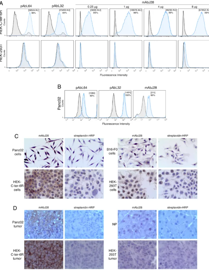
Immunoreactivity of murine pancreatic
adenocarcinoma Panc02 cells and tumors to mAbJ28
within the O-glycosylated C-ter of human pBSDL, we used transfected HEK-C-ter-6R cells. The polyclonal antibodies (pAbs) L64 and L32, reactive against physiological and pathological BSDLs (Figure 1A), and mAb J28, in a dose-dependent manner, detected C-ter-6R on non-permeabilized HEK-C-ter-6R cells but not on HEK-293T cells. We used these Abs to detect immunoreactivity on murine Panc02. All three Abs revealed a clear immunoreactivity (Figure 1B); intact or permeabilized Panc02 cells were stained with mAbJ28 indicating the presence of the epitope J28 in mouse as in human pancreatic tumor cells [19]. The by immunocytochemistry, using HEK-C-ter-6R as positive control (Figure 1C) and HEK-293T and melanoma tumor showed mAbJ28 immunoreactivity unlike those of normal pancreas under the same conditions (Figure 1D). As expected, tumors induced by HEK-C-ter-6R but not HEK-293T from nude mice displayed strong mAbJ28 staining. We also tested pancreatic tumors from genetically engineered mice (Pdx1-Cre; KrasG12D; Ink4a/
Arf and Pdx-1-Cre; LSL-KrasGD12; LSL-Trp53R172H)
developing PDAC, which showed no reactivity to mAbJ28 (unpublished data). Collectively these data demonstrate at the O-glycosylated C-terminal domain of human and murine pBSDL expressed by both pancreatic cancer cells and inoculated cell-induced tumors.
Immunogenicity of glycoprotein pBSDL-J28+
and C-ter-J28+: CD4+ and CD8+ T-cells from
mice immunized with full-length pBSDL-J28+
proliferated in the presence of either pBSDLs, from patients suffering PDAC, or the O-glycosylated C-terminal moiety
We investigated the ability of pBSDLs to promote adaptive immunity in mice. T-cells from mice immunized with pBSDL proliferated in the presence of either pBSDLs from different origins, or C-ter-J28+ (with optimal
Figure 1: Immunoreactivity of C-ter-6R-transfected HEK cells and murine pancreatic adenocarcinoma Panc02 cells to mAbJ28. A.
pAbL32 (4µg), pAbL64 (4µg) and mAbJ28 (at different concentrations). B. Expression of pBSDL and J28 epitope on Panc02 cells was C. on Panc02, HEK-C-ter-6R, B16-F0 and HEK-293T cells on polylysine-coated slides, and D.
concentrations likely below 0.16 µM), with increased Figure S1A-D). T-cells from mice immunized with C-ter-J28+ markedly proliferated in the presence of
pBSDL and glycosylated C-ter-J28+; no such proliferation
occurred in the presence of either a synthetic peptide encompassing an aa sequence identical to that of the C-ter or the short peptides EAT and GAP mimicking the most represented repeated sequences of BSDL (Figure 2A), thus underlining the requirement of glycosylated structures. C-ter-J28+ and pBSDL at the same molarity led to similar
levels of proliferation of CD4+ and CD8+ T-cells (Figure
2B). Thus, T-cells from mice immunized with either one of the immunogens recognize both glycosylated antigens, but not non-glycosylated peptides. T-cell proliferation was by C-ter-J28+ induced a greater level of secretion than
that by pBSDL (Figure 2C). These data imply that immunodominant glycoepitope J28-bearing pBSDLs and glycosylated C-ter trigger CD4+ and CD8+ T-cell
DC pulsed with the O-glycosylated C-ter-J28+
triggered activation of CD3+ T-cells from mice
immunized with C-ter-J28+ of pBSDLs
To delineate the ability of DC pulsed with C-ter-J28+
to activate T-cells, DC were loaded with C-ter-J28+ and
underwent maturation with lipopolysaccharide (LPS) and CD40L. Mature (m)DC presented enhanced expression of the co-stimulatory and Class II molecules by comparison with DC cultured in control medium so-called immature
among the factors secreted by C-ter-J28+-pulsed mDC
(Figure 3B). A positive spot for MCP-1 was found for iDC but less than that for C-ter-J28+-mDC. High amounts
of IL-12 were secreted only by mDC, whether antigen-pulsed or not (Figure 3C), while IL-15 was secreted by mDC and iDC (Figure 3D). Remarkably, antigen-loading of DC impaired neither the increase in co-stimulatory and Class II molecules nor IL-12 and IL-15 secretion (Figure 3A, 3C and 3D).
IL-12- and IL-15-secreting C-ter-pulsed mDC or control DC were co-cultured with CD3+ T-cells from mice
immunized with C-ter-J28+. Both CD4+ and CD8+ T-cell
populations proliferated upon encounter with C-ter-pulsed mDC as compared to unpulsed mDC (CD4+ T and CD8+
T increased by 66 and 62.5 % respectively) and to DC pulsed with the O-glycosylated peptide control TnMUC1 [20] (CD4+ T and CD8+ T increased by 19.8 and 20 %
respectively) (Figure 3E). The control TnMUC1 showed no immunoreactivity to mAbJ28 when compared to
rC-proliferation with mDC, pulsed or not with TnMUC1, was likely due to their cytokine production. Secretion levels
+
or TnMUC1 were normalized to that with unpulsed mean±SD = 1548±1389, n = 3).
culture with C-ter-J28+-pulsed mDC was around twice
that detected with unpulsed mDC. In contrast, secretion Thus mDC pulsed with the glycosylated tumor antigen J28+ hold potential to activate CD4+ and CD8+
T-cell responses mandatory to developing adaptive anticancer immunity.
Immune status of mice injected with tumor antigen-pulsed mDC
Splenocytes freshly collected from recipients phenotypic changes from controls (not depicted). However, after 4 days of culture, the percentage of granzymeB-expressing CD4+- and CD8+ T-cells and + cells had conspicuously increased in accordance
with C-ter-J28+ mDC injection number, by comparison
with percentages in unvaccinated mice injected with PBS (Figure 4A). Granzyme B-expressing CD4+ T-cells
are indeed reported [21] and may acquire killing activity in various conditions. Interestingly, in co-culture assays, splenocytes from mice DC-vaccinated twice, though not those from naïve controls (Figure 4B) or from Panc02-recipients (unpublished data), proliferated with Panc02 (P < 0.05) but not with B16-F0 cells. Identical results were obtained at a splenocyte: tumor cell ratio of 25:1. more pronounced with Panc02 than with B16-F0, and increased with DC-vaccination number. To detect whether C-ter-J28+ pulsed mDC could generate
C-ter-J28+
the immunized mice served as effectors in the presence of tumor cells. The abnormal round shape of Panc02 occurring upon culture with splenocytes from mice vaccinated with C-ter-J28+ pulsed mDC is illustrated
in Figure 4C. Also, compared to mDC-recipient mice, splenocytes from C-ter-J28+-mDC-vaccinated mice
+
+, + cells; not depicted) were able to lyse Panc02 but
not B16-F0 cells (Figure 4E) while CD8+ T-cells from
to melanoma B16-F0 and B16-F10, Panc02 expressed higher levels of CMH class I required for CTL-mediated lysis in the context of DC-vaccination; however they
Figure 2: Glycosylated C-ter-J28+ induced activation of CD4+ and CD8+ T-cells in LN from mice immunized with C-ter-J28+. A.
containing 20µg of C-ter-J28+ of pBSDL-Caro in PBS and incomplete Freund’s adjuvant supplemented with
toxin (Pt) (Sigma-Aldrich) IP, before the immunization, and again 24 h later. CFSE-labeled
+ from Bo, synthetic C-ter or synthetic peptides EAT or GAP. After
B. Cells were cultured at 2x10⁵
C-ter-J28+ of pBSDL-OG3 or full-length pBSDL-OG3 at different molarities. After 6 days, cells were labeled with anti-CD4 and anti-CD8.
C. Culture supernatants were collected after 24 and 72 h P < 0.05)
lacked CD40L, which interacts with DC for cytokine production signaling (unpublished data).
C-ter-J28+ DC-vaccinations hence led to enhanced
selective cellular immunoreactivity to Panc02, as demonstrated by increased immune cell proliferation and B-expressing CD4+ T-cells, CD8+ + cells,
and to selective T-cell cytotoxicity.
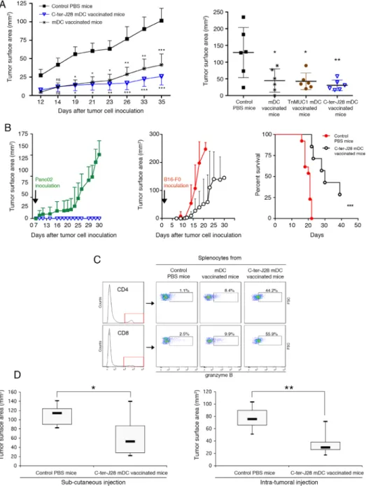
Vaccination with C-ter-J28+-pulsed-mDC prevents
Panc02 tumor development
Motivated by these results, we initiated preclinical trials of tumor DC-vaccination. Control Panc02-recipient mice injected with PBS developed large tumors (mean size of 101.52±45.20 mm2) at d35 (Figure 5A, left panel). In
contrast, three vaccinations with C-ter-J28+-pulsed-DC
conferred resistance to subsequent Panc02 challenge (P < 0.001 at the termination date). Striking protection from tumor development was obtained in 12 mice, 6 of which remained tumor-free indicating the high capacity of DC-vaccination to increase survival. Tumors in the other 6 were smaller (mean: 25.95±33.52 mm2 at d35) than those
in the unvaccinated Panc02 group (P
protection was also obtained in the mDC-vaccinated group. To study the effects of DC pulsed with another O-glysolated peptide, we loaded DC with TnMUC1 (25 the C-ter-J28+-pulsed mDC. Figure 5A (right panel) shows
that C-ter-J28+-pulsed mDC induced expected marked
protection against Panc02 challenge compared to PBS-controls (P < 0.01); the ranges of protection evaluated in mice vaccinated with either mDC or TnMUC1-mDC were similar (P < 0.05). A lower concentration of of rC-ter-17R-J28+) gave the same results. All these
towards a difference in the tumor development curves as a function of time, with that for C-ter-J28+-mDC recipients
being consistently below those of mDC and TnMUC1-mDC recipients (four experiments). This difference is likely more qualitative than quantitative, an assumption consolidated by the immune status of vaccinated mice with long-term protection (see below).
Tumor protection in C-ter-J28+ mDC-vaccinated
mice, shown in the experiment depicted in Figure 5A (left panel), correlated with a high level of spontaneous splenocyte concentration (Supplementary Figure S3 A and B). Thus C-ter-J28+
signature in spleen. Tumor protection was also associated with differentiation of CD4+ and CD8+ T-subpopulations + activation as measured by granzyme B
expression, not detected in the control conditions at day 35
after tumor challenge (Supplementary Figure S3C). At day 28 after tumor challenge, among protected DC-vaccinated mice, CD4+T-subpopulation expressing granzyme B
in C-ter-J28+DC recipient splenocytes had increased
compared to that in mDC- and TnMUC1-mDC recipient splenocytes, respectively by 69% and 77% (Figure S5B). All groups receiving mDC pulsed or not displayed similar
+ expressing granzyme B.
Vaccination with C-ter-J28+-pulsed-mDC
establishes long-term protection against Panc02-induced tumor development
The mice fully protected against tumor development (depicted in Figure 5A) were rechallenged with either inoculation. Whereas controls inoculated with Panc02 developed large tumors (mean 128.8±38 mm2 at d30),
vaccinated mice showed long-term survival without tumors. Interestingly upon challenge with melanoma B16-F0 (33-54 days later; 2 experiments) 3 out of 4 of these Panc02-resistant mice developed tumors similarly to the 10 control melanoma-recipients and died; one mouse remained tumor-free until the termination date. test; P = 0.143). At euthanasia, the C-ter-J28+
-mDC-vaccinated recipients challenged with Panc02 displayed high expansion of CD4+- and CD8+-T-cells expressing
granzyme B (more than 44%) in spleen compared to mDC-recipients (less than 10%) and a PBS-control mouse with late tumor development (2.5%) (Figure 5C). Tumor cell by the negligible or low level of cell activation observed in splenocytes from mice injected with Panc02 or B16-F0 (Supplementary Figure S4).
To strengthen these results, another experiment was performed with six 17R-C-ter-J28+ mDC-vaccinated mice
receiving either B16-F0 or Panc02. B16-F0 administration led to the development of melanoma in all three rC-ter-17R-J28+ mDC-vaccinated mice, with tumor surface areas
of 61.5, 143 and 250 mm2 (surface area mean±SD = 151±
90 mm2) compared to a mean surface area of 250 mm2 - the
B16-F0 controls. In contrast, all three rC-ter-17R-J28+
mDC-vaccinated mice showed complete resistance to Panc02 while the three non vaccinated Panc02 controls developed tumors with a mean surface area of 56 ± 8 mm2
at day 28. All the results of the experiments presented above were pooled (Figure 5B). To resume, one sub-group of C-ter-J28+-mDC vaccinated mice fully resisted
a second challenge by Panc02, while the other sub-group challenged instead with B16-F0 displayed only a slight delay in melanoma development and most did not survive.
Figure 3: Mature DC pulsed with the glycosylated C-ter-J28+ moiety trigger activation of CD3+ T-cells from mice immunized with C-ter-J28+. A. Quality control of DC maturation. At day 5, immature DC (iDC) were pulsed or not with C-ter-J28+ or
TnMUC1 (at equimolarity, 0.55 µM) for 10 h then cultured with LPS and CD40L for 22 h. The purity of the DC fraction was determined cytometry. Black histograms represent control isotype and colored histograms staining with anti-CD11c, -CD86, -CD40, -CD80 and -IA. B. Protein analysis of cytokines secreted by DC. Culture supernatants from immature DC (medium) and from DC pulsed with C-ter-J28+
and matured were collected after 22 h for cytokine detection using cytokine Ab array. Positive spots for GM-CSF and IL-4 were due to the addition of these cytokines in the culture medium. C. and D. IL-12 and IL-15 production. Culture supernatants were collected after 22 h of maturation for cytokine detection using ELISA assay. Representative results of at least three experiments. E. + T-cells
from mice immunized with C-ter-J28+ of pBSDL Caro and CFA were labeled with CFSE. CD3+ T-cells were plated at 2.5x10⁵
T-cell proliferation evaluated. Percentages of CD4+ and CD8+ daughter T-cells without addition of DC: 2.5±0.3 (not shown). Representative +
Vaccination with C-ter-J28+-pulsed-mDC treats
established tumors
reduced size of tumors in vaccinated mice compared to those in PBS-treated controls (mean tumor size of 52.8 mm2 in SC DC-vaccinated vs 114.3 mm2 in controls; P
< 0.01; and 29.5 mm2 in intratumorally DC-vaccinated
vs 75.6 mm2 in controls; P < 0.05) (Figure 5D). Thus,
intratumoral DC-injection was more effective than the SC route.
Vaccination with C-ter-J28+-pulsed-mDC
attenuates deleterious effects of pancreatic tumor
During the MRI follow-up, no sign of cachexia could be detected in vaccinated or non-vaccinated mice. Primary pancreatic tumors appeared solid with well-indicated a trend towards lower tumor volume in DC-vaccinated mice (Supplementary Figure S6A). However due to pleomorphic multi-lobulated appearance, volumetry of the primary tumor at d17-18 was only an estimate and only 3 out of the 6 tumors in each group were considered measurable. Figure 6A-6E shows representative images of the primary tumor and its spread within and beyond the peritoneal cavity in two non-vaccinated mice in contrast to limited peritoneal spread in a vaccinated DC-vaccination were observed on the MRI-based disease progression score (Supplementary Table S2 and S3), with non-vaccinated mice at d17-18 (disease progression score of 7.5±1.18 in vaccinated mice [n = 6] and 11±0.58 in non-vaccinated mice [n
0.7151, P = 0.4973 for interaction; F1,30 = 9.694, P = 0.0040 for treatment effect; F2,30 = 27.29, P < 0.0001 for time effect; Bonferroni post hoc test *P < 0.05 for group comparison at d17-18) (Figure 6F). MRI showed that despite direct extension to peripancreatic tissues (retroperitoneal and mesenteric fat, peritoneum) and often unresectable tumor observed in both groups, the C-ter-J28+-DC-vaccine delayed or prevented metastasis (distant
lymph nodes, liver, kidneys and lungs) and reduced secondary abdominal disease (organ displacement, splenomegaly, obstructed bile ducts and distended gall bladder) (Figure 6G, supplementary Figure S3B). Pleural effusion and pleural metastasis were detected at d17-18 only in non-vaccinated mice and resulted in rapid death after anesthesia induction. Thus, vaccination with C-ter-J28+-pulsed DC hampered growth, invasiveness and
metastasis of pancreatic tumors.
Source of BSDL-J28+
+,
we turned our attention towards bile salt-stimulated lipase (BSSL) present in human milk, which differs from BSDL only in glycosylation pattern [22]. We showed that BSSLs were both immunoreactive to mAbJ28 and immunogenic (Supplementary Figure S7).
DISCUSSION
We have validated pancreatic tumor glycoepitope C-ter-J28+ as a good inducer of anticancer adaptive
study from antigen discovery as a tumor marker in human tissue to immunogenic target for DC-vaccination in murine pancreatic cancer. Our data provide new strong evidence that (i) fucose-rich epitopes of pBSDL-J28+,
appearing during human pancreatic oncogenesis processes, are also expressed by murine PDAC cells Panc02; (ii) DC pulsed with the tumor pancreatic antigen skew adaptive immunity towards Th1 polarized responses; (iii) vaccination with C-ter-J28+ DC meets cancer vaccine
objectives by inducing anti-Panc02 CD8+
cells required to eradicate tumors and by promoting long-lasting resistance [23].
antigen by demonstrating the presence on C-ter-J28+ of
a glycosylation-dependent immunodominant epitope, common to various pBSDLs, that is able to trigger adaptive immune responses.
the hypothesis that the reported poor immunogenicity of Panc02 tumor could be due to lack of recognition of expression, lack of co-stimulatory signals and other factors may be partly responsible [4].
presentation of cancer antigens to T cells [4].We therefore investigated the potential of C-ter-J28+-pulsed mDC to
activate CD4+ and CD8+ T-cell responses mandatory to
develop adaptive anticancer immunity. We found that DC, high producers of two cytokines important for both innate and adaptive immunities, IL-12 and IL-15 [24, 25], when loaded with glycoantigen-J28+, drove CD3+ T-cells toward +
and CD8+
immunity by inducing or increasing Th1 cell and CTL responses [24].
cooperates to induce T-cell clones that expand in response cell activating factor IL-15 may skew naive CD8+ T-cell
Figure 4: Immune status of C-ter-J28+-pulsed mDC-injected mice. A.-B. Immune status of mice injected with tumor
antigen-+-pulsed mDC, once, twice or three times at weekly intervals or
received PBS (control mice). A. Intracellular expression of granzyme B in CD4+, CD8+ + cells was determined after 4 days
supplemented with IL-2. B. Reactivity of splenocytes to mitomycin C-treated Panc02 in co-culture. CFSE-labeled splenocytes were plated
+-pulsed mDC three times at weekly intervals or received PBS (control
mice). Four days following the 3rd DC injection, splenocytes were collected and cultured with mitomycin C-treated Panc02 cells at ratio
responder:stimulators of 50:1 with IL-2 for 4 days. C. Images of cells after 4 days of co-culture (x200). Co-cultures of splenocytes with tumor cells were examined using a phase-contrast microscope. Panc02 cultured with splenocytes from PBS-or mDC-treated mice exhibit their usual spindled shape; Panc02 cultured with splenocytes from vaccinated mice show dramatic changes in cell morphology and density.
D. E.
+ T-cells against Panc02 cells. After co-culture, splenocytes were collected and CD8+ T-cells
before incubation with tumor cells at various ratios for 5 hrs. Supernatants from triplicate or quadruplicate cultures were harvested P < 0.05).
differentiation towards effectors secreting type 1 cytokines [26].
Importantly, , tumor antigen J28+-pulsed mDC
mice, monitored by a T-cell proliferative response to epitope J28+
of CD4+ T-, CD8+
B, and CTL-lysis restricted to PDAC cells. Sustained C-ter-J28+-DC-vaccinated mice in response to Panc02 in
culture, with the number of C-ter-J28+-DC-vaccinations
was obtained in response to B16-F0, likely from cells other than CD4+- and CD8+
and natural killer DC [26, 27]. These distinct responses as reported for
responses elicited by TAA-loaded DC vaccination [28]. C-ter-J28+antigen-pulsed-DC vaccination prevents
ectopic growth of Panc02, in line with previous reports using different preparations of Panc02. In prophylactic conditions, increased survival was obtained in mice vaccinated with mDC pulsed with Panc02 heat-treated lysate [29] and irradiated Panc02 [30]. In therapeutic orthotopic tumors induced their regression [30]. While these preclinical trials show [29, 31] and [30] that loaded DC activate CTLs, none has controlled from other cancers. As long as the tumor antigens from target surface antigens common to tumor cell lines from various origins. In our test of long-term protection, we demonstrated that C-ter-J28+-pulsed-DC-vaccinated
mice, fully protected against Panc02-tumor development, all resisted a second Panc02 challenge while most did not survive melanoma challenge. This could be attributed to memory CD4+ and CD8+ T-cells, consistent with
the ability of IL-15 yielded by DC to induce increased percentages of effector memory CD8+ T-cells [25, 32].
cannot exclude their possible role. Yet, since C-ter-J28+
-mDC-vaccination resulted in an at least 5-fold increase in the percentage of CD4+- and CD8+-T-cells expressing
granzyme B in splenocytes of recipients by comparison to mDC-vaccination, one can reasonably assume that CD4+ and CD8+ T-cell activation is associated to the
long-lasting protection induced by Ag-pulsed-DC. Moreover, produced by patients with melanoma after DC vaccination
+
T-cell immunity [34].Overall, these results suggest the resistance to Panc02-tumor development, induced by tumor antigen-J28+-pulsed mDC.
Immunization with antigen-pulsed mature DCs,
which aims to elicit antitumor T cell responses, may in fact and CD4+ + T-cells as reported in melanoma + cells expressing granzyme B in mice receiving
unloaded mDC or mDC loaded with the O-glycosylated polypeptide control, TnMUC1.
Protective CTL responses requiring a contribution of both CD4+
vaccinations, though only in peculiar conditions, with CTL responses elicited by TAA-loaded DC has, to our knowledge, never as yet been reported. To summarize, C-ter-J28+ DC-vaccinations led to an immune status
characterized by expansion of anti-C-ter-J28+-tumor CD8+
T-cells, which
now needed to delineate the relative mechanistic contribution of CTL, CD4+
Furthermore, in control experiments using unloaded mDC-immunization, we and others [30, 38-40] have found some degree of protection against tumor challenge, which is in line with the demonstration that the unloaded We obtained similar results using the O-glycosylated polypeptide control, TnMUC1, for mDC pulsing. This correlates in particular with our mDC producing IL-12, unloaded DC to exert protection against tumor challenge, to prevent the development of tumor metastases and to establish long-term survival is proved to be dependent on Therapeutically, intratumoral immunization provided a more potent protective immunity than SC immunization, as reported by others [31, 42], but agents targeting different pathways is thus required [4]. Such combinations might synergistically generate more potent immune responses by activating DC and so fully exploiting their capacity to trigger anticancer responses.
Lastly, in an orthotopic model of pancreatic cancer, disease progression in the murine model is similar to clinically encountered adenocarcinomas [43]. Although both groups of mice presented with locally advanced disease, metastasis was delayed or prevented by the C-ter-J28+-DC-vaccine, and secondary abdominal disease was
reduced. Genetically-engineered mouse models
(Pdx-1-Cre/KrasG12D/Ink4a/Arf and Pdx-1-Cre; LSL-KrasGD12;
LSL-Trp53R172H mice), which develop pancreatic tumors
[44, 45], could not be used since the tested tumors showed no reactivity to mAbJ28. This might be due to the fact that
Figure 5: Prophylactic DC-vaccination in Panc02 pancreatic adenocarcinoma model. A.
vaccinated SC with C-ter-J28+-pulsed mDC, TnMUC1-pulsed mDC or mDC three times at weekly intervals or they received PBS (control
mice). Four days following the 3rd
performed twice on groups of six mice gave similar results (pooled experiments designed experiment I). Data are expressed as mean tumor surface area ± SEM. Six of the 12 mice vaccinated with C-ter-J28+ mDC remained free of tumors. Comparisons between groups
P P P < 0.01,
P < 0.001). Right panel: n = 6 per group. DC were loaded with C-ter-J28+
P P < 0.01; Holm-Sidak’s post-hoc test. Long-term protection provided by prophylactic
DC-vaccination. B. Mice receiving prophylactic DC-vaccination and remaining tumor-free were challenged after 43 (experiment I) or 35 (experiment III) days with Panc02 (left panel, open blue triangle, n = 7) or B16-F0 cells (middle panel; open black circle, n = 7) and
n n = 13). Data
pooled from experiments I and II are expressed as mean of tumor surface area ± SEM. Graph (right panel) shows a Kaplan-Meier survival
n = 13 ) or Cter-J28+ n = 7) followed by B16-F0
P = 0.0002; log-rang test. C. Fifty-nine days after the second challenge with Panc02 (experiment I), splenocytes were
collected and expression of granzyme B was determined on the same day (Day 0) without further culture. Therapeutic vaccination. D. Fourteen days after Panc02 cell challenge (once a palpable nodule had formed), mice were injected SC (left panel) or intratumorally (right panel) with C-ter-J28+-pulsed mDC three times at weekly intervals, or they received PBS (control mice; n = 6). Measurements at day 13
were subtracted from those at day 31 (termination date). Mann-Whitney test was used to compare tumor development of C-ter-J28+-pulsed
Figure 6: C-ter-J28+-mDC vaccination attenuates deleterious effects of orthotopically implanted pancreatic adenocarcinoma cell induced-tumor monitored by abdominal MRI at d17-18. A. C-ter-J28+-DC-vaccinated mouse (score: 3)
with a well-marginated primary tumor. B.-E. Control PBS mice. Mouse (score: 13) with a lobulated primary tumor, numerous contiguous and C.
(arrowheads), tumor tissue enveloping the spleen, metastasis of the kidney (mass below the kidney capsule), and an abnormal nodular aspect of the liver (axial image). D.
(thoracic axial image, arrowheads), spleen compression by surrounding tumor tissue, and SC tumors (arrowheads, axial image). E. Mouse (score: 12) with bilateral pleural effusion, pleural metastases in the left cavity (coronal and thoracic axial images, arrowheads), and in the F. MRI-based disease progression
*P < 0.05). G. Radar chart comparing
the total sum of each MRI feature of the score between vaccinated and non-vaccinated mice at d17-18. Abbreviations: d, dorsal; l, left; spleen; SP, spine; ST, stomach; T, primary tumor. Scale bars: 1.
in addition to these three genes showing high frequency of mutations, a great number ( > 50 in average) of gene mutations is found in human PDAC [46]. They affect at least a dozen key signaling pathways. Their direct or indirect consequences might therefore be responsible for the observed human pathological features such as hyperfucosylation of pBSDL.
In the proposed model, Panc02-cell-induced pancreatic tumor was associated with poorly developed stroma and caused neither the typical ductal lesions seen in human PDAC nor those found in the genetically engineered mice. Interestingly, it induced peritoneal carcinomatosis, commonly observed in patients suffering from PDAC [47] and considerably contributing to their demise [48], and metastasis. This model is widely explored for the development of novel therapeutic strategies, and here essentially provides relevant targets for tumor antigen-DC vaccination.
The main limitation of our model concerns the source of C-ter-J28+, more precisely the impossibility of
obtaining pathological pancreatic juices from patients
+
production. Yet, this production has today greatly improved. We have also presented one potential way of circumventing this problem by detecting the glycotope-J28 within BSSL from human milk.
the immunogenicity of glycosylated pancreatic TAA, and
that
the selective and spontaneous expression of TAA such as pBSDL-J28+ on PDAC cells and tissues makes them
pertinent targets of DC-immunotherapy. C-ter-J28+
DC-vaccination could represent a novel option for PDAC multiple adjuvant therapy in humans[for review see 49].
MATERIALS AND METHODS
Ethics statement
The investigation was conducted in accordance with the French guidelines for animal care and the directive approved by the local ethics committee of Aix-Marseille University.
Mice
b) mice and
mice were from Janvier (Le Genest-St. Isle, France).
Cell lines
highly tumorigenic murine pancreatic carcinoma cell line Schmitz and E. Raskopf (University of Bonn, Germany), and the metastatic clones of the B16 melanoma, B16-F0 and B16-F10. Also used were HEK-293T cells isolated with the pSecTag-2B plasmid (Life Technologies, Saint Aubin, France) encoding for hexahistidine-tagged 6 repeated sequences of the human C-terminal domain of the human C-terminal domain and thus called HEK-BSDL-17R.
Before injection, cells were tested negative for mycoplasma contamination.
Antigens
pBSDLs-J28+
of patients suffering from PDAC [11] and the pBSDL-J28+
C-terminal glycopolypeptide (C-ter-J28+) was then
obtained by cyanogen bromide cleavage of pBSDL-J28+
[14]. These C-ter-J28+ were used in all the experiments
unless indicated.Recombinant BSDL-17R-J28+
(rBSDL-17R-J28+
culture supernatant and its C-terminal glycopolypeptide (rC-ter-17R-J28+) obtained as mentioned above. EAT
and the synthetic pBSDL C-terminal polypeptide (77 mers), were purchased from Proteogenix (Oberhausbergen, France). The synthetic MUC1 polypeptide (100 mers) per repeat [20]. This glycosylated peptide, designated TnMUC1, was kindly provided by Pr O. Finn (Univ of salt-stimulated lipases (BSSL) were a generous gift of Pr O. Hernell (Umea University, Sweden).
Tumor induction and vaccination strategies
Panc02 (5x10⁵ to 1x10⁶) and B16-F0 (1 to 3x10⁵) 6)
as the product of perpendicular diameters, measured exceeded 250 mm2. For orthotopic conditions, cubes (2
mm3) of SC Panc02-tumors were surgically transplanted
For prophylactic vaccination, DC were injected SC into
.
For therapeutic vaccination, DC were injected SC or intratumorally when tumors had formed a palpable nodule.
Orthotopic tumor implantation
The pancreas of anesthetized mice was exposed after laparotomy on a sterile gauze. A cube (2 mm3) of a
SC tumor induced by pancreatic adenocarcinoma Panc02 cells, was placed on a sterile disc (diameter: 2 mm) of (Brown, Melsungen, Germany) was put next to the tumor. The paper was then stuck onto the pancreas with the tumor in between. After suturing, mice received SC injections of and again a few hours later.
Antibodies (Ab)
Control isotype Abs from mouse and rabbit were purchased from BD Pharmingen and Beckman Coulter, respectively. Polyclonal (p)AbL64 and pAbL32 Abs, directed against a mixture of human BSDL and pBSDL from pathological pancreatic juices, were produced in our laboratory. The mAbJ28 was a gift from Dr. M. J. Escribano (Inserm U260, Marseille, France). Anti-CD4-A647, anti-CD8-PE, anti-IA-A647, and anti-MHC-I (H-2Kb)-PE Abs were from Ozyme (St
Quentin-en-Yvelines, France); anti-CD11c-eFluor450, anti-granzyme
San Diego, CA); anti-CD40, anti-CD80 and anti-CD86 from BD Biosciences (Le Pont-de-Claix, France); anti-CD40L-FITC from Proteogenix (Oberhausbergen, France); and Biotec (Bergisch Gladbach, Germany).
Flow cytometry
Cells were labeled as described in [12] using monensin (GolgiStop, BD Pharmingen, San Diego, CA). Pharmingen) and staining with anti-CD4, anti-CD8and Factor Staining Buffer Set (Ebioscience, San Diego, CA
cytometer (Beckman Coulter, Roissy CDG, France). Data were analyzed using FlowJo software (Tree Star, San Carlos, CA). The results are expressed as percentages of
Immunohistocytochemistry
Immunohistocytochemistry was performed using the Dako ARKTM (Dako, Hamburg, Germany) according
to the manufacturer’s instructions. The staining was completed by incubation with substrate-chromogen 3,3-diaminobenzidine (DAB). Sections were counter-stained with hematoxylin-phloxin.
T-cell proliferation
succinimidyl ester (CFSE) (1µM, Invitrogen), cells were plated at 2-4 x10⁵
[RPMI-1640, 5% fetal bovine serum (FBS), 50µM (Invitrogen) and 1% sodium pyruvate (Invitrogen)] containing antigens. Cultures were carried out in triplicates or quadruplicates.
Generation of dendritic cells (DC), DC-antigen loading and maturation
marrow according to Inaba’s protocol [50],and cultured Germany At days 2 and 4, supernatant was removed was added when indicated. At days 5-6, DC were loaded with antigens. Maturation was induced with a combination
markers was controlled by cytometry analysis.
Cytokine detection in DC-culture supernatants
Culture supernatants were collected for cytokine detection by ELISA (for IL-12, Ozyme; for IL-15,
,
Mouse Cytokine Antibody Array I (RayBiotech), before being revealed using Gbox system (GeneSnap software, Syngene, Ozyme).
Cytotoxic assays
Cytotoxicity was assessed by lactate dehydrogenase Cytotoxicity kit (Promega Corporation, Madison, WI, USA) following the manufacturer’s protocol.
Abdominal MRI
DC-induced response was assessed on days 7-8, 9-10 and 17 or 18 after orthotopic tumor implantation by serial MRI. Twelve tumor-bearing and MR system (Bruker, Ettlingen, Germany) [51],with a The respiratory rate was kept at 70±20 breaths per minute (bpm) and monitored using a pneumatic pressure probe and an MRI compatible monitoring and gating system (PC-sam, Small Animal Instruments Inc., Stony Brook, magnet gradients. Multi-slice images were acquired, 15 France), in the sagittal, coronal and transverse planes using a 2D spin-echo sequence (repetition time, 448 ms; respiratory gating to reduce motion artefacts. Geometrical parameters were as follows: matrix, 300 x 240 for sagittal of view, 24 x 30 mm for sagittal and coronal planes and 24 x 24 mm for the axial plane; spatial resolution, 100 x 100 x 500 µm3, 20 contiguous slices. Total acquisition
time was 8 to 12 minutes per plane depending on the respiration rate. Tumor growth, morphology and spread were evaluated by two trained MRI scientists (ATP-B and slices in three orthogonal planes covering the entire primary tumor, using manual volumetry and a disease
Statistical analysis
The statistical analysis was performed using the a Bonferroni test, and the Mann-Whitney, the Kruskall-Wallis and the Fisher’s exact tests.
P < 0.05 were considered P
curves were determined using the Kaplan-Meier method. The log-rank test was used to compare curves between study and control groups. Principal component analysis (PCA) on variances of MRI parameters was performed with JMP 9 software.
ACKNOWLEDGMENTS
We are deeply indebted to Pr Olle Hernell (Paediatrics, Univ of Umea, Sweden) and Martine Armand, PhD (Centre de Résonance Magnétique
Campus Santé-Timone, Marseille, France) for the generous gift of BSSLs and to Pr Olivera J. Finn and Dr John R. McKolanis (Immunology, Univ of Pittsburgh School of Medicine, Pittsburgh, PA) for the generous gift of TnMUC1. We thank Pr David A. Tuveson, (Cambridge Research Institute, Univ of Cambridge, Cambridge, UK) and R. Tomasini, PhD (Cancer Research Center of for the gift of respectively PDX-Cre/Kras G12D/Ink4a/Arf
and Pdx-1-Cre ; LSL-KrasGD12; LSL-Trp53R172H mouse
Timone, Marseille, France) for helpful discussion on tumor imaging, Dr Jean Del Grande (Laboratoire d’Anatomie la Timone,Marseille, France) for helpful discussion on tumor pathology, and to Dr Anderson D. Loundou (Unité d’Aide Méthodologique à la Recherche clinique, Laboratoire de Santé Publique, Campus Santé-Timone, Marseille, France) for help with statistical analysis. We thank Guillaume Schmidt (Centre de Recherche en Oncologie biologique et Oncopharmacologie, Campus Santé-Timone, Marseille, France) for expert technical assistance, and Thierry Cheyrol and Perrine André (Centre Timone, Marseille, France) for their invaluable assistance in animal experiments.
This work was supported by institutional funding Marseille Université (Marseille, France) (DL, EM, EB), the town of Marseilles authorities (DL, EM, EB), and Recherche Intégrée sur le Cancer (SIRIC) (DL, EM, EB). AC is a doctoral fellow of Ministère de l’Enseignement Supérieur et de la Recherche, and the Ligue Contre le Cancer (Paris, France).
CONFLICTS OF INTEREST
interest.
Abbreviations
BSDL, bile salt-dependent lipase; C-ter, C-terminal moiety; DC, dendritic cells; iDC, immature DC; IP, intraperitoneally; mDC, mature DC; mABJ28, monoclonal antibody J28; PDAC, pancreatic ductal adenocarcinoma; SC, subcutaneously; TAAs, tumor-associated-carbohydrate antigens.
REFERENCES
1. Simard EP, Ward EM, Siegel R and Jemal A. Cancers with increasing incidence trends in the United States: 1999 through 2008. CA: a Cancer Journal for Clinicians. 2012; 62:118-128.
2. Rahib L, Smith BD, Aizenberg R, Rosenzweig AB, Fleshman JM and Matrisian LM. Projecting cancer incidence and deaths to 2030: the unexpected burden of thyroid, liver, and pancreas cancers in the United States. Cancer Research. 2011;74:2913-21.
3. Conroy T, Desseigne F, Ychou M, Bouché O, Guimbaud R, Bécouarn Y, Adenis A, Raoul JL, Gourgou-Bourgade S, de la Fouchardière C, Bennouna J, Bachet JB,
Khemissa-Medicine. 2011;364:1817-25.
4. Palucka K and Banchereau J. Dendritic-cell-based therapeutic cancer vaccines. Immunity. 2013;39:38-48. J, Cremer I, Galon J, Tartour E, Zitvogel L, Kroemer G and Galluzzi L. Trial watch: Dendritic cell-based interventions for cancer therapy. Oncoimmunology. 2013;2:e25771. 6. Dodson LF, Hawkins WG and Goedegebuure P. Potential
targets for pancreatic cancer immunotherapeutics. Immunotherapy. 2011;3:517-37.
7. Freire T, Lo-Man R, Bay S and Leclerc C. Tn glycosylation of the MUC6 protein modulates its immunogenicity and promotes the induction of Th17-biased T cell responses. Journal of Biological Chemistry. 2011;286:7797-7811. Tumor-associated MUC1 glycopeptide epitopes are not subject to self-tolerance and improve responses to MUC1 peptide epitopes in MUC1 transgenic mice. Biological Chemistry. 2009;390:611-8.
P. Differentiation antigens in fetal human pancreas. Reexpression in cancer. International Journal of Cancer. 1986;38:155-160.
characterization of FAP, a feto-acinar protein associated with the differentiation of human pancreas. Journal of Biological Chemistry. 1989;264:21865-71.
O, Figarella C, Escribano MJ and Lombardo D. Human fetoacinar pancreatic protein: an oncofetal glycoform of the normally secreted pancreatic bile-salt-dependent lipase. Biochem Journal. 1993;289:609-615.
A, Silvy F, Bernard JP, Lombardo D, Beraud E, Olive D and Mas E. A novel tumor-associated pancreatic glycoprotein is internalized by human dendritic cells and induces their maturation. Journal of Immunology. 2011;186:4067-77. 13. Panicot L, Mas E, Pasqualini E, Zerfaoui M, Lombardo
D, Sadoulet MO and El Battari A. The formation of the oncofetal J28 glycotope involves core-2 beta6-fucosyltransferase activities. Glycobiology. 1999;9(9):935-46.
14. Mas E, Crotte C, Lecestre D, Michalski JC, Escribano MJ, Lombardo D and Sadoulet MO. The oncofetal J28 epitope involves fucosylated O-linked oligosaccharide structures of the fetoacinar pancreatic protein. Glycobiology. 1997;7:745-52.
15. Panicot L, Mas E, Thivolet C and Lombardo D. Circulating antibodies against an exocrine pancreatic enzyme in type 1 diabetes. Diabetes. 1999;48:2316-23.
Y. Recognition of tumor glycans by antigen-presenting cells. Current Opinion in Immunology. 2006;18:105-111. Finn OJ. The mechanism of unresponsiveness to circulating tumor antigen MUC1 is a block in intracellular sorting and processing by dendritic cells. Journal of Immunology. 2000;165:3730-41.
18. Beraud E, Collignon A, Franceschi C, Olive D, Lombardo D and Mas E. Investigation of a new tumor-associated glycosylated antigen as target for dendritic cell vaccination in pancreatic cancer. Oncoimmunology. 2012;1:56-61. 19. Panicot-Dubois L, Aubert M, Franceschi C, Mas E,
Silvy F, Crotte C, Bernard J-P, Dominique Lombardo D and Sadoulet MO. Monoclonal antibody 16D10 to the C-terminal domain of the feto-acinar pancreatic protein binds to membrane of human pancreatic tumoral SOJ-6 cells and inhibits the growth of tumor xenografts.
Tumor-associated MUC1 glycopeptide epitopes are not subject to self-tolerance and improve responses to MUC1 peptide epitopes in MUC1 transgenic mice. Biological Chemistry. 2009;390:611-8.
exocytosis with T cells from mice expressing active 22. Hernell O and Bläckberg L. Human milk bile
salt-stimulated lipase: functional and molecular aspects. Journal of Pediatrics. 1994;125:56-61.
23. Steinman RM and Banchereau J. Taking dendritic cells into 24. Trinchieri G. Interleukin-12 and the regulation of innate
Immunology. 2003;3:133-46.
25. Ma A, Koka R and Burkett P. Diverse functions of IL-2, IL-15, and IL-7 in lymphoid homeostasis. Annual Review of Immunology. 2006;24:657-79.
Current Opinion in Immunology. 2008;20:558-65. 27. Tel J, Anguille S, Waterborg CE, Smits EL, Figdor CG and
. Tumoricidal activity of human dendritic cells.
Trends in Immunology. 2014;35:38-46.
28. Prins RM, Odesa SK and Liau LM. Immunotherapeutic targeting of shared melanoma-associated antigens in a murine glioma model. Cancer Research. 2003;63:8487-91. 29. Kim HS, Choo YS, Koo T, Bang S, Oh TY, Wen J and Song
SY. Enhancement of antitumor immunity of dendritic cells pulsed with heat-treated tumor lysate in murine pancreatic cancer. Immunology Letters. 2006;103:142-48.
30. Bauer C, Bauernfeind F, Sterzik A, Orban M, Schnurr M, Lehr HA, Endres S, Eigler A and Dauer M. Dendritic cell-based vaccination combined with gemcitabine increases survival in a murine pancreatic carcinoma model. Gut. 2007;56:1275-82.
Gorschlüter M, Schneider C, Sauerbruch T and Schmidt-pulsed dendritic cells confers antitumor immunity in a 2003;63:8962-67.
32. Eberl G, Brawand P and MacDonald HR. Selective bystander proliferation of memory CD4+ and CD8+ T cells
2000;165:4305-11.
L, Lanier LL, Yokoyama WM and Ugolini S. Innate or adaptive immunity? The example of natural killer cells. Science. 2011;331:44-9.
34. Carreno BM, Becker-Hapak M, Huang A, Chan M, Alyasiry A, Lie WR, Aft RL, Cornelius LA, Trinkaus KM and Linette GP. IL-12p70-producing patient DC vaccine elicits Tc1-polarized immunity. Journal of Clinical Investigation. 2013;123:3383-94.
Oncologist. 2012;17:1256-70.
Cancer Therapy. Clinical Cancer Research. 2014; 20:3390– 400.
37. Wargo JA, Schumacher LY, Comin-Anduix B, Dissette
immune response following immunization with melanoma-antigen-engineered dendritic cells. Cancer Gene Therapy. 2005;12:516-27.
38. Boudreau JE, Bridle BW, Stephenson KB, Jenkins KM, Brunellière J, Bramson JL, Lichty BD and Wan Y. Recombinant vesicular stomatitis virus transduction of dendritic cells enhances their ability to prime innate and adaptive antitumor immunity. Molecular Therapy. 2009;17:1465-72.
39. Ribas A, Wargo JA, Comin-Anduix B, Sanetti S,
tumor responses to dendritic cells in the absence of CD8-positive cells. Journal of Immunology. 2004;172:4762-69. 40. Shimizu K and Fujii S. DC therapy induces long-term
Immunology. 2009;39:457-68.
41. Boudreau JE, Stephenson KB, Wang F, Jenkins KM, Brunellière J, Bramson JL, Lichty BD and Wan Y. IL-15 and type I interferon are required for activation of Cancer Research. 2011;71:2497-506.
42. Candido KA, Shimizu K, McLaughlin JC, Kunkel R, Fuller JA, Redman BG,
Local administration of dendritic cells inhibits established breast tumor growth: implications for apoptosis-inducing agents. Cancer Research. 2001;61:228-36.
M. Pancreatic cancer. Lancet. 2011; 378:607-20.
ductal adenocarcinoma. Genes and Development. 2003;17:3112-26.
45. Hingorani SR, Wang L, Multani AS, Combs C, Deramaudt TB, Hruban RH, Rustgi AK, Chang S and Tuveson DA. Trp53R172H and KrasG12D cooperate to promote chromosomal instability and widely metastatic pancreatic ductal adenocarcinoma in mice. Cancer Cell. 2005;7:469-83.
46. Blackford A, Parmigiani G, Kensler TW, Wolfgang Eshleman JR, Goggins M, Jaffee EM, Iacobuzio-Donahue CA, et al. Genetic mutations associated with cigarette smoking in pancreatic cancer. Cancer Research. 2009 ;69:3681-3688.
47. Seelig SK, Burkert B, Chromik AM, Tannapfel A, Uhl Wand Seelig MH. Pancreatic resections for advanced M1-pancreatic carcinoma: the value of synchronous metastasectomy. HPB Surgery. 2010;2010:579-672. W, Aerts R and Topal B. Patterns of recurrence after curative resection of pancreatic ductal adenocarcinoma. European Journal of Surgical Oncology. 2009;35:600-4. 49. Melero I, Gaudernack G, Gerritsen W, Huber C, Parmiani
I and Mellstedt H. Therapeutic vaccines for cancer: an Oncology. 2014;11:509-24.
S, Muramatsu S and Steinman R M. Generation of large numbers of dendritic cells from mouse bone marrow colony-stimulating factor. Journal of Experimental
Medicine. 1992;176:1693-1702.
C, Maitre P, Hosseiny S, Petit-Paitel A, et al. Melanin-concentrating hormone regulates beat frequency