HAL Id: inserm-02877916
https://www.hal.inserm.fr/inserm-02877916
Submitted on 22 Jun 2020
HAL is a multi-disciplinary open access
archive for the deposit and dissemination of
sci-entific research documents, whether they are
pub-lished or not. The documents may come from
teaching and research institutions in France or
abroad, or from public or private research centers.
L’archive ouverte pluridisciplinaire HAL, est
destinée au dépôt et à la diffusion de documents
scientifiques de niveau recherche, publiés ou non,
émanant des établissements d’enseignement et de
recherche français ou étrangers, des laboratoires
publics ou privés.
caveolae is required for human skin pigmentation
Lia Domingues, Ilse Hurbain, Floriane Gilles-Marsens, Julia Sirés-Campos,
Nathalie André, Melissa Dewulf, Maryse Romao, Christine Viaris de Lesegno,
Anne-Sophie Macé, Cédric Blouin, et al.
To cite this version:
Lia Domingues, Ilse Hurbain, Floriane Gilles-Marsens, Julia Sirés-Campos, Nathalie André, et al..
Coupling of melanocyte signaling and mechanics by caveolae is required for human skin pigmentation.
Nature Communications, Nature Publishing Group, 2020, 11 (1), pp.2988.
�10.1038/s41467-020-16738-z�. �inserm-02877916�
Coupling of melanocyte signaling and mechanics
by caveolae is required for human skin
pigmentation
Lia Domingues
1
✉
, Ilse Hurbain
1,2
, Floriane Gilles-Marsens
1,5
, Julia Sirés-Campos
1
, Nathalie André
3
,
Melissa Dewulf
4
, Maryse Romao
1,2
, Christine Viaris de Lesegno
4
, Anne-Sophie Macé
2
, Cédric Blouin
4
,
Christelle Guéré
3
, Katell Vié
3
, Graça Raposo
1,2,6
, Christophe Lamaze
4,6
& Cédric Delevoye
1,2
✉
Tissue homeostasis requires regulation of cell
–cell communication, which relies on signaling
molecules and cell contacts. In skin epidermis, keratinocytes secrete factors transduced by
melanocytes into signaling cues promoting their pigmentation and dendrite outgrowth, while
melanocytes transfer melanin pigments to keratinocytes to convey skin photoprotection. How
epidermal cells integrate these functions remains poorly characterized. Here, we show that
caveolae are asymmetrically distributed in melanocytes and particularly abundant at the
melanocyte
–keratinocyte interface in epidermis. Caveolae in melanocytes are modulated by
ultraviolet radiations and keratinocytes-released factors, like miRNAs. Preventing caveolae
formation in melanocytes increases melanin pigment synthesis through upregulation of
cAMP signaling and decreases cell protrusions, cell–cell contacts, pigment transfer and
epidermis pigmentation. Altogether, we identify that caveolae serve as molecular hubs that
couple signaling outputs from keratinocytes to mechanical plasticity of pigment cells. The
coordination of intercellular communication and contacts by caveolae is thus crucial to skin
pigmentation and tissue homeostasis.
https://doi.org/10.1038/s41467-020-16738-z
OPEN
1Institut Curie, PSL Research University, CNRS, UMR144, Structure and Membrane Compartments, 75005 Paris, France.2Institut Curie, PSL Research
University, CNRS, UMR144, Cell and Tissue Imaging Facility (PICT-IBiSA), 75005 Paris, France.3Laboratoire Clarins, 5 rue Ampère, 95000 Pontoise, France.
4Institut Curie, PSL Research University, INSERM U1143, CNRS UMR 3666, Membrane Mechanics and Dynamics of Intracellular Signaling Laboratory,
75005 Paris, France.5Present address: Institut NeuroMyoGene, UCBL1, UMR 5310, INSERM U1217, Génétique et Neurobiologie de C. Elegans, Faculté de
Médecine et de Pharmacie, 8 Avenue Rockefeller, 69008 Lyon, France.6These authors contributed equally: Graça Raposo, Christophe Lamaze.
✉email:ldddomingues@gmail.com;cedric.delevoye@curie.fr
123456789
H
uman skin comprises a highly stratified epidermis and a
bottom dermis. The epidermis, the outermost and
photo-protective layer of the skin, is mainly composed of
mel-anocytes and keratinocytes that together create a structural and
functional epidermal unit
1. Melanocytes are neural crest-derived
cells
2that extend dendrites and allow them to contact up to 40
epidermal keratinocytes
1. The main role of melanocytes is to
produce the melanin pigments in a specialized organelle, called
melanosome, that undergoes maturation from early
non-pigmented to late non-pigmented stages
3,4. The maturing and
pig-mented melanosome moves toward the tip of the dendrites
5to be
transferred to keratinocytes where it protects the nuclei against
ultraviolet (UV) radiations. In melanocytes, the formation of
dendrites, melanosome biogenesis, synthesis and transfer of
melanin to keratinocytes is a tightly coordinated process under
the control of UV radiations, keratinocytes-secreted factors (e.g.
hormones) and secreted endosomal-derived vesicles called
exo-somes
6–8. These secreted hormones can trigger different
trans-duction pathways in melanocytes, including the cyclic adenosine
monophosphate (cAMP) signaling pathway through binding to
various G-protein coupled receptors (GPCRs) at the cell
surface
9,10. As a result, melanocytes increase pigment synthesis—
mainly through stimulating gene transcription of
melanin-synthesizing enzymes
11—and dendrite outgrowth—through
reg-ulation of Rho GTPases activity and remodeling of the actin
cytoskeleton
12,13. Also, we have recently shown that specific
miRNAs associated with exosomes released from keratinocyte
modulate human melanocyte pigmentation by enhancing the
expression of proteins associated with melanosome maturation
and trafficking
8. However, how environmental cues are spatially
and temporally controlled in melanocytes to be efficiently
translated into biochemical and physical cellular responses
remains mostly uncharacterized.
Caveolae are cup-shaped plasma membrane invaginations
firstly described in endothelial and epithelial cells
14,15. Their size
(50–100 nm) and the absence of an electron-dense coat
mor-phologically distinguish caveolae from other invaginated
struc-tures at the plasma membrane
16. Caveolae are mainly composed
of two groups of proteins, the caveolins (Cav1, -2 and -3)
17and
the more recently identified cavins (Cavin1, 2, 3 and 4)
18.
Caveolae biogenesis and functions are dependent on Cav1 and
Cavin1 in non-muscle cells, and on Cav3 in muscle cells
19.
Caveolae have various crucial functions including endocytosis,
lipid homeostasis, signal transduction and, the most recently
identified, mechanoprotection
19,20. As a transduction platform,
caveolae control the production of second messengers, such as
cAMP, through local confinement of different elements of this
signaling cascade
21. Cav1 and -3 contain a scaffolding domain
(CSD) located in the N-terminal region that could bind to
transmembrane adenylate cyclases (tmACs) to decrease their
activities, and thus to control intracellular cAMP levels
22. For
instance, caveolae in cardiomyocytes participate in the
compart-mentalization of intracellular cAMP, which can regulate cell
contractility in distal regions of the heart and, therefore, its
function
23,24. The mechanoprotective role of caveolae is
asso-ciated with the maintenance of plasma membrane integrity when
both cells and tissues experience chronical mechanical stress
25–28.
And caveolae were recently shown to couple mechanosensing
with mechanosignaling in human muscle cells, a process that is
impaired in caveolae-associated muscle dystrophies
29.
Epidermal melanocytes and keratinocytes are in constant
communication, not only via secreted factors and exosomes that
modulate cellular responses, but also by the physical contacts they
establish to maintain the tissue homeostasis and pigmentation.
Here, we report an additional function for caveolae, which by
integrating the biochemical and mechanical behavior of
melanocytes, control melanin transfer to keratinocytes and
epi-dermis pigmentation. Altogether, this study provides the
first
evidence for a physiologic role of caveolae as a molecular sensing
platform required for the homeostasis of one of the largest human
tissue, the skin epidermis.
Results
Caveolae distribute asymmetrically in melanocytes and are
positively regulated by keratinocyte-secreted factors.
Melano-cytes and keratinoMelano-cytes establish a complex intercellular dialog
required for skin photoprotection. 2D co-culture systems, where
these two cell types share the same medium, have been widely
used to study intercellular communication and pigment transfer
between epidermal cells
7,30. To evaluate the distribution of
caveolae within the epidermal unit in 2D, normal human
mela-nocytes and keratimela-nocytes were co-cultured and labeled for two
main constituents of caveolae, Cav1 or Cavin1.
Immuno-fluorescence microscopy revealed that both Cav1 and Cavin1, and
therefore caveolae, were asymmetrically distributed in
melano-cytes (Fig.
1
a, b; arrowheads) that were identified by the abundant
HMB45 staining of the processed fragment of the premelanosome
protein PMEL
4(Methods). This asymmetry was not observed in
keratinocytes.
Symmetry-breaking can occur in response to local external
chemical and/or mechanical cues such as signaling molecules
and/or cell–cell contacts, respectively
31. However, in the absence
of any type of spatial signaling, cells break their symmetry
randomly and spontaneously
32. When grown alone in the
absence of any pre-existing signaling cues, one third of
melanocytes presented an asymmetric distribution of caveolae,
as shown by the endogenous staining of Cav1 and Cavin1 (Fig.
1
c
and Supplementary Fig. 1a; arrowheads in top panels). This
asymmetry was restricted to caveolae as the distribution of
clathrin-coated pits (CCPs, see AP-2 staining; Supplementary
Fig. 1b), the canonical plasma membrane invaginated structures
mediating endocytosis
33was even. Interestingly, the number of
melanocytes
showing
caveolae
asymmetrically
distributed
doubled when co-cultured with keratinocytes (Fig.
1
a, c), whereas
co-culture with HeLa cells had no effect (Fig.
1
c and
Supplementary Fig. 1a; bottom panels). This shows that the
intrinsic asymmetry of caveolae in melanocytes is specifically
enhanced by keratinocytes, either by cell–cell contacts and/or by
keratinocytes-secreted factors. To address the role of extracellular
factors in caveolae distribution, melanocytes were incubated with
the medium recovered from a confluent culture of keratinocytes
(referred as keratinocytes-conditioned medium, Ker-CM). Under
this condition, we observed two to fourfold increase of the
number of melanocytes with an asymmetric distribution of
caveolae (Fig.
1
d and Supplementary Fig. 1c, arrowheads) as
compared to cells grown in their own medium (Mel
supplemen-ted medium) or in keratinocytes non-conditioned medium (Ker
medium). The proportion of melanocytes with caveolae
asymme-trically distributed was similar between cells co-cultured with
keratinocytes (Fig.
1
c) and cells incubated with Ker-CM (Fig.
1
d),
indicating that factors secreted from keratinocytes are the main
extracellular contributors to the increased asymmetric
distribu-tion of caveolae in melanocytes.
Caveolae
are
preferentially
located
at
the
melanocyte
–keratinocyte interface in human epidermis and
are more abundant in melanocytes during tissue pigmentation.
We
investigated
the
distribution
of
caveolae
at
the
melanocyte–keratinocyte interface in human skin samples.
The tissues were chemically
fixed or physically immobilized using
high-pressure freezing (HPF), which preserves membranes in
their native state
34, processed for ultrathin (60 nm) sectioning
and analyzed by 2D conventional transmission electron
micro-scopy (TEM) (Fig.
1
e and Supplementary Fig. 1d). The
melanocyte–keratinocyte interface revealed numerous plasma
membrane-associated cup-shaped invaginations, with a diameter
between 43 and 102 nm and an average size of 63.9 nm, which
lacked an electron-dense cytoplasmic coat (Fig.
1
e and
Supple-mentary Fig. 1d, arrowheads). Immunogold labeling on ultrathin
cryosections of human skin samples revealed that these
invagi-nations were positive for Cav1 in melanocytes (Supplementary
Fig. 1e) and were thus identified as caveolae. To access caveolae
3D ultrastructure, thick-sectioned (300 nm) human skin samples
were subjected to double-tilt electron tomography (Fig.
1
f and
Supplementary Fig. 1f). The reconstructed 3D model (Fig.
1
f and
Supplementary Video 1) depicts an epidermal area consisting of a
transversal section of a melanocyte dendrite (plasma membrane
in green) containing pigmented melanosomes (limiting
mem-brane in red and intraluminal pigment in black) and surrounded
by a keratinocyte (plasma membrane in blue, presenting keratin
bundles on the cytosol, white arrowheads in left panel). Caveolae
Human skin Chemical fixation HPF 1 1 2 2 3 Keratinocyte Keratinocyte Melanocyte Melanocyte 4 2 1 UV-B * Cav1 Calnexin kDa 20 75 0 1 2 3 Melanocytes Days Cavin Co-culture Cav1
a
Cavin1 Cav1 Cavin1Cav1 Cav1 Cavin1
HMB45 Merge Zoom Zoom 1 2 3 4 100 P < 0.0001 P < 0.0001 75 50 25
Mean fluorescent intensity (a.u.)
0
Box1 Box2 Box1 Box2
Melanocytes with asymmetr
ically distr
ib
uted
Ca
v1 or Ca
vin1 (% of the total)
Melanocytes with asymmetr
ically distr
ib
uted
Ca
v1 or Ca
vin1 (% of the total)
70 60 50 40 30 20 10 0 60 50 40 30 20 10 0 20 15 10 Ca v eolae/10 μ m 5 0 20 15 10 Ca v eolae/10 μ m 6 5 4 2 3 1 0 Relativ e Ca v1 e xpression 5 0 Day 4 Day 6 Mel/Ker Ker/Ker
Day 12 Day 4 Day 6 Day 12 P = 0.0136
P = 0.0294
P = 0.0041 Single culture
Mel supplemented medium Ker medium Ker-CM Co-culture with Ker
Co-culture with HeLa
P = 0.0079 P = 0.0001 P = 0.0004 P < 0.0001 P = 0.0001 P = 0.0079 Day 4 Day 6 Mel Ker
Day 12 Day 4 Day 6 Day 12
Day 0 Day 1 Day 2 Day 3 P = 0.0051 P = 0.0048 P = 0.0003 P = 0.0122 P = 0.0131 P = 0.0191 P = 0.0020
b
c
d
e
f
g
h
i
(white) were observed in the melanocyte as single (Fig.
1
f, black
arrowhead) or clustered structures known as rosettes (Fig.
1
f,
arrow) connected to the cell surface
16,35.
3D human reconstructed pigmented epidermis (3D-HRPE)
composed of normal human epidermal melanocytes (Mel) and
keratinocytes (Ker) are used to study epidermis stratification and
pigmentation
36. The development of the reconstructed tissue
includes the initial epidermis stratification at day 4, pigmentation
at day 6 and formation of a fully stratified and pigmented
epidermis at day 12. To address the distribution and modulation
of caveolae during human epidermis development, we specifically
examined the two cell–cell interfaces existing in 3D-HRPE; i.e.
the melanocyte–keratinocyte and the keratinocyte–keratinocyte
interfaces. Representative samples of each day were chemically
fixed, thin-sectioned and analyzed by conventional TEM (Fig.
1
g, h
and Supplementary Fig. 1g, h). From days 4 to 12, the
melanocyte–keratinocyte interface showed an increased number
of caveolae per 10 µm-length of plasma membrane when
compared to homologous keratinocyte–keratinocyte interface
(Fig.
1
g and Supplementary Fig. 1g). Although the number of
caveolae was constant at the melanocyte–keratinocyte interface
(Fig.
1
g), differences in caveolae density appeared with time for
each cell type (Fig.
1
h). At day 4, when the tissue stratified,
caveolae were fourfold higher in keratinocytes when compared to
melanocytes (Fig.
1
h). However, from days 4 to 6, when the tissue
started to color (Supplementary Table 1), caveolae density
increased
fivefold in melanocytes (Fig.
1
h). As a control, we
observed that the number of CCPs, identified by the presence of a
characteristic electron-dense coat
37, was similar at both interfaces
and cell types and constant over time (Supplementary Fig. 1h, top
and bottom panels, respectively). This shows that the density of
caveolae in the tissue is higher at the melanocyte–keratinocyte
interface than at the keratinocyte–keratinocyte one, and more
importantly that such density can vary. Because caveolae
numbers increase specifically in melanocytes as compared to
keratinocytes when the epidermis acquires its pigmentation, it
suggests that caveolae could participate in tissue pigmentation.
Ultraviolet (UV) radiation increases skin pigmentation by
stimulating melanocytes to synthesize and transfer the pigment
melanin, while modulating the secretion of keratinocyte signaling
factors as well as exosomes
7,8. We thus examined whether daily
low doses of UV-B, which mimic physiological solar exposure
8could modulate the expression levels of Cav1 in melanocytes and
keratinocytes (Fig.
1
i and Supplementary Fig. 1i, respectively).
Cav1 protein levels were increased sevenfold in melanocytes after
three consecutive irradiations (Fig.
1
i), whereas no difference was
observed in keratinocytes (Supplementary Fig. 1i). Thus, UV-B
exerts a positive role in modulating Cav1 expression in the
epidermal unit, yet more prominently in melanocytes. Altogether,
melanocytes modulate the levels and distribution of caveolae in
response to extracellular and physiological stimuli, such as
keratinocyte-secreted factors and UVs.
Caveolin-1 regulates cAMP production in melanocytes.
Con-sidering the prominent function of caveolae in intracellular
sig-naling
20and the significant impact of both keratinocyte-secreted
factors and UV-B on caveolae distribution, and Cav1 levels,
respectively, we investigated whether caveolae-mediated signaling
could contribute to pigmentation in melanocytes. Melanocytes
express different receptors that activate signal transduction
pathways and increase pigmentation
7,9. A key signaling molecule
in this process is the second messenger cAMP produced by
tmACs downstream of GPCR activation
11. Interestingly, Cav1
and Cav3 can control cAMP production and therefore would
contribute
to
the
compartmentalization
of
this
second
messenger
23,38. Melanocytes treated with control siRNA or
siR-NAs targeting Cav1 (Supplementary Fig. 2a) and grown without
any cAMP-stimulating molecules were stimulated with forskolin
(FSK; Fig.
2
a and Supplementary Fig. 2b), a cell-permeable direct
activator of tmACs
39. Upon FSK stimulation, Cav1-depleted
melanocytes increased the intracellular cAMP markedly by
6.5-fold, whereas the increase in cAMP was only threefold in control
cells (Fig.
2
a). Several studies have reported that caveolae could
regulate the activity of various signaling molecules, mostly in an
inhibitory fashion and in a process dependent of the caveolin-1
scaffolding domain
40,41(CSD). Indeed, the catalytic activity of
specific tmACs isoforms can be inhibited by a cell-permeable
synthetic peptide, which mimics the Cav1 CSD
22, and herein after
referred to as CavTratin. The stimulation with FSK of
CavTratin-treated melanocytes resulted in a 30% reduction of cAMP
intracellular levels (Fig.
2
b and Supplementary Fig. 2c). These
results strongly suggest that caveolin-1 through its CSD leads to
the decrease of the production of cAMP in melanocytes, possibly
by reducing the activity of tmACs.
Caveolin-1 controls pigmentation in melanocytes. Elevated
cAMP levels enhance the activity of protein kinase A (PKA),
which phosphorylates the cAMP responsive element binding
protein (CREB) and therefore upregulates the transcription of
genes associated with pigmentation
11. Stimulation of cells, either
by supplemented medium (containing factors known to promote
Fig. 1 Caveolae localization and modulation in human epidermis and 2D co-culture. a IFM images of melanocytes and keratinocytes co-cultured for 1 day and immunolabelled for Cav1 (caveolin-1) or Cavin1 (top or bottom, respectively; green) and HMB45 (premelanosome protein PMEL, red). Cav1 or Cavin1 polarization, arrowheads; melanocytes, asterisks. The boxed regions mark the zoomed area. Bars, 10µm. b Quantification of Cav1 or Cavin1 mean fluorescent intensity in boxes depicted in a. c, d Quantification of the frequency of melanocytes with Cav1 or Cavin1 polarization [as in a, arrowheads] in mono- or co-cultures (c), or maintained in different media (d). e Conventional 2D EM from human skin tissuefixed chemically (top) or immobilized by high-pressure freezing (HPF, bottom); plasma membranes of keratinocytes (red) and of melanocytes (blue), plasma membrane invaginations with morphological features of caveolae (arrowheads). The boxed regions mark the zoomed area. Bars: (main) 1µm; (zoom) 100 nm. f 3D-model reconstruction electron tomography of the melanocyte–keratinocyte interface in human skin epidermis; plasma membrane of melanocytes (green) and of keratinocytes (blue), limiting membrane of melanosomes (red), melanin (black), caveolae (white) in single (arrowhead) and clustered structures (arrow) and keratin bundles (white arrowheads). See also Supplementary Video 1 and Supplementary Fig. 1f.g, h Quantification of the number of caveolae [as identified in e] per 10µm of plasma membrane at the indicated interface (g) and of individual cell types at melanocyte–keratinocyte interface (h) during 3D-HRPE formation.i Immunoblot analysis and quantification of Cav1 protein levels (normalized to Calnexin) in melanocytes daily exposed to UV-B radiations (ultraviolet-B; 11 mJ/cm2, 3 days). Asterisk, Cav1 full-length protein and truncated form (upper and lower bands, respectively). Values are mean ± s.e.m.
b 12 cells. c, d 150 cells, three independent experiments. g, h 1 experiment. i Three independent experiments. b Two-tailed unpairedt-test with Welch’s correction;c, d one-way ANOVA with Tukey’s multiple-comparison test; g, h comparison between interface/cells at the same time point: two-tailed unpairedt-test with Welch’s correction; comparison between time points from the same cell type: one-way ANOVA with Tukey’s multiple-comparison test;i one-way ANOVA with Sidak’s multiple-comparison test. See also Supplementary Table 1.
intracellular cAMP production
42,43, Methods) or by FSK,
increased the fraction of phosphorylated CREB (p-CREB)
simi-larly in both control and Cav1-depleted melanocytes
(Supple-mentary Fig. 2d). Activation of CREB by phosphorylation leads
ultimately to the neo-synthesis of melanin-synthesizing enzymes
and increased melanin production
11. Stimulation of
Cav1-depleted melanocytes increased the mRNA expression levels
of the rate-limiting enzyme tyrosinase (Tyr, Fig.
2
c) and the
L
-dopachrome tautomerase
44(DCT, Supplementary Fig.2e). This
was followed by an increase in the levels of both proteins (Fig.
2
d
and Supplementary Fig. 2f) and in the intracellular melanin
content (Fig.
2
e). Within melanosomes, synthesized melanin
deposits onto a
fibrillar matrix that forms on the proteolytic
cleavage of the structural protein PMEL
45, and whereby
expres-sion levels remained unchanged in Cav1-depleted melanocytes
(Supplementary Fig. 2g). Similarly, the mRNA and protein
10a
P = 0.0047 P = 0.0318 P = 0.0145 P = 0.0083 P = 0.0032 P < 0.0001 1.25 100 80 60 40 20 0 2.00 1.75 1.50 1.25 1.00 0.50 0.25 0.00 0.75 1.00 0.75 0.50 0.25 0.00 cAMP f old change (nor maliz ed to DMSO) % melanosome stages (nor maliz ed total n umber) cAMP f old change (nor maliz ed to Ctr l) T yr mRNA e xpression (relativ e to siCtr l) 8 6 4 2 0 DMSO FSK siCtrl siCtr l kDa 75 Tyr Cav1 Tyr Cav1 β-Actin 20 50 DMSO Ctrl FSK CavTratin FSK siCav1 siCa v1 siCtr l 1 2 siCa v1 siCtr l siCa v1 siCtr l siCa v1 siCtrl Supplemented medium Poor medium + FSK siCav1 siCtrl siCav1 siCtrl siCav1 siCtrl siCav1 Relativ e protein e x pression 5 Estimation of melanin content (a.u.) 2.5 2.0 1.5 1.0 0.5 0.0 4 3 2 1 0 Stage I Unpigmented Pigmented Stage II Stage III Stage IV 1 2 3 4 IV IV IV III III II III f 4 3 II P = 0.0095 P = 0.0271 P = 0.0218 P = 0.0428 P = 0.0185b
c
d
e
f
g
Fig. 2 Caveolin-1 depletion in stimulated melanocytes increases cAMP production and pigmentation. a, b Quantification of intracellular cAMP fold-change in melanocytes.a Melanocytes were transfected with Ctrl (control) or Cav1 (caveolin-1) siRNA for 24 h and incubated with DMSO
(dimethylsulfoxide) or 30µM of FSK (forskolin) for 3 h. b Melanocytes were treated with Ctrl (scrambled) or CavTratin (Cav1- scaffolding domain) peptides for 7 h and incubated with DMSO or 30µM of FSK for 1 h. c Quantification of TYR (Tyrosinase) mRNA expression in siCtrl- or siCav1-treated cells maintained in supplemented medium or in poor medium+ FSK (3 h) and normalized to GAPDH (glyceraldehyde 3-phosphate dehydrogenase). d–g Melanocytes treated 5 days with siCtrl or siCav1. d Immunoblot analysis of melanocytes treated for 5 days with siCtrl or siCav1 using the indicated antibodies (left) and associated quantifications (right). e Estimation of intracellular melanin content normalized to control. f Conventional EM images representative of each condition with the respective zooms of the boxed regions; Bar: original 1µm, zoomed 0.5 µm; II to IV represent different stages of maturation of melanosomes.g Quantification of the number of non-pigmented and pigmented melanosomes from EM images as in f (n = 14 independent cells). Values are mean ± s.e.m.a Five independent experiments; b, c three independent experiments; d, e four independent experiments. g 19 cells, 4 independent experiments.a, g One-way ANOVA with Holm–Sidak’s multiple-comparison test; b, c, e two-tailed unpaired t-test with Welch’s correction; d two-tailed pairedt-test. See also Supplementary Table 2.
expression levels of the Rab27a GTPase, which regulates
mela-nosome transport to the cell periphery
5, were constant
(Supple-mentary Fig. 2e, h). These data indicate that Cav1 depletion
specifically affects pigment production in melanosomes, but not
their structure or their intracellular peripheral localization, as also
evidenced by conventional TEM of siCav1-treated melanocytes
(Supplementary Fig. 2i). Because pigment production is
accom-panied by melanosome maturation
4, we used TEM to quantify
the early unpigmented melanosomes (stages I and -II) and the
mature pigmented melanosomes (stages III and IV) in
Cav1-depleted melanocytes. Consistent with the biochemical analyses
(Fig.
2
d, e), the number of pigmented stage IV melanosomes
increased significantly with a concomitant decrease in
unpig-mented stage II melanosomes in Cav1-depleted melanocytes
(Fig.
2
f, g and Supplementary Table 2). Taken together,
caveolin-1 control early signaling events in melanocytes that affect the
transcriptional regulation of melanin-synthesizing enzymes,
melanin production and melanosome maturation.
Caveolae regulate the mechanical response of melanocytes to
increased cAMP, to cell
–cell contacts and to mechanical stress.
Local production of cAMP at the plasma membrane regulates
neuronal cell shape
46, epithelial cell polarity
47or cell migration
48.
In melanocytes and melanoma cells, the increase of cAMP levels
supports dendrite outgrowth
12. For the last few years, caveolae
mechanosensing and mechanoprotective functions have emerged
as a new major feature of caveolae in many cell types in vitro and
in vivo
25,28. In this context, caveolae were recently shown to
couple mechanosensing with mechanosignaling in human
myo-tubes
29. Because Cav1 regulates cAMP levels in melanocytes
(Fig.
2
a, b), we explored the role of caveolae in the mechanical
behavior of melanocytes in response to chemical stimulation.
Cav1-depleted melanocytes (Supplementary Fig. 3a) were grown
in four different media: (1) devoid of stimulating molecules (poor
medium), and three others containing stimulating molecules: (2)
supplemented medium (Methods), (3) forskolin (poor medium
+
FSK) or (4) keratinocyte-conditioned media (Ker-CM). The
shape of the cells was analyzed using
fluorescently labeled
phal-loidin that stained actin
filaments (F-Actin; Fig.
3
a). In the
absence of signaling molecules (poor medium), control and
Cav1-depleted melanocytes preferentially displayed a similar
mor-phology characterized by the presence of at most two protrusions
(*; Fig.
3
a, b). Interestingly, stimulation of control melanocytes
increased the number of protrusions, whereas the majority of
Cav1-depleted cells did not yield more than two protrusions
(Fig.
3
a, b). We then characterized cell morphology by measuring
the cell area, major and minor axis and by calculating the
length-to-width ratio (Fig.
3
c and Supplementary Fig. 3b–d). Without
stimulation, the length-to-width ratio was similar in control- and
Cav1-depleted melanocytes. After stimulation, only the area of
the cell and the minor axis increased in control cells
(Supple-mentary Fig. 3b–d). This caused a slight decrease in the
length-to-width ratio (Fig.
3
c), which is reflected by cell spreading and
formation of dendrite-like protrusions (Fig.
3
a, b). Conversely,
Cav1-depleted cells responded to stimulation by preserving the
cell area (Supplementary Fig. 3b) and by increasing the major axis
while decreasing the minor axis (Supplementary Fig. 3c, d), which
confirms their elongated shape (Fig.
3
a). This marked increase of
the length-to-width ratio (Fig.
3
c) suggests that cell spreading
occurs mainly along the major axis. In addition, time-lapse
microscopy of control- or Cav1-depleted-melanocytes
co-cultured with keratinocytes was performed to investigate the
impact of caveolae on the dynamic change of morphology of
melanocytes. In the absence of direct cell contact with
keratino-cytes, control melanocytes responded dynamically by extending
and retracting dendrite-like protrusions along time
(Supple-mentary Video 2). Conversely, Cav1-depleted melanocytes
dis-played an elongated shape and formed fewer projections
(Supplementary Video 3). The difference of response due to the
absence of caveolae was better evidenced by delineating the cell
boundaries during the 4 h acquisition (Fig.
3
d) and consistent
with the immunofluorescence microscopy data obtained for
sti-mulated melanocytes in monoculture (Fig.
3
a). Therefore, in
melanocytes devoid of caveolae, the sole elevation of intracellular
cAMP (poor medium+ FSK) or of other cAMP-independent
signaling pathways (supplemented medium and Ker-CM) is not
sufficient to support the outgrowth of protrusions. Overall,
caveolae are required to couple the physical response of
mela-nocytes to extracellular stimuli, more particularly the ones elicited
by keratinocytes.
In addition to the established role of extracellular signaling
molecules, direct contact between melanocytes and keratinocytes
might also promote dendrite outgrowth
49. As a result, we tested if
caveolae contribute to changes in the morphology of the
melanocytes in response to cell–cell interactions with
keratino-cytes. Control melanocytes responded by extending and
retract-ing dendrite-like protrusions when keratinocytes were in close
contact (Supplementary Video 4), while Cav1-depleted
melano-cytes were mostly unresponsive to any contact made by
keratinocytes; they formed fewer projections and displayed an
elongated shape (Supplementary Video 5). Interestingly,
Cav1-depleted melanocytes were more frequently deprived of physical
contact by keratinocytes during the total time of acquisition
(Fig.
3
e). In addition, the frequency of melanocytes–keratinocytes
contacts that were long-lasting (1–4 h) decreased (Fig.
3
e,
Supplementary Videos 4, 5 and Supplementary Table 3). As
caveolin-1 has been associated with cell migration
50, we
quantified the average velocity and the total traveled distance in
control- and Cav1-depleted melanocytes when co-cultured with
keratinocytes (Supplementary Fig. 3e). No difference was
observed among conditions. Thus, melanocytes devoid of
caveolae are unable to produce the outgrowth of protrusions in
response to keratinocyte-secreted factors or to direct contact with
keratinocytes. Altogether, these data show that caveolae in
melanocytes have a key role in melanocyte dendrite outgrowth
and the establishment and maintenance of contact with
keratinocytes.
Local changes in cell cortex mechanical properties can drive
cell contractility and changes in cell shape that are primarily
controlled by non-muscle myosin-II motors (NMMIIs)
51. As the
activity of NMMIIs is regulated by the phosphorylation of the
regulatory myosin light chain
52(p-MLC), we investigated the
changes in p-MLC levels in melanocytes stimulated with
supplemented medium or with FSK (Fig.
3
f, g). As compared
to control cells, the p-MLC/MLC ratio was decreased in
Cav1-depleted melanocytes after stimulation with either supplemented
medium or FSK, which suggests that melanocytes devoid of
caveolae have a decreased NMMIIs activity. This result indicates
that caveolae control the cAMP-dependent changes in shape and
dendricity of melanocytes by modulating the contractile force
generated by the actomyosin subcortical network.
The mechanical response of cells to changes in shape is also
correlated with adjustments in the plasma membrane tension to
the cytoskeletal architecture and dynamics
53. Under mechanical
stress, caveolae serve as a membrane reservoir that disassembles
rapidly to buffer variations of plasma membrane tension
25. To
address whether the mechanical function of caveolae during the
changes in morphology is also reflected in membrane tension
variations, we monitored the resistance of the plasma membrane
of melanocytes during membrane tension increase induced by
hypo-osmotic shock. Melanocytes were pre-incubated with the
membrane permeant cytoplasmic green-fluorescent dye
calcein-AM and exposed to a 30 mOsm hypo-osmotic shock in the
presence of propidium iodide (PI), a non-permeant
red-fluorescent DNA intercalating agent. A loss of plasma membrane
integrity is revealed by a decrease or absence of the calcein-AM
signal while acquiring a positive signal for propidium iodide.
After 10 min of hypo-osmotic shock, Cav1-depleted melanocytes
(Supplementary Fig. 3f) had burst more frequently than control
cells (Supplementary Videos 6, 7 and Fig.
3
h, i), confirming that
caveolae offer mechanoprotection to melanocytes experiencing
membrane tension variations. All in all, these data indicate that
caveolae in melanocytes translate extracellular stimuli originating
from keratinocytes, such as secreted factors and physical contacts,
into a mechanical response of the pigment cell. This is likely to
siCtrla
Cav1 P oor medium Melanocytes (% of total) Length-to-width r atio (a.u) P oor medium P oor medium P oor medium + FSK P oor medium +FSK P oor medium +FSK K er-CM F requency of k e ratinocytes incontact with melanocytes (%) F
requency of b ursting melanocytes (%) Ker-CM Supplemented medium Supplemented medium Supplemented medium Poor medium p-MLC/MLC 0 80 60 40 20 0 1 2 3 4 Poor medium Poor medium +FSK Poor medium +FSK Supplemented medium Supplemented medium Poor medium Poor medium + FSK Supplementedmedium
Cav1 Cav1 F-Actin
Cav1 F-Actin
siCav1
siCtrl siCav1
Mel siCtrl Mel siCav1
siCtrl kDa 20 p-MLC GAPDH GAPDH MLC 37 37 20 siCav1 siCtrl siCav1 siCtrl siCav1 siCtr l Calcein PI siCa v1 siCtr l siCa v1 siCtr l siCtr l siCa v1 siCa v1 siCtr l siCa v1 125 ≤ 2 protrusions >2 protrusions P = 0.0001 P < 0.0001 P < 0.0001 25 80 60 40 20 12 8 4 0 No contact Up to 1 h From 1 to 4 h 20 15 10 5 0 100 75 50 25 0 Ker-CM Poor medium Poor medium + FSK Supplementedmedium siCtr l siCa v1 siCtr l siCtr l siCa v1 siCa v1 siCtr l siCa v1 P < 0.0001 t = 0 min t = 10 min P < 0.0001 P < 0.0001 Contact P = 0.0425 P < 0.0001 P = 0.0019 P = 0.0301 P = 0.0086
b
c
d
e
f
g
h
i
occur through the NMMII-dependent control of the actomyosin
network and/or by adjusting membrane tension.
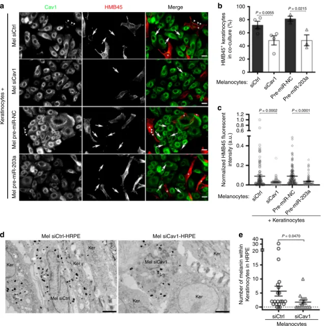
Loss of caveolae impairs melanin transfer in 2D co-culture and
3D-epidermis. Skin pigmentation relies on the synthesis of the
melanin within melanocytes, and its transfer and maintenance
within neighboring keratinocytes. Different mechanisms have
been proposed for melanin transfer to occur
5,54and each one
requires the local remodeling of the plasma membrane of
mela-nocytes at the near vicinity of keratimela-nocytes. To address the role
of caveolae in melanin transfer, siCtrl- and siCav1-treated
mel-anocytes were co-cultured with keratinocytes for 3 days, after
which the cells were analyzed by immunofluorescence using
HMB45 anti-PMEL antibody
30(Fig.
4
a). Keratinocytes
co-cultured with Cav1-depleted melanocytes were less frequently
positive for HMB45 staining (Fig.
4
b) and, when positive, showed
decreased mean
fluorescence intensity (Fig.
4
c), reflecting in
overall that fewer melanin-positive
fibrils have been transferred.
This result shows that caveolae are required for the efficient
transfer of melanin from melanocytes to keratinocytes in
co-culture.
Interestingly, the microRNA-203a (miR-203a) downregulates
Cav1 expression in melanoma cells
55. Likewise, normal human
melanocytes transfected with the pre-mir-203a showed decreased
Cav1 protein expression levels (Supplementary Fig. 4a). When
co-cultured with melanocytes treated with pre-miR-203a, melanin
transfer had occurred in fewer keratinocytes (Fig.
4
a, b) whose the
mean
fluorescence intensity (HMB45) was decreased (Fig.
4
c).
The miR-203a is secreted by keratinocytes together with
exosomes
8, which suggests that keratinocytes could regulate
Cav1 expression levels and caveolae biogenesis in melanocytes in
order to control their signaling and mechanical responses and,
ultimately, the transfer of melanin.
Finally, we sought to establish the importance of caveolae in
pigment transfer in vivo. For this purpose, we used the model of
skin epidermis (3D-HRPE) and generated three different types of
epidermises composed of normal human keratinocytes either
alone (Ker-HRPE) or associated with control- or Cav1-depleted
melanocytes. The expression of Cav1 mRNAs was efficiently
downregulated after siCav1 treatment in melanocytes
(Supple-mentary Fig. 4b). Macroscopic examination of the reconstructed
tissue showed unpigmented epidermis when composed of only
keratinocytes, and homogenous pigmented epidermis when
control melanocytes were added (Supplementary Fig. 4c; left
and middle panels, respectively). In contrast, a non-homogenous
pigmentation was observed in the epidermis reconstructed with
siCav1-treated melanocytes (Supplementary Fig. 4c, arrow; right
panel). The pigmentation defect was further characterized at the
ultrastructural level (Fig.
4
d) and revealed that keratinocytes
juxtaposed with Cav1-depleted melanocytes contained less
melanin than when adjacent to control cells (Fig.
4
e and
Supplementary Table 4). These data show that caveolae have a
novel and critical role in melanin transfer from melanocytes to
keratinocytes in the human epidermis.
Discussion
Pigmentation of the human epidermis represents a natural body
photo-protective screen that relies on melanocytes and
kerati-nocytes. To adapt to their environment, like during intense solar
exposure, these epidermal cells communicate to orchestrate
cel-lular responses important for producing and disseminating the
pigment through the tissue. In this study, we provide evidence for
a physiological role of caveolae in human epidermis
pigmenta-tion. By exploiting the signaling and mechanical functions of
caveolae, melanocytes respond to the extracellular signals sent by
keratinocytes to potentiate skin photoprotection. The capacity of
caveolae to modulate intracellular signals, to provide
mechan-oprotection and to support the morphological changes in
mela-nocytes defines them as a molecular platform required for human
skin pigmentation.
The asymmetric distribution (or enrichment) of caveolae in
melanocytes are positively regulated during the formation of skin,
by keratinocyte-secreted factors and by solar mimicking UV-B
radiation, indicating that specific plasma membrane domains of
melanocytes are preferentially dedicated to the biogenesis,
dis-tribution and/or stabilization of caveolae. In addition, the
miR-203a secreted together with keratinocytes extracellular vesicles
8can target Cav1 in both melanoma cells
55and normal
melano-cytes. This indicates that extracellular factors secreted by
kerati-nocytes contribute directly to
fine-tune Cav1 expression and
caveolae in melanocytes, so that cellular responses are organized
and coordinated. A downregulation of Cav1/caveolae would
promote pigment production in melanocytes whereas an
upre-gulation would favor changes in cell morphology and cell–cell
contacts, both leading to melanin transfer and skin pigmentation.
Melanocytes devoid of caveolae have higher production of
intracellular cAMP after stimulation, whereas treatment with the
Cav1-scaffolding domain (CSD) mimicking peptide, CavTratin,
has an opposite effect. A classical view of caveolae function in
signaling is associated with the intracellular
compartmentaliza-tion and concentracompartmentaliza-tion of different signaling transduccompartmentaliza-tion
path-way components
20. In this context, caveolin-1 could regulate the
Fig. 3 Caveolae contributes to changes in melanocyte morphology, contacts with keratinocytes and mechanoprotection. a IFM images of melanocytes treated with Ctrl (control) or Cav1 (caveolin-1) siRNA and incubated for 14 h with poor medium (+DMSO), supplemented medium (+DMSO), poor medium+ FSK (forskolin, 30 µM) or Ker-CM (keratinocytes-conditioned medium) for 14 h, immunolabelled for Cav1 (caveolin-1, green) and stained for F-actin (phalloidin, red). Cav1 polarization, arrowheads; cell protrusions, asterisks. Bars, 20µm. b Frequency of melanocytes [as in a] showing at most two (≤2) or more than two (>2) membrane protrusions (n = 150 cells, three independent experiments). c Quantification of the length-to-width ratio of melanocytes cultured as ina. d, e Melanocytes treated for 72 h with siCtrl or siCav1 were co-cultured with keratinocytes for 14 h prior to cell imaging. d Representative projection of time-lapse images with interpolated region of interest for the cell’s boundaries every 20 min. Bars, 10 µm. See also Supplementary Videos 2 and 3.e Frequency of keratinocytes contacting melanocytes for a total of 4 h. f Immunoblot analysis of p-MLC (phosphorylated myosin light chain) and MLC (myosin light chain) levels in siCtrl- and siCav1-treated melanocytes maintained for 4 h in poor medium, supplemented medium or poor medium+ FSK. g Quantification of MLC activation in f, corresponding to the ratio of p-MLC on MLC total expression level after GAPDH (glyceraldehyde 3-phosphate dehydrogenase) normalization.h, i Melanocytes treated with siCtrl or siCav1 for 72 h were incubated with calcein-AM (green) for 15 min, washed and subjected to hypo-osmotic shock (30 mOsm) in the presence of PI (propidium iodide, red) for 10 min. PI-positive cells (red nuclei) indicate melanocytes with ruptured plasma membrane. See also Supplementary Videos 6 and 7.h First (0 min) and last (10 min) still images from the time-lapse acquisition. Bars, 50µm. i Frequency of bursting melanocytes. Values are the mean ± s.e.m. b, c, e Three independent experiments. i siCtrl, n = 714 cells, siCav1, n = 958 cells; three independent experiments. g Six independent experiments. b, c One-way ANOVA with Tukey’s multiple-comparison test;e two-tailed pairedt-test; g one-way ANOVA with Dunnett’s multiple-comparison test; i two-tailed unpaired t-test with Welch’s correction. See also Supplementary Table 3.
activity of selected isoforms of tmACs in cells
56,57, whereas the
CavTratin peptide could negatively regulate these enzymes
in vitro, as reflected by the concomitant decrease of cAMP
pro-duction after stimulation
22. So, and even if the mode of action of
the CSD is still debated
58, this shows that caveolae mitigate the
cAMP-dependent signaling in melanocytes through the possible
interaction of Cav1 with tmACs to inhibit their catalytic activity.
In response to increased cAMP production, Cav1-depleted
melanocytes display lower NMMIIs activity, which results in
diminished contractile forces
51, similarly to Cav1-KO
fibro-blasts
59. Consequently, less cortical tension is generated on the
plasma membrane, which is necessary for changes in
morphol-ogy, and may explain the marked failure of melanocytes to extend
dendritic-like protrusions in the absence of caveolae. This
strongly suggests that caveolae couple cAMP-induced signaling to
the mechanical response of melanocytes. This feature of caveolae
might not only be restricted to melanocytes and is likely shared
by neural crest-derived cells. Indeed, the modulation of cAMP
levels in the vicinity of membrane lipid rafts controls dendritic
arborization in mice neurons
60, whereas neuron-targeted Cav1
enhances branching out of the dendrites
61. Dendrite outgrowth in
human melanocytes and murine melanoma cells is also
depen-dent on cAMP
12. In addition, non-stimulated Cav1-depleted
melanocytes seem to display higher p-MLC—yet without impact
on the cell shape. That would suggest that caveolin-1 and caveolae
might represent key elements modulating the activity of NMMIIs
in very different cell types, like melanocytes or
fibroblasts
59, and
under resting and stimulated conditions.
Mel siCtrl-HRPE Ker 100 P = 0.0055 P = 0.0215 P = 0.0002 P < 0.0001 80 60 40 20 0 1.2 1.0 0.8 0.6 0.4 0.2 0.0 40 30 20 15 10 5 0 Mel pre-miR-203a Mel pre-miR-NC Pre-miR-NCPre-miR-203a Mel siCa v1 K e ratinocytes + Mel siCtr l Mel siCav1-HRPE Cav1 HMB45 Merge Ker Ker Ker Ker Ker Mel siCav1 Mel siCtrl HMB45 + k e ra tinocytes in co-culture (%) Nor malized HMB45 fluorescent intensity (a.u.)
Number of melanin within K
e ratinocytes in HRPE siCtr l siCa v1 Melanocytes: Pre-miR-NCPre-miR-203a siCtr l siCa v1 siCtrl siCav1 Melanocytes: Melanocytes + Keratinocytes P = 0.0470
a
b
c
e
d
Fig. 4 Caveolae in melanocytes are necessary for melanin transfer in vitro and in tissue. a–c Melanocytes treated with Ctrl (control) or Cav1 (caveolin-1) siRNAs, pre-miR-NC (pre-miRNA-negative control) or pre-miR-203a (pre-miRNA-203a) for 5 days were co-cultured with keratinocytes for the last 2 days. a IFM images of the co-culture immunolabeled for Cav1 (caveolin-1, green) and HMB45 (premelanosome protein PMEL, red). Keratinocytes positive for HMB45-transferred melanin, arrows; melanocytes in merge panels, asterisks. Bars, 20µm. b Quantification of the frequency of keratinocytes positive for HMB45-positive structures in each condition.c Quantification of HMB45 fluorescent intensity in individual keratinocytes positive in b. d Conventional EM micrographs of 9 days 3D-HRPE (human reconstructed pigmented epidermis) composed of keratinocytes and siCtrl- or siCav1-treated melanocytes. Bars, 2µm. e Quantification of the number of pigmented melanosomes in keratinocytes at the vicinity of melanocytes. Values are the mean ± s.e.m. b, c Four independent experiments for siCtrl and siCav1 and three independent experiments for pre-miR-NC and pre-miR-203a;e one experiment. b Two-tailed pairedt-test; c, e, two-tailed unpaired t-test with Welch’s correction. See also Supplementary Table 4.
Endogenous Cav1 and Cavin1, and therefore caveolae,
dis-tribute asymmetrically and cell-autonomously in cultured human
melanocytes. And polarization of Cav1 and caveolae is observed
in different cells during cell migration
50,62. However and alike
immortalized mouse melanocytes
63, normal human melanocytes
are poorly motile, which suggests that caveolae asymmetric
dis-tribution in these cells should perform functions unrelated to cell
migration. In addition to caveolae asymmetry, melanocytes are
likely polarized cells because their shape consists of a cell body
facing the basal membrane with multiple dendrites extending
upwards and because they express proteins specific of epithelial
cells
64. Therefore, we propose that the intrinsic asymmetrical
distribution of caveolae imposes a spatial organization of
cAMP-dependent pathways and/or downstream targets in melanocytes
that contribute to its polarized organization and ensure its cellular
functions.
Caveolae are required for two crucial functions in melanocytes:
pigment production and transfer. Stimulation of Cav1-depleted
melanocytes causes increased cAMP levels and acceleration of
pigment production, through the upregulation of Tyrosinase and
DCT mRNA and protein expression levels. Pigment synthesis and
packaging into melanosomes rely on intracellular signaling
pathways, among which cAMP synthesis by tmACs is of key
importance
9. The activation of the GPCR-triggered cAMP
path-way increases Tyrosinase and DCT proteins content through
increased cell transcriptional activity
11or post-translational
events
43,65. This indicates that caveolae is key in regulating the
production of the pigment through the
fine control of cAMP
production and downstream pathways.
The fate of melanin in the epidermis is to be transferred to
keratinocytes, where it provides color to skin, in addition to
shielding the nuclei against UV radiation. Here, we establish a
correlation between caveolae formation and human skin
pig-mentation. Caveolae accumulate at melanocytes when the
epi-dermis becomes pigmented, while impaired caveolae formation in
melanocytes, through Cav1 depletion, decreases the transfer of
melanin in co-culture and reconstructed epidermis. The dendrites
of melanocytes are seen as conduits for melanin transfer and
points of contact with keratinocytes and, therefore, their plasticity
appears to be important to support these functions. Our results
show that caveolae are needed for dendricity of melanocytes, for
promoting contacts with keratinocytes and for protecting the
plasma membrane of melanocytes against acute rupture after a
mechanical stress, thus helping the cells adjust to tension
varia-tions. Indeed, both plasma membrane and cortical tension
51contribute to deform membranes during exo- and endocytosis or
changes in cell shape
53,66. Furthermore, the dynamic cycle of
caveolae mechanics, i.e. disassembly and reassembly in response
to tension variations that occur during cell morphological
chan-ges, could facilitate both dendrite outgrowth and pigment transfer
by melanocytes. Nonetheless, the formation of caveolae and
non-caveolae Cav1-positive clusters could also exert a spatiotemporal
control on melanin secretion by favoring the local remodeling of
the plasma membrane in response to signaling cues. Therefore,
the coupling of signaling and mechanical responses by caveolae in
melanocytes seems important for the regulation of pigment
transfer.
Dysregulation of Cav1 expression in the human skin is
asso-ciated with hyperproliferative diseases such as melanoma and
non-melanoma cancers as well as psoriasis
67,68. In melanoma,
Cav1 function remains very controversial, since it is recognized as
a tumor suppressor and an oncogene
69. Such discrepancy might
be explained by the variations of Cav1 expression during disease
progression, in that the balance between caveolae signaling and
mechanical functions in response to the extracellular
environ-ment changes during tumor mass growth
70. Long-term exposure
to UV radiation is a key factor causing skin cancers
71and high
levels of expression of the miR-203a occur in psoriatic lesions
72.
We therefore propose caveolae to be an additional modulator of
skin pigmentation that couple signaling with mechanical
responses in melanocytes. Thus, characterizing the physiology
and modulations in response to the extracellular context
under-lying the functions of caveolae in the skin is a key to decipher
their alterations in the disease.
Methods
Antibodies. The following antibodies were used for immunoblot (IB) or immu-nofluorescence (IFM): rabbit anti-Caveolin-1 (BD Transduction Laboratories; 1:5000 [IB]; 1:200 [IFM]); rabbit anti-PTRF (Cavin1; Abcam; 1:200 [IFM]); mouse anti-HMB45 (recognizing PMEL-positivefibrils onto which melanin deposits, used here as a‘melanin marker’; clone HMB45; abcam; 1:200 [IFM]); mouse anti-α adaptin (clone AP6; abcam; 1:50 [IFM]); mouse anti-Tyrosinase (clone T311; Santa Cruz biotechnology; 1:200 [IB]); mouse anti-DCT (clone C-9; Santa Cruz bio-technology; 1:200 [IB]); rabbit anti-Pep13h4(1:200 [IB]); goat anti-Rab27a
(SIC-GEN; 1:1000 [IB]); rabbit anti-phosphorylated CREB (Ser133) (clone 87G3; Cell Signaling technology; 1:1000 [IB]); rabbit anti-CREB (clone 48H2; Cell Signaling Technology; 1:1000 [IB]); mouse anti-phosphorylated MLC2 (Ser19) (Cell Sig-naling Technology; 1:1000 [IB]); rabbit anti-MLC2 (clone D18E2; Cell SigSig-naling Technology; 1:1000 [IB]); mouse anti-ACTB (β-actin; clone AC-74; Sigma; 1:2000 [IB]); rabbit anti-GAPDH (Sigma; 1:10000 [IB]); rabbit anti-Calnexin (Enzo Life Sciences; 1:1000 [IB]). Secondary antibodies coupled to horseradish peroxidase (HRP) were used at 1:10000 ([IB], Abcam). Secondary antibodies and phalloidin conjugated to 488, 555 and 647 Alexa dyes were used at 1:200 ([IFM], Invitrogen). Cell culture. Primary cells: Normal human epidermal melanocytes and normal human epidermal keratinocytes used in this study were isolated from neonatal foreskins and purchased from CellSystems, Sterlab or PromoCell. Melanocytes and keratinocytes were used from passage two andfive and maintained in culture in DermaLife Basal Medium supplemented with DermaLife M Life factors (melano-cytes-supplemented medium) or in DermaLife Basal Medium supplemented with DermaLife K Life, respectively.
Cell line: HeLa cells were cultured in DMEM supplemented with 10% (v/v) FBS, 100 U/ml penicillin G and 100 mg/ml streptomycin sulfate (Gibco). All cells were maintained at 37 °C in a 5% (v/v) CO2incubator.
Co-cultures and media incubation. Co-cultures: Melanocytes and keratinocytes or HeLa were seeded in the following ratio, respectively: 1:4 for 24 h beforefixation to quantify caveolae asymmetry (Fig.1); at 1:4 for 14 h before time-lapse acquisition (Fig.3); and 1:1 for 3 days beforefixation to quantify melanin transfer (Fig.4). All co-cultures were done in melanocyte-supplemented medium.
Media incubation (IFM and IB): Keratinocyte medium from a confluent flask in culture for 48 h was collected and centrifuged at 200×g to remove cell debris. The Keratinocyte-conditioned medium (Ker-CM) was immediately used or stored at −80 °C (Fig.1). Melanocytes were seeded and maintained in poor medium (DermaLife Basal Medium without the addition of StiMel8) for at least 3 h after which this medium was removed, the cells washed in phosphate-buffered saline (PBS) and fresh poor medium or poor medium supplemented with 30 µM of forskolin (FSK, Sigma) or with melanocyte-supplemented medium (see above), or Ker-CM was added and kept for ~14 h beforefixation for IFM or 15 min to probe for p-CREB/CREB or 4 h to probe for p-MLC/MLC by IB. Dimethylsulfoxide (DMSO, between 0.2 to 0.6%) was added to the medium as a control to FSK addition.
siRNA and miRNA transfections. For melanocytes siRNA and miRNA trans-fections, cells were seeded in the appropriate wells or plates and transfected with 0.2 µM of siRNA using Oligofectamine (Invitrogen) accordingly to manufacturer’s instructions using non-targeting siRNA (siCtrl; 5′-AATTCTCCGAACGTGT-CACGT-3′) and siRNA targeting Cav1 (SI00299635 and SI00299628) from Qiagen, or using pre-miR-NC (negative control; #AM17111) and pre-miR-203a (#AM17100) from ThermoFischer Scientific. In 3D-HRPE experiments, melano-cytes were transfected previously to reconstruction with 1 µM of siRNA using DharmaFECT and following the manufacturer’s protocol (Dharmacon, Horizon) using non-targeting siRNA (Accell non-targeting pool) or siRNA targeting Cav1 (SMARTpool: Accell Cav1) from Dharmacon.
UV treatment. Melanocytes and keratinocytes were seeded in six-well plates at day 0 and irradiated with a single shot of 11 mJ cm−2of ultraviolet-B (312 nm) during 3 consecutive days using a Biosun machine (Vilber Lourmat, Suarle´e, Belgium). Cell medium was replaced by PBS before irradiation and replaced by the culture medium just after the treatment. The cells were then incubated overnight and recovered by trypsinization at the indicated time points.
Skin samples. Healthy skin samples were obtained from surgical left-over residues of breast or abdominal reduction from healthy women. Written informed consent was obtained in accordance with the Helsinski Declaration and with article L.1243-4 of the French Public Health Code. Given its special nature, surgical residue is subject to specific legislation included in the French Code of Public Health (anonymity, gratuity, sanitary/safety rules etc). This legislation does not require prior authorization by an ethics committee for sampling or use of surgical waste
(http://www.ethique.sorbonne-paris-cite.fr/?q=node/1767).
Human reconstructed epidermis (3D-HRPE). The following protocol was adapted from Salducci et al.73. Briefly, dead de-epidermized dermis was prepared as
follows: Skin samples from healthy adults were obtained, cut in circular pieces (18 mm diameter) and incubated 20 min at 56 °C in HBSS (Invitrogen) containing 0.01% (v/v) Penicillin/Streptomycin (Invitrogen). Epidermis was removed and collected dermis fragments were sterilized in 70° ethanol, washed twice in HBSS, frozen in HBSS (−20 °C) and submitted to six cycles of freezing-thawing to eliminatefibroblasts. The de-epidermized dermis was placed at the bottom of a 6-well plate in 3D-HRPE culture medium composed of IMDM medium (Invitrogen) and keratinocyte medium (CellSystems) at a proportion of 2/3 to 1/3, respectively, and containing 10% (v/v) of calf fetal serum gold (PAA). siRNA-treated melano-cytes and non-treated keratinomelano-cytes were seeded at a proportion 1:20, respectively, in a culture insert of 8 mm of diameter affixed on the dermis to promote cell adhesion. After 24 h, the culture insert was removed and the de-epidermized dermis submerged for 3 days in 3D-HRPE culture medium to promote cell pro-liferation. Tissue stratification was initiated by moving up the de-epidermized dermis to the air–liquid interface. All the incubation steps were performed at 37 °C in a 5% CO2incubator. The number of melanocytes and keratinocytes counted on
EM cross-sections of 3D-HRPE provides an estimation of the ratio of the two cells types within the reconstructed tissue. The siCtrl-HRPE consisted of siCtrl-mela-nocyte:keratinocyte ratio of 1:3.6 (17 siCtrl-melanocytes, 61 keratinocytes); the siCav1-HRPE consisted of siCav1-melanocyte:keratinocyte ratio of 1:2.7 (13 siCav1-melanocytes, 35 keratinocytes).
Measurement of intracellular cAMP levels. Melanocytes were transfected once with the indicated siRNAs and maintained in poor medium for 24 h. DMSO or 30 µM of FSK were added to the respective wells for 3 h after which the cells were collected and the intracellular cAMP content measured using the cAMP complete ELISA kit (Enzo Life Sciences) following manufacturer’s instructions. Regarding the treatment with the peptides, NHEMs were maintained in poor medium for 14 h before the addition of the peptides Ctrl (scrambled sequence) or CavTratin (Cav1-scaffolding domain, CSD) for 7 h. Then the cells were incubated for 1 h with DMSO or 30 µM of FSK after which the cells were collected and the intracellular cAMP content measured.
Melanin assay. Melanocytes were transfected twice at days 1 and 3 for a total of 5 days with the indicated siRNAs. Cells were then collected, sonicated in 50 mM Tris-HCl pH 7.4, 2 mM EDTA, 150 mM NaCl, 1 mM dithiothreitol (with the addition of protease inhibitor cocktail, Roche) and pelleted at 20,000×g for 15 min at 4 °C. The pigment was rinsed once in ethanol:ether (1:1) and dissolved in 2 M NaOH with 20% (v/v) DMSO at 60 °C. Melanin content was measured by optical density at 490 nm (Spectramax 250, Molecular Devices).
Membrane bursting assay. Melanocytes were transfected twice with the indicated siRNAs at days 1 and 3 for a total of 3 days and seeded in 12-well plates for 24 h in supplemented medium. At day 4, cells were incubated in 5 µg/ml of Calcein-AM (Life technologies) for 15 min at 37 °C protected from light. The wells were washed once with melanocyte-supplemented medium and maintained until image acqui-sition. Melanocyte-supplemented medium was diluted in 90% (v/v) water, the equivalent of 30 mOsm hypo-osmotic shock, followed by the addition of 2 mg/ml of propidium iodide (PI, Sigma) and used to induce the rupture of the plasma membrane29. Immediately after the medium replacement, images were acquired
every minute for a total of 10 min in an inverted microscope (Eclipse Ti-E, Nikon), equipped with a CoolSnap HQ2 camera, using the 20×0.75 NA Plan Fluor dry objective together with MetaMorph software (MDS Analytical Technologies). Melanin transfer assay. The detailed protocol for the melanin transfer assay is described elsewhere74. Melanocytes were transfected twice with the indicated
siRNA or miRNA at days 1 and 3 for a total of 5 days. At day 3, melanocytes were co-cultured with keratinocytes for a total of 2 days. Images were acquired with an upright epi-fluorescence microscope (Eclipse Ni-E, Nikon) equipped with a CoolSnap HQ2 camera, using a 40×1.4 NA Plan Apo oil immersion objective together with MetaMorph software.
Immunofluorescence microscopy. Cell monolayers seeded on glass coverslips werefixed with 4% (v/v) paraformaldehyde in PBS at room temperature for 15 min, then washed three times in PBS and once in PBS containing 50 mM glycine. Primary and secondary antibody dilutions were prepared in the buffer A: PBS containing 0.2% (w/v) BSA and 0.1% (w/v) saponin. The coverslips were
washed once in buffer A and then incubated for 1 h at room temperature (RT) with the primary antibodies. Following one wash step in buffer A, the coverslips were incubated for 30 min at RT with the secondary antibodies. For phalloidin staining, the coverslips were washed in buffer A and incubated overnight in the same buffer with phalloidin at 4 °C. Thefinal wash step was done once in buffer A, once in PBS and once in water. The coverslips were mounted onto glass slides using ProLong™ Gold Antifade Mount with DAPI (ThermoFischer Scientific). Images were acquired on an Applied Precision DeltavisionCORE system (unless stated otherwise), mounted on an Olympus inverted microscope, equipped with a CoolSnap HQ2 camera (Photometrics), using the 40×1.3 NA UPLFLN or the 60×1.42 NA PLAPON-PH oil immersion objectives. Images were deconvolved with Applied Precision’s softWorx software (GE Healthcare).
Time-lapse microscopy. Melanocytes were transfected twice with the indicated siRNA molecules at days 1 and 3 for a total of 3 days and co-cultured with keratinocytes in an ibidi polymer coverslip µ-slide (Ibidi) for 14 h before imaging. Images were acquired every 5 min for a total of 240 min in an inverted microscope (Eclipse Ti-E, Nikon), equipped with a CoolSnap HQ2 camera, using the 40×0.75 NA Plan Fluor dry objective together with NIS-Elements software (Nikon).
Electron microscopy. Conventional EM: Human skin epidermis tissues and 3D-HRPE were prepared for EM as described. For high-pressure freezing, the tissue was high-pressure frozen using an HPM 100 (Leica Microsystems) in FBS serving asfiller and transferred to an AFS (Leica Microsystems) with precooled (−90 °C) anhydrous acetone containing 2% (v/v) osmium tetroxide and 1% (v/v) of water. Freeze substitution and Epon embedding was performed as described in Hurbain et al.75. For chemicalfixation, melanocytes seeded on coverslips and transfected
twice with the indicated siRNAs at days 1 and 3 for a total of 5 days werefixed in 2.5 % (v/v) glutaraldehyde in 0.1 M cacodylate buffer for 24 h, post-fixed with 1% (w/v) osmium tetroxide supplemented with 1.5% (w/v) potassium ferrocyanide, dehydrated in ethanol and embedded in Epon as described in Hurbain et al.76.
Ultrathin sections of cell monolayers or tissue were prepared with a Reichert UltracutS ultramicrotome (Leica Microsystems) and contrasted with uranyl acetate and lead citrate.
Electron tomography: 300-nm-thick sections were randomly labeled on the two sides with 10 nm Protein-A gold (PAG). Tilt series (2 perpendicular series, angular range from−60° to +60° with 1° increment) were acquired with à Tecnai 20 electron microscope (ThermoFischer Scientific). Projection images (2048 × 2048 pixels) were acquired with a TEMCAM F416 4k CMOS camera (TVIPS). Tilt series alignment and tomogram computing (resolution-weighted back projection) were performed using eTomo77(IMOD) software. PAG 10 nm at the surface of the
sections was used asfiducial markers. Manual contouring of the structures of interest was performed using IMOD78.
Immuno-EM: Cell samples werefixed with 2% PFA in a 0.1 M phosphate buffer pH 7.4 and processed for ultracryomicrotomy as described in Hurbain et al.76.
Ultrathin sections were prepared with an ultracryomicrotome UC7 FCS (Leica) and underwent single immunogold labeling with protein-A conjugated to gold particles 10 nm in diameter (Cell Microscopy Center, Department of Cell Biology, Utrecht University). All images were acquired with a Transmission Electron Microscope (Tecnai Spirit G2; ThermoFischer Scientific, Eindhoven, The Netherlands) equipped with a 4k CCD camera (Quemesa, EMSIS, Muenster, Germany).
Image analysis and quantifications. Conventional EM: Caveolae and clathrin-coated pits16, and melanosome stages were identified based on their ultrastructural
features4. Caveolae structures associated with plasma membranes of randomly
selected cell profiles were quantified from 2D ultrathin sections of 3D-HRPE. The length of the plasma membranes either of melanocytes or keratinocytes were measured using ITEM software (EMSIS) and the total number of caveolae found associated was reported to 10 µm of plasma membrane of the respective cell type. For melanosome stage quantification, the areas corresponding to the tips of the cells were not considered.
Immunoblot: Quantification of protein content on western blot was performed using Fiji software, the background subtracted and intensities were normalized to loading control.
Caveolae asymmetric distribution by IFM: Images of endogenous staining for Cav1 and Cavin1 asymmetrically distributed in co-culture were acquired and the background subtracted. Two identical boxes were positioned at the plasma membrane but on opposite sides of the cells and the averagefluorescent intensity retrieved. The frequency of Cav1 and Cavin1 asymmetry in melanocytes was defined by identifying cells with one side presenting enriched labeling closely associated with the plasma membrane.
Protrusions and cell morphology: A protrusion was defined as an actin-stained extension that originated from the soma of the cell. Isolated cells-treated with siCtrl and siCav1 were selected randomly, imaged and the size parameters (area, length-to-width ratio, major and minor axis) were retrieved. The contour of the cell was achieved by using the wand tool and corrected manually if needed using the tool OR (combine).