Fibronectin, Fibrinogen, and Laminin Act as Mediators of Adherence
of Clinical Staphylococcal Isolates to Foreign Material
Mathias Herrmann, Pierre E. Vaudaux, Didier Pittet, Raymond Auckenthaler, P. Daniel
Lew,
Franeolse Schumacher-Perdreau, Georg Peters, and Francis A. Waldvogel
From the Infectious Diseases Division, University Hospital, Geneva, Switzerland; and the University of Cologne, Cologne, Federal Republic of Germany
Bacterial adherence to polymer surfaces is a required early step in intravenous (iv) device infection. Wecollected eight strains of
Staphylococcus aureus
and 19of coagulase-negative staphylococci from patients with proven iv device bacteremia and studied the role of plasma or connective-tissue proteins in promoting bacterial adherence to polymethylmethacry-late (PMMA) coverslips. Although only a negligible percentage of organisms adhered to albumin-coated PMMA, surface-bound fibronectin significantly promoted adherence of all isolates. Fibrinogen markedly promoted adherence of allS. aureus
strains but of only four coagulase-negative strains. Thus, coagulase-negative staphylococci revealed a marked heterogeneity in adherence to fibrinogen-coated surfaces, a result suggesting the existence of heretofore unknown receptors for fibrinogen. Laminin promoted adherence of staphylococci to a much lower extent. Although strain specific, adherence of clinical staphylococcal isolates to foreign surfaces is significantly increased by fibronectin, fibrino-gen, and laminin, an observation suggesting the possible contribution of these proteins to the pathogenesis of iv device infection.Adherence of microorganisms to specific substrates
is presently considered to be a crucial step in the
ini-tiation of infections [1]. Many investigators have
searched for such ligands in staphylococcal
prosthe-sis infection. There is increasing evidence that
simi-lar specific interactions might also playa role in the
pathogenesis of foreign-body infections.
After contact with blood, a polymer surface (such
as a cannula inserted iv) is almost immediately coated
with a protein layer at the blood-polymer interface
[2-4]. Bacterial adherence to the protein-coated
sur-face is a prerequisite for initiating iv device
infec-tion. Fibrinogen and fibronectin are proteins known
to bind and to aggregate staphylococci [5,6]. More
recently, laminin, a large glycoprotein mainly found
Receivedfor publication 2 December 1987and in revised form 7 April 1988.
This paper was presented in part at the 27th Interscience Con-ference on Antimicrobial Agents and Chemotherapy (abstract 503), held on 4-7 October 1987, in New York, New York.
This work was supported by grant 3.829-0.87 from the Swiss National Research Foundation.
We thank Elzbieta Huggler for technical assistance, Dr. Inge-borg Filthuth and Chantal Genier for isolation and character-ization of the bacterial isolates, and Paule Schilling-Doriot for manuscript preparation.
Please address requests for reprints to Dr. Mathias Herrmann, Infectious Diseases Division, Department of Medicine, Univer-sity Hospital, CH-1211 Geneva 4, Switzerland.
693
in basal membranes and to a slight extent in plasma,
has also been described as possessing
staphylococcal-binding properties [7]. Several studies have
inves-tigated staphylococcal adherence [8-11] to polymer
surfaces. In particular, studies performed on
ex-planted foreign bodies [12, 13]and on in vitro models
[14, 15] strongly suggested that adherence of selected
staphylococcal strains was promoted by fibronectin.
The relative role of these various plasma proteins in
promoting adherence of clinically relevant strains has
not yet, however, been studied in detail. In this
pa-per we analyzed adherence of bacteremic strains of
Staphylococcus aureus
and coagulase-negative
staph-ylococci to polymer surfaces and, in particular, the
influence of surface-bound fibronectin, fibrinogen,
and laminin.
Materials and Methods
Chemicals andmaterials.
PBS solutions
contain-ing 1
mM
Ca" and 0.5
mM
Mg'" were purchased
from GIBCO (Paisley,Scotland); [3H]thymidine was
purchased from Amersham (Buckinghamshire,
En-gland).
Fibronectin was purified as previously described
[13]. Fibrinogen (provided by Dr. Haeberli,
Univer-sity Hospital, Bern, Switzerland) was further
puri-fied from trace amounts of fibronectin by affinity
chromatography on gelatin-Sepharose 4B [13], and
the final protein concentration was determined by
the Bio-Rad" protein assay (Bio-Rad Laboratories,
Glattbrugg, Switzerland). Laminin was purchased
from Bioreba-Diagnostica (Basel, Switzerland).
Ex-amination of the different proteins by using 7070
SDS-PAGE under reducing conditions [16] revealed the
expected bands; no significant contaminants were
seen. All protein solutions were dispensed into
ali-quots and stored at - 70 C. Coverslips (1
x
1 em)
made of polymethylmethacrylate (PMMA) were
cleaned with 100070 ethanol and sterilized by
heat-ing at 49 C for 30 min.
Staphylococcal isolates. We investigated 62
strains of staphylococci that, according to the
clini-cal data obtained, were subdivided into three
differ-ent groups. These strains were isolated from patidiffer-ents
at the University Hospital of Geneva (Geneva), from
patients at the University Hospital of Cologne
(Co-logne, FRG), and from healthy volunteers.
Twoad-ditional clinical strains were provided by Dr. G.
Christensen (University of Tennessee, Memphis).
Tlie three groups were defined as follows. Group
1 included strains isolated from iv device infection.
Twenty-seven isolates were obtained from 27 patients
with proven septicemia associated with an iv device.
This classification was based on the following
criteria:
(1)
two or more positive cultures of blood
yielding the same organism;
(2)
fever >38 C that was
frequently associated with chills and sometimes
as-sociated with hemodynamic alterations (tachycardia,
hypotension, low central-venous pressure);
(3)leu-kocytosis (>10 OOO/mm
3) ;
(4)
positive culture of the
catheter tip yielding the same organism and
anti-biogram as found in the cultures of blood (available
in 24 of 27 cases); and
(5)presence of an iv device,
inserted before the onset of infection, in a patient
showing clinical signs of acute inflammation and
who improved after removal of the device and
initi-ation of antibiotic therapy. The clinical data were
evaluated twice and independently by two
investi-gators (M. H. and D. P.).
Group 2 included strains from patients with
sep-ticemia. Nineteen isolates were obtained from 18
pa-tients who met the aforementioned criteria, except
the septicemia was not associated with an iv device
infection.
Group 3 included strains from controls. Sixteen
isolates were derived from the skin of healthy
volun-teers.
All isolates were identified as staphylococci in the
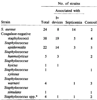
Table 1. Number of isolates and identification of in-vestigated staphylococcal strains.
No. of strains Associated with Iv
Strain Total devices Septicemia Control
S. aureus 24 8 14 2 Coagulase-negative staphylococci 38 19 5 4 Staphylococcus epidermidis 22 14 3 5 Staphylococcus haemolyticus 5 3 2 Staphylococcus hyicus Staphylococcus xylosus Staphylococcus warneri 4 3 Staphylococcus simulans 1 1 Staphylococcus spp.
*
4 2*
These strains could not be identified.diagnostic microbiology laboratories of the
Univer-sity Hospital of Geneva and the UniverUniver-sity
Hospi-tal of Cologne. Species identification was performed
according to the procedure described by Kloos and
Schleifer [17]. The results of bacterial and clinical
determinations are shown in table 1.
Slime production was assessed qualitatively by
using the tube method and quantitatively by
adher-ence of organisms to tissue culture plates and by
spec-trophotometric determination, as previously
de-scribed by Christensen et al. [18, 19].All strains were
kept on Mueller-Hinton agar plates at 4 C.
Preparation of bacteria for adherence
experi-ments. Each clinical or laboratory isolate of a fresh
overnight culture was grown and radiolabeled with
[3H]thymidine, as previously described [15], in
Mueller-Hinton broth for 3 hat 37 C (final
concen-tration, 2
X10
7-2X
10
8cfu). After the labeling
period and rinsing procedures [15], in most
experi-ments the strains were sonicated to remove possible
bacterial exopolymers [20] by using a B-12 sonifier
(Branson, Danbury, Conn) for 10 s (with 50 watts)
and were centrifuged. The strains were finally
sus-pended in saline and standardized for cfu and cpm
[15].
Attachment of strains to protein-coated
cover-slips. Adherence conditions were found to be
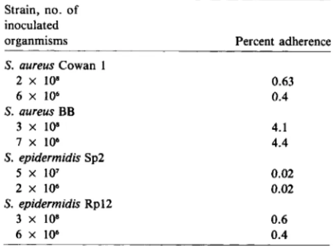
op-Table 2. Adherence of different numbers of inoculated organisms to polymethylmethacrylate.
NOTE. Staphylococcal organisms at the indicated concen-trations (20-fold differences) were incubated with coverslips precoated with fibronectin (25 IJ.g/mL) in the presence of 0.5% albumin; adherence was assessed as described in Materials and Methods. The results are averages of triplicate experiments.
shown). Variations in the quantity of proteins
ad-sorbed on PMMA did not affect adherence, because
staphylococci adhered to almost the same extent to
surfaces coated with a fivefold lower concentration
of proteins (results not shown).
Statistical analysis.
The Mann-Whitney test for
unrelated scores, the Wilcoxon test for related
rank-able scores, and the Fisher's exact test were
per-formed by using specific programs adapted to a
com-puter (model HP-41C; Hewlett-Packard, Geneva; or
MacIntosh; Apple, Cubertino, Calif).
Results
Adherence to uncoatedcoverslips and influence
of albumin on adherence.
In vitro assays, testing
characteristics of bacterial adherence to artificial
sur-faces in the absence of blood proteins, have shown
that there is adherence due to electrical charge and
to other physicochemicalbinding forces [11]. To
evalu-ate staphylococcal adherence to uncoevalu-ated PMMA
coverslips, in the absence of serum proteins, we
in-cubated 23 clinical isolates obtained from septicemic
patients with iv device infection (seven isolates of
S. aureus
and 16 of coagulase-negative strains) with
uncoated coverslips in PBS without albumin.
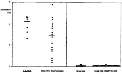
Ad-herence is shown in figure lA.
S. aureus
strains
ad-hered more homogeneously and more strongly to
PMMA (median, 2.1070; range, 1.3070-2.3(70) than did
4.1 4.4 0.63 0.4 0.02 0.02 0.6 0.4 Percent adherence Strain, no. of inoculated organmisms S.aureusCowan 1 2 x 10⁸ 6 X 10⁶ S.aureusBB 3
x
lQ8 7x
106 S. epidermidisSp2 5 X 10⁷ 2 X 10⁶ S. epidermidisRp12 3x
108 6 X 10⁶timal (0.5 I-Lg of fibronectin/cm") after coating
PMMA coverslips with 25 I-Lg of fibronectin/mL of
buffer solution [15]. Others have shown that
fibrino-gen binds at a concentration of 35 I-Lg/mL to a nearly
saturated extent to polymethacrylate (0.25 I-Lg of
fibrinogen/em") [21] and that optimal concentrations
of laminin for adherence of
S. aureus
to surfaces were
found to be 25 I-Lg/mL [22]. To assess the amount
of deposited laminin, we immersed multiple
cover-slips in a solution containing 25 I-Lg of laminin/mL
and measured the protein concentration in the
solu-tion before and after immersion. The average deposit
was found to be 0.3 I-Lg of laminin/cm". With indirect
immunofluorescence, we could demonstrate that
laminin was homogeneously deposited on PMMA
coverslips.
Each coverslip was immersed in a 1-mL solution
of PBS containing either fibronectin (25 I-Lg/mL),
fibrinogen (35 I-Lg/mL), or laminin (25 I-Lg/mL) for
1 hat 37 C. After being rinsed as previously described
[15], protein-coated coverslips were incubated with
10
6-107radiolabeled bacteria (as determined by cfu
counts) for 60 min at 37 C. The adherence medium
was PBS containing divalent cations with or
with-out 0.5070 human serum albumin (as indicated in the
text). At the end of the attachment period, the fluids
containing unbound bacteria were removed, and the
coverslips were rinsed and counted for
radioactiv-ity. Background radioactivity was subtracted, and
adherence was expressed as the percentage of
radio-activity remaining on the coverslips divided by the
amount of radioactivity used to label the coverslips.
All experiments were performed in triplicate. and
were repeated on at least two different days.
Validation ofthe adherence assay.
To minimize
bacterial aggregation and to remove residual
frag-ments of exopolymeric substances or slime [18,20],
we sonicated organisms, because adherence
experi-ments with sonicated and nonsonicated organisms
yielded no significant differences (median, 2.3070 and
2.4070, respectively;
n
=
23). To rule out possible
day-to-day variations in the number of inoculated
bac-teria, we tested four staphylococcal strains, each with
20-fold different concentrations of inoculated
organ-isms (table 2). The number of bacteria bound to
PMMA was proportional to the number of bacteria
inoculated; thus no significant difference in the
per-centage of bound bacteria (depending on the
inocu-lum size) could be found in our assay. Time courses
revealed that optimal adherence conditions were
ob-served after incubation for 60 min {results not
Adherence (%)
..
•
-
•
•
•
•
•
•
•
•
•
•
•
-.-•
•
a
•
•
•
coag.neg. staphylococci.
coag.neg. staphylococciFigure 1. Staphylococcal adherence
to uncoated polymethylmethacrylate
(PMMA). Sterile PMMA coverslips
were incubated for 60 min at 37 C in
PBS in the absence
(left)or presence
(right)
of
0.5070albumin with 5 x 10
6cfu of radiolabeled strains obtained
from patients with iv device infections
(S.
aureus, seven isolates;
coagulase-negative staphylococci,
16isolates).
Each dot indicates the average of
ad-herent radioactivity of triplicate
deter-minations performed twice. The
bars
indicate the median of the plotted
values.
without albumin with albumin
the coagulase-negative staphylococci (median,
1.25070; range, 0.2%-2.9070), but this difference was
not significant.
Implanted or inserted foreign bodies, such as iv
catheters, are rapidly covered to a variable degree
with serum proteins and blood cell deposits [23,24].
Thus, 0.5% human serum albumin was added to the
medium (figure IB). We found a striking reduction
of adherence: S.
aureus
strains had 0.0090/0 (median)
adherence to PMMA and the coagulase-negative
strains, 0.003% adherence. Therefore, albumin
in-hibited nonspecific adherence to >99%. We
consid-ered these observations to be maximum inhibition
of nonspecific bacterial adherence and performed
all further experiments in the presence of 0.5%
hu-man serum albumin in the incubation assay.
Adherence of
S.
aureus and coagulase-negative
staphylococci to surface-boundproteins.
S.
aureus
and coagulase-negative strains that had been
ob-tained from cultures of blood from patients with iv
device infection weretested for adherence to
protein-coated PMMA substrates (table 3). S.
aureus
adhered
significantly more than did coagulase-negative
staphylococci to fibronectin-coated
(P<
.02) and
fi-brinogen-coated
(P<
.002) PMMA. In comparison,
adherence to surface-bound laminin did not differ
as much; however, the S.
aureus
strains also adhered
more to laminin than did the coagulase-negative
strains
(P<
.02).
Promotion of adherence by
surface-boundpro-teins.
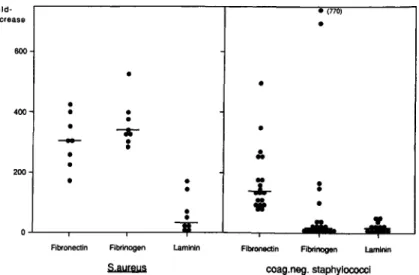
Adherence of the eight strains of S.
aureus
from iv device infections was strongly promoted by
surface-bound fibronectin (median increase,
307-fold; range,
177-425-fold) and surface-bound
fibrinogen (median, 335-fold; range, 289-525-fold;
figure 2, left) when compared with albumin-coated
PMMA. In contrast, promotion of adherence by
surface-bound laminin was significantly
(P<
.02)
Table 3.
Adherence of strains from iv devices to protein-coated polymethylmethacrylate (PMMA).
Strain (no. of isolates) S.aureus(8) Coagulase-negative staphylococci (19) Fibronectin 2.6 (1.5-3.6)* 0.4 (0.25-1.5)Percent adherence to PMMA coated with Fibrinogen 2.85 (2.25-4.45)t 0.05 (0.01-2.24) Laminin 0.32 (0.04-1.38)* 0.02 (0.00-0.16) NOTE: Data are the medians (ranges) of triplicate experiments. S.aureusand coagulase-negative isolates were incubated with coverslips precoated with either fibronectin (25 ug/ml.), fibrinogen (35 ug/rnl.), or laminin (25 ug/rnl.) in the presence of 0.5070 human serum albumin and were assessed for adherence, as described in Materials and Methods.
*
p
<
.02, compared with coagulase-negative staphylococci, by using the two-tailed Mann-Whitney test. t p<
.002, compared with coagulase-negative staphylococci, by using the two-tailed Mann-Whitney test.
fold-• (770)
increase
•
Figure 2. Promotion of adherence to
polymethylmethacrylate (PMMA) of 600
iv device-associated staphylococci by
surface-bound proteins. Radiolabeled
•
•
organisms (5
x
lQ6 cfu) wereincubated•
with PMMA coverslips precoated with 400
•
•
the indicated proteins in the presence
•
•
--a-
•
of 0.5070 albumin. Results are expressed
-
••
as fold increase of adherence compared
•
Iewith incubation with albumin-coated 200
•
coverslips, as described in Materials
•
•
•
-.L-
•
•
•
and Methods. The dots indicate the av-
;;
•
erage results of triplicate determina-
••
tions. Thebarsindicate the median of
the plotted values. Fibroneetin Fibrinogen Laminin Fibroneetin Fibrinogen laminin
~ coag.neg. staphylococci
i . v . _ .oplic control L v . _ soptic control _no S1raino S1raino .tralno _no Blraino
Figure 3. Promotion of adherence of staphylococci from various sources by surface-bound proteins.S. aureusand coagulase-negative strains obtained from the blood of pa-tients with an iv device infection or with sepsis of another origin, as well as strains obtained from controls, were as-sessed for adherence to polymethylmethacrylate coverslips precoated with the indicated proteins, as described in Materials and Methods.
found that adherence of only 3 of 14 control strains
was >10-fold promoted, whereas adherence of 12 of
19 iv device-associated
(P<
.05) and 4 of 5
sepsis-associated
(P<
.05) strains was >10-fold promoted.
Slime production and promotion ofadherence of
coagulase-negative staphylococci.
Slime
produc-tion was first quantitatively examined with the
spec-Fibrinogen Fibronectin
I
li...IIIwIJ. :•
·
4 -·
-
:
-t
...
•
-,.
.
·
0.
cOlIg.nog. ltaphylococcl 0·
·
·
0·
·
-
·
A-
.
-t-
.
T-
.z;
·
...:.... OJ 50 100 200 loki 400 increase 600lower (median, 37-fold; range, 4-163-fold).
Further-more, laminin-promoted adherence showed greater
strain variability, with half of the strains being poorly
promoted «20-fold) and the other half being more
strongly promoted (>50-fold).
Adherence of the 19 coagulase-negative strains
from iv device infection was more strongly promoted
by surface-bound fibronectin (median increase,
140-fold; range, 80-495-fold) than by surface-bound
fibrinogen (median, 16-fold; range, 3-722-fold;
fig-ure 2, right). Closer analysis of these data revealed
a heterogeneous response of coagulase-negative
staphylococci to surface-bound fibrinogen.
Adher-ence of 15 coagulase-negative strains (10 of S.
epider-midis,
3 of S.
haemolyticus,
and 2
Staphylococcus
spp.) was slightly (median, 13-fold) promoted. In
contrast, adherence of four coagulase-negative
strains (three of S.
epidermidis
and one of S.
hy-icus)
was promoted to a larger extent by fibrinogen
(median, 430-fold), and the two most-adherent
strains were agglutinated by purified fibrinogen.
Ad-herence of the coagulase-negative staphylococci to
laminin-coated PMMA was only slightly promoted
(median, ll-fold; range, I-SO-fold).
Adherence of iv device-associated, septicemic,
and control isolates.
Adherence of strains that had
been obtained from cultures of blood from patients
with iv device infection did not differ significantly
from adherence of strains obtained from patients
with septicemia of other origin (figure 3).
In contrast, we found adherence of
coagulase-negative, clinical isolates versus control isolates to
fibrinogen to be significantly different (figure 3,
bot-tom right). Defining an arbitrary breakpoint, we
trophotometric method [19]. Four of 35
coagulase-negative clinical and control isolates were found to
be negative for slime production (Ol), at
A
570 ,<0.24),
whereas 31 of 35 isolates were positive (Ol), at
A
5 7 0 ,>0.24) when tested on tissue culture dishes. Both
positive and negative strains adhered, however, to
a similar extent to surface-bound fibronectin
(me-dian of promotion, 115- and 3Ot-fold, respectively)
or fibrinogen (median, 10- and 82-fold, respectively);
no significant differences between the groups were
found. Furthermore, no quantitative correlation
could be found when values of slime production
as-sessed by Ol) were compared with adherence to
fibronectin-coated PMMA
(r = .1)or to
fibrinogen-coated PMMA
(r = .1).In addition, we assessed
slime production with a more qualitative test (tube
test [18]) and found that nine of 35
coagulase-negative strains were coagulase-negative for slime production.
Organisms positive for slime production adhered to
fibronectin-coated PMMA to the same extent as did
organisms negative for slime production (median
promotion, 115-and 156-fold, respectively); the same
was true for adherence to fibrinogen-coated PMMA
(median, 263- and 137-fold, respectively). The
differ-ences were not significant.
Discussion
The present study focused on the early events of
ad-herence of bacteremic staphylococcal isolates to
ar-tificial surfaces. For this purpose we used a
previ-ously described [15] in vitro assay established for
S.
aureus
strain Wood 46. Because many materials
are used for bioprosthesis and iv devices, we used
PMMA, a polymer widely used in orthopedic
sur-gery and proven to adsorb proteins to a similar
ex-tent when compared with other polymers used in iv
catheters [25].
Bacteria adhere to uncoated polymer surfaces to
a different extent depending on the surface charge
and hydrophobicity of the material [10] and the
or-ganisms [26]. We found no significant difference in
adherence to uncoated PMMA when comparing
S.
aureus
with coagulase-negative staphylococci; the
differences in adherence between the strains might
be due to surface properties of the microorganisms
influencing charge or hydrophobicity. Furthermore,
we confirmed previously published results [15, 27,
28] showing a striking inhibition of adherence of all
our isolates to polymers by either human serum or
albumin, probably due to a decreased
hydrophobic-ity of the interacting surfaces.
Staphylococcal adherence was studied primarily
on surfaces coated by a variety of host proteins
pre-sumed to be deposited on foreign surfaces and to
react with staphylococcal surface receptors.
Bind-ing of staphylococci to fibronectin has been
de-scribed as a specific, receptor-mediated process.
Fibronectin-binding molecules of different
molec-ular weight have been described [29-31], and recently
a fibronectin-binding protein from
S.
aureus
has
been cloned and expressed [32]. Our findings
demon-strated that adherence of all coagulase-positive and
-negative clinical staphylococcal strains was
signifi-cantly promoted by fibronectin. These results are
consistent with previous studies investigating selected
laboratory strains and showing staphylococcal
bind-ing to specific bindbind-ing sites on soluble [6, 33-35] and
surface-bound fibronectin [14, 15, 28].
Fibrinogen is deposited, perhaps preferentially, on
surfaces of iv devices, as demonstrated with
1251_
labeled fibrinogen in human [36] and animal [37]
in vivo studies. Development of
fibrinogen-asso-ciated thrombosis is a frequent complication of iv
devices [38-40] and catheter infection [41, 42].
Ad-herence of all of the
S.
aureus
isolates tested was
highly promoted by surface-bound fibrinogen, and
no significant differences were observed between the
iv device-associated, septicemic, or control strains.
S.
aureus
binds specifically to soluble fibrinogen in
a reaction described as staphylococcal clumping
reac-tion [43, 44] and to surface-bound fibrinogen [35],
possibly via a recently described receptor protein
[45]. Our observation of the extent of increased
ad-herence of
S.
aureus
to foreign surfaces because of
fibrinogen therefore suggests a contribution of this
protein to colonization and infection of catheters
with
S.
aureus.
In contrast,
S.
epidermidis
is
gener-ally considered to be nonadherent to fibrinogen [46],
a belief suggesting less importance of this protein
in the initiation of foreign-body infection. So far,
adherence has only been described for some
coagu-lase-negative strains, such as
S. hyicus
and
Staphy-lococcus intermedius [47].
We found adherence of coagulase-negative
staph-ylococci to fibrinogen to be heterogeneous. Although
the majority of strains was only slightly adherent to
fibrinogen, some coagulase-negative staphylococci
strains were highly adherent to fibrinogen-coated
surfaces. Bacteremic strains adhered to fibrinogen
to a higher extent than did control strains. No
differ-ence was found between iv device-associated and
septicemic strains in the bacteremic group. The
na-ture of this interaction remains to be explored, as
does the question of whether coagulase-negative
staphylococci strains that bind to fibrinogen are more
virulent in an animal model. In summary, our
experi-ments suggest the existence of heretofore unknown
ligands for fibrinogen on some coagulase-negative
staphylococci associated with iv device infection.
In comparison with fibronectin and fibrinogen,
much less is known concerning the interaction of
staphylococci with surface-bound laminin, a large
glycoprotein that has a molecular mass of 900
kilodaltons and that is found mainly in basal
mem-branes [48]. Laminin has been shown to promote
cel-lular [49-51] and bacterial [22] attachment to solid
surfaces. Furthermore, soluble laminin binds to
s.
aureus
but not to S.
epidermidis,
and a tentative
receptor of
S.
aureus
for laminin has been described
[7]. Laminin is present in the serum of healthy
per-sons in a concentration of 1"\.10.2 J,1g/mL, and the
se-rum concentration of laminin is increased in the
pres-ence of liver diseases [52]. We found that adherpres-ence
was promoted for four of eight S.
aureus
strains,
whereas adherence of all the tested
coagulase-nega-tive strains was uniformly weak. This finding is not
due to a significantly lower adsorption of laminin
on PMMA, compared with fibronectin or
fibrino-gen. In addition, coating PMMA with a fivefold or
lower concentration of laminin produced no
signifi-cant difference in adherence of a moderately
promoted S.
aureus
strain. Because staphylococci
ad-hered to laminin-coated PMMA to a lower degree
and because the concentration of laminin in serum
is >100-fold lower than in our adherence assay, it
might be suggested that laminin contributes to a
mi-nor extent to the colonization of catheter surfaces.
Certain S.
aureus
and coagulase-negative strains
produce an extracellular fibrous matrix of either
polysaccharides or glycoproteins; this matrix has
been designated as extracellular slime [18, 53, 54],
glycocalyx[55], or bacterial exopolymeric substances
[56]. Several published studies investigating the
cor-relation between clinical invasiveness of
staphylococ-cal strains and adherence properties to artificial
sur-faces have focused on their slime production as a
causative factor [12, 18, 56-63]. Bacterial
produc-tion of exopolymers is, however, a time-dependent
process [18]. In contrast, bacteria must, in order to
initiate colonization, quite instantly and under
slime-negative conditions bind to artificial surfaces like
catheters. Our experiments yielded no significant
correlation between slime production and adherence
to fibronectin- or fibrinogen-coated surfaces.
There-fore, the conceivably early organism-substrate
inter-action might not be conditioned by the ability of the
organism to produce slime. Thus, slime production
may playa minor role during the initial steps of
bac-terial adherence leading to the permanent
coloniza-tion of implants or inserted devices [20].
Recent evidence from our laboratory suggests that
adherence of S.
aureus
and, to a lesser extent also,
of coagulase-negative staphylococci to ex vivo
cath-eter material is promoted by surface-adherent plasma
proteins, probably due to a significant amount of
deposited fibrinogen and/or fibronectin, as
deter-mined by a specific RIA (P. E. V.,unpublished data).
These data might indicate relevance of our findings
in the in vivo situation.
In conclusion, our results imply an important role
for two major plasma proteins, namely fibronectin
and fibrinogen, and a minor role for laminin in
staphylococcal adherence to polymer surfaces and
thus in mediating the early steps of colonization of
catheter surfaces. Further studies, however, are
neces-sary to relate these results to the in vivo conditions
of inserted iv devices.
References
1. Quie PG, Belani KK. Coagulase-negative staphylococcal ad-herence and persistence. J Infect Dis1987;156:543-7 2. Baier RE. The organization of blood components near
in-terfaces. Ann NY Acad Sci1977;283:17-36
3. Cottonaro CN, Roohk HV, Shimizu G, Sperling DR. Quan-titation and characterization of competitive protein bind-ing to polymers. Transactions of the American Society for Artifical Internal Organs 1981;27:391-5
4. Kochwa S, Litwak RS, Rosenfield RE, Leonard EF. Blood elements at foreign surfaces: a biochemical approach to the study of the adsorption of plasma proteins. Ann NY Acad Sci1977;283:37-49
5. Hawiger J, Hammond OK, Timmons S, Budzynski AZ. In-teraction of human fibrinogen with staphylococci: pres-ence of a binding region on normal and abnormal fibrino-gen variants and fibrinofibrino-gen derivatives. Blood 1978;51: 799-812
6. Kuusela P. Fibronectin binds to Staphylococcus aureus. Na-ture 1978;276:718-20
7. Lopes JD, dos Reis M, Brentani RR. Presence of laminin receptors in Staphylococcus aureus. Science1985;229:275-7 8. Ashkenazi S, Mirelman D. Adherence of bacteria to pedi-atric intravenous catheters and needles and its relation to phlebitis in animals. Pediatr Res1984;18:1361-6 9. Franson TR, Sheth NK, Rose HD, Sohnle PG. Quantitative
adherence in vitro of coagulase-negative staphylococci to intravascular catheters: inhibition with D-mannosamine. J Infect Dis1984;149:116
10. Hogt AH, Dankert J, FeijenJ. Adhesion of coagulase-negative staphylococci to methacrylate polymers and copolymers. J Biomed Mater Res1986;20:533-45
bac-terial adhesion, colonization, and infection. CRC Criti-cal Reviews of Biocompatibility 1986;2:219-301 12. Peters G, Locci R, Pulverer G. Adherence and growth of
coagulase-negative staphylococci on surfaces of intrave-nous catheters. J Infect Dis 1982;146:479-82
13. Vaudaux P, Suzuki R, Waldvogel FA, Morgenthaler JJ, Nydegger UE. Foreign body infection: role of fibronectin as a ligand for the adherence ofStaphylococcus aureus.
J Infect Dis 1984;150:546-53
14. Maxe I, Ryden C, Wadstrom T, Rubin K. Specific attach-ment ofStaphylococcus aureusadherence. Infect Immun 1986;54:695-704
15. Vaudaux PE, WaldvogelFA, Morgenthaler JJ, Nydegger UE. Adsorption of fibronectin onto polymethylmethacrylate and promotion ofStaphylococcus aureusadherence. In-fect Immun 1984;45:768-74
16. Laemmli UK. Cleavage of structural proteins during the as-sembly of the head of bacteriophage T4. Nature 1970; 227:680-5
17. Kloos WE, Schleifer KH. Genus IV. Staphylococcus. In: Sneath PHA, Mair NS, Sharpe ME, Holt JG, eds. Ber-gey'smanual of systematic bacteriology. Vol.2. Baltimore: William&Wilkins, 1986:1013-35
18. Christensen GO, Simpson WA, Bisno AL, Beachey EH. Ad-herence of slime-producing strains of Staphylococcus epidermidisto smooth surfaces. Infect Immun 1982;37: 318-26
19. Christensen GO, Simpson WA, YoungerJJ, Baddour LM, Barrett FF, Melton OM, Beachey EH. Adherence of coagulase-negative staphylococci to plastic tissue culture plates: a quantitative model for the adherence of staphy-lococci to medical devices. J Clin Microbiol 1985;22: 996-1006
20. Falcieri E, Vaudaux P, Huggler E, Lew0, Waldvogel F. Role of bacterial exopolymers and host factors on adherence and phagocytosis ofStaphylococcus aureusin foreign body infection. J Infect Dis 1987;155:524-31
21. Lindon IN, McManama G, Kushner L, Merrill EW, Salz-man EW. Does the conformation of adsorbed fibrinogen dictate platelet interactions with artificial surfaces? Blood 1986;68:355-62
22. Vercellotti GM, McCarthy JB, Lindholm P, Peterson PK, Jacob HS, Furcht LT.Extracellular matrix proteins (fibro-nectin, laminin, and type IV collagen) bind and aggregate bacteria. Am J Pathol 1985;120:13-21
23. Fuller RA, RosenJJ. Materials for medicine. Sci Am 1986; 255:118-25
24. Murabayashi S, Nose Y. Biocompatibility: bioengineering aspects. Artificial Organs 1986;10:114-21
25. Vaudaux P, Lerch P, Velazco MI, Nydegger UE, Waldvogel FA. Role of fibronectin in the susceptibility of biomaterial implants to bacterial infections. In: Christel P, Meunier A, Lee AJC, eds. Biological and biomedical performance of biomaterials. Amsterdam: Elsevier, 1986:355-60 26. Hogt AH, Dankert J, Feijen J. Adhesion ofStaphylococcus
epidermidisandStaphylococcus saprophyticusto a hydro-phobic biomaterial. J Gen Microbiol 1985;131:2485-91 27. Fletcher M, Marshall KC. Bubble contact angle method for
evaluating substratum interfacial characteristics and its rel-evance to bacterial attachment. Appl Environ Microbiol 1982;44:184-92
28. Vaudaux PE, Lew0, WaldvogelFA. Host-dependent patho-genic factors in foreign body infection. A comparison betweenStaphylococcus epidermidisandS. aureus. In: Pulverer G, Quie PG, Peters G, eds. Pathogenicity and clinical significance of coagulase-negative staphylococci. Stuttgart, FRG: Gustav Fisher Verlag, 1987:183-93 29. Ryden C, Rubin K, Speziale P, Hook M, Lindberg M,
Wad-strom T. Fibronectin receptorsfromStaphylococcus aureus.
J BioI Chem 1983;258:3396-401
30. Espersen F, ClemmensenI.Isolation of a fibronectin-binding protein fromStaphylococcus aureus.Infect Immun 1982; 37:526-31
31. Froman G, Switalski LM, Speziale P, Hook M. Isolation and characterization of a fibronectin receptor from Staphylo-coccus aureus. J BioI Chem 1987;262:6564-71
32. Flock J-I, Froman G, Jonsson K, Guss B, Signas C, Nilsson B, Raucci G, Hook M, Wadstrom T, Lindberg M. Clon-ing and expression of the gene for a fibronectin-bindClon-ing protein from Staphylococcus aureus. EMBO J 1987;6: 2351-7
33. Switalski LM, Ryden C, Rubin K, Ljungh
A,
Hook M, Wad-strom T. Binding of fibronectin toStaphylococcusstrains. Infect Immun 1983;42:628-3334. Proctor RA, Prendergast E, Mosher OF. Fibronectin medi-ates attachment ofStaphylococcus aureusto human neu-trophils. Blood 1982;59:681-7
35. Kuusela P, Vartio T, Vuento M, Myhre EB. Attachment of staphylococci and streptococci on fibronectin, fibronec-tin fragments, and fibrinogen bound to a solid phase. In-fect Immun 1985;50:77-81
36. Lindblad B, JohanssonA.1251-Fibrinogen uptake on periph-eral venous cannulas: a comparison between different can-nula materials and coatings. J Biomed Mater Res 1987; 21:99-105
37. Horbett TA, Cheng CM, Ratner BD, Hoffman AS, Hanson SR. The kinetics of baboon fibrinogen adsorption to poly-mers: in vitro and in vivo studies. J Biomed Mater Res 1986;20:739-72
38. Peters WR, Bush WH Jr, McIntyre RD, Hill LD. The devel-opment of fibrin sheath on indwelling venous catheters. Surg Gynecol Obstet 1973;137:43-7
39. Welch GW, McKeel OW Jr, Silverstein P, Walker HL. The role of catheter composition in the development of throm-bophlebitis. Surg Gynecol Obstr 1974;138:421-4 40. Brismar B, Hardstedt C, Jacobson S. Diagnosis of
throm-bosis by catheter phlebography after prolonged central ve-nous catheterization. Ann Surg 1981;194:779-83 41. Stillman RM, Soliman F, Garcia L, Sawyer PN. Etiology of
catheter-associated sepsis.Correlation with thrombogenic-ity. Arch Surg 1977;112:1497-9
42. Ratcliffe FM. Suppurative thrombosis of the superior vena cava: a lethal complication of central venous catheters. In-tensive Care Med 1985;11:265-6
43. Doolittle RF. Fibrinogen and fibrin. Annu Rev Biochem 1984;53:195-229
44. Allington MJ. Fibrinogen and fibrin degradation products and the clumping of staphylococci by serum. Br J Hae-matol 1967;13:550-67
45. Usui Y. Biochemical properties of fibrinogen binding pro-tein (clumping factor) of the staphylococcal cell surface. Zentralbl Bakteriol Mikrobiol Hyg [A] 1986;262:289-97
46. ToyPTCY, Lai L-W,Drake TA, Sande MA. Effect of fibronec-tin on adherence ofStaphylococcus aureus to fibrin throm-bi in vitro. Infect Immun 1985;48:83-6
47. Leammler C, de Freitas JC, Chhatwal GS, Blobel H. Inter-actions of immunoglobulin F, fibrinogen and fibronectin with Staphylococcus hyicus and Staphylococcus inter-medius. Zentralbl Bakteriol Mikrobiol Hyg [A] 1985;260:232-7
48. Timpl R, Rohde H, Robey PG, Rennard SI, Foidart J-M, Mar-tin GR. Laminin - a glycoprotein of basement membranes. J BioI Chern 1979;254:9933-7
49. Terranova VP, DiFlorio R, Hujanen ES, Lyall RM, Liotta LA, Thorgeirsson U, Siegal GP, Schiffmann E. Laminin promotes rabbit neutrophil motility and attachment. J Clin Invest 1986;77:1180-6
50. Nakatsuji N. Presumptive mesoderm cells from Xenopus laevisgastrulae attach to and migrate on substrata coated with fibronectin or laminin. J Cell Sci 1986;86:109-18 51. Perri RT, Vercellotti G, McCarthy J, Vessella RL, Furcht LT.
Laminin selectivelyenhances monocyte-macrophage-medi-ated tumoricidal activity. J Lab Clin Med 1985;105:30-5 52. Gressner AM, Tittor W, Negwer A, Pick-Kober K-H. Serum concentrations of laminin and aminoterminal pro peptide of type III procollagen in relation to the portal venous pres-sure of fibrotic liver diseases. Clin Chim Acta 1986;161: 249-58
53. Peters G, Schumacher-Perdreau F, Jansen B, Bey M, Pulverer G. Biology of S.epidermidis extracellular slime. In: Pul-verer G, Quie PG, Peters G, eds. Pathogenicity and clini-cal significance of coagulase-negative staphylococci. Stutt-gart, FRG: Gustav Fischer Verlag, 1987:15-32 54. Ludwicka A, Uhlenbruck G, Peters G, Seng PN, Gray ED,
Jeljaszewicz J, Pulverer G. Investigation on extracellular slime substance produced by staphylococcus epidermidis. Zentralbl Bakteriol Mikrobiol Hyg [A] 1984;258:256-67
55. Costerton JW, Irvin RT, Cheng K-J. The bacterial glycoca-lyx in nature and disease. Annu Rev Microbiol 1981;35: 299-324
56. Geesey GG. Microbial exopolymers: ecological and economic considerations. American Society for Microbiology News-letter 1982;48:9-14
57. Peters G, Saborowski F, Locci R. Pulverer G. Investigations on staphylococcal infection of transvenous endocardial pacemaker electrodes. Am Heart J 1984;108:359-65 58. Kristinsson KG, Spencer RC, Brown CB. Clinical importance
of production of slime by coagulase negative staphylococci in chronic ambulatory peritoneal dialysis. J Clin Pathol 1986;39:117-8
59. Davenport DS, Massanari RM, Pfaller MA, Bale MJ, Streed SA, Hierholzer WJ Jr. Usefulness of a test for slime produc-tion as a marker for clinically significant infecproduc-tions with coagulase-negative staphylococci. J Infect Dis 1986;153: 332-9
60. Christensen GD, Simpson WA, Bisno AL, Beachey EH. Ex-perimental foreign body infections in mice challenged with slime-producingStaphylococcus epidermidis. Infect Im-mun 1983;40:407-10
61. Ishak MA, Groschel DHM, Mandell GL, Wenzel RP. Associ-ation of slime with pathogenicity of coagulase-negative staphylococci causing nosocomial septicemia. J Clin Micro-biol 1985;22:1025-9
62. Diaz-Mitoma F, Harding GKM, Hoban DJ, Roberts RS, Low DE. Clinical significance of a test for slime production in ventriculoperitoneal shunt infections caused by coagu-lase-negative staphylococci. J Infect Dis 1987; 156:555-60 63. YoungerJJ, Christensen GD, Bartley DL, Simmons JCH, Barrett FE Coagulase-negative staphylococci isolated from cerebrospinal fluid shunts: importance of slime produc-tion, species identificaproduc-tion, and shunt removal to clinical outcome. J Infect Dis 1987;156:548-54