. . . .
. . . .
Early diagnosis of acute myocardial infarction in
patients with pre-existing coronary artery disease
using more sensitive cardiac troponin assays
Miriam Reiter
1†, Raphael Twerenbold
1†, Tobias Reichlin
2, Benjamin Benz
1,
Philip Haaf
1, Julia Meissner
1, Willibald Hochholzer
1, Claudia Stelzig
1,
Michael Freese
1, Corinna Heinisch
1, Cathrin Balmelli
1, Beatrice Drexler
1,
Heike Freidank
3, Katrin Winkler
4,5, Isabel Campodarve
5, Joaquim Gea
4,
and Christian Mueller
1*
1
Department of Internal Medicine, University Hospital, Petersgraben 4, CH-4031 Basel, Switzerland;2
Department of Cardiology, University Hospital, Basel, Switzerland; 3
Department of Laboratory Medicine, University Hospital, Basel, Switzerland;4
Servicio de Pneumologia, Hospital del Mar—IMIM, UPF, CIBERES, ISC III, Barcelona, Spain; and5
Servicio de Urgencias, Hospital del Mar—IMIM, Barcelona, Spain
Received 9 March 2011; revised 20 July 2011; accepted 13 September 2011; online publish-ahead-of-print 31 October 2011
See page 944 for the editorial comment on this article (doi:10.1093/eurheartj/ehs009)
Aims We sought to examine the diagnostic and prognostic utility of sensitive cardiac troponin (cTn) assays in patients with
pre-existing coronary artery disease (CAD).
Methods and results
We conducted a multicentre study to examine the diagnostic accuracy of one high-sensitive and two sensitive cTn assays in 1098 consecutive patients presenting with symptoms suggestive of acute myocardial infarction (AMI), of whom 401 (37%) had pre-existing CAD. Measurements of Roche high-sensitive cTnT (hs-cTnT), Siemens cTnI-Ultra, Abbott-Architect cTnI and the standard assay (Roche cTnT) were performed in a blinded fashion. The final diagnosis was adjudicated by two independent cardiologists. Acute myocardial infarction was the final diagnosis in 19% of CAD patients. Among patients with diagnoses other than AMI, baseline cTn levels were elevated above the 99th percentile with Roche hs-cTnT in 40%, with Siemens TnI-Ultra in 15%, and Abbott-Architect cTnI in 13% of them. In patients with pre-existing CAD, the diagnostic accuracy at presentation, quantified by the area under the receiver operator characteristic curve (AUC), was significantly greater for the sensitive cTn assays com-pared with the standard assay (AUC for Roche hs-cTnT, 0.92; Siemens cTnI-Ultra, 0.94; and Abbott-Architect cTnI, 0.93 vs. AUC for the standard assay, 0.87; P , 0.01 for all comparisons). Elevated levels of cTn measured with the sensitive assays predicted mortality irrespective of pre-existing CAD, age, sex, and cardiovascular risk factors.
Conclusion Sensitive cTn assays have high-diagnostic accuracy also in CAD patients. Mild elevations are common in non-AMI patients and test-specific optimal cut-off levels tend to be higher in CAD patients than in patients without history of CAD. Sensitive cTn assays also retain prognostic value. (ClinicalTrials.gov number, NCT00470587).
-Keywords Acute myocardial infarction † Coronary artery disease † Troponin † Diagnosis † Prognosis
Introduction
Acute myocardial infarction (AMI) is a major cause of death and disability. Its rapid and accurate diagnosis is critical for effective
evidence-based medical management and treatment,1,2 but still
an unmet clinical need. Delayed ‘rule-in’ increases morbidity and mortality, particularly in patients with pre-existing coronary
artery disease (CAD).3,4 Delayed ‘rule-out’ prolongs the time
spent in the emergency department (ED), increasing patients’
anxiety, and causes enormous costs for the health-care system.5
†Both authors have contributed equally and should be considered first author.
*Corresponding author. Tel:+41 3286549, Fax: +41 2657552, Email:[email protected]
More sensitive cardiac troponin (cTn) assays with a limit of detection (LoD) below the 99th percentile of a reference popu-lation and improved precision have recently become available in
clinical practice.6–8 These assays improved the early diagnosis of
AMI in unselected patients with acute chest pain.9,10 However,
their diagnostic accuracy in patients with pre-existing CAD is uncertain, as recently elevated cTn levels were found in .10%
of patients with stable CAD.11,12
Also for several other reasons, patients with pre-existing CAD merit particular attention. First, they are at increased risk for both AMI as well as anxiety related to non-cardiac causes of chest pain. Secondly, interpretation of a 12-lead electrocardiogra-phy (ECG) is challenging in these patients: pre-existing ST-segment and T-wave alterations are frequent, and new ST-segment
elevation is less common in patients with pre-existing CAD.13
Thirdly, the utility of CT angiography is considerably reduced in
such patients.14,15Fourthly, the impact of myocardial loss is
par-ticularly devastating when the ventricles have already suffered pre-vious assaults, and delayed diagnosis of AMI yields especially severe
consequences.3,4We therefore examined the diagnostic
perform-ance of more sensitive cTn assays for the early diagnosis of AMI in patients with pre-existing CAD, presenting with acute chest pain to the ED.
Methods
Study design and population
The Advantageous Predictors of Acute Coronary Syndrome Evaluation (APACE) Study is an ongoing prospective multicentre study designed, coordinated by the University Hospital Basel. From April 2006 to June 2009, a total of 1247 consecutive patients presenting to the ED with chest pain suggestive of AMI with onset or peak within the last 12 h were recruited. Patients with end-stage renal failure requiring dialysis were excluded. Pre-existing CAD was defined as history of previous AMI, previous coronary revascularization for obstructive CAD, or known coronary artery stenosis exceeding 50%. For analysis, patients were included if baseline values of all four cTn assays were available.
The study was carried out according to the principles of the Declaration of Helsinki and approved by the local ethics committees. Written informed consent was obtained from all patients. The authors designed the study, gathered and analysed the data, vouch for the data and analysis, wrote the paper, and made the decision to submit it for publication. The assays were donated by the manufac-turers, who had no role in the design of the study, data analysis, manu-script, or decision to submit for publication.
Routine clinical assessment
All patients underwent an initial clinical assessment that included history-taking, a physical examination, 12-lead ECG, continuous ECG monitoring, pulse oximetry, standard blood tests, and chest radiogra-phy. Cardiac troponin I or cTnT, CK-MB, and myoglobin were measured at presentation and 6 – 9 h after, or as long as clinically indi-cated. The precise timing of clinical post-baseline measurements and the treatment of patients were left to the discretion of the attending physician.
Adjudicated final diagnosis
To determine the final diagnosis for each patient, two independent cardiologists reviewed all available medical records from the time of
the patient’s arrival in the ED to the end of the 90-day follow-up period. When there was disagreement about the diagnosis, cases were reviewed and adjudicated in conjunction with a third cardiologist.
An AMI, ST-elevation or Non-ST-elevation myocardial infarction, was defined in accordance with current guidelines.16In brief, an AMI was diagnosed when there was evidence of myocardial necrosis in association with clinical signs of myocardial ischaemia and/or ECG find-ings suggestive of myocardial ischaemia. Necrosis was diagnosed by a 30% rising and/or falling pattern of the local cTn level, with at least one value above the 99th percentile, at a level of imprecision of ,10% (for detailed information see Supplementary material online, Appendix).6,17The following cTn assays were used for the adjudication of the final diagnosis at participating hospitals: Abbott-Axsym cTnI ADV, Beckmann Coulter Accu cTnI, and Roche cTnT. All three are well-validated current cTn assays with comparable performance in the diagnosis of AMI.6,17Unstable angina (UA) was diagnosed when a patient had normal cTn levels and typical angina at rest, a deterio-ration of a previously stable angina, in cases of positive cardiac exercise testing or cardiac catheterization showing coronary arteries with ste-nosis of 70% or more of the vessel diameter, or when the diagste-nosis was uncertain but follow-up information showed that the patient had an AMI or a sudden cardiac death within 60 days after presen-tation. Further predefined diagnostic categories included cardiac but not coronary symptoms (e.g. tachyarrhythmias), non-cardiac causes, and symptoms of unknown origin. If AMI was ruled out in the ED but no sufficient diagnostic procedures were performed to establish a conclusive diagnosis, symptoms were classified as being of unknown origin.
Cardiac troponin analysis
Blood samples for determination of cTn levels with four cTn assays one high-sensitive cTnT (hs-cTnT) assay: Roche high-sensitive-cTnT;18 two sensitive cTnI assays: Siemens cTnI-Ultra,7,8 Abbott-Architect cTnI;18and one standard cTnT assay: Roche cTnT,18,19were collected into tubes containing potassium EDTA or serum within the first hour of the patient’s presentation to the ED. Additional samples were col-lected at 1, 2, 3, and 6 h. Serial sampling was discontinued when the diagnosis of AMI was certain and treatment required transferring the patient to the catheter laboratory or coronary care unit. After cen-trifugation, samples were frozen at 2808C until they were assayed in a blinded fashion in two batches in a dedicated core laboratory. In contrast to the standard assay, the more sensitive cTn assays have a LoD below the 99th percentile of a normal reference population.7,8,18 All Roche assays were performed with the use of the Elecsys 2010 system (Roche Diagnostics): cTnT (fourth generation) with a LoD of 0.01 ng/mL, a 99th percentile cut-off point of ,0.01 ng/mL, and a coef-ficient of variation of ,10% at 0.035 ng/mL; and high-sensitive-cTnT with a LoD of 0.003 ng/mL (3 ng/L), a 99th percentile cut-off point of 0.014 ng/mL (14 ng/L), and a coefficient of variation of ,10% at 0.013 ng/mL (13 ng/L).20The Siemens cTnI-Ultra assay was performed with the use of the ADVIA Centaur immunoassay system (Siemens), with a LoD of 0.006 ng/mL (6 ng/L), a 99th percentile cut-off point of 0.04 ng/mL (40 ng/L), and a coefficient of variation of ,10% at 0.03 ng/mL (30 ng/L), as specified by the manufacturer.7–9 The Abbott-Architect cTnI assay was performed with the use of the Archi-tect system (Abbott Diagnostics), with a LoD of 0.01 ng/mL (10 ng/L), a 99th percentile cut-off point of 0.028 ng/mL (28 ng/L), and a coeffi-cient of variation of ,10% at 0.032 ng/mL (32 ng/L), as specified by the manufacturer.
Statistical analysis
Continuous variables are presented as means (+SD) or medians (with the inter-quartile range), and categorical variables as numbers and per-centages. Continuous variables were compared with the use of the Mann – Whitney test and categorical variables with the use of the Pearson-x2-square test. Receiver operating characteristic (ROC) curves were constructed to assess the sensitivity and specificity of cTn measurements obtained at specific times with the four assays and to compare their ability to diagnose AMI. Logistic regression was used to combine cTn levels at presentation with early changes in cTn levels. The comparison of areas under the ROC curves (AUC) was performed as recommended by DeLong et al.21 The optimal cut-off values were determined by the minimal distance of the ROC-curve to the point (0;1) of the graph. We used the relevant cross table at this cut-off point to calculate sensitivity and its 95% con-fidence interval (95% CI), and determined the troponin values around this cut-off, that corresponded to the 95% CI.22Sensitivities and spe-cificities were compared with a Mc Nemar x2test in the case of paired binary outcomes.23In the case of independent binary outcomes, we used the x2test to compare sensitivity, specificity, and positive, and negative predictive values. For the analysis of the prognostic value of the sensitive cTn assays, we did Kaplan – Meier analysis and presented cumulative survival rates at 1 year, subgrouping for pre-existing CAD, diagnosed AMI and elevated sensitive cTn levels above the 99th per-centile. We estimated 95% CIs estimated by the standard error. Fur-thermore, we performed a separate Cox regression analysis for each assay including the cTn elevation above the 99th percentile, pre-existing CAD, age, sex, and cardiovascular risk factors that represented independent predictors for death (arterial hypertension and diabetes) and for AMI during follow-up (arterial hypertension and hypercholes-terolaemia) in univariate regression models. All hypothesis testing was two-tailed, and P-values of ,0.05 were considered to indicate statisti-cal significance. All statististatisti-cal analyses were performed with the use of SPSS for Windows, version 15.0 (SPSS), MedCalc software, version 10.3.0 (MedCalc), and the R statistical package (online athttp://www. R-project.org).
Results
Characteristics of the patients
Of the 1247 consecutively enrolled patients, measurement of all four cTn assays was obtained at presentation from 1098 patients, of whom 401 (37%) had existing CAD. Patients with pre-existing CAD differed in several baseline characteristics from
those without pre-existing CAD (Table1).
Acute myocardial infarction was the adjudicated final diagnosis in 19% of patients with pre-existing CAD when compared with 14% in patients without pre-existing CAD (P , 0.01). In patients with pre-existing CAD, other adjudicated diagnoses included UA in 27%, cardiac symptoms from causes other than CAD in 10%, non-cardiac causes in 34%, and symptoms of unknown origin in 10%
(Table2).
Cardiac troponin levels at presentation
Among the patients, whose final diagnosis was not an AMI, patients with pre-existing CAD had significantly higher baseline levels of all three more sensitive cTn compared with patients without a history of CAD: median levels in CAD patients were 0.014 mg/dL (IQR: 0.009 – 0.024), with hs-cTnT; 0.01 mg/dL (IQR: 0.004 – 0.025),with cTnI-Ultra; and 0.003 mg/dL (IQR: 0 – 0.011), with
Abbott-Architect cTnI; compared with 0.005 mg/dL (IQR:
0.003 – 0.009), with hs-cTnT; 0.004 mg/dL (IQR: 0.001 – 0.011),
with cTnI-Ultra; and 0.000 mg/dL (IQR: 0.000 – 0.002),
with Abbott-Architect cTnI in patients without a history of CAD (P , 0.001 for all comparisons).
Forty per cent of the CAD patients, with a final diagnosis other than AMI, had elevated baseline levels above the 99th percentile with the hs-cTnT, 15% had elevated baseline levels above the 99th percentile with the Siemens cTnI-Ultra, and 13% had elevated
baseline levels above the 99th percentile with the
Abbott-Architect cTnI assay. Among patients without a history of CAD the percentages were significantly smaller (18%, 9 and 7%; P , 0.001, P ¼ 0.002, and P ¼ 0.004, respectively; see
Figure1). Among all patients with elevated cTn levels above the
99th percentile measured with the hs-cTnT, the Siemens cTnI-Ultra, and the Abbott-Architect cTnI assay 24%, 10 and 9%, had UA while 34%, 9 and 7% had non-cardiac chest pain,
respectively (see Table3).
Diagnostic accuracy of cardiac troponin
in the early diagnosis of acute myocardial
infarction
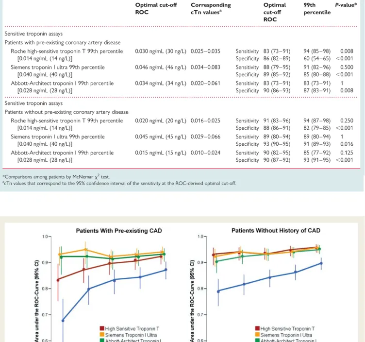
In patients with pre-existing CAD, the diagnostic accuracy for AMI, quantified by the AUC, was significantly higher with the sensitive cTn assays than that with the standard assay (AUC for Roche hs-cTnT, 0.92; 95% CI: 0.89 – 0.95; for Siemens cTnI-Ultra, 0.94; 95% CI: 0.91 – 0.96; and for Abbott-Architect cTnI, 0.93; 95% CI: 0.90 – 0.95; vs. AUC for the standard assay, 0.87; 95% CI: 0.83 – 0.90; P ¼ 0.01, P ¼ 0.003, P ¼ 0.007, respectively, for comparisons;
Table4, Supplementary material online, Table S4B and Figure2A).
Overall, the diagnostic accuracy was similar among the three sen-sitive assays (P . 0.05).
Optimal cut-off for cardiac troponin in
the early diagnosis of acute myocardial
infarction determined by receiver
operating characteristic curve
The optimal cut-off value to separate AMI from non-AMI deter-mined by ROC analysis in CAD patients was more than twice the 99th percentile for hs-cTnT [0.030 ng/mL (30 ng/L)], and close to the 99th percentile for both sensitive cTnI assays [0.034 ng/mL (34 ng/L) for Abbott-Architect cTnI and 0.046 ng/
mL (46 ng/L) for Siemens cTnI-Ultra; see Table5and Figure 2A].
The optimal cut-off value to separate AMI from non-AMI in
patients without a history of CAD was close to the 99thpercentile
for hs-cTnT [0.020 ng/mL (20 ng/L)] and half the 99th percentile
for Abbott-Architect cTnI [0.015 ng/mL (15 ng/L); see Table 5
and Figure2C].
Diagnostic performance in the early
diagnosis of acute myocardial infarction
at the 99th percentile
Overall, at the 99th percentile, all cTn assays showed lower speci-ficity in patients with pre-existing CAD when compared with
. . . . . . . . . . . . . . . . . . . . . . . . . . . . . . . . . . . . Table 1 Baseline characteristics of the patients
All patients (n 5 1098) Patients with a history of CADa (n 5 401) Patients without a history of CAD (n 5 697)
P-value* Patients with a history of CAD
Acute myocardial infarction P-value Yes (n 5 77) No (n 5 324) Male gender, no (%) 756 (67) 307 (77) 424 (61) ,0.001 58 (75) 249 (77) 0.78 Age, year Median 64 72 59 ,0.001 75 70 ,0.001 Inter-quartile range 51 – 75 59 – 79 47 – 72 68 – 84 57 – 78 Risk factors, no (%) Hypertension 693 (63) 320 (82) 361 (53) ,0.001 63 (82) 267 (82) 0.90 Hypercholesterolaemia 492 (45) 282 (71) 210 (30) ,0.001 49 (64) 233 (72) 0.15 Diabetes 217 (20) 126 (31) 91 (13) ,0.001 26 (34) 100 (31) 0.59 Current smoking 265 (24) 74 (19) 191 (27) 0.001 18 (23) 56 (17) 0.21 History of smoking 391 (36) 194 (48) 197 (28) ,0.001 31 (40) 163 (50) 0.11 History, no (%) Previous myocardial infarction 271 (25) 271 (68) 0 ,0.001 54 (70) 217 (67) 0.60 Previous revascularization 296 (27) 295 (74) 1 (0)b ,0.001 49 (64) 245 (76) 0.03 Previous PCI 254 (23) 253 (63) 1 (0)b ,0.001 37 (48) 216 (67) 0.002 Previous CABG 111 (10) 111 (28) 0 ,0.001 24 (31) 87 (27) 0.45 Peripheral artery disease 76 (7) 57 (14) 19 (3) ,0.001 15 (20) 42 (13) 0.14 Impaired kidney function 115 (11) 88 (22) 27 (4) ,0.001 23 (30) 65 (20) 0.06 Previous stroke 64 (6) 32 (8) 32 (5) 0.02 11 (14) 21 (7) 0.02 Vital Status, median (IQR)
Heart rate, b.p.m. 75 (66 – 89) 71 (62 – 82) 78 (68 – 92) ,0.001 80 (65 – 97) 69 (61 – 79) ,0.001 Systolic blood pressure,
mmHg
142 (127 – 160) 138 (124 – 157) 144 (129 – 161) ,0.001 140 (118 – 162) 138 (124 – 155) 0.87 Diastolic blood pressure,
mmHg
84 (74 – 93) 80 (70 – 89) 86 (77 – 95) ,0.001 78 (67 – 88) 80 (71 – 89) 0.28 Body mass index 26 (24 – 30) 27 (24 – 30) 26 (24 – 29) 0.062 26 (24 – 29) 27 (24 – 30) 0.05 Medication ACE inhibitors/AT-II blockers 536 (49) 270 (67) 266 (38) ,0.001 53 (69) 203 (63) 0.33 ASA 420 (38) 297 (74) 123 (18) ,0.001 56 (73) 241 (75) 0.77 Beta-blockers 411 (37) 288 (72) 123 (18) ,0.001 47 (61) 241 (74) 0.02 Calcium antagonists 184 (17) 120 (30) 64 (9) ,0.001 21 (5) 99 (30) 0.57 Diuretics 299 (27) 170 (42) 129 (19) ,0.001 45 (58) 125 (38) 0.002 Lipid-lowering drugs 393 (36) 293 (73) 100 (14) ,0.001 48 (62) 245 (76) 0.02 Nitrates/molsidomin 132 (12) 109 (27) 23 (3) ,0.001 29 (38) 80 (25) 0.02 ECG
Potential ischaemic ECG changes
247 (23) 109 (27) 138 (20) 0.004 41 (53) 68 (21) ,0.001 ST-segment elevation 58 (5) 19 (5) 39 (6) 0.556 12 (16) 7 (2) ,0.001 ST-segment depression 131 (12) 57 (14) 74 (11) 0.071 29 (38) 28 (8) ,0.001 Abnormal Q-wave 112 (10) 75 (19) 37 (5) ,0.001 14 (18) 61 (19) 0.90 Left bundle branch
block
42 (4) 25 (6) 17 (2) 0.001 13 (17) 12 (4) ,0.001 T-wave inversion 147 (13) 71 (18) 76 (11) 0.001 24 (31) 47 (14) 0.001
a
CAD, coronary artery disease. b
Patient with aortal dissection and consecutive coronary dissection but without relevant coronary artery disease.
patients without a history of CAD (Table4). The decrease in speci-ficity was particularly pronounced with hs-cTnT (59% in CAD patients vs. 81% in patients without a history of CAD, P , 0.001).
Diagnostic accuracy of cardiac troponin in
the diagnosis of ACS (acute myocardial
infarction or unstable angina)
The diagnostic accuracy for acute coronary syndroms (ACS), quan-tified by the AUC, was similarly low with the three sensitive cTn
assays in patients with pre-existing CAD, (AUC for Roche hs-cTnT, 0.66; 95% CI: 0.61 – 0.70; for Siemens cTnI-Ultra, 0.67; 95% CI: 0.62 – 0.72; and for Abbott-Architect cTnI, 0.67; 95% CI: 0.63 – 0.72), but moderate to high in patients without a history of CAD (AUC for Roche hs-cTnT 0.89; 95% CI: 0.86 – 0.91; AUC for Siemens cTnI-Ultra,0.86, 95% CI: 0.83 – 0.89; AUC for Abbott-Architect cTnI, 0.86, 95% CI: 0.83 – 0.88; P , 0.001 for all comparisons of AUC in patients with vs. without a history of CAD). For the diagnosis of acute coronary syndromes (AMI or UA), the negative predictive value of a measured cTn-value
Figure 1 Baseline levels of sensitive troponin assays at presentation. Cardiac troponin levels at presentation displayed as multiples of the 99th percentile. Boxes represent inter-quartile ranges, while whiskers display ranges (without outliers further than 1.5 inter-quartile ranges). CAD denotes coronary artery disease. Left side: in patients with final diagnosis of acute myocardial infarction, troponin levels compared within assays were similar in patients with pre-existing coronary artery disease compared with patients without a history of coronary artery disease (all P . 0.05). Right side: in patients with final diagnosis other than acute myocardial infarction, troponin levels compared within assays were sig-nificantly higher in patients with pre-existing coronary artery disease (all P , 0.001).
. . . .
. . . . Table 2 Final diagnoses of the patients
All patients (n 5 1098) History of CADa(n 5 401) No history of CAD (n 5 697) P-value*
Acute myocardial infarction 173 (16) 77 (19) 96 (14) 0.02 ST-segment elevation 41 (4) 15 (4) 26 (4) 0.99 Non-ST-segment elevation 132 (12) 62 (16) 70 (10) 0.01 UA 152 (14) 108 (27) 44 (6) ,0.001 Cardiac cause, but not CAD 147 (13) 39 (10) 108 (16) 0.01 Non-cardiac cause 528 (48) 136 (34) 392 (56) ,0.001 Unknown 98 (9) 41 (10) 57 (8) 0.25
a
CAD, coronary artery disease. *x2
below the 99th percentile was 64% (57 – 71%) for the Roche hs-cTnT, 65% (60 – 71%) for the Siemens cTnI-Ultra, and 64% (58 – 69%), for the Abbott-Architect cTnI assay. In patients without a history of CAD, the negative predictive value was 93% (91 – 95%), 92% (89 – 94%), and 91% (88 – 93%), respectively.
Cardiac troponin levels in patients
with recent onset of chest pain
In patients with pre-existing CAD the superiority of the sensitive cTn assays in the diagnosis of AMI was most pronounced among
patients with recent onset of chest pain (Figure 2B, Figure3, and
Supplementary material online, Table S4A).
Among CAD patients who presented within 3 h after the onset of chest pain (n ¼ 167), the AUCs for the four assays were as follows: Roche hs-cTnT, 0.86 (95% CI: 0.80 – 0.91); Siemens cTnI-Ultra, 0.96 (95% CI: 0.92 – 0.98); Abbott-Architect cTnI, 0.93 (95% CI: 0.89 – 0.97); and the standard assay, 0.76 (95% CI: 0.64 – 0.88) (P , 0.01, P , 0.001, P , 0.001, respectively, for the comparisons of the sensitive assays with the standard assay). The AUCs of the sensitive cTnI assays, Siemens cTnI-Ultra and Abbott-Architect cTnI, were higher than the AUC of the Roche
hs-cTnT assay (P ¼ 0.012 and P ¼ 0.051, respectively; Figure 2B
and Figure3).
For patients without a history of CAD, who presented within 3 h to the ED, the three sensitive cTn assays had comparable
. . . .
. . . . Table 4 Diagnostic performance of sensitive troponin assays at the 99th percentile; at 10% coefficient of variation for the standard assay (95% CI)
History of CAD No history of CAD P-value* Sensitive troponin assays
Roche high-sensitive troponin T 99th percentile [0.014 ng/mL (14 ng/L)]
Sensitivity 94 (85 – 98) 94 (87 – 98) 0.998 Specificity 59 (54 – 65) 81 (78 – 84) ,0.001 Negative predictive value 97 (94 – 99) 99 (97 – 100) 0.452 Positive predictive value 35 (29 – 41) 45 (39 – 52) 0.137 Siemens troponin I ultra 99th percentile
[0.040 ng/mL (40 ng/L)]
Sensitivity 91 (82 – 96) 89 (81 – 94) 0.880 Specificity 85 (80 – 88) 91 (88 – 93) 0.007 Negative predictive value 98 (95 – 99) 98 (97 – 99) 0.883 Positive predictive value 58 (49 – 67) 62 (53 – 69) 0.832 Abbott-Architect troponin I 99th percentile
[0.028 ng/mL (28 ng/L)]
Sensitivity 83 (73 – 91) 85 (77 – 92) 0.918 Specificity 87 (83 – 91) 93 (91 – 95) 0.016 Negative predictive value 96 (93 – 98) 98 (96 – 99) 0.292 Positive predictive value 61 (51 – 70) 66 (57 – 74) 0.719 Standard troponin assay
Roche troponin T 4th generation 99th percentile (unknown) 10% CV (0.035 ng/mL)
Sensitivity 69 (57 – 79) 83 (57 – 77) 0.988 Specificity 97 (94 – 99) 95 (96 – 99) 0.788 Negative predictive value 93 (90 – 95) 95 (93 – 96) 0.417 Positive predictive value 84 (73 – 92) 83 (73 – 90) 0.958
CAD, coronary artery disease; CV, coefficient of variation.
*x2test for comparison of proportions of patients with a history of coronary artery disease and patients without coronary artery disease.
. . . .
. . . . Table 3 Final diagnoses of patients with cardiac troponin levels above the 99th percentile
High-sensitive troponin T (n 5 402)
Siemens troponin I ultra (n 5 257)
Abbott-Architect troponin I (n 5 229)
Acute myocardial infarction 162 (40) 155 (60) 146 (64) ST-segment elevation 125 (31) 119 (46) 114 (50) Non-ST-segment elevation 37 (9) 36 (14) 32 (14)
UA 58 (14) 26 (10) 21 (9)
Cardiac cause, but not CAD 71 (18) 44 (17) 39 (17) Non-cardiac cause 81 (20) 23 (9) 16 (7) Unknown 30 (8) 9 (4) 7 (3)
accuracy (AUC for Roche hs-cTnT, 0.93; 95% CI: 0.90 – 0.96;
Siemens cTnI-Ultra, 0.93; 95% CI: 0.90 – 0.96;
Abbott-ArchitectcTnI, 0.91; 95% CI: 0.88 – 0.94; Figure2D and Figure3).
Serial cardiac troponin levels
During serial sampling the AUC for all cTn assays increased (Sup-plementary material online, Table S4B). Absolute values of changes in high-sensitive cTn levels from presentation to 1 and 2 h alone had similar diagnostic accuracy as the baseline high-sensitive cTn levels. The combination of baseline levels plus early changes improved the performance of the baseline level for all cTn assays. With the hs-cTnT assay and the Siemens cTnI-Ultra assay this increase in accuracy was statistically significant for the combi-nation of the baseline level and the change already within the first hour after presentation (P ¼ 0.032 and P ¼ 0.039, respectively); with the Abbott-Architect cTnI assay this increase was only signifi-cant for the combination of the baseline level and the change within 2 h after presentation (P ¼ 0.02; Supplementary material online, Table S4C ).
With the standard assay the diagnostic performance of the com-bination of baseline levels plus early change at 2 h was superior to
that of the combination of baseline levels and early change at 1 h. The diagnostic performance of the combinations was higher than that of early changes alone, and superior to the single measure-ment at presentation (all P-values , 0.05; see Supplemeasure-mentary material online, Table S4C ).
Prognostic value of sensitive cardiac
troponin assays
Median follow-up was 379 days (IQR: 107 – 721) days. Among the whole cohort, 58 patients died and 53 patients sustained an AMI during follow-up. Cumulative survival rates for patients with pre-existing CAD were 0.88 at 1 year vs. 0.98 in patients without a history of CAD (log-rank test: P , 0.001). In patients with AMI, survival rates at 1 year were 0.82 vs. 0.97 in patients with other diagnoses than AMI (log-rank test: P , 0.001). In patients with elevated levels of Roche hs-cTnT, Siemens cTnI-Ultra, and Abbott-Architect cTnI above the 99th percentile, survival rates were 0.87, 0.83, 0.85 vs. 0.99, 0.98, 0.97 in patients with cTn levels below the 99th percentile (all comparisons by log-rank test ,0.001; for details see Supplementary material
Figure 2 Diagnostic performance of cardiac troponin at presentation. Receiver operating characteristic curves describing the diagnostic per-formance of different cardiac troponin assays at presentation for the diagnosis of acute myocardial infarction in patients with a history of cor-onary artery disease (top), and in those without a history of corcor-onary artery disease (bottom). Left side: presenting within 12 h from chest pain onset/maximum. Right side: presenting within 3 h. (A) and (C) The calculated optimal cut-off values for patients with and without a history of coronary artery disease are defined by the point farthest from the point (0,1) of the graph.
online, Table S5A and B in the Supplementary material online, Appendix).
Elevated levels of Roche hs-cTnT as well as Siemens cTnI-Ultra above the 99th percentile strongly predicted mortality
independent of the presence of pre-existing CAD, age, sex, arterial hypertension, and diabetes in all patients (HR: 2.3, 95% CI: 1.1 – 5.1, P ¼ 0.034; HR: 2.3, 95% CI: 1.2 – 4.4, P ¼ 0.009; respectively).
. . . .
. . . . Table 5 Diagnostic performance of sensitive troponin assays in patients with pre-existing coronary artery disease at the optimal cut-off determined by the receiver operating characteristic curve (95% CI)
Optimal cut-off ROC Corresponding cTn valuesa Optimal cut-off ROC 99th percentile P-value*
Sensitive troponin assays
Patients with pre-existing coronary artery disease Roche high-sensitive troponin T 99th percentile
[0.014 ng/mL (14 ng/L)]
0.030 ng/mL (30 ng/L) 0.025 – 0.035 Sensitivity 83 (73 – 91) 94 (85 – 98) 0.008 Specificity 86 (82 – 89) 60 (54 – 65) ,0.001 Siemens troponin I ultra 99th percentile
[0.040 ng/mL (40 ng/L)]
0.046 ng/mL (46 ng/L) 0.034 – 0.083 Sensitivity 88 (79 – 95) 91 (82 – 96) 0.500 Specificity 89 (85 – 92) 85 (80 – 88) ,0.001 Abbott-Architect troponin I 99th percentile
[0.028 ng/mL (28 ng/L)]
0.034 ng/mL (34 ng/L) 0.020 – 0.061 Sensitivity 83 (73 – 91) 83 (73 – 91) 1 Specificity 90 (86 – 93) 87 (83 – 91) 0.008 Sensitive troponin assays
Patients without pre-existing coronary artery disease Roche high-sensitive troponin T 99th percentile
[0.014 ng/mL (14 ng/L)]
0.020 ng/mL (20 ng/L) 0.016 – 0.025 Sensitivity 91 (83 – 96) 94 (87 – 98) 0.250 Specificity 88 (86 – 91) 82 (79 – 85) ,0.001 Siemens troponin I ultra 99th percentile
[0.040 ng/mL (40 ng/L)]
0.045 ng/mL (45 ng/L) 0.029 – 0.066 Sensitivity 89 (80 – 94) 89 (80 – 94) 1 Specificity 93 (90 – 95) 91 (89 – 93) 0.016 Abbott-Architect troponin I 99th percentile
[0.028 ng/mL (28 ng/L)]
0.015 ng/mL (15 ng/L) 0.010 – 0.024 Sensitivity 90 (82 – 95) 85 (77 – 92) 0.125 Specificity 90 (87 – 92) 93 (91 – 95) ,0.001
*Comparisons among patients by McNemar x2test. a
cTn values that correspond to the 95% confidence interval of the sensitivity at the ROC-derived optimal cut-off.
Figure 3 Diagnostic accuracy at presentation according to chest pain onset. Area under the receiver operating characteristic curves and 95% confidence intervals for the different cardiac troponin assays at presentation in the diagnosis of acute myocardial infarction according to the time from the onset of chest pain. Left: patients with a history of coronary artery disease. Right: patients without a history of coronary artery disease.
In Cox regression analyses, adjusting for pre-existing CAD, age, sex, arterial hypertension, and hypercholesterolaemia, none of the sensitive cTn assays predicted AMI during follow-up (all P-values not significant).
Discussion
In this prospective multicentre study, we address important issues related to the clinical application of sensitive cTn assays and exam-ined the impact of pre-existing CAD on their diagnostic and prog-nostic accuracy. We provide seven important findings with impact on their best possible use in the early diagnosis of AMI:.
First, the prevalence of elevated sensitive cTnI and hs-cTnT levels above the 99th percentile in CAD patients with a final diag-nosis other than AMI was high and differed largely among the three novel cTn assays, ranging from 13 to 40%. Clinically, the high inci-dence of elevated cTn levels in CAD patients challenges the appli-cation of the 99th percentile for the decision limit for the diagnosis of AMI, as suggested in current guidelines. Careful clinical assess-ment and thoughtful differential diagnosis are required to separate AMI from a variety of acute and chronic disorders also associated
with low-level myocardial necrosis.24 In addition, the difference
regarding the incidence of cTn levels above the 99th percentile in CAD patients without AMI might indicate the presence of important differences in the release of cTnI and cTnT in these
non-AMI settings, which is further supported by recent data.11,12
An alternative explanation for the difference regarding the inci-dence of elevated cTn levels is the fact that the 99th percentiles for the three sensitive assays were not determined in the same reference population.
Second, for all three sensitive cTn assays, the diagnostic accuracy at presentation was nevertheless significantly higher than with the standard assay in CAD patients as well as in patients without a history of CAD.
Third, the ROC-derived optimal cut-off levels for CAD patients tended to be higher than in patients without a history of CAD, although the AUC of sensitive cTn assays did not differ significantly comparing CAD and non-CAD patients. All cTn assays showed higher sensitivity but lower specificity in CAD patients when com-pared with patients without a history of CAD, reflecting the higher baseline levels in CAD patients without AMI. These findings high-light the clinical need to develop test-specific algorithms that fine tune the application of these novel tests in patients with acute
chest pain.12
Fourthly, the superiority of the sensitive cTn assays was most pronounced among CAD patients with a recent onset of chest pain, offering the opportunity to minimize myocardial damage by extending early treatment options to AMI patients without
ST-segment elevation.1,2
Fifthly, the sensitive cTnI assays seemed to outperform the hs-cTnT assay in early presenters.
Sixthly, the accuracy of sensitive cTn assays to diagnose ACS (AMI or UA) was significantly lower in patients with pre-existing CAD when compared with patients without pre-existing CAD. Further research is necessary to identify biomarkers that reliably detect myocardial ischaemia irrespective of necrosis, particularly
in patients with pre-existing CAD.25
Seventhly, the Roche hs-cTnT and the sensitive Siemens cTnI-Ultra assay predict mortality independent of age, sex, pre-existing CAD, and cardiovascular risk factors. Our findings extend the results of previous studies, investigating the mortality of apparently healthy subjects with elevated levels measured
with sensitive cTnI assays.11,26,27
The following limitations of the current study merit consider-ation. First, we evaluated three sensitive cTn assays. We hypoth-esize that our findings can be generalized to other cTn assays with similar sensitivity and precision. However, additional studies need to confirm this hypothesis. Secondly, in this ongoing prospec-tive study, the subgroup analysis of patients with pre-existing CAD was not predefined at the time of the initial protocol written in 2005. It was added while we were still blinded to the results in 2009, with regard to recent investigations, challenging the
diagnos-tic accuracy of sensitive cTn assays in CAD patients.11,12Therefore,
e.g. our analysis of the assay-specific ROC-derived optimal cut-off values to differentiate AMI from other causes of acute chest pain should be considered exploratory and requires confirmation in additional studies. Third, this observational study cannot quantify exactly the clinical benefit associated with the increase in early diagnostic accuracy. To add this important information, interven-tion studies seem warranted. Fourth, the first 800 blood samples for the hs-cTnT assay were collected into tubes containing serum, while all other blood samples were collected into tubes containing potassium EDTA, which might lead to slightly different concentration values. Fifthly, some of the patients with positive sensitive cTn values classified as non-AMIs might have had small AMIs below the decision value of conventional cTn. Presumably, this contributed to the reduced specificity of the sensitive assays. In conclusion, sensitive cTn assays introduce diagnostic improve-ments as well as challenges. The excellent diagnostic performance of sensitive cTn assays in the early diagnosis of AMI can be extended to patients with pre-existing CAD. However, elevated cTn levels at presentation are common also in CAD patients with diagnoses other than AMI, challenging differential diagnosis. Accordingly, the accuracy to diagnose ACS was lower in patients with existing CAD when compared with patients without pre-existing CAD and optimal cut-off levels tend to be higher. Sensitive cTn assays have prognostic value in patients with a final diagnosis other than AMI.
Supplementary material
Supplementary material is available at European Heart Journal online.
Acknowledgements
We are indebted to the patients who participated in the study and to the ED staff as well as the laboratory technicians for their most valuable efforts, and we thank Kris Denhaerynck and Dr Schindler for expert statistical advice.
Funding
The study was supported by research grants from the Swiss National Science Foundation (PP00B-102853), the Swiss Heart Foundation,
Abbott, Roche, Siemens, and the Department of Internal Medicine, University Hospital Basel.
Conflict of interest: We disclose that C.M. has received research support from the Swiss National Science Foundation (PP00B-102853), the Swiss Heart Foundation, the Novartis Foun-dation, the Krokus FounFoun-dation, Abbott, Astra Zeneca, Biosite, Brahms, Nanosphere, Roche, Siemens, and the Department of Internal Medicine, University Hospital Basel, as well as speaker honoraria from Abbott, Biosite, Brahms, Roche, and Siemens.
References
1. Bassand JP, Hamm CW, Ardissino D, Boersma E, Budaj A, Fernandez-Aviles F, Fox KA, Hasdai D, Ohman EM, Wallentin L, Wijns W. Guidelines for the diagnosis and treatment of non-ST-segment elevation acute coronary syndromes. Eur Heart J 2007;28:1598 – 1660.
2. Kushner FG, Hand M, Smith SC Jr, King SB III, Anderson JL, Antman EM, Bailey SR, Bates ER, Blankenship JC, Casey DE Jr, Green LA, Hochman JS, Jacobs AK, Krumholz HM, Morrison DA, Ornato JP, Pearle DL, Peterson ED, Sloan MA, Whitlow PL, Williams DO. 2009 focused updates: ACC/AHA guidelines for the management of patients with ST-elevation myocardial infarction (updating the 2004 guideline and 2007 focused update) and ACC/AHA/SCAI guidelines on per-cutaneous coronary intervention (updating the 2005 guideline and 2007 focused update) a report of the American College of Cardiology Foundation/American Heart Association Task Force on Practice Guidelines. J Am Coll Cardiol 2009;54: 2205 – 2241.
3. Antman EM, Cohen M, Bernink PJ, McCabe CH, Horacek T, Papuchis G, Mautner B, Corbalan R, Radley D, Braunwald E. The TIMI risk score for unstable angina/non-ST elevation MI: a method for prognostication and therapeutic decision making. JAMA 2000;284:835 – 842.
4. Morrow DA, Antman EM, Charlesworth A, Cairns R, Murphy SA, de Lemos JA, Giugliano RP, McCabe CH, Braunwald E. TIMI risk score for ST-elevation myocar-dial infarction: a convenient, bedside, clinical score for risk assessment at presen-tation: an intravenous nPA for treatment of infarcting myocardium early II trial substudy. Circulation 2000;102:2031 – 2037.
5. Tiemann O. Variations in hospitalisation costs for acute myocardial infarction: a comparison across Europe. Health Econ 2008;17(Suppl. 1):S33 – S45.
6. Apple FS, Jesse RL, Newby LK, Wu AH, Christenson RH. National Academy of Clinical Biochemistry and IFCC Committee for Standardization of Markers of Cardiac Damage Laboratory Medicine Practice Guidelines: analytical issues for biochemical markers of acute coronary syndromes. Circulation 2007;115: e352 – e355.
7. Apple FS, Smith SW, Pearce LA, Ler R, Murakami MM. Use of the Centaur TnI-Ultra Assay for detection of myocardial infarction and adverse events in patients presenting with symptoms suggestive of acute coronary syndrome. Clin Chem 2008;54:723 – 728.
8. Melanson SE, Morrow DA, Jarolim P. Earlier detection of myocardial injury in a preliminary evaluation using a new troponin I assay with improved sensitivity. Am J Clin Pathol 2007;128:282 – 286.
9. Keller T, Zeller T, Peetz D, Tzikas S, Roth A, Czyz E, Bickel C, Baldus S, Warnholtz A, Frohlich M, Sinning CR, Eleftheriadis MS, Wild PS, Schnabel RB, Lubos E, Jachmann N, Genth-Zotz S, Post F, Nicaud V, Tiret L, Lackner KJ, Munzel TF, Blankenberg S. Sensitive troponin I assay in early diagnosis of acute myocardial infarction. N Engl J Med 2009;361:868 – 877.
10. Reichlin T, Hochholzer W, Bassetti S, Steuer S, Stelzig C, Hartwiger S, Biedert S, Schaub N, Buerge C, Potocki M, Noveanu M, Breidthardt T, Twerenbold R, Winkler K, Bingisser R, Mueller C. Early diagnosis of myocardial infarction with sensitive cardiac troponin assays. N Engl J Med 2009;361:858 – 867.
11. Omland T, de Lemos JA, Sabatine MS, Christophi CA, Rice MM, Jablonski KA, Tjora S, Domanski MJ, Gersh BJ, Rouleau JL, Pfeffer MA, Braunwald E. A sensitive cardiac troponin T assay in stable coronary artery disease. N Engl J Med 2009;361: 2538 – 2547.
12. Eggers KM, Lind L, Venge P, Lindahl B. Will the universal definition of myocardial infarction criteria result in an overdiagnosis of myocardial infarction? Am J Cardiol 2009;103:588 – 591.
13. Kloner RA, Shook T, Przyklenk K, Davis VG, Junio L, Matthews RV, Burstein S, Gibson M, Poole WK, Cannon CP, McCabe CH, Braunwald E. Previous angina alters in-hospital outcome in TIMI 4. A clinical correlate to preconditioning? Cir-culation 1995;91:37 – 45.
14. Janne d’Othee B, Siebert U, Cury R, Jadvar H, Dunn EJ, Hoffmann U. A systematic review on diagnostic accuracy of CT-based detection of significant coronary artery disease. Eur J Radiol 2008;65:449 – 461.
15. Pontone G, Andreini D, Quaglia C, Ballerini G, Nobili E, Pepi M. Accuracy of mul-tidetector spiral computed tomography in detecting significant coronary stenosis in patient populations with differing pre-test probabilities of disease. Clin Radiol 2007;62:978 – 985.
16. Thygesen K, Alpert JS, White HD, Jaffe AS, Apple FS, Galvani M, Katus HA, Newby LK, Ravkilde J, Chaitman B, Clemmensen PM, Dellborg M, Hod H, Porela P, Underwood R, Bax JJ, Beller GA, Bonow R, Van der Wall EE, Bassand JP, Wijns W, Ferguson TB, Steg PG, Uretsky BF, Williams DO, Armstrong PW, Antman EM, Fox KA, Hamm CW, Ohman EM, Simoons ML, Poole-Wilson PA, Gurfinkel EP, Lopez-Sendon JL, Pais P, Mendis S, Zhu JR, Wallentin LC, Fernandez-Aviles F, Fox KM, Parkhomenko AN, Priori SG, Tendera M, Voipio-Pulkki LM, Vahanian A, Camm AJ, De Caterina R, Dean V, Dickstein K, Filippatos G, Funck-Brentano C, Hellemans I, Kristensen SD, McGregor K, Sechtem U, Silber S, Tendera M, Widimsky P, Zamorano JL, Morais J, Brener S, Harrington R, Morrow D, Lim M, Martinez-Rios MA, Steinhubl S, Levine GN, Gibler WB, Goff D, Tubaro M, Dudek D, Al-Attar N. Universal definition of myocardial infarction. Circulation 2007;116:2634 – 2653. 17. Apple FS, Wu AH, Jaffe AS. European Society of Cardiology and American
College of Cardiology guidelines for redefinition of myocardial infarction: how to use existing assays clinically and for clinical trials. Am Heart J 2002;144: 981 – 986.
18. Mingels A, Jacobs L, Michielsen E, Swaanenburg J, Wodzig W, van Dieijen-Visser M. Reference population and marathon runner sera assessed by highly sensitive cardiac troponin T and commercial cardiac troponin T and I assays. Clin Chem 2009;55:101 – 108.
19. McCann CJ, Glover BM, Menown IB, Moore MJ, McEneny J, Owens CG, Smith B, Sharpe PC, Young IS, Adgey JA. Novel biomarkers in early diagnosis of acute myo-cardial infarction compared with cardiac troponin T. Eur Heart J 2008;29: 2843 – 2850.
20. Giannitsis E, Kurz K, Hallermayer K, Jarausch J, Jaffe AS, Katus HA. Analytical vali-dation of a high-sensitivity cardiac troponin T assay. Clin Chem 2010;56:254 – 261. 21. DeLong ER, DeLong DM, Clarke-Pearson DL. Comparing the areas under two or more correlated receiver operating characteristic curves: a nonparametric approach. Biometrics 1988;44:837 – 845.
22. Simel DL, Samsa GP, Matchar DB. Likelihood ratios with confidence: sample size estimation for diagnostic test studies. J Clin Epidemiol 1991;44:763 – 770. 23. Trajman A, Luiz RR. McNemar Chi2
test revisited: comparing sensitivity and speci-ficity of diagnostic examinations. Scand J Clin Lab Invest 2008;68:77 – 80. 24. Januzzi JL Jr, Bamberg F, Lee H, Truong QA, Nichols JH, Karakas M,
Mohammed AA, Schlett CL, Nagurney JT, Hoffmann U, Koenig W. High-sensitivity troponin T concentrations in acute chest pain patients evaluated with cardiac computed tomography. Circulation 2010;121:1227 – 1234. 25. Staub D, Jonas N, Zellweger MJ, Nusbaumer C, Wild D, Pfisterer ME,
Mueller-Brand J, Perruchoud AP, Mueller C. Use of N-terminal pro-B-type natriuretic peptide to detect myocardial ischemia. Am J Med 2005;118:1287. 26. Eggers KM, Lagerqvist B, Venge P, Wallentin L, Lindahl B. Persistent cardiac
tro-ponin I elevation in stabilized patients after an episode of acute coronary syn-drome predicts long-term mortality. Circulation 2007;116:1907 – 1914. 27. Reiter M, Twerenbold R, Reichlin T, Haaf P, Peter F, Meissner J, Hochholzer W,
Stelzig C, Freese M, Heinisch C, Breidthardt T, Freidank H, Winkler K, Campodarve I, Gea J, Mueller C. Early diagnosis of acute myocardial infarction in the elderly using more sensitive cardiac troponin assays. Eur Heart J 2011;32: 1379 – 1389.