Assessment of high sensitive
Assessment of high sensitive troponin
troponin T and I
T and I
immunoassays in patients with acute chest pain
immunoassays in patients with acute chest pain
without ST
without ST--segment elevation
segment elevation
Le Goff C.1, Garweg C.2, Laurent T.1, Kaux J-F.3, Deroyer C.1, Merville M.P.1, Melon P.2, Fillet M.1, Legrand V.2, Chapelle J-P.1 1Department of Clinical Chemistry, University Hospital of Liege, Belgium
2Department of Cardiology, University Hospital of Liege, Belgium 3Department of Clinical Sciences, University of Liege, Belgium
Background:
Cardiac troponin I and T are specific markers of myocardial injury that are widely used for the diagnosis of acute coronary syndrome (ACS). In acute chest pain without ST-segment elevation, they are used to differentiate unstable angina from non ST-segment elevation myocardial infarction (NSTEMI). Recently, troponin assays with higher analytical sensitivities became available to enable the detection of minor myocardial damage and identify individuals at higher risk for ACS. As a result of its high tissue-specificity, cardiac troponin T and I are cardio-specific, highly sensitive markers for myocardial damage. The aim of this study was to evaluate the new higher sensitive troponin (T and I) in patients with stable angina and acute chest pain without ST-segment elevation.
Materials and methods:
Sixty subjects (mean age : 65.5± 11 years), were included: 20 healthy controls, 20 patients with stable angina, 9 with unstable angina (troponin-) and 18 patients with NSTEMI myocardial infarction (troponin+). The protocol was approved by the ethic committee of the University of Liège (Belgium). High sensitive troponin T (hsTnT) determination was realized on heparin plasma by electrochemiluminescence immunoassay on Modular E (Roche Diagnostic). Troponin I II (TnI II) is a chemiluminescent microparticle immunoassay for the quantitative determination of cardiac troponin-I in heparine plasma on the ARCHITECT i System (Abbott Diagnostic). The lower detection limit of these assays was 0.005µg/L for hsTnT and 0.01µg/L for TnI II.
Stastistical analysis was performed using t test. P value <0.05 was considered significant.
10 100
Results:
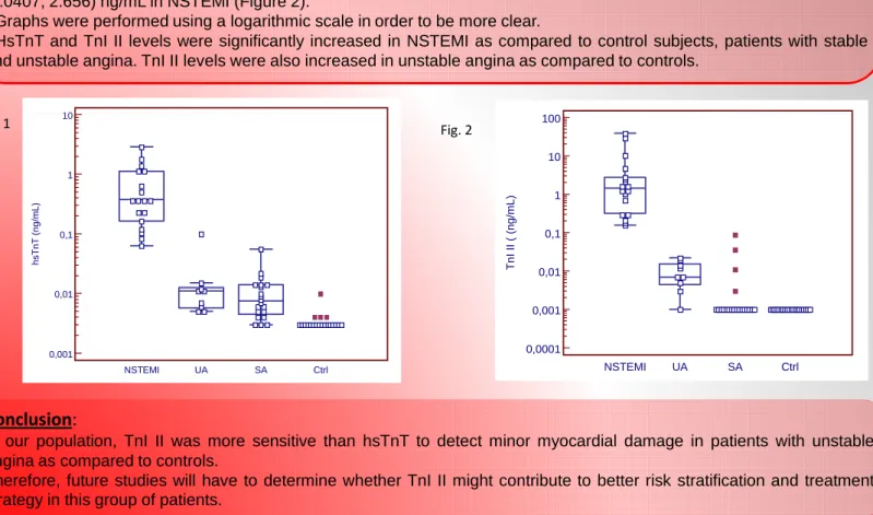
• HsTnT levels were 0.003(0.003, 0.004) [median baseline (1st, 3rd quartile)] ng/mL in controls (Ctrl), 0.0075 (0.00475, 0.014) ng/mL in stable angina (SA), 0.011(0.006, 0.012) ng/mL in unstable angina (UA) and 0.3715 (0.1795, 1.00725) ng/mL in NSTEMI (Figure 1).
• TnI II levels were 0 (0, 0.001) ng/mL in Ctrl and in patients with SA, 0.07 (0.005, 0.014) ng/mL in UA and 1.4475 (0.0407, 2.656) ng/mL in NSTEMI (Figure 2).
• Graphs were performed using a logarithmic scale in order to be more clear.
• HsTnT and TnI II levels were significantly increased in NSTEMI as compared to control subjects, patients with stable and unstable angina. TnI II levels were also increased in unstable angina as compared to controls.
Conclusion:
In our population, TnI II was more sensitive than hsTnT to detect minor myocardial damage in patients with unstable angina as compared to controls.
Therefore, future studies will have to determine whether TnI II might contribute to better risk stratification and treatment strategy in this group of patients.
Fig. 1 10 Fig. 2 1 0,1 0,01 0,001 h s T n T ( n g /m L ) NSTEMI UA SA Ctrl 100 10 1 0,1 0,01 0,001 0,0001 T n I II ( (n g /m L ) NSTEMI UA SA Ctrl