HAL Id: inserm-00663642
https://www.hal.inserm.fr/inserm-00663642
Submitted on 27 Jan 2012
HAL is a multi-disciplinary open access
archive for the deposit and dissemination of
sci-entific research documents, whether they are
pub-lished or not. The documents may come from
teaching and research institutions in France or
abroad, or from public or private research centers.
L’archive ouverte pluridisciplinaire HAL, est
destinée au dépôt et à la diffusion de documents
scientifiques de niveau recherche, publiés ou non,
émanant des établissements d’enseignement et de
recherche français ou étrangers, des laboratoires
publics ou privés.
High-sensitivity versus conventional troponin in the
emergency department for the diagnosis of acute
myocardial infarction.
Yonathan Freund, Camille Chenevier-Gobeaux, Pascale Bonnet, Yann-Erick
Claessens, Jean-Christophe Allo, Benoit Doumenc, François Leumani,
Claudine Cosson, Bruno Riou, Patrick Ray
To cite this version:
Yonathan Freund, Camille Chenevier-Gobeaux, Pascale Bonnet, Yann-Erick Claessens,
Jean-Christophe Allo, et al.. High-sensitivity versus conventional troponin in the emergency department for
the diagnosis of acute myocardial infarction.. Critical Care, BioMed Central, 2011, 15 (3), pp.R147.
�10.1186/cc10270�. �inserm-00663642�
R E S E A R C H
Open Access
High-sensitivity versus conventional troponin in
the emergency department for the diagnosis of
acute myocardial infarction
Yonathan Freund
1*, Camille Chenevier-Gobeaux
2, Pascale Bonnet
1, Yann-Erick Claessens
3, Jean-Christophe Allo
3,
Benoit Doumenc
4, François Leumani
1, Claudine Cosson
5, Bruno Riou
1,6and Patrick Ray
1,6Abstract
Introduction: Recently, newer assays for cardiac troponin (cTn) have been developed which are able to detect changes in concentration of the biomarker at or below the 99th percentile for a normal population. The objective of this study was to compare the diagnostic performance of a new high-sensitivity troponin T (HsTnT) assay to that of conventional cTnI for the diagnosis of acute myocardial infarction (AMI) according to pretest probability (PTP). Methods: In consecutive patients who presented to our emergency departments with chest pain suggestive of AMI, levels of HsTnT were measured at presentation, blinded to the emergency physicians, who were asked to estimate the empirical PTP of AMI. The discharge diagnosis was adjudicated by two independent experts on the basis of all available data.
Results: A total of 317 patients were included, comprising 149 (47%) who were considered to have low PTP, 109 (34%) who were considered to have moderate PTP and 59 (19%) who were considered to have high PTP. AMI was confirmed in 45 patients (14%), 22 (9%) of whom were considered to have low to moderate PTP and 23 (39%) of whom were considered to have high PTP (P < 0.001). In the low to moderate PTP group, HsTnT levels ≥ 0.014 μg/ L identified AMI with a higher sensitivity than cTnI (91%, 95% confidence interval (95% CI) 79 to 100, vs. 77% (95% CI 60 to 95); P = 0.001), but the negative predictive value was not different (99% (95% CI 98 to 100) vs. 98% (95% CI 96 to 100)). There was no difference in area under the receiver operating characteristic (ROC) curve between HsTnT and cTnI (0.93 (95% CI 0.90 to 0.98) vs. 0.94 (95% CI 0.88 to 0.97), respectively).
Conclusions: In patients with low to moderate PTP of AMI, HsTnT is slightly more useful than cTnI. Our results confirm that the use of HsTnT has a higher sensitivity than conventional cTnI.
Introduction
Early detection of acute myocardial infarction (AMI) remains a major concern, with approximately 15 million patients per year presenting to US emergency departments (EDs) with symptoms suggestive of the diagnosis [1,2]. Among such patients, a strong association between ele-vated cardiac troponin (cTn) levels and myocardial necro-sis has been clearly demonstrated [3-5]. Conventional cTn has revolutionised the management of patients presenting
with suspected acute coronary syndrome (ACS), including risk stratification of ACS, and the use of cTn measure-ments is recommended by current guidelines [6]. A cutoff point at the 99th percentile has been endorsed, as values above this level have repeatedly proven to be associated with adverse cardiovascular outcomes, including death [7-13]. However, the delay (4 to 6 hours, and 12 hours for peak level) in its elevation remains of concern, since it can delay AMI diagnosis and its treatment and increases the burden on EDs. Thus, cTn measurement does not reliably exclude AMI without repeated negative measurements over the course of 4 to 6 hours. These last years, newer assays have been developed, and High Sensitivity Troponin (HsTn) has been associated with higher sensitivity and
* Correspondence: yonatman@gmail.com
1
Department of Emergency Medicine and Surgery, Hôpital Pitié-Salpétrière, Assistance Publique-Hôpitaux de Paris (APHP), Université Pierre et Marie Curie-Paris 6 (UPMC), 47-83 boulevard de l’hôpital, F-75651 Paris cedex 13, France
Full list of author information is available at the end of the article
© 2011 Freund et al.; licensee BioMed Central Ltd. This is an open access article distributed under the terms of the Creative Commons Attribution License (http://creativecommons.org/licenses/by/2.0), which permits unrestricted use, distribution, and reproduction in any medium, provided the original work is properly cited.
NPV than conventional cTn. Recent studies have shown excellent diagnostic performance, even with early presen-tation to the ED [14], and a better diagnostic accuracy than cTn [15]. However, the latter studies did not evaluate the diagnostic accuracy of high-sensitivity troponin T (HsTnT) according to the pretest probability (PTP) of AMI. For example, ST elevation on an electrocardiogram of a patient with chest pain would be diagnosed as AMI, and then the patient would undergo cardiac catheteriza-tion without any measurement of a cardiac biomarker. Furthermore, one of the potential strengths of HsTnT might be the exclusion of AMI earlier than it would be with conventional cTn measurement as suggested by pre-vious studies [15]. Therefore, the objectives of the current study were to confirm whether HsTnT is more sensitive than conventional cTnI to detect AMI according to the patient’s PTP.
Materials and methods
Clinical setting
During the period from August 2005 to January 2007 in three urban teaching hospitals, we prospectively enrolled consecutive hospital outpatients (> 18 years of age) who presented to the ED with chest pain suggestive of ACS with the onset or peak occurring within the previous 6 hours. Patients with acute or chronic kidney failure requir-ing dialysis were excluded. The study was performed according to the principles of the Declaration of Helsinki and approved by the local ethics committee (Comité de Protection des Personnes Ile-de-France VI, CHU Pitié-Salpétrière Hospital, Paris, France). Because routine medi-cal care was unchanged, waiver of informed consent was authorised. We followed most of the recommendations concerning the reporting of diagnostic studies set forth by the Standards for Reporting of Diagnostic Accuracy initia-tive [16].
Routine assessment
As part of the routine assessment in our institutions, all patients underwent an initial clinical evaluation that included clinical history, a physical examination, 12-lead electrocardiography (ECG), pulse oximetry, routine blood tests and chest X-rays. After these routine tests were done, and before cardiac biomarker results were available, ED physicians were asked to offer an ‘empiri-cal’ clinical probability of AMI (low, medium or high PTP) based on cardiovascular risk factors, type of chest pain, physical findings and electrocardiogram abnormal-ities [17,18]. Conventional cardiac troponin I (cTnI) was measured at presentation and, if needed, was repeated after 3 to 9 hours as long as it was clinically indicated. Thus, according to the diagnosis of non-ST elevation MI (NSTEMI) or ST elevation MI (STEMI), the patients were admitted either to the cardiology unit for further
evaluation and treatment or directly to the catheteriza-tion laboratory for primary percutaneous coronary inter-vention. However, the timing and treatment of patients were left to the discretion of the attending physicians according to the suspected diagnosis. ED physicians in charge were blinded to the results of HsTnT, and biolo-gists were blinded to the emergency diagnosis suspected by physicians.
To determine the etiologic diagnosis of chest pain at presentation for each patient, two independent experts (ED physicians) who were blinded to the results of HsTnT reviewed all available medical records (including patient history, physical findings, results of laboratory and radiolo-gic testing, ECG, echocardiography, cardiac exercise test, coronary angiography and summary chart at discharge) pertaining to the patient from the time of ED presentation to 30-day follow-up. In the event of diagnostic disagree-ment, cases were reviewed and adjudicated in conjunction with a third expert (also an ED physician).
AMI was diagnosed according to the joint European Society of Cardiology/American College of Cardiology/ American Heart Association/World Heart Federation Task Force redefinition of MI guidelines [6]. Diagnosis of AMI required a cTnI increase above the 10% coefficient of variation (CV) value associated with at least one of the fol-lowing: symptoms of ischaemia, new ST-T changes or a new Q wave on an electrocardiogram, imaging of new loss of viable myocardium or normal cTnI on admission. Unstable angina was diagnosed in patients with constant normal cTnI levels and a history or clinical symptoms consistent with ACS. Predefined further diagnostic cate-gories included AMI (STEMI with the presence of ST-segment elevation in at least two continuous leads on ECG, new onset of left bundle branch block or NSTEMI), unstable angina, and a third group including cardiac but not coronary symptoms (for example, stable angina, myo-carditis, arrhythmias and heart failure), noncardiac symp-toms (for example, pulmonary embolism) and chest pain of unknown origin.
To assess the influence of renal function on cTn mea-surement accuracy, the creatinine level was measured in each patient and then renal function was estimated using the Modification of Diet in Renal Disease study equation [19].
Biochemical analysis
In two EDs (Cochin Hospital and La Pitié Salpêtrière Hos-pital, Paris, France), plasmatic cTnI concentrations were routinely measured on an Xpand HM analyzer using the Cardiac Troponin I one-step enzyme immunoassay system (Siemens Healthcare Diagnostics Inc., Newark, NJ, USA). The measurement range extended from 0.04 to 40.00 μg/L. The threshold for this method (0.14 μg/L) corresponds to the lowest substrate concentration that can be reproducibly
Freund et al. Critical Care 2011, 15:R147 http://ccforum.com/content/15/3/R147
measured with a CV ≤ 10%. In the remaining ED (Bicêtre Hospital, Le Kremlin-Bicêtre, France), plasmatic cTnI con-centrations were routinely measured on an Access analyser (Beckman Coulter, Inc., Brea, CA, USA). The measurement range of this one-step chemiluminescence immunoassay extends from 0.01 to 100.00 μg/L. The threshold (10% CV) given by the manufacturer is 0.06 μg/L.
HScTnT measurement
Heparinised samples collected upon admission and, if available, samples collected 3 to 9 hours later were ana-lysed. Plasmatic highly sensitive cardiac TnT (HScTnT) concentrations were measured using the HScTnT one-step electrochemiluminescence immunoassay on an Elecsys 2010 analyzer (Roche Diagnostics, Meylan, France). The measuring range extended from 0.003 to 10 μg/L. The threshold for this method is 0.014 μg/L and corresponds to the 99th percentile. The CV was found to be < 10% at 0.014 μg/L. In our laboratory, CVs obtained in Roche Diagnostics quality controls containing 0.027 and 2.360 μg/L of HScTnT were < 4%. These analytical performance levels were in accordance with data pro-vided by the manufacturer.
Statistical analysis
Continuous variables are presented as means ± SD or medians (25th to 75th percentile), and categorical variables are expressed as numbers and percentages. Continuous variables were compared by using the Mann-Whitney U test, and categorical variables were assessed using Pear-son’s c2test. Correlations among continuous variables
were assessed using the Spearman’s rank correlation coef-ficient. Receiver operating characteristic (ROC) curves were constructed to assess the sensitivity and specificity, positive predictive value (PPV) and negative predictive value (NPV), positive likelihood ratio (LR+) and negative likelihood ratio (LR-) (all data presented with their 95% confidence intervals (95% CIs)) throughout the concentra-tions of cTnI and HScTnT to compare the accuracy of these markers in the diagnosis of AMI. Comparison of areas under the ROC curve was performed [20]. As this comparison is recognised as potentially insensitive, the net reclassification index (NRI) method was used as recently described [21]. For tests with binary outcomes (such as cTn for the diagnosis of AMI), NRI is defined as the gain in certainty of the first test (cTnI) minus the gain in cer-tainty of the second test (HScTnT) or, alternatively stated, the difference of the sum of the sensitivity and specificity expressed as follows:
NRIHScTnT vs. cTnI= (sensitivity + specificity)HScTnT− (sensitivity + specificity)cTnI.
NRI is the combination of four components: the pro-portion of individuals with events who move up or
down in a category and the proportion of individuals with nonevents who move up or down in a category. Table 1 is a contingency table comparing diagnostic classifications according to cTnI and HsTnT, with shifts between the two classifications, to represent the possible benefit of HScTnT in terms of the number of patients correctly reclassified. As stated in the Routine assess-ment subsection above, we separated the study popula-tion into two groups: one included the patients assessed as having low or moderate PTP of AMI and the other assessed as having high PTP of AMI.
All hypothesis testing was two-tailed, and P < 0.05 was considered statistically significant. Statistical analysis was performed using StatView for Windows version 5.0 ware (SAS Institute, Cary, NC, USA) and MedCalc soft-ware for ROC analysis (MedCalc Softsoft-ware, Mariarkerke, Belgium). Graphs were built with GraphPad Prism 5 software (GraphPad Software Inc., La Jolla, CA, USA).
Results
After 18 months, 317 consecutive patients were enrolled in the study. The baseline characteristics of the patients are shown in Table 2. The mean age of the patients was 57 ± 17 years (range, 40 to 90 years), and 205 (65%) were men. There were significant proportions of older adult patients (31% patients were age 65 years or older, n = 98) and patients with a history of cardiovascular events (26%, n = 83). Chest pain was considered typical of ACS in 43% (n = 136) of the patients. In our study
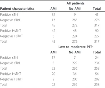
Table 1 Contingency data according to pretest
probabilitya
All patients
Patient characteristics AMI No AMI Total Positive cTnI 32 9 41 Negative cTnI 13 263 276 Total 45 272 317 Positive HsTnT 42 48 90 Negative HsTnT 3 224 227 Total 45 272 317 Low to moderate PTP AMI No AMI Total Positive cTnI 17 7 24 Negative cTnI 5 229 234 Total 22 236 258 Positive HsTnT 20 36 56 Negative HsTnT 2 200 202 Total 22 236 258
aNet reclassification improvement (NRI) from the use of highly sensitive
troponin T (HsTnT) was 7.9% (95% CI = 0.9 to 14.9; P = 0.034). Comparison of the model including HsTnT with cTnI was significant for low PTP patients (NRI = 10.3%, 95% CI = 1.9 to 18.7; P = 0.027), but NRI was not significantly different in moderate PTP patients (NRI = 11.6%, 95% CI = -0.5 to 23.7; P = 0.084) or in high PTP patients (NRI = -14.4%, 95% CI = -32.6 to -3.6; P = 0.181).
population, 149 patients (47%) were assessed as having a low PTP of AMI, 109 patients (34%) were assessed as moderate and 59 patients (19%) were assessed as high. AMI was confirmed in 45 patients (14%), 13 of whom had sustained STEMI, and all of these 13 patients were in the high PTP group; 32 of the patients had sustained NSTEMI. Table 2 shows that patients in the two groups (high PTP and low or moderate PTP) had significantly different characteristics. There was a higher rate of a personal history of AMI in the high PTP group and a
higher final diagnosis of AMI (39% vs. 9%) in the high PTP group (P < 0.001). At 30 days after admission, there were three deaths (two in the AMI group and one in the other cause group) and four relapses of ACS (all in the AMI group).
HsTnT diagnostic performances
The area under the ROC curve (AUC) for the diagnosis of AMI was 0.940 (95% Confidence Intervall 0.901 to 0.980) (P < 0.001) for initial cTnI compared to 0.926
Table 2 Baseline characteristics of the population according to the pretest probabilitya
Population characteristics All patients Low or moderate PTP High PTP P value* Number of patients 317 258 59
Age, years 57 ± 17 56 ± 17 60 ± 17 0.168 Men 205 (65) 166 (64) 39 (66) 0.88 Systolic BP, mmHg 141 ± 28 141 ± 27 144 ± 30 0.396 Diastolic BP, mmHg 80 ± 16 80 ± 16 82 ± 16 0.428 Heart rate, beats/minute 85 ± 45 84 ± 23 80 ± 19 0.177 Pulse oxymetry, % 97 ± 3 97 ± 3 97 ± 2 0.651 TIMI risk score 1 (0 to 3) 1 (0 to 2) 2 (1 to 4) < 0.001 Family history of CAD 100 (32) 77 (30) 23 (59) 0.161 Personal history of CAD 83 (26) 56 (22) 27 (46) 0.0003 Dyslipidemia 113 (36) 86 (33) 27 (46) 0.069 Smoking 128 (40) 99 (38) 29 (49) 0.145 Diabetes 44 (14) 31 (12) 13 (22) 0.059 Hypertension 116 (37) 89 (34) 27 (46) 0.134 History of heart failure 21 (7) 14 (5) 7 (12) 0.083 Typical thoracic pain 136 (43) 105 (41) 31 (53) 0.11 Positive cTnI at admission 41 (13) 24 (9) 17 (29) < 0.001** eGFR, mL/minute/1.73 m2 77 (62 to 94) 77 (64 to 94) 76 (56 to 91) 0.187
Treatment within first 24 hours after admission
Aspirin 119 (38) 79 (31) 40 (68) <0.001 Clopidogrel 54 (17) 29 (11) 25 (42) < 0.001 LMWH 68 (21) 41 (16) 27 (46) < 0.001 Anti GPIIb/IIIa 3 (1) 1 (0) 2 (3) 0.09 Coronarography 83 (26) 51 (20) 32 (54) < 0.001 Outcomes Hospital admission 192 (61) 140 (54) 52 (88) < 0.001 Admission to CCU 134 (42) 88 (34) 46 (78) < 0.001 Final diagnosis AMI 45 (14) 22 (9) 23 (39) < 0.001 STEMI 13 (4) 0 (0) 13 (22) < 0.001 NSTEMI 32 (10) 22 (9) 10 (17) < 0.001 Unstable angina 11 (3) 4 (2) 7 (12) < 0.001 Other diagnosis 261 (82) 232 (90) 29 (49) < 0.001***
aAMI, acute myocardial infarction; BP, blood pressure; CAD, coronary artery disease; cTnI, conventional troponin I; eGFR, estimated glomerular filtration rate;
LMWH, low-molecular-weight heparin; anti-GPIIb/IIIa, Anti-glycoprotein IIb-IIIa; CCU, cardiologic care unit; NSTEMI, non-ST elevation myocardial infarction; PTP, pretest probability; STEMI, ST elevated myocardial infarction. TIMI, Thrombolysis in Myocardial Infarction. Results are expressed as means ± standard deviations, medians (25th to 75th percentile) or n (%); *statistical comparisons are between low to moderate PTP and high PTP groups unless otherwise indicated; **P > 0.14 μg/L in Pitie-Salpetriere and Cochin, P > 0.06 μg/L in Bicêtre; ***Statistical comparison including stable angina (n = 63), pulmonary embolism (n = 16),
myopericarditis (n = 43), heart failure (n = 5) and others.
Freund et al. Critical Care 2011, 15:R147 http://ccforum.com/content/15/3/R147
(0.881 to 0.971) (P < 0.001) for HsTnT. However, there was no significant difference between AUCs (Figure 1). ROC analysis indicated an optimal threshold of HsTnT for the diagnosis of AMI at 0.014 μg/L, with a high sen-sitivity of 89% (78 to 98) and a high specificity of 82% (78 to 87). The overall diagnostic accuracy of HsTnT was not significantly different compared to that of cTnI, regardless of PTP. Similar results (data not shown) were observed when we considered only NSTEMI patients (that is, after exclusion of the 13 STEMI patients). For the diagnosis of AMI, the sensitivities of HsTnT were higher and the specificities were lower than those of cTnI, regardless of PTP (Table 3). When we assessed the low and moderate PTP populations, the sensitivity of HsTnT was higher (91% (79 to 100) vs. 77% (60 to 95)) but NPV was not (99% (96 to 100) vs. 98% (95 to 99) for cTnI).
Net reclassification improvement
Table 3 shows patient classification on the basis of using cTnI or HsTnT to diagnose AMI and highlights the shifts between the two classifications.
Influence of renal function on cTn performances
Patients were classified into tertiles: tertile 1 (estimated glomerular filtration rate (eGFR) < 67.2 ml-1 minute-1 1.73 m-2), tertile 2 (eGFR from 67.2 to 86.8 ml-1 min-ute-1 1.73 m-2) and tertile 3 (eGFR ≥ 86.9 ml-1 minute-1 1.73 m-2). Cardiac TnI levels were not significantly dif-ferent across tertiles. However, HsTnT increased signifi-cantly across tertiles (P < 0.001): the lower the eGFR, the higher the HsTnT value. However, in each eGFR tertile, cTnI and HsTnT levels remained significantly
Figure 1 ROC curves for the diagnosis of AMI. Values were log-transformed before analysis. AUC: area under the curve; cTnI: conventional troponin I; HSTnT: highly sensitive troponin T.
Table 3 Diagnostic accuracy of HScTnT compared to that of cTnI for the diagnosis of AMI according to pretest probability a Pat ient ch aracter istics Sens itivity Specific ity PPV NPV Acc LR + LR -A ll pa tients (N = 317 ) Positiv e cTnI 71 (55-84 ) 97 (94 to 98) 78 (62 to 89) 95 (92 to 97) 93 (90 to 96) 21.5 (20 .1 to 22.9) 0.3 2 (0.23 to 0.36) Positiv e HScTnT 93 (89 to 100)* 82 (77 to 87)* 47 (36 to 58)* 99 (96 to 100 ) 84 (79 to 88)* 5.3 (4.8 to 5.8) 0.0 8 (0.04 to 0.12) Low to mod erate PTP grou p (n = 258 ) Positiv e cTnI 77 (54 to 92) 97 (94 to 99) 71 (49 to 87) 98 (95 to 99) 95 (92 to 97) 26.1 (24 .0 to 28.1) 0.2 3 (0.17 to 0.30) Positiv e HScTnT 91 (69 to 98) 85 (79 to 89)* 36 (24 to 49)** 99 (96 to 100 ) 85 (80 to 89)* 6.0 (5.3 to 6.6) 0.1 1 (0.06 to 0.15) Hi gh PTP grou p (n = 59) Positiv e cTnI 65 (43 to 83) 94 (79 to 99) 88 (62 to 98) 81 (65 to 91)* 83 (71 to 91)** 11.7 (10 .1 to 13.4) 0.3 7 (0.18 to 0.55) Positiv e HScTnT 96 (76 to 100)** * 67 (49 to 81)*** 65 (47 to 81) 96 (78 to 100 ) 78 (65 to 87) 2.9 (2.3 to 3.4) 0.0 7 (0 to 0.1 7) aHScTnT, highly sensitive cardiac tropon in T; PPV: positive predicti ve value; NPV: negative predictive value; Acc: diagno stic accuracy; LR: likeli hood ratio. Values are expressed as percen tages (except for LR) and their 95% confidence interva ll. Positive cTnI > 0.14 μ g/L in Pitie-Sal petriere and Ccochin, > 0.06 μ g/L in Bicetre; positive HScTnT >0.014 μ g/mL. * P < 0.05 versus positive cTnI in all patients; ** P < 0.05 versus cTnI in low to moderate PTP group; *** P < 0.05 versus cTnI in high PTP group.
different between AMI and no AMI (P < 0.001 for both) (Figure 2). We found no significant differences in the AUCs of cTnI and HsTnT regarding eGFR tertiles, and the optimal threshold value of cTnI did not change across tertiles. Conversely, the optimal threshold value of HsTnT increased only in tertile 1 (0.036 μg/L com-pared to 0.014 μg/L).
Discussion
During the past two decades, cTn has been adopted as the preferred biomarker for the diagnosis of acute MI, a position reaffirmed in recent consensus guidelines [14,22]. However, until recently, cTn methods were unable to deliver the requisite analytic performance at the 99th percentile, an extremely low cutoff point within the range of analytic ‘noise’ in most conventional assays. The present prospective multicenter study of unselected patients who presented to the ED with chest pain of < 6
hours’ duration produced major different findings about the new HsTnT assay.
First, the sensitivity of the HsTnT assay remains high at all PTP levels. The excellent sensitivity of 93% was comparable to that found in a previous study (84% to 90% [22]) and significantly higher than conventional cTn (69% in our study and 72% previously described [14]). However, despite its good sensitivity of 91% in the low and moderate PTP groups, the use of HsTnT assays would not allow physicians to rule out AMI in these patients with a unique measurement of HsTnT, as the NPV is not quite perfect, that is, a unique value < 0.014 μg/L cannot avoid a second blood test several hours later to control HsTnT level. It should be noted that in the high PTP group, HsTnT showed excellent diagnostic accuracy, with 93% sensitivity (compared to 80% for cTnI) and 96% NPV (compared to 93% for cTnI). Recently, Januzzi et al. [15] showed that HsTnT was able to detect ACS more sensitively than a correspond-ing conventional cTnT method in a population of low to moderate PTP patients with chest pain.
Second, we confirmed the value of 0.014 μg/L as an optimal threshold [14,22]. We confirmed the high diag-nostic accuracy of HsTnT; the AUC of HsTnT was 0.93, similar to that found by investigators in previous studies. Thus, Keller et al. [22] and Reichlin et al. [14] found AUCs that ranged from 0.94 to 0.96. However, and con-versely to other reports, our findings do not show a better AUC for HsTnT than for conventional cTnI measure-ments. Several reasons could explain this discrepancy.
First, we used cTnI (from Siemens and Beckman Coulter) instead of cTnT as the comparator, thus with a different assay than was previously used, and our com-parator cTnI could have slightly better analytical quali-ties than the one called the ‘standard assay’ that was used in the Reichlin et al. study [14]. Second, in our study, the AUC for cTnI, or ‘conventional troponin’, that is, the comparator, was 0.94 (95% CI, 0.90 to 0.98), which in fact is included in the 95% CIs of the AUCs of other comparators previously used. For example, Christ et al.[23] found an AUC of the standard fourth-genera-tion cTnT assay, that is, its comparator, of 0.89 (95% CI, 0.81 to 0.98). Unfortunately, Keller et al. [22] did not detail the 95% CIs of their AUCs for cTn, and Reichlin et al. [14] used an old standard assay which in fact underestimated the diagnostic performance of the cTn assay. Other reasons could explain this discrepancy in the AUC of ROC curves for cTnI. Our inclusion criteria differ from those of Reichlin et al. [14], Keller et al. [22] and others who included patients with chest pain of less than 12 hours’ duration with high rates of AMI and unstable angina. Our population markedly differs from those in previous studies. Thus, other conventional cTnT assays (also called third-generation cTnTs, from
0,001 0,01 0,1 1 10 100 eGFR tertiles c T nI µg/ L 1 2 3 AMI 0 1 *** *** *** 0.04 0.04 0.04 1.10 0.12 1.30 0,001 0,01 0,1 1 10 eGFR tertiles c T nT µg/ L 1 2 3 AMI 0 1 *** *** *** 0.007 0.004 0.003 0.241 0.034 0.225 a) b)
Figure 2 Boxplots for cTnI (A) and HSTnT (B) values as a function of AMI and according to eGFR tertiles. ***P < 0.001 versus AMI patients in the same eGFR tertile. Tertile 1 (eGFR < 67.2 mL-1minute-1 1.73 m-2), tertile 2 (eGFR from 67.2 to 86.8 mL-1minute-11.73 m-2) and
tertile 3 (eGFR ≥ 86.9 mL-1minute-11.73 m-2). Medians are indicated
for each box.
Freund et al. Critical Care 2011, 15:R147 http://ccforum.com/content/15/3/R147
Roche Diagnostics) that could be used in studies as comparators for HSTnT have been reported to have excellent AUCs. Collinson et al. [24] found that at 6 hours postpain, the AUC of cTnT was 0.989 (95% CI, 0.966 to 1.0). However, although the comparison of AUCs remains the most popular metric by which to capture discrimination, it appears that for models con-taining clinical risk and possessing reasonably good dis-crimination, very important associations between the biomarker and the end point are required to provide significantly different AUCs. In other words, compari-sons of AUCs might be considered powerless in identi-fying biomarkers of interest in such situations [20]. To address this problem, new ways of evaluating the useful-ness of biomarkers have been described, but they are used very rarely in studies evaluating diagnostic tests or biomarkers [14,22]. In the present study, reclassification, for example, NRI, demonstrated that the use of HsTnT with a clinical assessment (including ECG findings) only slightly improved the discriminative power and perfor-mance in predicting AMI [14,22,25]. As described in previous studies, we have demonstrated a worsening of specificity and lower PPV of HsTnT measurement com-pared to those of conventional cTn; that is, we observed an increase in false-positive findings. Last, the present study is the first to investigate the impact of kidney function on HsTnT levels. We found no significant dif-ference in the AUCs of HsTnT regarding eGFR tertiles. Only in tertile 1 was the optimal threshold value of HsTnT increased (0.036 μg/ml compared to 0.014 μg/L). Conventional cTn is widely used and is recommended for the management of patients presenting with sus-pected ACS [6]. However, the delay in detecting its ele-vation prevents early, safe discharge from the ED without repeated negative measurements during the course of 4 to 6 hours. Recent studies have shown excel-lent diagnostic performance of HsTnT measurement, even with early presentation to the ED [14], and better diagnostic accuracy than cTn [15]. Despite its higher sensitivity, we did not find that HsTnT had better NPV, diagnostic accuracy or AUC, conversely to the findings of previous studies [15]. Furthermore, as expected, spe-cificity and PPV were lower. The clinical setting, time of inclusion, rate of AMI in our patient population and our focus on low or moderate PTP of AMI could explain this discrepancy.
The emergency medicine field would greatly benefit from a new biomarker that eases and hastens the triage of non-cardiac chest pain patients. The main incremental value that could have provided a new highly sensitive assay for Tn would have allowed emergency physicians to rule out AMI and discharge patients with a normal Tn value. This study suggests that even when considering only low to moderate PTP patients, the better sensitivity of HsTnT
cannot translate into a real clinical improvement. A NPV of 99% can be interpreted as excellent, but this slight gain from that of cTnI is not sufficient to change the conven-tional method of chest pain investigation in our ED, even in low to moderate PTP patients. This subgroup is the one of most interest in our study, as high PTP patients (and even more so for STEMI patients) are not to be promptly discharged and will more easily undergo further investiga-tions and care.
To rapidly and reliably rule out AMI, the answer may be assessment of a combination of different biomarkers, as suggested by Reichlin et al. [26] in their study, where they found that with a copeptin level < 14 pmol/L and a TnT level < 0.01 μg/L, AMI was excluded with 99.7% NPV in an unselected population of chest pain patients.
Limitations
The main limitation of our study is the small sample of patients, especially patients with AMI. We cannot exclude the possibility that better results might have been found with a larger sample. Our sample is compar-able to those used in previous studies, however, and most of all, we believe that the imperfect NPV that we describe herein is the major result of our study, which could not have been corrected by including more patients.
Our study has some other limitations. First, we per-formed only a single measurement of HsTnT. We did not evaluate its kinetics, which would have been interesting, especially in the ‘grey zone’ (between 0.014 μg/L and 0.050 μg/L). A second value could have provided more data, as previously described in the Giannitsis et al. study [27], which reported that a doubling in the HsTnT concentra-tion within 3 hours of chest pain (with first negative HsTnT and no electrocardiogram abnormality) was asso-ciated with a 100% PPV of a diagnosis of NSTEMI.
Second, we used empirical PTP and not a standardised, validated one [17,18]. However, outcomes in the low and moderate PTP population (only nine with confirmed NSTEMI), and differences in clinical characteristics at admission suggested that even though empirical, this eva-luation by the clinician was accurate. Furthermore, one of the strengths of our study was that it evaluated differ-ences in diagnostic performance for the HsTnT regarding PTP as demonstrated for D-dimers and empirical suspi-cion of pulmonary embolism [28]. Another limitation of our study is that different conventional Tn assays have been used at the two study sites with different threshold values and CVs. These assays were used because they were both local and well-understood methods at the time of the study.
Third, we used two different assays for the comparator (that is, conventional TnI): a Siemens cTnI assay in two centres (CCH and PSL) and a Beckman Coulter assay in
the third centre (BCT). The ROC curve for the cTnI is, then, a combined ROC curve of two different assays, making it imprecise. However, the two different ROC curves (for each assay) have similar AUCs.
Last, this study was underpowered to find any signifi-cant change in the detection of AMI in the low to mod-erate PTP patients. However, as the NPV is not perfect in our patient population, we expect that this would remain the case with a larger sample.
Conclusions
We have confirmed that HsTnT is accurate for diagno-sis of AMI, with a sensitivity slightly higher than that of conventional cTnI, regardless of PTP of AMI in patients with chest pain presenting to an ED. However, we did not show a better NPV. Intervention studies are clearly warranted to support the use of HsTnT to help ED phy-sicians achieve clinical improvement in treating patients with chest pain and providing them with an early, safe discharge from the hospital.
Key messages
• Fast and reliable detection of ACS remains a great concern in the ED.
• Novel assays for troponin have been developed and tested recently.
• HsTnT is more sensitive than cTn.
• In this study, the weak gains realised by measuring HsTnT rather than cTn in terms of NPV is not suffi-cient to change daily clinical practice.
Abbreviations
ACS: acute coronary syndrome; AMI: acute myocardial infarction; AUC: area under the curve; cTn: conventional troponin; CV: coefficient of variation; ED: emergency department; HsTn: high-sensitivity troponin; LR: likelihood ratio; NPV: negative predictive value; NRI: net reclassification improvement; NSTEMI: non-ST elevation myocardial infarction; PPV: positive predictive value; PTP: pretest probability; ROC: receiver operating characteristic; SD: standard deviation; STEMI: ST elevation myocardial infarction.
Acknowledgements
We thank Roche Diagnostics France (Meylan, France) for providing free reagents and kits for HsTnT assays. The tests and kits for the HsTnT assays were provided free of charge by Roche Diagnostics France. Other sources of support were provided solely from departmental sources.
We also thank Dr DJ Baker (Department of Anaesthesiology, CHU Necker-Enfants Malades, Assistance Publique des Hôpitaux de Paris (AP-HP), Paris, France) for reviewing the manuscript. This study was partially presented at the research forum of the 2010 scientific assembly of the American College of Emergency Physicians, Las Vegas, NV, USA, 29 September 2010. Author details
1Department of Emergency Medicine and Surgery, Hôpital Pitié-Salpétrière,
Assistance Publique-Hôpitaux de Paris (APHP), Université Pierre et Marie Curie-Paris 6 (UPMC), 47-83 boulevard de l’hôpital, F-75651 Paris cedex 13, France.2Department of Biochemistry, Hôpital Cochin-Hôtel Dieu, APHP, 27 rue du Faubourg Saint-Jacques, F-75679 Paris cedex 14, France.3Department
of Emergency Medicine, Hôpital Cochin-Hôtel Dieu, APHP, Université Paris Descartes-Paris 5, 27 rue du Faubourg Saint-Jacques, F-75679 Paris cedex 14, France.4Department of Emergency, Hôpital Bichat, APHP, 46 rue Henri
Huchard, F-75018, Paris, France.5Department of Biochemistry, Hôpital Bicêtre,
APHP, 78 rue du Général Leclerc 94270, Le Kremlin-Bicêtre, France.6INSERM
UMRS 956, UPMC, 91 Boulevard de l’Hôpital, F-75013 Paris, France. Authors’ contributions
CCG, BR and PR designed the study. PB, YEC, JCA, BD, FL and CC helped in collecting the data. CC and YF carried out the statistical analyses and the biochemical assays. YF, CCG, BR and PR wrote the paper. All authors read and approved the final manuscript.
Competing interests
CCG, PR and BR received honoraria from Thermo Fisher Scientific B.R.A.H.M.S. (Hennigsdorf, Germany). PR received an honorarium from bioMérieux, Roche Diagnostics France (Lyon, France).
Received: 1 February 2011 Revised: 19 April 2011 Accepted: 10 June 2011 Published: 10 June 2011 References
1. Task Force for Diagnosis and Treatment of Non-ST-Segment Elevation Acute Coronary Syndromes of European Society of Cardiology, Bassand JP, Hamm CW, Ardissino D, Boersma E, Budaj A, Fernández-Avilés F, Fox KA, Hasdai D, Ohman EM, Wallentin L, Wijns W: Guidelines for the diagnosis and treatment of non-ST-segment elevation acute coronary syndromes. Eur Heart J2007, 28:1598-1660.
2. Nawar EW, Niska RW, Xu J: National Hospital Ambulatory Medical Care Survey: 2005 emergency department summary. Adv Data 2007, 386:1-32. 3. Hamm CW, Ravkilde J, Gerhardt W, Jørgensen P, Peheim E, Ljungdahl L,
Goldmann B, Katus HA: The prognostic value of serum troponin T in unstable angina. N Engl J Med 1992, 327:146-150.
4. Lindahl B, Toss H, Siegbahn A, Venge P, Wallentin L, for the FRISC Study Group: Markers of myocardial damage and inflammation in relation to long-term mortality in unstable coronary artery disease. N Engl J Med 2000, 343:1139-1147.
5. Antman EM, Tanasijevic MJ, Thompson B, Schactman M, McCabe CH, Cannon CP, Fischer GA, Fung AY, Thompson C, Wybenga D, Braunwald E: Cardiac-specific troponin I levels to predict the risk of mortality in patients with acute coronary syndromes. N Engl J Med 1996, 335:1342-1349.
6. Thygesen K, Alpert JS, White HD, Joint ESC/ACCF/AHA/WHF Task Force for the Redefinition of Myocardial Infarction: Universal definition of myocardial infarction. J Am Coll Cardiol 2007, 50:2173-2195.
7. Eggers KM, Jaffe AS, Lind L, Venge P, Lindahl B: Value of cardiac troponin I cutoff concentrations below the 99th percentile for clinical decision-making. Clin Chem 2009, 55:85-92.
8. Apple FS, Pearce LA, Smith SW, Kaczmarek JM, Murakami MM: Role of monitoring changes in sensitive cardiac troponin I assay results for early diagnosis of myocardial infarction and prediction of risk of adverse events. Clin Chem 2009, 55:930-937.
9. Apple FS, Smith SW, Pearce LA, Ler R, Murakami MM: Use of the Centaur TnI-Ultra assay for detection of myocardial infarction and adverse events in patients presenting with symptoms suggestive of acute coronary syndrome. Clin Chem 2008, 54:723-728.
10. Eggers KM, Lagerqvist B, Venge P, Wallentin L, Lindahl B: Persistent cardiac troponin I elevation in stabilized patients after an episode of acute coronary syndrome predicts long-term mortality. Circulation 2007, 116:1907-1914.
11. James SK, Lindahl B, Armstrong P, Califf R, Simoons ML, Venge P, Wallentin L: A rapid troponin I assay is not optimal for determination of troponin status and prediction of subsequent cardiac events at suspicion of unstable coronary syndromes. Int J Cardiol 2004, 93:113-120. 12. Venge P, Lagerqvist B, Diderholm E, Lindahl B, Wallentin L: Clinical
performance of three cardiac troponin assays in patients with unstable coronary artery disease (a FRISC II substudy). Am J Cardiol 2002, 89:1035-1041.
13. Morrow DA, Cannon CP, Rifai N, Frey MJ, Vicari R, Lakkis N, Robertson DH, Hille DA, DeLucca PT, DiBattiste PM, Demopoulos LA, Weintraub WS, Braunwald E: Ability of minor elevations of troponins I and T to predict benefit from an early invasive strategy in patients with unstable angina and non-ST elevation myocardial infarction: results from a randomized trial. JAMA 2001, 286:2405-2412.
Freund et al. Critical Care 2011, 15:R147 http://ccforum.com/content/15/3/R147
14. Reichlin T, Hochholzer W, Bassetti S, Steuer S, Stelzig C, Hartwiger S, Biedert S, Schaub N, Buerge C, Potocki M, Noveanu M, Breidthardt T, Twerenbold R, Winkler K, Bingisser R, Mueller C: Early diagnosis of myocardial infarction with sensitive cardiac troponin assays. N Engl J Med2009, 361:858-867.
15. Januzzi JL Jr, Bamberg F, Lee H, Truong QA, Nichols JH, Karakas M, Mohammed AA, Schlett CL, Nagurney JT, Hoffmann U, Koenig W: High-sensitivity troponin T concentrations in acute chest pain patients evaluated with cardiac computed tomography. Circulation 2010, 121:1227-1234.
16. Bossuyt PM, Reitsma JB, Bruns DE, Gatsonis CA, Glasziou PP, Irwig LM, Moher D, Rennie D, de Vet HC, Lijmer JG, Standards for Reporting of Diagnostic Accuracy: The STARD statement for reporting studies of diagnostic accuracy: explanation and elaboration. Ann Intern Med 2003, 138:W1-W12.
17. Chandra A, Lindsell CJ, Limkakeng A, Diercks DB, Hoekstra JW, Hollander JE, Kirk JD, Peacock WF, Gibler WB, Pollack CV, EMCREG i*trACS Investigators: Emergency physician high pretest probability for acute coronary syndrome correlates with adverse cardiovascular outcomes. Acad Emerg Med2009, 16:740-748.
18. Pollack CV Jr, Braunwald E: 2007 update to the ACC/AHA guidelines for the management of patients with unstable angina and non-ST-segment elevation myocardial infarction: implications for emergency department practice. Ann Emerg Med 2008, 51:591-606.
19. Levey AS, Bosch JP, Lewis JB, Greene T, Rogers N, Roth D: A more accurate method to estimate glomerular filtration rate from serum creatinine: a new prediction equation. Modification of Diet in Renal Disease Study Group. Ann Intern Med 1999, 130:461-470.
20. Ray P, Le Manach Y, Riou B, Houle TT: Statistical evaluation of a biomarker. Anesthesiology 2010, 112:1023-1040.
21. Pencina MJ, D’Agostino RB Sr, D’Agostino RB Jr, Vasan RS: Evaluating the added predictive ability of a new marker: from area under the ROC curve to reclassification and beyond. Stat Med 2008, 27:157-172, discussion 207-112.
22. Keller T, Zeller T, Peetz D, Tzikas S, Roth A, Czyz E, Bickel C, Baldus S, Warnholtz A, Fröhlich M, Sinning CR, Eleftheriadis MS, Wild PS, Schnabel RB, Lubos E, Jachmann N, Genth-Zotz S, Post F, Nicaud V, Tiret L, Lackner KJ, Münzel TF, Blankenberg S: Sensitive troponin I assay in early diagnosis of acute myocardial infarction. N Engl J Med 2009, 361:868-877.
23. Christ M, Popp S, Pohlmann H, Poravas M, Umarov D, Bach R, Bertsch T: Implementation of high sensitivity cardiac troponin T measurement in the emergency department. Am J Med 2010, 123:1134-1142.
24. Collinson PO, Gaze DC, Morris F, Morris B, Price A, Goodacre S: Comparison of biomarker strategies for rapid rule out of myocardial infarction in the emergency department using ACC/ESC diagnostic criteria. Ann Clin Biochem2006, 43:273-280.
25. Levinson SS: Clinical validation of biomarkers for predicting risk. Adv Clin Chem2009, 48:1-25.
26. Reichlin T, Hochholzer W, Stelzig C, Laule K, Freidank H, Morgenthaler NG, Bergmann A, Potocki M, Noveanu M, Breidthardt T, Christ A, Boldanova T, Merki R, Schaub N, Bingisser R, Christ M, Mueller C: Incremental value of copeptin for rapid rule out of acute myocardial infarction. J Am Coll Cardiol2009, 54:60-68.
27. Giannitsis E, Becker M, Kurz K, Hess G, Zdunek D, Katus HA: High-sensitivity cardiac troponin T for early prediction of evolving non-ST-segment elevation myocardial infarction in patients with suspected acute coronary syndrome and negative troponin results on admission. Clin Chem2010, 56:642-650.
28. Carrier M, Righini M, Djurabi RK, Huisman MV, Perrier A, Wells PS, Rodger M, Wuillemin WA, Le Gal G: VIDAS D-dimer in combination with clinical pre-test probability to rule out pulmonary embolism: a systematic review of management outcome studies. Thromb Haemost 2009, 101:886-892.
doi:10.1186/cc10270
Cite this article as: Freund et al.: High-sensitivity versus conventional troponin in the emergency department for the diagnosis of acute myocardial infarction. Critical Care 2011 15:R147.
Submit your next manuscript to BioMed Central and take full advantage of:
• Convenient online submission
• Thorough peer review
• No space constraints or color figure charges
• Immediate publication on acceptance
• Inclusion in PubMed, CAS, Scopus and Google Scholar
• Research which is freely available for redistribution
Submit your manuscript at www.biomedcentral.com/submit