Dietary supplementation with
Bifidobacterium lactis NCC2818 from
weaning reduces local immunoglobulin production in lymphoid-associated
tissues but increases systemic antibodies in healthy neonates
Marie C. Lewis
1*, Dilip V. Patel
1, Jenni Fowler
1, Swantje Duncker
2, Adrian W. Zuercher
2,
Annick Mercenier
2and Mick Bailey
11School of Veterinary Science, Bristol University, Langford House, Langford BS40 5DU, UK
2Allergy Group, Nutrition and Health Department, Nestle´ Research Center, Vers-chez-les-Blanc, CH-1000 Lausanne 26, Switzerland
(Submitted 12 July 2012 – Final revision received 9 November 2012 – Accepted 7 January 2013 – First published online 11 March 2013)
Abstract
Weaning is associated with a major shift in the microbial community of the intestine, and this instability may make it more acquiescent than the adult microbiota to long-term changes. Modulation achieved through dietary interventions may have potentially beneficial effects on the developing immune system, which is driven primarily by the microbiota. The specific aim of the present study was to determine whether immune development could be modified by dietary supplementation with the human probiotic Bifidobacterium lactis NCC2818 in a tractable model of weaning in infants. Piglets were reared by their mothers before being weaned onto a solid diet supplemented with B. lactis NCC2818, while sibling controls did not receive supplementation. Probiotic supplementation resulted in a reduction in IgA (P, 0·0005) and IgM (P, 0·009) production by mucosal tissues but had no effect on IgG production (P. 0·05). Probio-tic-supplemented pigs had more mast cells than unsupplemented littermates (P, 0·0001), although numbers in both groups were low. In addition, the supplemented piglets made stronger serum IgG responses to fed and injected antigens (P, 0·05). The present findings are consistent with B. lactis NCC2818 reducing intestinal permeability induced by weaning, and suggest that the piglet is a valuable intermediate between rodent models and human infants. The results also strongly suggest that measures of the effect of probiotic sup-plementation on the immune system need to be interpreted carefully as proxy measures of health benefit. However, they are useful in developing an understanding of the mechanism of action of probiotic strains, an important factor in predicting favourable health outcomes of nutritional intervention.
Key words:Dietary supplementation: Immune development: Mucosal immunology: Weaning
It is clear that the intestinal microbiota provides the first line of defence against pathogenic organisms(1,2). However, it is becoming more apparent that it also exerts a major influence over host homeostasis in healthy humans and animals(3 – 6).
The microbiota can be altered by factors such as diet(7) and
environment(4 – 6), but in adults, the mature microbiota tends
to re-establish itself once the external influence is removed(8). More long-term alterations may be generated during early life, when this intestinal ecosystem is still fluctuating(9)and highly susceptible to change(10,11). The process of microbial colonisa-tion and succession in the intestine is a major factor in driving maturation of the immune system(12 – 16), and the composition of the microbiota can affect the function of the immune system in neonates and adults(17). Some specific modifications of the microbiota have been correlated with disease(10,18,19),
and clinical trials have suggested that strain-specific probiotic therapy can confer a health benefit for specific disease situ-ations(20 – 24). The process of weaning is associated with a major shift in the gut microbial community in both humans(25) and other mammals(26), and therefore may present a target for beneficial manipulation of the microbiota. Intervention with probiotics during weaning may have a more pronounced impact on the subsequent function of the immune system than administration of probiotics to adults.
Assessing the likely value of a probiotic in a specific clinical situation requires either direct measurement of health benefit as part of a clinical trial, or an understanding of the mechanism of action of the probiotic. The size and robustness of the evidence base will allow rational selection of specific pro-biotic strains and the clinical situations in which trials are
* Corresponding author: Dr M. C. Lewis, fax þ 44 1179 289 505, email [email protected] Abbreviations: MLN, mesenteric lymph node; PP, Peyer’s patches.
qThe Authors 2013
British
Journal
of
likely to have positive outcomes. However, the extent to which mechanistic studies can be carried out in human subjects is limited. Neonates of altricial species such as rodents are not easily manipulated: in contrast, the omnivorous pig is not only similar to humans in terms of anatomical, physiological, immune and metabolic characteristics(27 – 31) but, in addition, their precocial development makes them appropriate candi-dates for manipulation around weaning.
Effects of early-life environment on microbial colonisation have been identified in young piglets(4), and intra-individual stability and inter-individual variability of the microbiota are more similar between humans and pigs than between humans and mice(32). Further, full genome studies have demonstrated
less differences between humans and pigs than between humans and rodents(33,34). These factors suggest that piglets are a valuable intermediate between highly reductionist, mechanistic studies in mice, and human epidemiological studies and clinical trials, especially with regard to weaning and nutritional intervention. Here we use a healthy piglet wean-ing model to identify the effects of intervention with the human probiotic Bifidobacterium lactis at weaning on immunological development and function, and question how well generally accepted proxy measures of health truly reflect the physiologi-cal status of an individual.
Materials and methods Animals
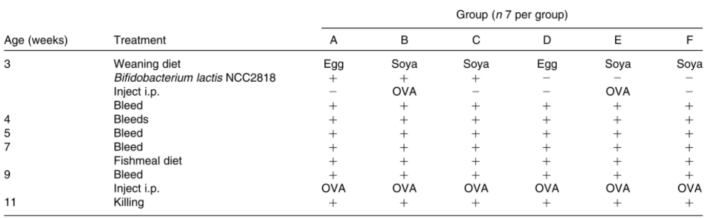
Animal housing and experimental procedures were all performed according to local ethical guidelines: all experi-ments were performed under a UK Home Office License and were approved by the local ethical review group. A total of seven outbred sows were artificially inseminated using semen from a single boar (supplied by Hermitage-Sea-borough Limited). Sows were transported to the Department of Clinical Veterinary Science 6 weeks before parturition and fed on a wheat-based diet (BOCM Pauls Limited). At 3 weeks of age, piglets were weaned and litter-matched into six groups (Table 1), each group being housed in a separate room, on straw, in standard large animal facilities.
At this point, three groups received the B. animalis subsp. lactis (CNCM I-3446) probiotic diet supplementation in the form of spray-dried culture mixed into the formula at a concen-tration of 4·2 £ 106 colony-forming units/ml (approximately 2 £ 109colony-forming units/kg metabolic weight per d). The required quantity of feed supplemented with fresh probiotics was fed twice per d to the appropriate groups (A – C) until the experiment concluded when the pigs were 11 weeks old. The remaining groups (D – E) did not receive the probiotic sup-plement. The probiotic-fed and control animals were in differ-ent suites separated by a biosecurity barrier. Of the piglets receiving probiotics, two groups were weaned onto a soya-based diet and one onto an egg-soya-based diet (Table 2). All diets were supplemented with appropriate levels of vitamins and minerals and were manufactured to order by Volac (Parnutt) Foods Limited. The weaning diets were designed such that the only differences were that each contained 21 % of the stated protein. One of the two groups that weaned onto the soya-based diet also received an intraperitoneal injection of 2 mg soluble ovalbumin from chicken egg-white (systemic exposure; Sigma) and 2 mg Quil A adjuvant (Brenntag Biosector A/S) in 2 ml PBS to investigate the immune response against a systemically administered novel protein. The treatment of these three probiotic-supplemented groups (A – C) was repli-cated in the probiotic-free control groups D to F. From 7 weeks of age, all six groups were fed a fishmeal-based diet, free of egg and soya, either with or without probiotic as appro-priate. The fishmeal diet was used to ensure that the serum anti-body response was to the injected egg protein and not to the dietary egg protein. The egg- and soya-based diets were designed to meet the nutritional requirements of piglets between 3 and 7 weeks old, whereas the fishmeal-based diet was designed for piglets between 7 and 11 weeks old. For this reason, the fishmeal-based diet cannot be compared with the egg- and soya-based diets. The composition of the different diets is shown in Table 2. At 9 weeks old, all piglets received an intraperitoneal injection of 2 mg ovalbumin and 2 mg Quil A adjuvant in 2 ml PBS.
All piglets were bled by venepuncture at 3, 4, 5, 7 and 9 weeks old for collection of serum. At 11 weeks old, piglets Table 1. Experimental design*
Group (n 7 per group)
Age (weeks) Treatment A B C D E F
3 Weaning diet Egg Soya Soya Egg Soya Soya
Bifidobacterium lactis NCC2818 þ þ þ 2 2 2
Inject i.p. 2 OVA 2 2 OVA 2
Bleed þ þ þ þ þ þ 4 Bleeds þ þ þ þ þ þ 5 Bleed þ þ þ þ þ þ 7 Bleed þ þ þ þ þ þ Fishmeal diet þ þ þ þ þ þ 9 Bleed þ þ þ þ þ þ
Inject i.p. OVA OVA OVA OVA OVA OVA
11 Killing þ þ þ þ þ þ
i.p., Intraperitoneally; OVA, ovalbumin.
* Forty-two (six piglets from seven litters) piglets were litter-matched into six treatment groups; three groups received the B. lactis NCC2818 intervention at weaning at 3 weeks of age.
British
Journal
of
were sedated with azaperone and killed with an overdose of barbiturate. At post-mortem, heart blood and tissues were recovered.
Tissue culture
At killing, 4 cm2samples of intestinal mucosa (proximal and distal jejunum, excluding Peyer’s patches (PP), distinct jejunal PP, caecum and descending colon), and 1 cm3 of spleen and mesenteric lymph node (MLN) were collected and placed in cold sterile medium. Organ fragment culture was carried out as described in detail by Logan et al.(35). Briefly, the samples were vigorously washed three times in Ca2þ and Mg2þ-free Dulbecco’s PBS (Sigma) containing 0·5 mM-EDTA (Sigma),
1M-HEPES (Invitrogen) and 50 mg gentamycin/ml (Gibco),
followed by three further washes in Ca2þ and Mg2þ-free Dulbecco’s PBS containing 1 % HEPES and 50 mg gentamycin/ml before being placed in Roswell Park Memorial Institute-1640 medium (Sigma) containing 10 % fetal calf serum (PAA), 200 mM-L-glutamine (Invitrogen), 10 units penicillin/ml
and 10 mg streptomycin/ml (Invitrogen) and 50 mg gentamy-cin/ml (complete medium). Intestinal tissues were cut into frag-ments approximately 3 mm2, while spleen and MLN were cut into 2 mm cubes, and one fragment of tissue was placed in each of six individual wells of a twenty-four-well culture plate (Corning, Inc.) containing 1 ml of complete medium. Cultures were incubated at 378C, 5 % CO2, 100 % humidity for 96 h,
after which they were frozen at 2 208C. The plates were defrosted and the spent medium from each of the six duplicate
wells for each sample was pooled and refrozen for analysis of Ig content.
Immunoglobulin assays
Catching ELISA was carried out to determine total IgG1, IgG2,
IgA and IgM in spent medium from organ fragment cultures and IgA in serum. Briefly, ninety-six well microplates were coated with either affinity-purified goat anti-pig IgG (H þ L), goat anti-pig IgA or goat anti-pig IgM (Bethyl Laboratories). Serial dilutions of serum samples and reference standard were added to coated plates and incubated for 2 h at room tempera-ture. Bound Ig were detected using isotype-specific monoclonal antibodies (anti-pig IgA K61.1B4, anti-pig IgM K52.1C3, anti-pig IgG1K139.3C8 and anti-pig IgG2K68.1G2; all from Serotec)
fol-lowed by horseradish peroxidase-conjugated goat anti-mouse IgG1. Concentrations of Ig subclasses were determined by
interpolation of samples onto the reference standards.
Antigen-specific immunoglobulin assays
Serum samples were analysed for anti-ovalbumin IgG1and IgG2
antibodies by ELISA as described in detail by Bailey et al.(36). Briefly, ninety-six-well microplates were coated with oval-bumin from chicken egg-white (Sigma) before non-specific binding sites were blocked with 2 % bovine serum albumin (Sigma) in PBS – Tween 20. After washing, serial dilutions of serum samples and reference standard were added to the plates. Reference standard was porcine serum obtained Table 2. Composition of the weaning diets and supplements
Ingredients (%) Egg-based Soya-based Fishmeal-based
Whole dried egg 24·3 2 2
Extruded full-fat soya (unmodified, 35 % protein, 19 % fat)
2 17·6 2
High-protein soya (48 % protein, 2·7 % fat) 2 12·2 2
LT94 Fishmeal 2 2 12·5
Wheat 2 2 30·0
Full-fat whey (50 % lard) 2 2 3·5
Potato protein (Roquette) 2 2 2·5
Barley 2 2 10·0
Cooked wheat (MASHM) 21·0 19·4 16·7
Presco maize 21·0 19·7 10·0
Cooked naked oats 11·7 9·2 2
Dairy crest tint whey 9·4 8·8 5·0
Denatured skimmed milk-A 7·7 6·7 5·0
Dextrose 1·7 1·6 1·5
Vitamin and mineral mix* 1·0 1·0 1·0
SNOWCAL chalk 10 2 2 0·6 Dicalcium phosphate 0·9 1·5 0·3 Limestone Trical 130 0·5 0·5 0·5 L-Lys 0·4 0·4 0·3 L-Thr 0·1 0·1 0·1 Salt 0·1 0·1 0·1 Protein 21·3 21·2 21·5 Oil 12·5 11·2 7·1 Fibre 1·1 2·3 1·7 Ash 4·5 5·3 5·1 Moisture 8·6 9·9 10·8 N-free extract 52·0 50·1 53·8
* Vitamin and mineral mix (calculated units in finished feed): vitamin A, 16 mg/kg; vitamin D3, 2 mg/kg; vitamin E, 250 mg/kg; vitamin K (menadione), 4 mg/kg; vitamin B1, 10 mg/kg; vitamin B2, 16 mg/kg; vitamin B6, 10 mg/kg; vitamin B12, 0·05 mg/kg; nicotinic acid, 50 mg/kg; pantothenic acid, 30 mg/kg; biotin (vitamin K), 0·2 mg/kg; vitamin C, 200 mg/kg; folic acid, 3 mg/kg; choline chloride, 300 mg/kg. Trace minerals: Cu, 155 mg/kg; Fe, 375 mg/kg; Zn, 110 mg/kg, Mn, 100 mg/kg; Co, 0·5 mg/kg; I, 1·2 mg/kg; Se, 0·3 mg/kg.
British
Journal
of
following hyperimmunisation with ovalbumin. Bound anti-soya IgG1 and IgG2 antibodies were detected using
isotype-specific monoclonal antibodies followed by HRP-conjugated goat anti-mouse as mentioned previously, and relative concentrations of antibody were determined by interpolation of samples onto the reference standards.
In order to compare changes in serum antibody generated by weaning and by the injection of novel proteins in outbred animals, in which the starting levels differ, results are expressed as the ratio of antibody after manipulation to that before manipulation (fold change in antibody).
Immunohistology
Sample collection. MLN and caecum tissue was removed
shortly after death from each of the experimental piglets. Tissues were embedded in OCT (Tissue TEK; BDH), snap-frozen in iso-pentane and pre-cooled to approximately 2 708C in the vapour phase of liquid N2. Samples were stored at 2 808C until
section-ing. Serial 5 mm sections of these tissues were cut using a Model
OTF cryotome (Bright Instrument Company Limited). Sections were air-dried for 24 h and then fixed by immersion in acetone for 15 min. Slides were allowed to dry before storage at 2 808C. Fluorescence immunohistology. For two-colour fluor-escence immunohistology, mouse pig monoclonal anti-bodies (IgA and IgM, as for ELISA) were used to identify free and cell-bound IgA- and IgM-positive cells and B-lymphocytes (anti-CD21, clone IAH CC55). The conjugated secondary reagents used were as follows: goat anti-mouse IgG1conjugated
to fluorescein isothiocyanate (FITC) (Southern Biotechnology, AMS Biotechnology) and goat anti-mouse IgG2bconjugated to
tetramethyl rhodamine isothiocyanate (TRITC) (Southern Bio-technology). Tissue staining, image capture and automated image analyses were carried out as described by Inman et al.(37)with the exception that fracture crystallography (Fc) receptor blocking was achieved using 10 % goat serum in PBS.
Histochemistry
Small-intestinal samples were obtained as described in the immunohistology section and processed the same up to and
–0·7 –0·5 –0·3 –0·1 0·1 0·3 0·5 0·7 0·9 (a) (b) (c) (d) Spleen MLN JPP Lo g10 total IgA ( µ g/ml) 0·2 0·3 0·4 0·5 0·6 0·7 0·8 0·9 1·0 1·1 1·2 Spleen MLN JPP Lo g10 total IgM ( µ g/ml) –0·2 0·0 0·2 0·4 0·6 0·8 1·0 1·2
Pro SI Dis SI Caecum Colon
Lo g10 total IgM ( µ g/ml) 0·1 0·3 0·5 0·7 0·9 1·1 1·3
Pro SI Dis SI Caecum Colon
Lo g10 total IgA ( µ g/ml) b b a a a a b
Fig. 1. Total (a, b) IgA and (c, d) IgM production (mg/ml, log-transformed) by organ fragment cultures from organised (a, c) lymphoid tissues and (b, d) non-lymphoid tissues from piglets supplemented with Bifidobacterium lactis NCC2818 (groups A, B and C) or unsupplemented (groups D, E and F). Values are means, with their standard errors represented by vertical bars (n 21).a,bMean value with unlike letters was significantly different from that of probiotic
supplemen-tation (P, 0·01; t test with Bonferroni correction). MLN, mesenteric lymph node; JPP, jejunal Peyer’s patches; Pro SI, proximal small intestine; Dis SI, distal small intestine. B, B. lactis; A, control.
British
Journal
of
including the acetone fixation step. Fixed slides were stained for mast cells using 2·5 % toluidine blue O solution (Sigma-Aldrich) for 15 s followed by dehydration through increasing concentrations of alcohol culminating in a histoclearw
(National Diagnostics) wash and mounted in distyrene plasti-ciser xytene (DPX) mounting medium (Fisher). Image capture was carried out using a Colour Coolview camera and Image-Pro Plus software (Photonic Science). Thereafter, ten fields of view were obtained from each piglet and ImageJ software (National Institutes of Health) was used to allow quantification of mast cells per cm2tissue.
Statistical analysis
Statistical analysis was carried out using SPSS statistics (SPSS, Inc.). Univariate linear regression was carried out using piglet as the experimental unit and litter, tissue and probiotic treat-ment as variables. Individual differences between the treattreat-ment groups were determined by least significant differences as in our previous experiments(3).
Results
Local immunoglobulins
Bifidobacterium lactis NCC2818 supplementation caused a reduction in local immunoglobulin production in lymphoid-associated organ fragment cultures. Total IgG1, IgG2, IgA
and IgM were quantified in organ fragment culture medium from all animals. There were highly significant differences in the amounts of the four isotypes produced between tissues (P, 0·0001), spleen producing less IgA (2 0·16 (SEM0·04) log10
mg/ml) than mucosal tissues in the control animals (mean range 2 0·5 – 1·16 log10mg/ml). Highly significant effects of
pro-biotic intervention were observed for IgA (P, 0·0005; Fig. 1(a) and (b)) and IgM (P, 0·009; Fig. 1(c) and (d)), but not for IgG1or IgG2(data not shown). IgA and IgM were lower in the
probiotic supplemented animals than in the unsupplemented animals (for IgA from MLN, 2 0·56 (SEM0·09) and 0·34 (SEM
0·05) log10 mg/ml, respectively; from proximal jejunum, 0·75
(SEM 0·03) and 0·86 (SEM 0·01) log10 mg/ml; from jejunal PP,
0·02 (SEM0·09) and 0·80 (SEM0·09) log10mg/ml; from caecum,
0·74 (SEM0·06) and 1·17 (SEM0·05) log10mg/ml; for IgM from
MLN, 0·38 (SEM0·08) and 0·72 (SEM0·03) log10mg/ml,
respect-ively; from caecum, 0·13 (SEM0·18) and 0·85 (SEM 0·03) log10
mg/ml; from colon, 0·60 (SEM0·04) and 0·81 (SEM0·03)) log10
mg/ml). There was also a significant interaction between pro-biotic treatment and tissue (P, 0·0001 for both classes), such that this effect was more marked for some tissues than others. Specifically, probiotic intervention appeared to have the most marked effect on IgA production by the organised tissues of MLN and jejunal PP (Fig. 1(a)), and to a lesser extent by the dif-fuse lymphoid tissue present in caecal mucosa (Fig. 1(b)). Although supplementation also resulted in significantly decreased IgA production by tissue from the proximal small intestine, the effect was much smaller. IgA production in the spleen, distal small intestine and colon showed no difference between the probiotic supplemented and non-supplemented animals (from spleen, 2 0·25 (SEM 0·04) and 2 0·16 (SEM
0·04) log10mg/ml, respectively; from distal jejunum, 0·46 (SEM
0·03) and 0·44 (SEM 0·07) log10 mg/ml; from colon, 1·16 (SEM
0·02) and 1·06 (SEM0·05) log10mg/ml). There was also no
differ-ence as a result of dietary supplementation in spleen or small-intestinal IgM (for spleen, 0·62 (SEM 0·09) and 2 0·79 (SEM
0·06) log10 mg/ml, respectively; from proximal jejunum, 0·36
(SEM0·07) and 0·36 (SEM0·07) log10mg/ml; for distal jejunum,
0·27 (SEM0·06) and 0·40 (SEM0·05) log10mg/ml; for discrete
jeju-nal PP, 0·75 (SEM0·05) and 0·35 (SEM0·05) log10mg/ml). It should
be noted that IgA in serum taken at time points throughout the experiment remained unaltered by the supplementation with B. lactis NCC2818 (P. 0·05).
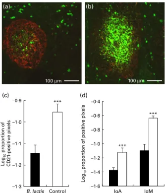
Local IgA and IgM proteins were reduced in caecal tissue, mesenteric lymph node-associated B-cells and B-cell follicles following probiotic intervention. In order to examine the mechanisms by which probiotic administration reduced Ig secretion in organ fragment cultures, levels of IgA, IgM and CD21 were examined in MLN (Figs. 2 and 3) and caecum (Fig. 4) samples from groups B and E (soya diet, supplemented with the probiotic and control, respectively). These tissues and groups were chosen as they had previously produced the most consistent differences in organ fragment cultures. Consistent with the organ fragment culture data, there was reduced expression of IgA (Fig. 2(a) and (d)), IgM (Fig. 2(d)) and also
–1·3 –1·2 –1·1 –1·0 –0·9 Lo g10 propor tion of CD21 -positi ve pixels –1·6 –1·4 –1·2 –1·0 –0·8 –0·6 –0·4 IgA IgM Lo g10 propor tion of positi ve pixels B. lactis Control *** *** *** 100 µm 100 µm (a) (b) (c) (d)
Fig. 2. Fluorescence immunohistology of the mesenteric lymph node from treatment groups B (ovalbumin priming and recall and Bifidobacterium lactis NCC2818 intervention; B) and E (ovalbumin priming and recall without B. lactis NCC2818 intervention; A). (a) Example field from treatment group B: green fluorescence indicates binding of anti-pig IgA monoclonal antibody and red, anti-pig CD21 monoclonal antibody. (b) Example field from treat-ment group E stained similarly. (c) Proportional area of expression of CD21 and (d) IgA and IgM in the same treatment groups (group B, B. lactis NCC2818; group E, control). Values are means, with their standard errors represented by vertical bars (n 7). *** Mean value was significantly different from that of group B (P, 0·0001 in all cases). (A colour version of this figure can be found online at http://www.journals.cambridge.org/bjn)
British
Journal
of
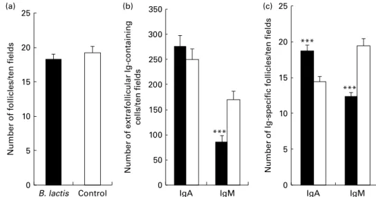
CD21 (Fig. 2(c)), within B-cell follicles in the MLN of the B. lactis NCC2818-treated animals compared with the control group (P, 0·0001). There was no effect of intervention with B. lactis NCC2818 on the total number of MLN follicles (P. 0·05; Fig. 3(a)), but the number of IgM-specific follicles (Fig. 3(c)) and extrafollicular IgM-producing B-cells (Fig. 3(b)) was reduced in the animals receiving the intervention (P, 0·0001). In contrast, no change was seen in the number of extrafollicular IgA-positive cells (Fig. 3(b), P. 0·05), whereas the number of IgA-specific follicles was actually significantly increased (Fig. 3(c)) in animals fed with B. lactis NCC2818 when com-pared with the controls (P, 0·0001). Reductions in the expression of IgA (Fig. 4(a) and (c)) and IgM (Fig. 4(b) and (d)) in situ in the caecum were also apparent (Fig. 4), both in the subepithelial lamina propria (associated with production) and in the caecal crypt epithelium (associated with transport) (Fig. 4(c) and (d)).
A reduction in lymphoid-associated IgA and IgM production was associated with increased mast cell numbers in the intestinal mucosa
In contrast to the decreases observed in IgA and IgM in the supplemented animals, there were significantly greater num-bers of mast cells in the small intestine (P, 0·001) of animals which received B. lactis NCC2818 (n 7) when compared with the control (Fig. 5).
Systemic antibody
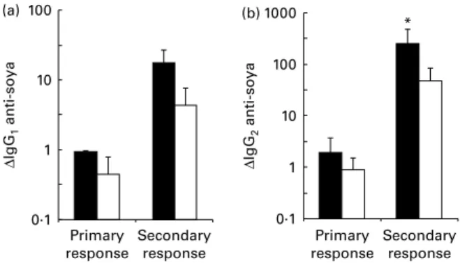
Primary systemic response to novel fed protein at weaning was increased following Bifidobacterium lactis NCC2818 administration. At weaning, there was a significant increase in IgG2 anti-soya antibody in animals which received a soya
diet with probiotic supplementation when compared with both the animals fed soya without probiotic and the egg-fed
animals (P¼ 0·021; Fig. 6(a)). The supplemented animals also mounted a significantly greater IgG1 antibody response to
soya compared with the egg-fed controls (P¼ 0·03), and a greater response than the unsupplemented animals fed soya, although this was not significant (Fig. 6(b)).
–1·6 Crypt epithelium Lamina propria Crypt epithelium Lamina propria –1·4 –1·2 –1·0 –0·8 –0·6 –0·4 (a) (b) (c) (d) Lo g10 propor tion of IgA-positi ve pixels –2·0 –1·8 –1·6 –1·4 –1·2 –1·0 –0·8 –0·6 –0·4 Lo g10 propor tion of IgM-positi ve pixels 100 µm 100 µm
Fig. 4. Fluorescence immunohistology of caecum crypts and lamina propria from soya-fed piglets which received intraperitoneal ovalbumin priming and recall (treatment groups B and E) and either (a) Bifidobacterium lactis NCC2818 supplementation or (b) not; fluorescence indicates the binding of anti-pig IgA monoclonal antibody. (c) IgA and (d) IgM fluorescence quantified (P, 0·0001 in all cases). Values are means, with their standard errors rep-resented by vertical bars (n 7). B, B. lactis; A, control. (A colour version of this figure can be found online at http://www.journals.cambridge.org/bjn)
0 5 10 15 20 25 (a) (b) (c)
Number of follicles/ten fields
B. lactis Control 0 5 10 15 20 25 IgA IgM
Number of Ig-specific follicles/ten fields
*** 0 50 100 150 200 250 300 350 IgA IgM
Number of extrafollicular Ig-containing
cells/ten fields
***
***
Fig. 3. B-cell follicles in the mesenteric lymph node from soya-fed piglets that received intraperitoneal ovalbumin priming and recall and Bifidobacterium lactis NCC2818 intervention (treatment group B; B) or no B. lactis NCC2818 intervention (treatment group E; A), identified by fluorescence immunohistology. (a) Total number of follicles present in tissues: no significant difference between the animals receiving the B. lactis NCC2818 intervention and those that did not. (b) Extra-follicular cells containing either IgA or IgM. (c) Number of IgA- or IgM-positive follicles. Values are means, with their standard errors represented by vertical bars (n 7). *** Mean value was significantly different from that of the control group (P, 0·0001).
British
Journal
of
Primary and secondary responses to injected antigens were increased following probiotic intervention at weaning. Fig. 7(a) and (b) shows the increase in serum IgG1and IgG2
anti-body, respectively, during the primary and secondary responses to systemically injected antigens (with the adjuvant). During the primary response (3 – 5 weeks old), there were trends towards an increased serum IgG1and IgG2anti-ovalbumin response in
the B. lactis NCC2818-fed animals (n 7) compared with the con-trol group. During the secondary response (9 – 11 weeks old), there was a significantly greater response in both isotypes in the supplemented group (IgG1, P¼ 0·05 and IgG2, P¼ 0·02).
Discussion
The common definition of a probiotic (given by the WHO in 2001) is ‘a live microorganism that when administered in adequate amounts confers a health benefit on the host’. Thus, the definitive outcome measure necessary when testing novel strains for probiotic activity is health: in normal or diseased humans or animals, this may be measured, for example, by susceptibility to disease. However, understanding the mechanisms by which probiotics function requires detailed measurement of a wider range of immunological and physio-logical parameters, which may then also be used as proxy measures of health. The strain of B. lactis NCC2818 used in the present experiments has been identified as having probiotic activity, as defined above, in human subjects and in rodent models. These benefits include reducing pathogen load and prevention or reduction of antibiotic-associated diarrhoea(38,39).
However, the effects of probiotics, including B. lactis NCC2818, on immune development at weaning, a time when the resident microbiota is changing rapidly, are largely unknown. The immunological measures reported here, then, relate to the mechanisms of action of the probiotic strain and to the iden-tification of proxy measures of the probiotic effect. The results presented clearly demonstrate that administration of B. lactis to piglets at weaning had marked effects on the structure and function of the mucosal immune system. In that respect, the present results are comparable with mechanistic experiments in rodents and with the data from human clinical trials.
In our system, intervention with B. lactis NCC2818 resulted in reduced IgA in mucosal-associated lymphoid tissues (associated with a reduction in plasma cell numbers by immu-nohistology). In contrast, preterm infants, which received B. lactis NCC2818 for 3 weeks following birth, showed a 2-fold increase in faecal IgA levels from 2 weeks onwards(40),
and IgA production by MLN and PP cells from adult mice was increased when cultured in the presence of Bifidobacterium bifidum(41). In a mouse study, IgA in the intestinal fluids of the supplemented animals was also higher than that in the controls. In these and previous studies, elevated secretory IgA has been presumed to be a mechanism or a proxy measure for a beneficial effect of probiotics(42), presumably by increasing the potential for neutralisation of allergens or pathogen, thus preventing or reducing disease. The disparity between the present results and those reported in human sub-jects and rodents may be more apparent than real. Where intervention has resulted in increased IgA levels in vivo, it should be noted that faecal IgA levels and total intestinal washes, as normally carried out in these species, may be more reflective of jejunal and/or colonic mucosal IgA levels, where there was no effect of supplementation in pigs of the present study, rather than the MLN, PP and caecum, where there was an effect. In addition, studies in human subjects have frequently involved compromised individuals(40),
–0·5 –0·4 –0·3 –0·2 –0·1 0·0 0·1 0·2 0·3 0·4 Lo g10
number of mast cells
B. lactis Control
Fig. 5. Mast cell counts in the lamina propria of the proximal small intestine from piglets (n 7) weaned onto a soya diet and given intraperitoneal oval-bumin priming and recall (treatment groups B and E). Mast cells identified by toluidine blue staining. Symbols indicate litter-matched animals (P, 0·0001).
–1·0 –0·8 –0·6 –0·4 –0·2 0·0 0·2 0·4 0·6 0·8 ∆ IgG 1 anti-soya Time post-weaning (d) –1·0 –0·5 0·0 0·5 1·0 1·5 2·0 2·5 3·0 (a) (b) 0 7 14 0 7 14 ∆ IgG 2 anti-soya Time post-weaning (d)
Fig. 6. Increased serum soya-specific (a) IgG2 and (b) IgG1 antibody in
response to weaning onto a soya-based diet in piglets receiving Bifidobacter-ium lactis NCC2818 supplementation (groups B and C) compared with the non-supplemented groups (groups E and F). Values are means, with their standard errors represented by vertical bars (n 14). IgG1anti-soya increase
was not significant on its own, but combined analysis of changes with anti-soya IgG2 increased the P value to P¼ 0·05. For IgG2, P¼ 0·03. ,
Soya þ B. lactis; , soya diet; , egg diet.
British
Journal
of
whereas the present study used normal, outbred healthy pig-lets. While an increase in faecal IgA has been correlated with protection, the same correlation has not been established for local tissue IgA. Interestingly, an increase in intestinal IgA can be linked to various disease states in humans(43 – 45), and a local increase of IgA in a healthy individual can also be an indication of the loss of barrier function. We thus suggest that the present observation that probiotic supplementation decreased, rather than increased, local IgA production in intes-tinal tissue reflects a reinforcement of the intesintes-tinal barrier (preventing exposure to luminal antigens) rather than a sup-pression of mucosal immunity, and a breakdown in barrier function is often associated with disease. It also suggests that while elevated IgA in the faeces is accepted as a proxy measure for health, the same interpretation cannot necessarily be applied to local IgA production in tissues.
Similarly, although elevated numbers of mast cells have been associated with allergic sensitisation(46), the increases in mast cell numbers seen here were within the normal ranges previously reported in young piglets(47,48), and are in line with physiological numbers in adult pigs(49) and were not comparable with those seen in disease states(50,51). An increase in mast cell numbers within the normal physiological range may be a consequence of increased recruitment to the intestinal mucosa, decreased mast cell exit or the inhibition of mast cell degranulation. Previous studies have suggested that mast cell degranulation contributes to impaired barrier function after weaning in young piglets, and several probiotic species have been shown to reduce IgE-mediated degranulation in an RBL-2H3 cell line(39,51). A reduction in antigen-induced mast cell degranulation may also occur as a consequence of elevated IgG antibody responses to fed and injected antigens in probiotic supplemented animals: elevated serum IgG antibody responses to food proteins have been associated with decreased susceptibility to IgE-mediated aller-gic disease in human subjects and to post-weaning diarrhoea in pigs(52,53). Further, since active, primary responses to intestinal antigens are largely mediated through PP(54) while
tolerance is mediated by the transfer of antigens from the
intestinal mucosa to the MLN(55), stronger responses to fed antigens in supplemented piglets may also indicate reduced uptake across the intestinal epithelium compared with PP. The present results strongly suggest caution in interpreting specific measures of the immune system (in this case, IgA pro-duction, mast cell numbers and antibody to food proteins) as linear, proxy measures for the health benefit of probiotic sup-plementation in the diet without taking the specific animal model and, more importantly, the specific intervention window into account.
Mechanistically, the present results are largely consistent with B. lactis NCC2818 intervention, increasing barrier func-tion between the lumen and the intestinal lamina propria. Specifically, a reduction in IgA production in organ fragment cultures is entirely consistent with a reduced exposure of the mucosal immune system to antigens derived from the intesti-nal lumen. Previously, certain probiotics, including bifidobac-teria, have been shown to enhance the barrier function of human intestinal epithelial cells in vitro(56), but not in vivo, in part by the stabilisation of tight cell junctions(57). B. bifidum, for example, was demonstrated to increase barrier integrity in a rat model of neonatal necrotising enterocolitis(58). In conclusion, the present results demonstrate clear effects of probiotic supplementation in the weaning diets of conven-tionally reared animals which do not have any diseases or unusual pathology. Mechanistically, these effects are consist-ent with increased barrier function. However, the results also strongly suggest that while measures of the effect of probiotic supplementation on the immune system are of value in devel-oping an understanding of the mechanism of action, we may need to interpret with caution. While studies of health benefits are appropriately conducted in human subjects, mechanistic studies require tractable animal models from which sufficient tissue samples can be easily recovered. Such mechanistic studies should, perhaps, be carried out in several mammalian species in order to establish generally applicable principles for predicting the activities of probiotic strains.
Acknowledgements
The present study was supported by Nestec Limited, which also supplied the probiotic B. lactis NCC2818. We wish to thank Stuart Ham for his assistance with the pigs. The authors’ contri-butions are as follows: M. C. L., M. B., A. W. Z., A. M. and S. D. developed the overall research plan; M. C. L., D. V. P., M. B. and J. F. conducted the research; M. C. L. and M. B. analysed the data, performed the statistical analysis and wrote the paper; M. C. L. had primary responsibility for the overall direction and final content. All authors read and approved the final manuscript. M. C. L., M. B., D. V. P. and J. F. declare no conflict of interest. A. W. Z., A. M. and S. D. are employees of Nestec Limited.
References
1. Falk PG, Hooper LV, Midtvedt T, et al. (1998) Creating and maintaining the gastrointestinal ecosystem: what we know and need to know from gnotobiology. Microbiol Mol Biol Rev 62, 1157 – 1170. ∆ IgG 2 anti-soya ∆ IgG 1 anti-soya 100 (a) (b) 10 1 0·1 Primary response Secondary response Primary response Secondary response 100 1000 10 1 0·1 *
Fig. 7. Increased piglet serum ovalbumin-specific (a) IgG1and (b) IgG2
anti-body in response to priming and recall with intraperitoneal ovalbumin with the Quil A adjuvant in piglets weaned onto the soya diet, with and without the Bifidobacterium lactis NCC2818 intervention. Results for antibodies are given as a proportion of the standard. Values are means, with their standard errors represented by vertical bars (n 7). * P values for IgG1and IgG2are 0·05 and
0·02, respectively. Combined analysis of changes gives a P value of 0·004. B, B. lactis; A, control.
British
Journal
of
2. Stecher B & Hardt WD (2008) The role of microbiota in infectious disease. Trends Microbiol 16, 107 – 114.
3. Lewis MC, Inman CF, Patel D, et al. (2012) Direct experimen-tal evidence that early-life farm environment influences regulation of immune responses. Pediatr Aller Immunol 23, 265 – 269.
4. Mulder I, Schmidt B, Stokes C, et al. (2009) Environmentally-acquired bacteria influence microbial diversity and natural innate immune responses at gut surfaces. BMC Biol 7, 79. 5. Mulder IE, Schmidt B, Lewis M, et al. (2011) Restricting
microbial exposure in early life negates the immune benefits associated with gut colonization in environments of high microbial diversity. PLoS One 6, e28279.
6. Schmidt B, Mulder IE, Musk CC, et al. (2011) Establishment of normal gut microbiota is compromised under excessive hygiene conditions. PLoS One 6, e28284.
7. Noverr MC & Huffnagle GB (2004) Does the microbiota regulate immune responses outside the gut? Trends Micro-biol 12, 562 – 568.
8. Moore WEC & Moore LH (1995) Intestinal floras of popu-lations that have a high-risk of colon-cancer. Appl Environ Microbiol 61, 3202 – 3207.
9. Palmer C, Bik EM, DiGiulio DB, et al. (2007) Development of the human infant intestinal microbiota. PLoS Biol 5, 1556 – 1573.
10. West CE, Hammarstrom ML & Hernell O (2009) Probiotics during weaning reduce the incidence of eczema. Pediatr Aller Immunol 20, 430 – 437.
11. Cox MJ, Huang YJ, Fujimura KE, et al. (2010) Lactobacillus casei abundance is associated with profound shifts in the infant gut microbiome. PLoS One 5, e8745.
12. Carter PB & Pollard M (1971) Host responses to normal microbial flora in germ-free mice. J Reticuloendothel Soc 9, 580 – 587.
13. Cebra JJ (1999) Influences of microbiota on intestinal immune system development. Am J Clin Nutr 69, 1046S – 1051S. 14. Cebra JJ, Periwal SB, Lee G, et al. (1998) Development and
maintenance of the gut-associated lymphoid tissue (GALT): the roles of enteric bacteria and viruses. Dev Immunol 6, 13 – 18.
15. Talham GL, Jiang HQ, Bos NA, et al. (1999) Segmented fila-mentous bacteria are potent stimuli of a physiologically normal state of the murine gut mucosal immune system. Infect Immun 67, 1992 – 2000.
16. Butler JE, Weber P, Sinkora M, et al. (2000) Antibody repertoire development in fetal and neonatal piglets. II. Characterization of heavy chain complementarity-determining region 3 diversity in the developing fetus. J Immunol 165, 6999 – 7010.
17. Ivanov II, Frutos Rde L, Manel N, et al. (2008) Specific micro-biota direct the differentiation of IL-17-producing T-helper cells in the mucosa of the small intestine. Cell Host Microbe 4, 337 – 349.
18. Bjorksten B, Sepp E, Julge K, et al. (2001) Allergy develop-ment and the intestinal microflora during the first year of life. J Aller Clin Immunol 108, 516 – 520.
19. Sekirov I, Tam NM, Jogova M, et al. (2008) Antibiotic-induced perturbations of the intestinal microbiota alter host susceptibility to enteric infection. Infect Immun 76, 4726 – 4736.
20. Iannitti T & Palmieri B (2010) Therapeutical use of probiotic formulations in clinical practice. Clin Nutr 29, 701 – 725. 21. Kalliomaki M, Antoine JM, Herz U, et al. (2010) Guidance
for substantiating the evidence for beneficial effects of pro-biotics: prevention and management of allergic diseases by probiotics. J Nutr 140, 713s – 721s.
22. Paton AW, Morona R & Paton JC (2000) A new biological agent for treatment of Shiga toxigenic Escherichia coli infec-tions and dysentery in humans. Nat Med 6, 265 – 270. 23. Villena J, Oliveira MLS, Ferreira PCD, et al. (2011) Lactic acid
bacteria in the prevention of pneumococcal respiratory infection: future opportunities and challenges. Int Immuno-pharmacol 11, 1633 – 1645.
24. Zakostelska Z, Kverka M, Klimesova K, et al. (2011) Lysate of probiotic Lactobacillus casei DN-114 001 ameliorates colitis by strengthening the gut barrier function and changing the gut microenvironment. PLoS One 6, e27961.
25. Magne F, Hachelaf W, Suau A, et al. (2006) A longitudinal study of infant faecal microbiota during weaning. FEMS Microbiol Ecol 58, 563 – 571.
26. Konstantinov SR, Awati AA, Williams BA, et al. (2006) Post-natal development of the porcine microbiota composition and activities. Environ Microbiol 8, 1191 – 1199.
27. Gandarillas M & Bas F (2009) The domestic pig (Sus scrofa domestica) as a model for evaluating nutritional and metabolic consequences of bariatric surgery practiced on morbid obese humans. Cienc Investig Agraria 36, 163 – 176.
28. Kararli TT (1995) Comparison of the gastrointestinal anatomy, physiology, and biochemistry of humans and commonly used laboratory-animals. Biopharm Drug Dispos 16, 351 – 380.
29. Merrifield CA, Lewis M, Claus SP, et al. (2011) A metabolic system-wide characterisation of the pig: a model for human physiology. Mol BioSyst 7, 2577 – 2588.
30. Pittner A, Nalos M, Asfar P, et al. (2003) Mechanisms of inducible nitric oxide synthase (iNOS) inhibition-related improvement of gut mucosal acidosis during hyperdynamic porcine endotoxemia. Intensive Care Med 29, 312 – 316. 31. Tadros T, Traber DL, Heggers JP, et al. (2003) Effects of
inter-leukin-1 alpha administration on intestinal ischemia and reperfusion injury, mucosal permeability, and bacterial trans-location in burn and sepsis. Ann Surg 237, 101 – 109. 32. Thompson CL, Hofer MJ, Campbell IL, et al. (2010)
Commu-nity dynamics in the mouse gut microbiota: a possible role for IRF9-regulated genes in community homeostasis. PLoS One 5, e10335.
33. Wernersson R, Schierup MH, Jorgensen FG, et al. (2005) Pigs in sequence space: a 0.66X coverage pig genome survey based on shotgun sequencing. BMC Genomics 6, 70. 34. Jorgensen FG, Hobolth A, Hornshoj H, et al. (2005)
Com-parative analysis of protein coding sequences from human, mouse and the domesticated pig. BMC Biol 3, 2.
35. Logan AC, Chow KPN, George A, et al. (1991) Use of Peyer patch and lymph-node fragment cultures to compare local immune-responses to Morganella morganii. Infect Immun 59, 1024 – 1031.
36. Bailey M, Haverson K, Miller B, et al. (2004) Effects of infec-tion with transmissible gastroenteritis virus on concomitant immune responses to dietary and injected antigens. Clin Diagn Lab Immunol 11, 337 – 343.
37. Inman CF, Rees LEN, Barker E, et al. (2005) Validation of computer-assisted, pixel-based analysis of multiple-colour immunofluorescence histology. J Immunol Methods 302, 156 – 167.
38. Ishibashi N & Shimamura S (1993) Bifidobacteria – research and development in Japan. Food Technology [Proceedings Paper] 47, 126 – 135.
39. Harata G, He F, Takahashi K, et al. (2010) Bifidobacterium suppresses IgE-mediated degranulation of rat basophilic leukemia (RBL-2H3) cells. Microbiol Immunol 54, 54 – 57.
British
Journal
of
40. Mohan R, Koebnick C, Schildt J, et al. (2008) Effects of Bifidobacterium lactis Bb12 supplementation on body weight, fecal pH, acetate, lactate, calprotectin, and IgA in preterm infants. Pediatr Res 64, 418 – 422.
41. Park JH, Um JI, Lee BJ, et al. (2002) Encapsulated Bifidobac-terium bifidum potentiates intestinal IgA production. Cell Immunol 219, 22 – 27.
42. Kukkonen K, Kuitunen M, Haahtela T, et al. (2010) High intestinal IgA associates with reduced risk of IgE-associated allergic diseases. Pediatr Aller Immunol 21, 67 – 73. 43. Berstad AE, Kilian M, Valnes KN, et al. (1999) Increased
mucosal production of monomeric IgA1 but no IgA1 pro-tease activity in Helicobacter pylori gastritis. Am J Pathol 155, 1097 – 1104.
44. Kett K, Scott H, Fausa O, et al. (1990) Secretory immunity in celiac-disease – cellular expression of immunoglobulin – a subclass and joining chain. Gastroenterology 99, 386 – 392. 45. Valnes K, Brandtzaeg P, Elgjo K, et al. (1986) Quantitative
distribution of immunoglobulin-producing cells in gastric-mucosa – relation to chronic gastritis and glandular atophy. Gut 27, 505 – 514.
46. Sun P, Li DF, Li ZJ, et al. (2008) Effects of glycinin on IgE-mediated increase of mast cell numbers and histamine release in the small intestine. J Nutr Biochem 19, 627 – 633. 47. Smith F, Clark JE, Overman BL, et al. (2010) Early weaning
stress impairs development of mucosal barrier function in the porcine intestine. Am J Physiol Gastrointest Liver Physiol 298, G352 – GG63.
48. Che C, Pang X, Hua X, et al. (2009) Effects of human fecal flora on intestinal morphology and mucosal immunity in human flora-associated piglet. Scand J Immunol 69, 223 – 233.
49. Duncker SC, Lorentz A, Schroeder B, et al. (2006) Effect of orally administered probiotic E-coli strain Nissle 1917 on
intestinal mucosal immune cells of healthy young pigs. Vet Immunol Immunopathol; 111, 239 – 250.
50. Gelbmann CM, Mestermann S, Gross V, et al. (1999) Strictures in Crohn’s disease are characterised by an accu-mulation of mast cells colocalised with laminin but not with fibronectin or vitronectin. Gut 45, 210 – 217.
51. Crowe SE, Luthra GK & Perdue MH (1997) Mast cell mediated ion transport in intestine from patients with and without inflammatory bowel disease. Gut 41, 785 – 792. 52. Li DF, Nelssen JL, Reddy PG, et al. (1991) Interrelationship
between hypersensitivity to soybean proteins and growth-performance in early-weaned pigs. J Anim Sci 69, 4062 – 4069.
53. Strait RT, Mahler A, Hogan S, et al. (2011) Ingested allergens must be absorbed systemically to induce systemic anaphy-laxis. J Allergy Clin Immunol 127, 982 – 9e1.
54. Snoeck V, Verfaillie T, Verdonck F, et al. (2006) The jejunal Peyer’s patches are the major inductive sites of the F4-specific immune response following intestinal immunis-ation of pigs with F4 (K88) fimbriae. Vaccine 24, 3812 – 3820. 55. Hadis U, Wahl B, Schulz O, et al. (2011) Intestinal tolerance requires gut homing and expansion of FoxP3(þ ) regulatory t cells in the lamina propria. Immunity 34, 237 – 246. 56. Resta-Lenert S & Barrett KE (2006) Probiotics and
commen-sals reverse TNF-alpha- and IFN-gamma-induced dysfunc-tion in human intestinal epithelial cells. Gastroenterology 130, 731 – 746.
57. Otte JM & Podolsky DK (2004) Functional modulation of enterocytes by gram-positive and gram-negative micro-organisms. Am J Physiol Gastrointest Liver Physiol 286, G613 – G626.
58. Khailova L, Dvorak K, Arganbright KM, et al. (2009) Bifidobacterium bifidum improves intestinal integrity in a rat model of necrotizing enterocolitis. Am J Physiol Gastro-intest Liver Physiol 2009 297, G940 – G949.