HAL Id: hal-03091771
https://hal.archives-ouvertes.fr/hal-03091771
Submitted on 31 Dec 2020
HAL is a multi-disciplinary open access
archive for the deposit and dissemination of sci-entific research documents, whether they are pub-lished or not. The documents may come from teaching and research institutions in France or abroad, or from public or private research centers.
L’archive ouverte pluridisciplinaire HAL, est destinée au dépôt et à la diffusion de documents scientifiques de niveau recherche, publiés ou non, émanant des établissements d’enseignement et de recherche français ou étrangers, des laboratoires publics ou privés.
Multi-autoantibody Signature and Clinical Outcome in
Membranous Nephropathy
Gian Ghiggeri, Barbara Seitz-Polski, Joana Justino, Christelle Zaghrini,
Christine Payré, Vesna Brglez, Guillaume Dolla, Alberto Sinico, Francesco
Scolari, Augusto Vaglio, et al.
To cite this version:
Gian Ghiggeri, Barbara Seitz-Polski, Joana Justino, Christelle Zaghrini, Christine Payré, et al.. Multi-autoantibody Signature and Clinical Outcome in Membranous Nephropathy. Clinical Journal of the American Society of Nephrology, American Society of Nephrology, 2020, 15 (12), pp.1762-1776. �10.2215/CJN.02500220�. �hal-03091771�
1
Multi-autoantibody Signature and Clinical Outcome in Membranous Nephropathy
Gian Marco Ghiggeri1*, Barbara Seitz-Polski2,3*, Joana Justino2, Christelle Zaghrini2, Christine Payré2, Vesna Brglez2,3, Guillaume Dolla2, Alberto Sinico4, Francesco Scolari5, Augusto Vaglio6,
Marco Prunotto7, Giovanni Candiano8, Antonella Radice9, The Italian Study Group for Membranous Nephropathy10, Maurizio Bruschi8*and Gérard Lambeau2*
1Istituto Giannina Gaslini IRCCS, Department of Pediatric and Hemato-Oncology Science,
Nephrology, Dialysis and Transplantation Unit, Genoa, Italy;
2Université Côte d’Azur, CNRS, Institut de Pharmacologie Moléculaire et Cellulaire (IPMC),
Valbonne Sophia Antipolis, France; 3Université Côte d’Azur, Département d'Immunologie, CHU de Nice, France; 4University Bicocca, Department of Medicine and Surgery, Milan, Italia; 5University of Brescia, Montichiari Hospital, Nephrology and Dialysis Unit, Montichiari, Italy; 6University of Parma, Department of Clinical Medicine, Nephrology and Health Sciences, Parma, Italy; 7School of Pharmaceutical Sciences, University of Geneva, Geneva, Switzerland; 8Istituto Giannina Gaslini IRCCS, Molecular Nephrology Laboratory, Genoa, Italy; 9Microbiology Institute, ASST Santi Paolo e Carlo, S. Carlo Borromeo Hospital, Milan, Italy 10The Italian Study Group for Membranous Nephropathy (see expanded list). **GMG, BSP, MB and GL contributed equally to this work. 10The Italian Study Group for Membranous Nephropathy:
Gian Marco Ghiggeri, Nephrology, Dialysis, and Transplantation, Istituto Giannina Gaslini IRCCS, Genoa, Italy; coordinator of the group and PI of the study;
Antonella Radice, Microbiology Institute, ASST Santi Paolo e Carlo, S. Carlo Borromeo Hospital, Milan, Italy;
Alberto Sinico, Department of Medicine and Surgery, University Bicocca, Milan, Italy;
Francesco Scolari, Nephrology and Dialysis Unit, University of Brescia and Montichiari Hospital, Brescia, Italy;
2
Gabriella Moroni, Division of Nephrology and Dialysis, IRCCS Foundation-Ospedale Maggiore, Mangiagalli, Regina Elena, Milan, Italy; Riccardo Magistroni, Department of Nephrology, University of Modena, Modena, Italy; Domenico Santoro, Division of Nephrology, University of Messina, Messina, Italy; Antonello Pani, Division of Nephrology and Dialysis, Brotzu Hospital, Cagliari, Italy; Augusto Vaglio, Landino Allegri and Isabella Pisani, Department of Clinical Medicine, Nephrology, and Health Sciences, University of Parma, Parma, Italy;
Antonio Granata, Division of Nephrology and Dialysis, San Giovanni di Dio Hospital, Agrigento, Italy; Giuseppe Grandagliano, Ospedali Riuniti, Foggia, Italy; Davie Rolla, Division of Nephrology and Dialysis, San Andrea Hospital, La Spezia, Italy; Giuliano Boscutti, Division of Nephrology and Dialysis, Cattinara Hospital, Trieste, Italy; Lucia Mardin bianco, Division of Nephrology and Dialysis, Gorizia Hospital, Italy; Statistical control and analysis Maurizio Bruschi, Molecular Nephrology Laboratory, Istituto Giannina Gaslini IRCCS, Via Gerolamo Gaslini 5, 16147 Genova;
Pietro Ravani, Division of Nephrology, University of Calgary, 1403-29th Street, NW,Calgary, AlbertaT2N 2T9, Canada.
Local coordination
Monica Bodria, Division of Nephrology, Dialysis and Transplantation, Istituto Giannina Gaslini IRCCS, Genoa, Italy;
Giovanni Candiano, Division of Nephrology, Dialysis and Transplantation, Istituto Giannina Gaslini IRCCS, Genoa, Italy.
Correspondence:
Dr Gian Marco Ghiggeri, Dipartimento Integrato di Scienze Pediatriche e Emato-Oncologiche, U.O.C. Nefrologia, Dialisi e Trapianto, Istituto Giannina Gaslini IRCCS, Via Gerolamo Gaslini 5, 16147 Genova, Email: [email protected]
Dr Gérard Lambeau, Institut de Pharmacologie Moléculaire et Cellulaire, UMR 7275 CNRS et Université Côte d'Azur, Valbonne Sophia Antipolis, France. Email: [email protected]
3
Abstract
Background and objectives Patients with membranous nephropathy can have circulating autoantibodies against membrane-bound (PLA2R1 and THSD7A) and intracellular (AR, SOD2 and αENO) podocyte autoantigens. We studied their combined association with clinical outcome. Design, setting, participants, & measurements Serum levels of anti-PLA2R1, anti-THSD7A, anti-AR, anti-SOD2 and anti-αENO autoantibodies were determined in 285 patients at diagnosis and during follow-up using standardized and homemade assays. Maintenance of eGFR >60 mL/min/1.73 m2 and remission of proteinuria (<0.3/<3.5 g/day) after 12 months were the clinical outcome targets. Results At diagnosis, 182 (64%), 8 (3%) and 95 (33%) patients were anti-PLA2R1+, anti-THSD7A+ and double-negative, respectively. The prevalence of a detectable antibody to at least one intracellular antigen was similarly distributed in anti-PLA2R1+ (n=118, 65%) and double negative (n=64, 67%) patients. Positivity for anti-PLA2R1, anti-SOD2 and anti-αENO antibodies and higher titers at diagnosis were independently to each other associated with poor clinical outcome. Combined positivity for anti-PLA2R1, anti-SOD2 and anti-αENO was associated with maximal risk (OR 5.5[1.2-24], p=0.01). In Kaplan-Meier analysis, patients PLA2R1+/SOD2+ or anti-PLA2R1+/anti-αENO+ had reduced eGFR at 12 months compared to anti-PLA2R1+/anti-SOD2- or anti-αENO- patients. Similar associations were observed when considering treatment. Predictive tests (NRI and ROC-AUC) showed that combined assessment of antibodies improved classification of outcome in 22-34% of cases for partial remission of proteinuria and maintenance of normal eGFR. For patients with nephrotic syndrome at diagnosis, SOD2 positivity and high anti-PLA2R1 titer were associated with lack of complete remission. Patients anti-PLA2R1-/intracellular- had the lowest proteinuria and the highest eGFR at diagnosis and the lowest risk of reducing eGFR at 12 months. Epitope spreading was present in 81% of anti-PLA2R1+ patients and was associated
with increased positivity for intracellular antigens and poor eGFR at diagnosis and 12 months. Conclusions Combined serological analysis of autoantibodies targeting membrane-bound and intracellular autoantigens identifies patients with poor clinical outcome.
4
Introduction
Membranous nephropathy is a common cause of nephrotic syndrome in adults (1-3). It is characterized by thickening of the glomerular basement membrane with a well-defined pattern of subepithelial immune complex deposits, usually including IgG4 autoantibodies against various autoantigens. The clinical outcome of membranous nephropathy is variable (4). About one third of patients does not require immunosuppressive therapy and reach spontaneous remission while two thirds require immunosuppressive treatment (such as cyclosporine, cytotoxic agents or anti-CD20 antibodies) and respond or not to treatment, with possible progression to end-stage kidney disease over 5-10 years (5-10).
Antibodies against various podocyte autoantigens have been shown to play a role in the pathogenesis of various forms of membranous nephropathy. In rare familial cases of alloimmune membranous nephropathy, autoantibodies were identified against neutral endopeptidase (11). These autoantibodies developed in mothers deficient for neutral endopeptidase and crossed the placenta to induce membranous nephropathy in the fetus.
This elegant demonstration of a first causative autoantibody of membranous nephropathy was followed by the identification of phospholipase A2 receptor 1 (PLA2R1) as the major autoantigen in adult membranous nephropathy, with circulating autoantibodies present in about 70% of patients (12). Genetic studies further supported a causal role of PLA2R1 based on association of the disease with 6p21 HLA-DQA1 and 2q24 PLA2R1 loci in cohorts of Caucasian (13) and Asian (14) patients. Multiple epitopes have been identified in the CysR, CTLD1, CTLD7 and CTLD8 domains of PLA2R1 (15-20), which may be reminiscent of a mechanism of epitope spreading reflecting the maturation of the immune response from CysR to other PLA2R1 domains (21, 22). Epitope spreading has also been documented in the Heymann rat nephritis model where megalin is the major autoantigen (23). Thrombospondin type-1 domain containing 7A (THSD7A) was reported as a second autoantigen in adult membranous nephropathy, with circulating anti-THSD7A autoantibodies present in about 3% of a different group of patients (24). Autoantibodies targeting different intracellular podocyte autoantigens including aldose reductase (AR) (25), superoxide dismutase (SOD2) (26) and α−enolase (αENO) (27) have been identified in a significant number of patients with membranous nephropathy (28).
Finally, three new antigenic markers of autoimmunity (exostosin1/exostosin2 and NELL-1) have been very recently identified in immune deposits, with exostosins being more present in
5
secondary cases of membranous nephropathy (i.e. class V lupus nephritis) (29, 30). The exact prevalence and significance of autoantibodies against these new autoantigens in the pathogenesis of membranous nephropathy remain to be determined, with circulating autoantibodies currently found only for NELL-1, and with different prevalence between American and European cohorts of patients (29, 30).
In this study, we aimed to 1) detect autoantibodies directed against the well-characterized membrane-bound autoantigens PLA2R1 and THSD7A and the intracellular autoantigens SOD2, AR and αENO in a large retrospective cohort of 285 patients with membranous nephropathy at diagnosis and after 6 and 12 months of follow-up, and 2) evaluate the clinical outcome of patients stratified into different groups based on their combined positivity or not for multiple autoantibodies directed against the different autoantigens.
6 Materials and Methods Study design and patient population This is a cross-sectional study in patients with MN assessing the relationships between detection of different autoantibodies at baseline and eGFR and proteinuria at 12 months. We included 285 patients at the time of a histopathological finding of idiopathic or primary membranous nephropathy (Table S1) and followed them with clinical visits for at least 12 months. They were enrolled in the frame of two studies: 244 patients were enrolled in Italy (Italian Study Group on Membranous Nephropathy-EudraCT 2011–003942–41) and 41 in France (Cohort SOURIS NCT02199145). Criteria for enrollment were (1) biopsy-proven diagnosis of membranous nephropathy associated with proteinuria >0.3 g/day, (2) normal complement profile, (3) negative tests for ANA, anti-dsDNA, ANCA, cryoglobulins and absence of viral markers (hepatitis B surface antigen and HIV), (4) absence of clinical and biochemical signs of cancer and (5) serum samples available at diagnosis and during follow up for determination of antibody levels. Clinical studies were approved by the relevant institutional review boards in the different hospitals and were conducted according to the principles of the Declaration of Helsinki, with written informed consent obtained from all patients. The study was presented at the Ethical Committee of IRCCS Giannina Gaslini. Serum was also obtained from 50 healthy donors recruited at the same institutions that participated to the sample recruitment. Healthy donors had at least one normal urine analysis and normal clinical tests in the prior six months (Table S1).
Detection of circulating autoantibodies
Anti-PLA2R1 autoantibodies (detection of total IgG) against the full-length extracellular domain of PLA2R1 were measured with the standardized ELISA from Euroimmun (Medizinische Labordiagnostika AG, Lübeck, Germany) (33). Anti-PLA2R1 autoantibodies (detection of IgG4 subclass) against the specific epitope-containing domains CysR, CTLD1 and CTLD7 were determined as described (19) using recombinant soluble forms of each domain harboring a HA-tag and produced in HEK293 cells. Anti-THSD7A autoantibodies (detection of IgG4 subclass) against the full-length extracellular domain of THSD7A were determined by ELISA as described (34). Positivity was confirmed by western-blot (24) and indirect immunofluorescence (35) assays. Autoantibodies against the intracellular antigens AR, SOD2 and αENO (detection of IgG4 subclass) were determined by dot-blot using recombinant proteins as immobilized antigens as described (26, 27). Further details on the detection methods and source of antigens are given in the supplemental material. For anti-PLA2R1 total IgG, we used the normal limit of positivity of
7
20 RU/mL given by the manufacturer (33). For anti-THSD7A and intracellular autoantibodies, normal limits were calculated from ROC curves as described (34, 36). High and low levels of autoantibodies were defined as the titers above median and between median and limit of normality, respectively (Table S2).
Outcome
Clinical outcome was evaluated on the basis of proteinuria levels at diagnosis versus after 12 months of follow-up, and was defined as complete remission (proteinuria <0.3 g/day) or partial remission (proteinuria <3.5 g/day) according to Thompson et al (31). Failure to achieve clinical remission was defined by persistence of proteinuria >3.5 g/day. Kidney function (eGFR) was evaluated based on the CKD-EPI creatinine equation (32).
Statistical analysis
Autoantibody levels were expressed as median and interquartile range. The differences in serum concentration of all autoantibodies within patient groups were analyzed using non-parametric U Mann-Whitney test for unpaired samples; Spearman’s correlation was used to evaluate correlations among circulating levels of the various types of autoantibodies. The receiver-operating characteristic (ROC) curve analysis was used to discriminate between healthy people and MN patients based on levels of each autoantibody at diagnosis. The best cut-offs were selected as the values that minimized the geometric distance from 100% sensitivity and 100% specificity on the ROC curves. In these analyses, discrimination is assessed as the ability of the test (performance) to distinguish the presence of the outcome beyond chance, defined when the area under the curves (AUC) are significantly >0.5. Odds ratios (OR) and 95% confidence intervals (CI) were tested to determine the associations between autoantibodies positivity or high titer and proteinuria and eGFR using 2x2 contingency tables. Fisher's exact test was used to determine the statistical significance. The P-values were corrected for multiple comparisons with alpha error values of 5%. We used the Kaplan-Meier method to construct survival curves for time to events according to the level of proteinuria. Differences in outcomes were estimated using the log-rank (Mantel-Cox) test and their hazard ratio; p-values ≤0.05 were considered as significant. The performance of single antibody and their synergism for predicting clinical outcome were assessed by two-category net reclassification index (NRI) and area under the curve of receiver operating characteristic analysis (AUC-ROC) (36). We used R for all statistical analyses (http://www.R-project.org/).
8
Results
Patients and predictors of clinical outcome
We included 285 patients with biopsy-proven membranous nephropathy whose the clinical characteristics are shown in Tables 1 and S1, according to PLA2R1 and intracellular antibody positivity Most patients were men (n=194, 68%). At diagnosis, 67% of patients had nephrotic syndrome. Seventy-nine percent of patients were treated with immunosuppressive treatments: steroids (33%), cytotoxic drugs (32%), rituximab (12%), and cyclosporine (25%). Antiproteinuric treatment with angiotensin converting enzyme inhibitors (ACEi) was added to the above therapy. Twenty-one percent of patients received only ACEi. Patients with nephrotic syndrome (n=177, 62%) had persistent high proteinuria after antiproteinuric treatment, with evidence for deterioration of kidney function (eGFR). When the cohort was analyzed as a whole, gender and nephrotic proteinuria at diagnosis were the only two clinical factors associated with the risk of failure to achieve complete remission of proteinuria after 12 months of follow-up (Figure S1), in agreement with previous findings (31).
Individual autoantibodies: distribution and levels
All patients were tested at diagnosis for the presence of circulating autoantibodies recognizing PLA2R1 and THSD7A by specific ELISAs (33, 34). The presence of circulating autoantibodies specifically recognizing the CysR, CTLD1 and CTLD7 domains of PLA2R1 were also analyzed, defining non-spreaders (patients with detectable reactivity limited to CysR) and spreaders (patients with detectable reactivity to CysR, CTLD1 and/or CTLD7 domains) (19, 37). Patients were also tested for the presence of autoantibodies recognizing the intracellular autoantigens SOD2, AR and αENO (26, 27). Patients with single or multiple positivities to any of these intracellular antigens were referred to as Intracellular+ while patients with no detectable reactivity were
referred to as Intracellular-
. The same analyses were repeated after 6 and 12 months (Figures S2a-S2e).
Among all patients, 182 (64%) were positive for anti-PLA2R1 (PLA2R1+ patients, Figures 1a and S2a). The median level of anti-PLA2R1 autoantibodies was 122 RU/mL (IQR 60-267) at baseline and decreased after 6 and 12 months of follow-up (Figure S2a). At baseline, 118 (65%) PLA2R1+
patients were positive for one or more intracellular antigens, defining PLA2R1+/Intracellular+ patients while 64 (35%) were negative, defining PLA2R1+/Intracellular- patients (Table 1, Figures 1a
and S3a). Interestingly, the percentage of Intracellular+ patients was higher in patients with high titer of anti-PLA2R1 autoantibodies (Figure 1b). Furthermore, PLA2R1+ patients defined as
9
spreaders had higher anti-PLA2R1 titers than non-spreaders (Figure S3b) and were also more positive for intracellular autoantigens (Figure S3c and Table S3), in particular for αENO (Figure S3d).
Among patients, only eight (3%) were positive for anti-THSD7A (THSD7A+, Figures 1a and S2b). One
was double-positive for PLA2R1 and THSD7A (Figure S2b). Seven of these patients had additional autoantibodies against at least one intracellular autoantigen (Figures 1a and S3e). Because of their low prevalence, anti-THSD7A+ patients were not considered for further analysis.
Thus, among all patients, 95 patients (33%) were identified as double-negative for PLA2R1 and THSD7A. Among these latter patients, 64 (67%) were also positive for one or more intracellular antigens, defining double-negative/Intracellular+ patients while 31 (33%) were negative, defining double-negative/Intracellular- patients (Table 1, Figure 1a and S3f).
Overall, autoantibodies against AR, SOD2 and αENO were detected in 85, 85 and 117 patients, respectively (Figures S2c, S2d and S2e). The median levels of each intracellular autoantibody decreased at six and 12 months of follow-up (Figures S2c, S2d and S2e). Positivity and titer levels for anti-PLA2R1, anti-SOD2, anti-AR and anti-αENO autoantibodies at baseline did not correlate to each other (Figure S3g).
Association of the different antibodies with clinical outcome
Considering all patients, positivity for anti-PLA2R1, anti-SOD2 and anti-αENO autoantibodies at diagnosis was variably and independently associated with complete or partial remission of proteinuria and eGFR after 12 months (Table 2). Concomitant positivity of the three antibodies had the highest odds ratio for complete remission of proteinuria after 12 months (OR 5.5 [1.2-24], p=0.01). High titers of anti-PLA2R1 and anti-SOD2 above the median (Table S2, n=91 and n=43, respectively) were similarly associated with lack of partial or complete remission of proteinuria after 12 months compared to those with low titers (Table 2). Kaplan-Meier analysis showed that combined positivity for anti-PLA2R1 and anti-SOD2 or anti-αENO was associated with a higher risk of low eGFR (<60 ml/min/1.73 m2) after 12 months compared to positivity for only anti-PLA2R1 (Figure 2). Additionally, high titers of anti-PLA2R1 and anti-αENO were associated with reduced eGFR after 12 months, as compared to low titers (Table 2). Considering only patients with nephrotic syndrome at diagnosis (67% of the whole cohort), the associations were different (Table 3). Only positivity for anti-SOD2 antibodies and concomitant positivity for anti-PLA2R1 and SOD2 were associated with persistence of proteinuria after 12 months. High titers of anti-PLA2R1 were also associated with persistence of proteinuria. Considering treatment, positivity and
10 high levels of anti-PLA2R1 and anti-SOD2 were the main features associated with lack of partial and complete remission after 12 months in patients who had been treated with cytotoxic drugs, cyclosporine A or rituximab (Table S4). We further compared the added value of anti-SOD2 and anti-αENO over anti-PLA2R1 to predict clinical outcome using net reclassification improvement (NRI) and area under the receiver-operating-characteristic curve (AUC-ROC) analysis (38). In NRI analysis, the association of two antibodies allowed the reclassification of patients who developed or maintained proteinuria <3.5 g/day after 12 months in 34% and 22% of cases negative for both PLA2R1 and SOD2 or αENO, respectively (Table 4). The same association allowed reclassification of eGFR >60 mL/min/1.73 m2 in 27% and 24% of patients at 12 months, respectively (Table 4). AUC-ROC analysis discriminated the outcome after 12 months in respect to the titer of a single antibody or its association. In this case, the association of more than one antibody slightly increased the discrimination for patients who developed eGFR <60 mL/min/1.73 m2 (Table 4).
Stratification of patients based on combined positivity for multiple autoantibodies and association with clinical outcome
Based on combined positivity for autoantibodies against PLA2R1 and intracellular antigens at diagnosis and excluding THSD7A+ patients, we stratified the remaining 277 patients into four groups (Table 1). Hundred eighteen patients were anti-PLA2R1+/Intracellular+ (i.e. with additional
single or multiple positivity for autoantibodies against intracellular antigens), 64 patients were anti-PLA2R1+/Intracellular-, 64 patients were anti-PLA2R1-/Intracellular+, and 31 patients were
anti-PLA2R1-/Intracellular-. At diagnosis, the four groups did not differ for age or sex and had the
same percentage of histology classes (Table 1). All patients received comparable therapy (Table 1). Proteinuria at diagnosis was higher in the two groups anti-PLA2R1+ versus those anti-PLA2R1-
(Table 1 and Figure 3a). Proteinuria after 12 months was lower and eGFR higher in anti-PLA2R1
-/Intracellular- patients versus others (Table 1 and Figures 3a and 3b).
Differences in outcomes were estimated using Kaplan-Meier curves and the log-rank (Mantel-Cox) test and their hazard ratio. Forty-five percent of anti-PLA2R1-/Intracellular- patients reached complete remission of proteinuria after 12 months, as compared to 16, 16 and 28% for the other groups (Figure 4a). More anti-PLA2R1-/Intracellular+ patients had a worse clinical outcome after 12 months as compared to anti-PLA2R1-/Intracellular- patients (Figure 4b). Odds ratios and 95% confidence intervals were utilized to determine the association with reduced eGFR after 12 months. Patients anti-PLA2R1+/Intracellular+ and anti-PLA2R1-/Intracellular+ had about four-fold
11
higher risk of developing eGFR <60 mL/min/1.73 m2 after 12 months,
as compared to patients
PLA2R1-/Intracellular- (Figure 4c). We finally analyzed whether the combined positivity for PLA2R1
epitope spreading and intracellular antigens may be associated with clinical outcome (Table S5). The concomitance of spreading and anti-αENO positivity was associated with low eGFR at diagnosis and after 12 months (Figure S4) whereas eGFR at diagnosis in non-spreaders was only barely modified by positivity for anti-αENO autoantibodies (not shown).
12
Discussion
This is the largest retrospective study cohort of membranous nephropathy patients with the combined detection of autoantibodies against the two major membrane-bound antigens PLA2R1 and THSD7A and the three intracellular autoantigens AR, SOD2 and αENO. As expected, PLA2R1+
patients represent the majority of the cohort (64%) while THSD7A+ patients represent only 3%, in line with recent studies (34, 35, 39). Our results confirmed that high titers of anti-PLA2R1 autoantibodies are associated with poor outcome in terms of remission of proteinuria and eGFR (40-45). We also confirmed that epitope spreading was associated with reduced eGFR after 12 months (19, 37).
In this study, we add to the above points that the presence of circulating anti-SOD2 and anti-αENO autoantibodies was independently associated with poor outcome in terms of partial and complete remission of proteinuria and eGFR. Furthermore, anti-SOD2, anti-αENO and anti-AR autoantibodies are not correlated to anti-PLA2R1 autoantibodies, and similar percentages of patients were positive for one or more intracellular antigens, irrespective of positivity (n=118, 65%) or not (n=64, 67%) for PLA2R1. Further analysis by Kaplan-Meier curves showed worse clinical outcome for patients positive for anti-PLA2R1 and anti-SOD2 or anti-αENO while NRI and AUC-ROC suggested that intracellular autoantibodies might have an added value over anti-PLA2R1 for refinement of association with clinical outcome. We thus conclude that the positivity for intracellular autoantibodies has an added value over anti-PLA2R1 positivity to predict adverse clinical outcome. It remains to be seen if this might be also true for other membrane-bound antigens such as THSD7A or the new extracellular autoantigens, in particular NELL-1.
Based on the concomitant presence or absence of anti-PLA2R1 and anti-intracellular autoantibodies, we stratified patients into four groups. Extending what was reported above for anti-PLA2R1 positivity, another novel finding is that patients negative for the two categories of autoantibodies have a reduced proteinuria at diagnosis and a better clinical outcome after 12 months of follow-up, in comparison to other patients. Thus, in anti-THSD7A negative patients, the simultaneous measurement of the two categories of autoantibodies (PLA2R1 and anti-SOD2 and anti-αENO) at baseline might be an additive factor to better evaluate clinical outcome. In this setting, positivity to intracellular antigens may represent per se a negative factor associated with poor clinical outcome. However, we acknowledge limitations in our study including the use of a retrospective cohort with patients with different ages, different clinical characteristics and
13
treatments, and homemade assays to measure autoantibodies to intracellular autoantigens. Our results should thus be confirmed in prospective studies and/or randomized clinical trials.
The second main finding reported here is that positivity for autoantibodies targeting the intracellular autoantigens, especially anti-αENO autoantibodies, is maximal in PLA2R1+ patients
with high anti-PLA2R1 titer or those defined as spreaders. The combined positivity associates with a reduction of eGFR, being worse in patients with high anti-PLA2R1 titer (Table 1) and in spreaders (Figure S4) positive for anti-αENO autoantibodies. The lower positivity of anti-αENO autoantibodies or other intracellular autoantigens in patients with low anti-PLA2R1 titer or defined as non-spreaders suggests that the formation of autoantibodies against intracellular antigens represents a secondary phenomenon that occurs after the formation of autoantibodies against PLA2R1 (and possibly THSD7A) as primary triggers of membranous nephropathy. We thus propose that anti-SOD2 and anti-αENO autoantibodies may appear as a second wave of autoantibodies in the pathogenesis of membranous nephropathy. This hypothesis is also supported by the in vitro and in vivo data obtained in the mouse model of THSD7A-associated membranous nephropathy where SOD2 was induced after podocyte injury and in carbonic anhydrase II-associated membranous nephropathy (46, 47). Anti-SOD2 and anti-αENO autoantibodies are not unique to membranous nephropathy and their occurrence has been described in lupus nephritis (27, 48). However, the IgG subclass is different in membranous nephropathy and lupus nephritis, with IgG4 in membranous nephropathy but IgG2 in lupus nephritis. Therefore, the same autoantigen can trigger the formation of autoantibodies with different IgG isotypes depending on the type of disease. For lupus nephritis, it has been speculated that exposure of methyl-oxidized αENO and/or the DNA-oxidized αENO by neutrophils to TLR9 is a possible mechanism leading to the production of anti-αENO antibodies (48, 49).
SOD2 is a key antioxidant enzyme playing a major role in protecting cells from oxidative injury (50). It is activated as the main antioxidant pathway after formation of autoimmune deposits in glomeruli. Formation of anti-SOD2 autoantibodies may modify the antioxidant protective effect of SOD2 and worsen the clinical outcome.
αENO is a glycolytic enzyme that catalyzes dehydration of 2-phospho-D-glycerate to phosphoenol-pyruvate and produces αENO is a glycolytic enzyme that catalyzes dehydration of 2-phospho-D-glycerate to phosphoenol-pyruvate and ATP in the Embden-Meyerhof-Parnas pathway (51). αENO has also an extracellular localization where it functions as a plasminogen receptor (52). Upon binding to αENO, plasminogen is transformed into plasmin that degrades extracellular matrix
14
components and activates metalloproteases (51, 53). The presence of anti-αENO autoantibodies may reduce its anti-fibrotic role and favor the accumulation of ECM.
Overall, the results of this study show a multi-autoantibody composition in membranous nephropathy, with antibodies that may act in concert, leading to high proteinuria and low eGFR. Based on the possible functions of SOD2 and αENO after podocyte injury both in vitro and in vivo (46, 47), we propose a multi-hit pathogenic mechanism of membranous nephropathy that first involves the formation of anti-PLA2R1 (or anti-THSD7A or autoantibodies to another similar "primary" autoantigen), that then progresses by a mechanism of both intramolecular (such as within PLA2R1) and intermolecular epitope spreading along which i) the anti-PLA2R1 titer increases and ii) is exacerbated by a second wave of autoantibodies targeting intracellular autoantigens, leading for instance to the production of anti-SOD2 and/or anti-αENO autoantibodies. In patients negative for anti-PLA2R1 and anti-THSD7A autoantibodies, an autoantibody targeting a still unknown membrane-bound autoantigen would take a role similar to anti-PLA2R1 or anti-THSD7A antibodies, possibly represented by patients positive for NELL-1, exostosins or other unknown antigens (29, 30).
In conclusion, our study first confirms the relevance of detecting and measuring anti-PLA2R1 (and anti-THSD7A) autoantibodies as fundamental tools to diagnose and identify patients with membranous nephropathy and at risk of severe disease. The study further shows the added value of assessing autoantibodies targeting intracellular podocyte antigens, combined or not with positivity for the above membrane-bound autoantigens. Altogether, our findings pave the way for
15 Author Contributions G.M.G, B.S.P. and G.L. designed the study; J.J., C.Z., C.P., V.B., G.D., C.M., M.B., G.C and A.S. carried out experiments; G.M.G, B.S.P., M.B., M.P. and G.L. analyzed the data; G.M.G, B.S.P., J.J., M.B. and G.L. made the figures; G.M.G, B.S.P., J.J., M.B. and G.L. drafted and revised the paper; A.S., F.S., A.V., M.P. and B.S.P. had a major role in the inclusion of patients; all authors approved the final version of the manuscript. Funding The Istituto Giannina Gaslini IRCCS (trial sponsor) had provided logistic and financial support to the study through grant from the Ministero della Salute (Ricerca Corrente). People working on the membranous nephropathy project belong to the “Fondazione Malattie Renali del Bambino” of which we acknowledge the financial support. GMG received a grant from Compagnia di San Paolo (ROL 9849). This work was supported by grants from CNRS, the Fondation Maladies Rares (LAM-RD_20170304, GL), the National Research Agency (ANR, grants MNaims (ANR-17- CE17-0012-01, GL) and “Investments for the Future” Laboratory of Excellence SIGNALIFE, a network for innovation on signal transduction pathways in life sciences (ANR-11-LABX-0028-01) with allocated PhD fellowships for CZ and JJ) and the Fondation pour la Recherche Médicale (FRM ING20140129210, SPF20150934219, DEQ20180339193 and FDT201805005509 to GL, VB and JJ).
Disclosures
Some coauthors are co-inventors on the patents “Diagnostics for membranous nephropathy” (GL), “Methods and kits for monitoring membranous nephropathy” (BSP and GL), and “Prognosis and monitoring of membranous nephropathy based on the analysis of PLA2R1 epitope profile and spreading” (BSP, GD and GL). GMG is inventor on a patent on the use of anti-SOD2 and anti-ENO antibodies as biomarkers in membranous nephropathy (EP 3097419). Supplemental Material This article contains the following supplemental material: 1. Supplemental Methods 2. Supplemental references 3. Supplemental Tables and Figures Table S1. Epidemiology, histological stage, clinical characteristics and treatment of patients with membranous nephropathy (MN) versus controls (Healthy donors) at diagnosis and after 12 months of follow-up.
16
Table S2. Quantitative data for limits of positivity and distribution of serum levels for each autoantibody.
Table S3. Detailed autoantibody positivity for intracellular antigens in non-spreaders and spreaders PLA2R1+ patients.
Table S4. Two-way contingency table showing the association of antibodies levels with indexes of kidney outcome and the interaction between autoantibodies against PLA2R1 and intracellular antigens for patients treated with cytotoxic drugs, cyclosporine A or rituximab.
Table S5. Clinical characteristics at diagnosis and after 12 months of follow-up for PLA2R1+ patients stratified according to epitope profiles.
Figure S1. Prognosis clinical factors associated with complete remission.
Figure S2. Circulating levels of the various autoantibodies at diagnosis and during follow-up.
Figure S3. Multi-autoantibody composition in the cohort of patients with membranous nephropathy.
Figure S4. Kidney function (eGFR) in spreaders positive (n=63) or not (n=84) for anti-αENO autoantibodies. References 1. Ponticelli C, Glassock RJ: Glomerular diseases: membranous nephropathy--a modern view. Clin J Am Soc Nephrol, 9: 609-616, 2014 2. Ronco P, Debiec H: Pathophysiological advances in membranous nephropathy: time for a shift in patient's care. Lancet, 385: 1983-1992, 2015 3. Couser WG: Primary Membranous Nephropathy. Clin J Am Soc Nephrol, 12: 983-997, 2017 4. Schieppati A, Mosconi L, Perna A, Mecca G, Bertani T, Garattini S, Remuzzi G: Prognosis of
untreated patients with idiopathic membranous nephropathy. N Engl J Med, 329: 85-89, 1993 5. Fervenza FC, Sethi S, Specks U: Idiopathic membranous nephropathy: diagnosis and treatment. Clin J Am Soc Nephrol, 3: 905-919, 2008 6. Polanco N, Gutierrez E, Covarsi A, Ariza F, Carreno A, Vigil A, Baltar J, Fernandez-Fresnedo G, Martin C, Pons S, Lorenzo D, Bernis C, Arrizabalaga P, Fernandez-Juarez G, Barrio V, Sierra M, Castellanos I, Espinosa M, Rivera F, Oliet A, Fernandez-Vega F, Praga M, Grupo de Estudio de las Enfermedades Glomerulares de la Sociedad Espanola de N: Spontaneous remission of nephrotic syndrome in idiopathic membranous nephropathy. J Am Soc
Nephrol, 21: 697-704, 2010
7. van de Logt AE, Hofstra JM, Wetzels JF: Pharmacological treatment of primary membranous nephropathy in 2016. Expert review of clinical pharmacology, 9: 1463-1478, 2016
17
8. Ruggenenti P, Fervenza FC, Remuzzi G: Treatment of membranous nephropathy: time for a paradigm shift. Nat Rev Nephrol, 13: 563-579, 2017 9. Hoxha E, von Haxthausen F, Wiech T, Stahl RAK: Membranous nephropathy-one morphologic pattern with different diseases. Pflugers Archiv : European journal of physiology, 469: 989-996, 2017 10. Cattran DC, Brenchley PE: Membranous nephropathy: integrating basic science into improved clinical management. Kidney Int, 91: 566-574, 2017 11. Debiec H, Guigonis V, Mougenot B, Decobert F, Haymann JP, Bensman A, Deschenes G, Ronco PM: Antenatal membranous glomerulonephritis due to anti-neutral endopeptidase antibodies. N Engl J Med, 346: 2053-2060, 2002
12. Beck LH, Jr., Bonegio RG, Lambeau G, Beck DM, Powell DW, Cummins TD, Klein JB, Salant DJ: M-type phospholipase A2 receptor as target antigen in idiopathic membranous nephropathy. N Engl J Med, 361: 11-21, 2009
13. Stanescu HC, Arcos-Burgos M, Medlar A, Bockenhauer D, Kottgen A, Dragomirescu L, Voinescu C, Patel N, Pearce K, Hubank M, Stephens HA, Laundy V, Padmanabhan S, Zawadzka A, Hofstra JM, Coenen MJ, den Heijer M, Kiemeney LA, Bacq-Daian D, Stengel B, Powis SH, Brenchley P, Feehally J, Rees AJ, Debiec H, Wetzels JF, Ronco P, Mathieson PW, Kleta R: Risk HLA-DQA1 and PLA2R1 alleles in idiopathic membranous nephropathy. N Engl J Med, 364: 616-626, 2011 14. Kim S, Chin HJ, Na KY, Kim S, Oh J, Chung W, Noh JW, Lee YK, Cho JT, Lee EK, Chae DW: Single Nucleotide Polymorphisms in the Phospholipase A(2) Receptor Gene Are Associated with Genetic Susceptibility to Idiopathic Membranous Nephropathy. Nephron Clin Pract, 117: c253-c258, 2010
15. Behnert A, Fritzler MJ, Teng B, Zhang M, Bollig F, Haller H, Skoberne A, Mahler M, Schiffer M: An anti-phospholipase A2 receptor quantitative immunoassay and epitope analysis in membranous nephropathy reveals different antigenic domains of the receptor. PloS one, 8: e61669, 2013
16. Kao L, Lam V, Waldman M, Glassock RJ, Zhu Q: Identification of the immunodominant epitope region in phospholipase A2 receptor-mediating autoantibody binding in idiopathic membranous nephropathy. J Am Soc Nephrol, 26: 291-301, 2015
18
17. Fresquet M, Jowitt TA, Gummadova J, Collins R, O'Cualain R, McKenzie EA, Lennon R, Brenchley PE: Identification of a major epitope recognized by PLA2R autoantibodies in primary membranous nephropathy. J Am Soc Nephrol, 26: 302-313, 2015
18. Seitz-Polski B, Dolla G, Payre C, Tomas NM, Lochouarn M, Jeammet L, Mariat C, Krummel T, Burtey S, Courivaud C, Schlumberger W, Zorzi K, Benzaken S, Bernard G, Esnault VL, Lambeau G: Cross-reactivity of anti-PLA2R1 autoantibodies to rabbit and mouse PLA2R1 antigens and development of two novel ELISAs with different diagnostic performances in idiopathic membranous nephropathy. Biochimie, 118: 104-115, 2015
19. Seitz-Polski B, Dolla G, Payre C, Girard CA, Polidori J, Zorzi K, Birgy-Barelli E, Jullien P, Courivaud C, Krummel T, Benzaken S, Bernard G, Burtey S, Mariat C, Esnault VL, Lambeau G: Epitope Spreading of Autoantibody Response to PLA2R Associates with Poor Prognosis in Membranous Nephropathy. J Am Soc Nephrol, 27: 1517-1533, 2016
20. Reinhard L, Zahner G, Menzel S, Koch-Nolte F, Stahl RAK, Hoxha E: Clinical Relevance of Domain-Specific Phospholipase A2 Receptor 1 Antibody Levels in Patients with Membranous Nephropathy. J Am Soc Nephrol, 31: 197-207, 2020
21. Salant DJ: Does Epitope Spreading Influence Responsiveness to Rituximab in PLA2R-Associated Membranous Nephropathy? Clin J Am Soc Nephrol, 14: 1122-1124, 2019
22. Beck LH, Jr., Salant DJ: Refining Our Understanding of the PLA2R-Antibody Response in Primary Membranous Nephropathy: Looking Forward, Looking Back. J Am Soc Nephrol, 31: 8-11, 2020
23. Shah P, Tramontano A, Makker SP: Intramolecular epitope spreading in Heymann nephritis. J
Am Soc Nephrol, 18: 3060-3066, 2007
24. Tomas NM, Beck LH, Jr., Meyer-Schwesinger C, Seitz-Polski B, Ma H, Zahner G, Dolla G, Hoxha E, Helmchen U, Dabert-Gay AS, Debayle D, Merchant M, Klein J, Salant DJ, Stahl RA, Lambeau G: Thrombospondin type-1 domain-containing 7A in idiopathic membranous nephropathy. N Engl J Med, 371: 2277-2287, 2014
25. Murtas C, Bruschi M, Carnevali ML, Petretto A, Corradini E, Prunotto M, Candiano G, degl'Innocenti ML, Ghiggeri GM, Allegri L: In vivo characterization of renal auto-antigens involved in human auto-immune diseases: the case of membranous glomerulonephritis.
Proteomics Clin Appl, 5: 90-97, 2011
26. Prunotto M, Carnevali ML, Candiano G, Murtas C, Bruschi M, Corradini E, Trivelli A, Magnasco A, Petretto A, Santucci L, Mattei S, Gatti R, Scolari F, Kador P, Allegri L, Ghiggeri GM:
19
Autoimmunity in membranous nephropathy targets aldose reductase and SOD2. J Am Soc
Nephrol, 21: 507-519, 2010
27. Bruschi M, Carnevali ML, Murtas C, Candiano G, Petretto A, Prunotto M, Gatti R, Argentiero L, Magistroni R, Garibotto G, Scolari F, Ravani P, Gesualdo L, Allegri L, Ghiggeri GM: Direct characterization of target podocyte antigens and auto-antibodies in human membranous glomerulonephritis: Alfa-enolase and borderline antigens. J Proteomics, 74: 2008-2017, 2011 28. Murtas C, Bruschi M, Candiano G, Moroni G, Magistroni R, Magnano A, Bruno F, Radice A, Furci L, Argentiero L, Carnevali ML, Messa P, Scolari F, Sinico RA, Gesualdo L, Fervenza FC, Allegri L, Ravani P, Ghiggeri GM: Coexistence of different circulating anti-podocyte antibodies in membranous nephropathy. Clin J Am Soc Nephrol, 7: 1394-1400, 2012 29. Sethi S, Madden BJ, Debiec H, Charlesworth MC, Gross L, Ravindran A, Hummel AM, Specks U, Fervenza FC, Ronco P: Exostosin 1/Exostosin 2-Associated Membranous Nephropathy. J Am Soc Nephrol, 30: 1123-1136, 2019 30. Sethi S, Debiec H, Madden B, Charlesworth MC, Morelle J, Gross L, Ravindran A, Buob D, Jadoul M, Fervenza FC, Ronco P: Neural epidermal growth factor-like 1 protein (NELL-1) associated membranous nephropathy. Kidney Int, 97: 163-174, 2020 31. Thompson A, Cattran DC, Blank M, Nachman PH: Complete and Partial Remission as Surrogate End Points in Membranous Nephropathy. J Am Soc Nephrol, 26: 2930-2937, 2015
32. Levey AS, Stevens LA: Estimating GFR using the CKD Epidemiology Collaboration (CKD-EPI) creatinine equation: more accurate GFR estimates, lower CKD prevalence estimates, and better risk predictions. Am J Kidney Dis, 55: 622-627, 2010 33. Dahnrich C, Komorowski L, Probst C, Seitz-Polski B, Esnault V, Wetzels JF, Hofstra JM, Hoxha E, Stahl RA, Lambeau G, Stocker W, Schlumberger W: Development of a standardized ELISA for the determination of autoantibodies against human M-type phospholipase A2 receptor in primary membranous nephropathy. Clin Chim Acta, 421C: 213-218, 2013 34. Zaghrini C, Seitz-Polski B, Justino J, Dolla G, Payre C, Jourde-Chiche N, Van de Logt AE, Booth C, Rigby E, Lonnbro-Widgren J, Nystrom J, Mariat C, Cui Z, Wetzels JFM, Ghiggeri G, Beck LH, Jr., Ronco P, Debiec H, Lambeau G: Novel ELISA for thrombospondin type 1 domain-containing 7A autoantibodies in membranous nephropathy. Kidney Int, 95: 666-679, 2019 35. Hoxha E, Beck LH, Jr., Wiech T, Tomas NM, Probst C, Mindorf S, Meyer-Schwesinger C, Zahner
20
Immunofluorescence Method Facilitates Detection of Thrombospondin Type 1 Domain-Containing 7A-Specific Antibodies in Membranous Nephropathy. J Am Soc Nephrol, 28: 520-531, 2017
36. Zweig MH, Campbell G: Receiver-operating characteristic (ROC) plots: a fundamental evaluation tool in clinical medicine. Clin Chem, 39: 561-577, 1993
37. Seitz-Polski B, Debiec H, Rousseau A, Dahan K, Zaghrini C, Payre C, Esnault VLM, Lambeau G, Ronco P: Phospholipase A2 Receptor 1 Epitope Spreading at Baseline Predicts Reduced Likelihood of Remission of Membranous Nephropathy. J Am Soc Nephrol, 29: 401-408, 2018
38. Pencina MJ, D'Agostino RB, Sr., D'Agostino RB, Jr., Vasan RS: Evaluating the added predictive ability of a new marker: from area under the ROC curve to reclassification and beyond.
Statistics in medicine, 27: 157-172; discussion 207-112, 2008
39. Sharma SG, Larsen CP: Tissue staining for THSD7A in glomeruli correlates with serum antibodies in primary membranous nephropathy: a clinicopathological study. Mod Pathol, 2017
40. Hofstra JM, Beck LH, Jr., Beck DM, Wetzels JF, Salant DJ: Anti-phospholipase A2 receptor antibodies correlate with clinical status in idiopathic membranous nephropathy. Clin J Am
Soc Nephrol, 6: 1286-1291, 2011
41. Qin W, Beck LH, Jr., Zeng C, Chen Z, Li S, Zuo K, Salant DJ, Liu Z: Anti-phospholipase A2 receptor antibody in membranous nephropathy. J Am Soc Nephrol, 22: 1137-1143, 2011
42. Kanigicherla D, Gummadova J, McKenzie EA, Roberts SA, Harris S, Nikam M, Poulton K, McWilliam L, Short CD, Venning M, Brenchley PE: Anti-PLA2R antibodies measured by ELISA predict long-term outcome in a prevalent population of patients with idiopathic membranous nephropathy. Kidney Int, 83: 940-948, 2013
43. Hoxha E, Thiele I, Zahner G, Panzer U, Harendza S, Stahl RA: Phospholipase A2 receptor autoantibodies and clinical outcome in patients with primary membranous nephropathy. J
Am Soc Nephrol, 25: 1357-1366, 2014
44. Bech AP, Hofstra JM, Brenchley PE, Wetzels JF: Association of Anti-PLA2R Antibodies with Outcomes after Immunosuppressive Therapy in Idiopathic Membranous Nephropathy. Clin
J Am Soc Nephrol, 9: 1386-1392, 2014
45. Ruggenenti P, Debiec H, Ruggiero B, Chianca A, Pelle T, Gaspari F, Suardi F, Gagliardini E, Orisio S, Benigni A, Ronco P, Remuzzi G: Anti-Phospholipase A2 Receptor Antibody Titer Predicts
21
Post-Rituximab Outcome of Membranous Nephropathy. J Am Soc Nephrol, 26: 2545-2558, 2015
46. Tomas NM, Hoxha E, Reinicke AT, Fester L, Helmchen U, Gerth J, Bachmann F, Budde K, Koch-Nolte F, Zahner G, Rune G, Lambeau G, Meyer-Schwesinger C, Stahl RA: Autoantibodies against thrombospondin type 1 domain-containing 7A induce membranous nephropathy. J
Clin Invest, 126: 2519-2532, 2016
47. Buelli S, Perico L, Galbusera M, Abbate M, Morigi M, Novelli R, Gagliardini E, Tentori C, Rottoli D, Sabadini E, Saito T, Kawano M, Saeki T, Zoja C, Remuzzi G, Benigni A: Mitochondrial-dependent Autoimmunity in Membranous Nephropathy of IgG4-related Disease.
EBioMedicine, 2: 456-466, 2015
48. Bonanni A, Vaglio A, Bruschi M, Sinico RA, Cavagna L, Moroni G, Franceschini F, Allegri L, Pratesi F, Migliorini P, Candiano G, Pesce G, Ravelli A, Puppo F, Martini A, Tincani A, Ghiggeri GM: Multi-antibody composition in lupus nephritis: isotype and antigen specificity make the difference. Autoimmunity reviews, 14: 692-702, 2015
49. Bruschi M, Petretto A, Santucci L, Vaglio A, Pratesi F, Migliorini P, Bertelli R, Lavarello C, Bartolucci M, Candiano G, Prunotto M, Ghiggeri GM: Neutrophil Extracellular Traps protein composition is specific for patients with Lupus nephritis and includes methyl-oxidized alphaenolase (methionine sulfoxide 93). Scientific reports, 9: 7934, 2019 50. Che M, Wang R, Li X, Wang HY, Zheng XFS: Expanding roles of superoxide dismutases in cell regulation and cancer. Drug Discov Today, 21: 143-149, 2016 51. Pancholi V: Multifunctional alpha-enolase: its role in diseases. Cell Mol Life Sci, 58: 902-920, 2001 52. Redlitz A, Fowler BJ, Plow EF, Miles LA: The role of an enolase-related molecule in plasminogen binding to cells. Eur J Biochem, 227: 407-415, 1995
53. Diaz-Ramos A, Roig-Borrellas A, Garcia-Melero A, Lopez-Alemany R: alpha-Enolase, a multifunctional protein: its role on pathophysiological situations. Journal of biomedicine &
biotechnology, 2012: 156795, 2012
22
Legend to Figures
Figure 1. Distribution of circulating autoantibodies against the membrane-bound autoantigens (PLA2R1 and THSD7A) further divided according to the combined positivity for intracellular autoantigens (AR, SOD2 and αENO). (a) Patients were first divided according to the presence of circulating autoantibodies targeting the membrane-bound autoantigens PLA2R1 or THSD7A. The three groups of patients (PLA2R1+, THSD7A+ and PLA2R1-/THSD7A-) were further divided according
to positivity for autoantibodies targeting any of the three intracellular podocyte autoantigens (AR, SOD2, αENO). (b) Stratification of PLA2R1+ patients according to anti-PLA2R1 titers below and
above the median. *The percentage of patients positive for intracellular autoantibodies was higher in those with high anti-PLA2R1 titers (p=0.02, Fisher exact test).
Figure 2. Additive effect of anti-SOD2+ and anti-αENO+ to anti-PLA2R1+ for decreased eGFR after 12 months of follow-up. Kaplan-Meier curve showing the proportion of patients in the different groups who reached the endpoint of reduction of kidney function (eGFR <60 mL/min/1.73 m2) after 12 months of follow-up based on the concomitant positivity or not for anti-PLA2R1 and anti-SOD2 (a) or anti-αENO (b). The Kaplan-Meier curve of the difference was calculated with the Log-rank (Mantel-Cox) test and the hazard ratios with confidence intervals for the different groups.
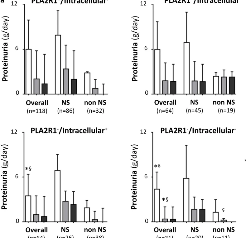
Figure 3. Clinical outcome of proteinuria and eGFR according to circulating autoantibodies defining four groups of patients. (a) Proteinuria and (b) eGFR values (data are presented as median with IQR) at diagnosis and after 12 months of follow-up for patients divided in four groups based on the concomitant positivity or not for anti-PLA2R1 (PLA2R1+ or PLA2R1-) and anti-intracellular antigen antibodies (Intracellular+ or Intracellular-). Proteinuria (a) and eGFR (b) values
are presented at diagnosis (open bars) and after 12 months (grey bars) for all patients (overall), for patients with nephrotic syndrome (NS) or not (non NS) at diagnosis and for patients who had been treated with an immunosuppressive treatment (IS-treated with cyclophosphamide, cyclosporine A or rituximab) after 12 months of follow-up (black bars).
Figure 4. Clinical outcome (complete remission and eGFR) according to circulating autoantibodies defining four groups of patients. (a) Kaplan-Meier curve showing the proportion of patients in the different groups (based on the concomitant positivity or not for anti-PLA2R1
23
(P+/P-) and anti-intracellular antibodies (IC+/IC-) who reached the endpoint of complete remission (proteinuria <0.3 g/day) after 12 months of follow-up. The Kaplan-Meier curve of the difference was calculated with the Log-rank (Mantel-Cox) test and the hazard ratios with confidence intervals for the different groups. (b) Kaplan-Meier curve showing the proportion of patients in the different groups (based on the concomitant negativity for anti-PLA2R1 (P-) and anti-intracellular antigen antibodies negativity or positivity (IC+/IC-) who reached the endpoint of reduction of kidney function (eGFR <60 mL/min/1.73 m2) after 12 months of follow-up. (c) Odds ratios and
confidence intervals were calculated for the association between the different groups and the risk of not reducing kidney function (eGFR <60 mL/min/1.73 m2) after 12 months of follow-up.
24
Table 1. Comparison of clinical characteristics at baseline (T0) and at clinical outcome (T12) of
MN patients stratified into four groups according to their antibody profile. Patients both PLA2R1 and intracellular antibodies positive; patients PLA2R1 positive and intracellular antibodies negative; patients PLA2R1 negative and intracellular antibodies positive; and patients both PLA2R1 negative and intracellular antibodies negative. Data are reported as number and percentage or median with interquartile range. Mann-Whitney or Kruskal–Wallis tests were used for continuous variables, respectively for two or more of two unpaired samples, and chi-squared or Fisher’s exact tests were used for categorical variables. Two-tailed p-values ≤0.05 were considered as significant. *p=0.001 versus A and B; ** p=0.001 versus A, B and C. ACEi: Angiotensin converting enzyme inhibitors.
Characteristics PLA2R1(n=118) +/Intracellular+ PLA2R1+(n=64) /Intracellular- PLA2R1-(n=64) /Intracellular+ PLA2R1-(n=31) /Intracellular-
Sex (M/F) 84/34 45/19 47/17 21/10 Age (years) 61 (50-72) 63 (46-70) 57 (42-67) 55 (43-71) MN histological stage (%) I 18.8 23.6 25.9 44.5 II 44.2 49.0 50.0 33.3 III 28.6 23.5 14.8 11.0 IV 8.4 3.9 9.3 11.1 Treatment (%) Steroids/ACEi§ 80 78 85 78 Cyclosporine A 18 30 14 16 Cytotoxic 43 44 33 32 Rituximab 7 22 16 19 Clinical characteristics Proteinuria (g/day) T0 5.6(3.3-9.7) 6.0 (3.3-9.8) 3.4 (1.8-6.4)* 4.4 (2.6-6.4)* T12 1.7 (0.4-4.4) 1.8(0.6-4.3) 1.3 (0.3-3.5) 0.5 (0.1-2.7)** CKD-EPI (mL/min/1.73m2) T0 69.4(46-99) 65.9(47-100) 72.4 (43-99) 89.2 (63-103)** T12 73.5(48-99) 77.9(59-99) 67.2 (44-101) 95.5 (72-103)** §Steroids and ACEi were given alone or in association with other drugs
25
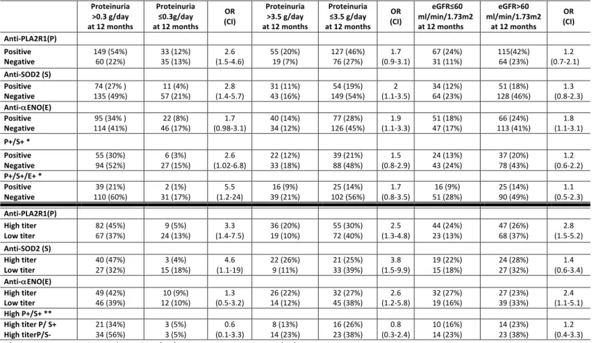
Table 2. Two-way contingency table showing the association of antibody levels with indexes of kidney outcome and the interaction between autoantibodies against PLA2R1 and intracellular antigens for the whole cohort. The 2x2 contingency table reports the association with odds ratios (OR) and confidence interval (CI) between antibody titer and clinical outcome. Data are calculated from all patients excluding the eight anti-THSD7A positive patients (n=277). The upper section shows the association between positivity versus negativity of each autoantibody at diagnosis with clinical outcome of proteinuria (complete (≤0.3 g/day) or partial (<3.5 g/day) remission) and eGFR after 12 months. The lower section shows the association of high versus low titers of each antibody with the same parameters of proteinuria and eGFR. The additive effect of positivity for more than one antibody is indicated as P+/S+ and P+/S+/E+ (anti-PLA2R1+/anti-SOD2+/anti-αENO+). Proteinuria >0.3 g/day at 12 months Proteinuria ≤0.3g/day at 12 months OR (CI) Proteinuria >3.5 g/day at 12 months Proteinuria ≤3.5 g/day at 12 months OR (CI) eGFR≤60 ml/min/1.73m2 at 12 months eGFR>60 ml/min/1.73m2 at 12 months OR (CI) Anti-PLA2R1(P) Positive Negative 149 (54%) 60 (22%) 33 (12%) 35 (13%) (1.5-4.6) 2.6 55 (20%) 19 (7%) 127 (46%) 76 (27%) (0.9-3.1) 1.7 67 (24%) 31 (11%) 115(42%) 64 (23%) (0.7-2.1) 1.2 Anti-SOD2 (S) Positive Negative 135 (49%) 74 (27% ) 57 (21%) 11 (4%) (1.4-5.7) 2.8 31 (11%) 43 (16%) 149 (54%) 54 (19%) (1.1-3.5) 2 34 (12%) 64 (23%) 128 (46%) 51 (18%) (0.8-2.3) 1.3 Anti-αENO(E) Positive Negative 114 (41%) 95 (34% ) 46 (17%) 22 (8%) (0.98-3.1) 1.7 40 (14%) 34 (12%) 126 (45%) 77 (28%) (1.1-3.3) 1.9 51 (18%) 47 (17%) 113 (41%) 66 (24%) (1.1-3.1) 1.8 P+/S+ * Positive Negative 55 (30%) 94 (52%) 27 (15%) 6 (3%) (1.02-6.8) 2.6 22 (12%) 33 (18%) 39 (21%) 88 (48%) (0.8-2.9) 1.5 24 (13%) 43 (24%) 37 (20%) 78 (43%) (0.6-2.2) 1.2 P+/S+/E+ * Positive Negative 110 (60%) 39 (21%) 31 (17%) 2 (1%) (1.2-24) 5.5 39 (21%) 16 (9%) 102 (56%) 25 (14%) (0.8-3.5) 1.7 51 (28%) 16 (9%) 25 (14%) 90 (49%) (0.5-2.3) 1.1 Anti-PLA2R1(P) High titer Low titer 82 (45%) 67 (37%) 24 (13%) 9 (5%) (1.4-7.5) 3.3 36 (20%) 19 (10%) 55 (30%) 72 (40%) (1.3-4.8) 2.5 44 (24%) 23 (13%) 47 (26%) 68 (37%) (1.5-5.2) 2.8 Anti-SOD2 (S) High titer Low titer 40 (47%) 27 (32%) 15 (18%) 3 (4%) (1.1-19) 4.6 22 (26%) 9 (11%) 21 (25%) 33 (39%) (1.5-9.9) 3.8 19 (22%) 15 (18%) 24 (28%) 27 (32%) (0.6-3.4) 1.4 Anti-αENO(E) High titer Low titer 49 (42%) 46 (39%) 12 (10%) 10 (9%) (0.5-3.2) 1.3 26 (22%) 14 (12%) 32 (27%) 45 (38%) (1.2-5.8) 2.6 32 (27%) 19 (16%) 27 (23%) 39 (33%) (1.1-5.1) 2.4 High P+/S+ ** High titer P/ S+ High titerP/S- 21 (34%) 34 (56%) 3 (5%) 3 (5%) (0.1-3.3) 0.6 14 (23%) 8 (13%) 16 (26%) 23 (38%) (0.3-2.4) 0.8 10 (16%) 14 (23%) 14 (23%) 23 (38%) (0.4-3.3) 1.2 *In these cases, P+/S+ were compared to P+/S- and P+/S+/E+ were compared to P+/S-/E-. ** In this case, high P/S (highP titer and S positivity) were compared to high P titer and S negativity.
26
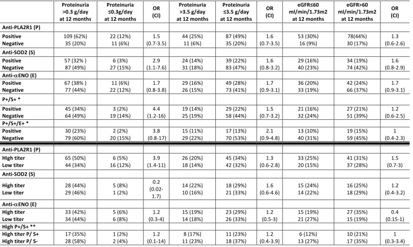
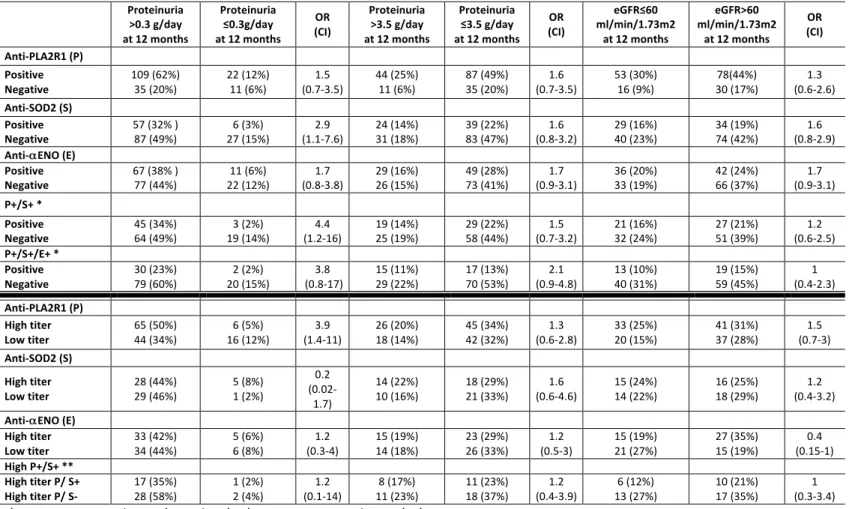
Table 3. Two-way contingency table showing the association of antibody levels with indexes of kidney outcome and the interaction between autoantibodies against PLA2R1 and intracellular antigens for nephrotic patients at diagnosis. The 2x2 contingency table reports the association with odds ratios (OR) and confidence interval (CI) between antibody titer and clinical outcome. Data are calculated from the subset of patients with nephrotic syndrome excluding the eight anti-THSD7A positive patients (n=177). The upper section shows the association between positivity versus negativity of each autoantibody at diagnosis with clinical outcome of proteinuria (complete (≤0.3 g/day) or partial (<3.5 g/day) remission) and eGFR after 12 months. The lower section shows the association of high versus low levels of each antibody with the same parameters of proteinuria and eGFR. The additive effect of positivity for more than one antibody is indicated as P+/S+ and P+/S+/E+. Proteinuria >0.3 g/day at 12 months Proteinuria ≤0.3g/day at 12 months OR (CI) Proteinuria >3.5 g/day at 12 months Proteinuria ≤3.5 g/day at 12 months OR (CI) eGFR≤60 ml/min/1.73m2 at 12 months eGFR>60 ml/min/1.73m2 at 12 months OR (CI) Anti-PLA2R1 (P) Positive Negative 109 (62%) 35 (20%) 22 (12%) 11 (6%) (0.7-3.5) 1.5 44 (25%) 11 (6%) 87 (49%) 35 (20%) (0.7-3.5) 1.6 53 (30%) 16 (9%) 30 (17%) 78(44%) (0.6-2.6) 1.3 Anti-SOD2 (S) Positive Negative 57 (32% ) 87 (49%) 27 (15%) 6 (3%) (1.1-7.6) 2.9 24 (14%) 31 (18%) 39 (22%) 83 (47%) (0.8-3.2) 1.6 29 (16%) 40 (23%) 34 (19%) 74 (42%) (0.8-2.9) 1.6 Anti-αENO (E) Positive Negative 67 (38% ) 77 (44%) 22 (12%) 11 (6%) (0.8-3.8) 1.7 29 (16%) 26 (15%) 49 (28%) 73 (41%) (0.9-3.1) 1.7 36 (20%) 33 (19%) 42 (24%) 66 (37%) (0.9-3.1) 1.7 P+/S+ * Positive Negative 45 (34%) 64 (49%) 19 (14%) 3 (2%) (1.2-16) 4.4 19 (14%) 25 (19%) 29 (22%) 58 (44%) (0.7-3.2) 1.5 21 (16%) 32 (24%) 27 (21%) 51 (39%) (0.6-2.5) 1.2 P+/S+/E+ * Positive Negative 30 (23%) 79 (60%) 20 (15%) 2 (2%) (0.8-17) 3.8 15 (11%) 29 (22%) 17 (13%) 70 (53%) (0.9-4.8) 2.1 13 (10%) 40 (31%) 19 (15%) 59 (45%) (0.4-2.3) 1 Anti-PLA2R1 (P) High titer Low titer 65 (50%) 44 (34%) 16 (12%) 6 (5%) (1.4-11) 3.9 26 (20%) 18 (14%) 42 (32%) 45 (34%) (0.6-2.8) 1.3 33 (25%) 20 (15%) 41 (31%) 37 (28%) (0.7-3) 1.5 Anti-SOD2 (S) High titer Low titer 28 (44%) 29 (46%) 5 (8%) 1 (2%) 0.2 (0.02-1.7) 14 (22%) 10 (16%) 18 (29%) 21 (33%) (0.6-4.6) 1.6 15 (24%) 14 (22%) 16 (25%) 18 (29%) (0.4-3.2) 1.2 Anti-αENO (E) High titer Low titer 33 (42%) 34 (44%) 5 (6%) 6 (8%) (0.3-4) 1.2 15 (19%) 14 (18%) 23 (29%) 26 (33%) (0.5-3) 1.2 15 (19%) 21 (27%) 27 (35%) 15 (19%) (0.15-1) 0.4 High P+/S+ ** High titer P/ S+ High titer P/ S- 17 (35%) 28 (58%) 1 (2%) 2 (4%) (0.1-14) 1.2 11 (23%) 8 (17%) 11 (23%) 18 (37%) (0.4-3.9) 1.2 13 (27%) 6 (12%) 10 (21%) 17 (35%) (0.3-3.4) 1 *In these cases, P+/S+ were compared to P+/S- and P+/S+/E+ were compared to P+/S-/E-. ** In this case, high P/S (high P titer and S positivity) were compared to high P titer and S negativity.
27
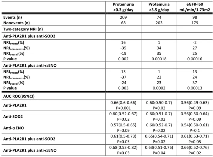
Table 4. Two-category net reclassification improvement (NRI) and area under the receiver-operating-characteristic curve (AUC-ROC) analysis for predictors of clinical outcome after 12 months in MN patients by adding intracellular autoantibodies to anti-PLA2R1 positivity. Data are calculated from all patients excluding the eight anti-THSD7A positive patients (n=277). For NRI, combined negativity of anti-PLA2R1 and anti-SOD2 antibodies allowed the reclassification of 34% of patients with proteinuria <3.5g/day (non-event) and 27% of cases with eGFR>60 mL/min/1.73 m2(non-event) after 12 months. Similar results were found for combined negativity of anti-PLA2R1 and anti-αENO. In both cases, the reclassification improvement was in comparison with anti-PLA2R1 positivity alone.
Proteinuria >0.3 g/day Proteinuria >3.5 g/day mL/min/1.73 m2 eGFR<60 Events (n) Nonevents (n) 209 68 203 74 179 98 Two-category NRI (n) Anti-PLA2R1 plus anti-SOD2 NRIevents(%) NRInon-events(%) NRIoverall(%) P value 16 -35 -19 0.002 1 34 35 0.00018 -2 27 25 0.00016 Anti-PLA2R1 plus anti-αENO NRIevents(%) NRInon-events(%) NRIoverall(%) P value 13 -37 -24 0.003 1 22 23 0.0002 13 24 37 0.00013 AUC ROC(95%CI) Anti-PLA2R1 0.66(0.6-0.66) P=0.001 0.60(0.50-0.7) P=0.02 0.56(0.49-0.63) P=0.09 Anti-SOD2 0.60(0.52-0.67) P=0.02 0.60(0.51-0.7) P=0.02 0.56(0.50-0.64) P=0.09 Anti-αENO 0.57(0.5-0.65) P=0.09 0.60(0.52-0.7) P=0.02 0.54(0.50-0.61) P=0.1 Anti-PLA2R1 plus anti-SOD2 0.61(0.5-0.73) P=0.03 0.65(0.54-0.71) P=0.02 0.61(0.53-0.71) P=0.05 Anti-PLA2R1 plus anti-αENO 0.68(0.53-0.82) P=0.03 0.63(0.51-0.76) P=0.04 0.66(0.52-0.76) P=0.02
Table 1. Comparison of clinical characteristics at baseline (T0) and at clinical outcome (T12) in
patients with iMN stratified into four subgroups according to their antibody profile: patients both PLA2R1 and intracellular antibodies positive; patients PLA2R1 positive and intracellular antibodies negative; patients PLA2R1 negative and intracellular antibodies positive; and patients both PLA2R1 negative and intracellular antibodies negative. Data are reported as number and percentage or median with interquartile range. Mann-Whitney or Kruskal–Wallis tests were used for continuous variables, respectively for two or more of two unpaired samples, and chi-squared or Fisher’s exact tests were used for categorical variables. Two-tailed p-values ≤0.05 were considered as significant. *p=0.001 versus A and B; ** p=0.001 versus A, B and C. ACEi: Angiotensin converting enzyme inhibitors.
Characteristics PLA2R1(n=118) +/Intracellular+ PLA2R1+(n=64) /Intracellular- PLA2R1-(n=64) /Intracellular+ PLA2R1-(n=31) /Intracellular-
Sex (M/F) 84/34 45/19 47/17 21/10 Age (years) 61 (50-72) 63 (46-70) 57 (42-67) 55 (43-71) MN histological stage (%) I 18.8 23.6 25.9 44.5 II 44.2 49.0 50.0 33.3 III 28.6 23.5 14.8 11.0 IV 8.4 3.9 9.3 11.1 Treatment (%) Steroids/ACEi§ 80 78 85 78 Cyclosporine A 18 30 14 16 Cytotoxic 43 44 33 32 Rituximab 7 22 16 19 Clinical characteristics Proteinuria (g/day) T0 5.6(3.3-9.7) 6.0 (3.3-9.8) 3.4 (1.8-6.4)* 4.4 (2.6-6.4)* T12 1.7 (0.4-4.4) 1.8(0.6-4.3) 1.3 (0.3-3.5) 0.5 (0.1-2.7)** CKD-EPI (mL/min/1.73m2) T0 69.4(46-99) 65.9(47-100) 72.4 (43-99) 89.2 (63-103)** T12 73.5(48-99) 77.9(59-99) 67.2 (44-101) 95.5 (72-103)** §Steroids and ACEi were given alone or in association with other drugs