HAL Id: tel-01552176
https://tel.archives-ouvertes.fr/tel-01552176
Submitted on 1 Jul 2017
HAL is a multi-disciplinary open access archive for the deposit and dissemination of sci-entific research documents, whether they are pub-lished or not. The documents may come from teaching and research institutions in France or abroad, or from public or private research centers.
L’archive ouverte pluridisciplinaire HAL, est destinée au dépôt et à la diffusion de documents scientifiques de niveau recherche, publiés ou non, émanant des établissements d’enseignement et de recherche français ou étrangers, des laboratoires publics ou privés.
remodeling functions in embryonic stem cells
Daria Bou Dargham
To cite this version:
Daria Bou Dargham. Genome-wide analysis of ATP-dependent chromatin remodeling functions in embryonic stem cells. Biomolecules [q-bio.BM]. Université Paris-Saclay, 2015. English. �NNT : 2015SACLS033�. �tel-01552176�
THESE DE DOCTORAT
DE L’UNIVERSITE PARIS-SACLAY,
préparée à L’Université Paris Sud
ÉCOLE DOCTORALE 577
Structure et Dynamique des Systèmes Vivants
Spécialité de doctorat : Sciences de la Vie et de la Santé
Par
Mme Daria Bou Dargham
Analyse de la fonction des facteurs de remodelage de chromatine ATP-dépendants
dans le contrôle de l’expression du génome des cellules souches embryonnaires
Thèse présentée et soutenue à Saclay, le 13/10/2015 : Composition du Jury :
Mme Fabienne Malagnac Professeur, Université Paris Sud Présidente M Saadi Khochbin Directeur de Recherche, CNRS Rapporteur M Slimane Ait-Si-Ali Directeur de Recherche, CNRS Rapporteur M Daan Noordermeer Chargé de Recherche, CNRS Examinateur M Eric Soler Chargé de Recherche, INSERM Examinateur M Matthieu Gérard Chercheur CEA, CEA Saclay Directeur de thèse
Titre : Analyse de la fonction des facteurs de remodelage de chromatine ATP-dépendants dans le contrôle de l’expression du génome des cellules souches embryonnaires
Mots clés : Cellules Souches Embryonnaires, remodeleurs de la chromatine, ChIP-seq Résumé:
Les cellules souches embryonnaires (cellules ES) constituent un excellent système modèle pour étudier les mécanismes épigénétiques contrôlant la transcription du génome mammifère. Un nombre important de membres de la famille des facteurs de remodelage de chromatine ATP-dépendants ont une fonction essentielle pour l’auto-renouvellement des cellules ES, ou au cours de la différentiation. On pense que ces facteurs exercent ces rôles essentiels en régulant l’accessibilité de la chromatine au niveau des éléments régulateurs de la transcription, en modulant la stabilité et le positionnement des nucléosomes. Dans ce projet, nous avons conduit une étude génomique à grande échelle du rôle d’une dizaine des remodeleurs (Chd1, Chd2, Chd4, Chd6, Chd8, Chd9, Ep400, Brg1, Smarca3, Smarcad1, Smarca5, ATRX et Chd1l) dans les cellules ES. Une double stratégie expérimentale a été utilisée.
Des expériences d’immunoprécipitation de la chromatine suivi par un séquençage à haute-débit (ChIP-seq) sur des cellules ES étiquetées pour les différents remodeleurs pour étudier leur distribution sur le génome, et un approche transcriptomique sur des cellules déplétées de chaque remodeleur par traitement avec des vecteurs shRNA (knockdown). Nous avons établi les profils de liaison des remodeleurs sur des éléments régulateurs (promoteurs, enhancers et sites CTCF) sur le génome, et montré que ces facteurs occupent toutes les catégories d’éléments régulateurs du génome. La corrélation entre les données ChIP-seq et les données transcriptomiques nous a permis d’analyser le rôle des remodeleurs dans les réseaux de transcription essentiels des cellules ES. Nous avons notamment démontré l’importance particulière de certains remodeleurs comme Brg1, Chd4, Ep400 et Smarcad1 dans la régulation de la transcription chez les cellules ES.
Title : Genome-wide analysis of ATP-dependent chromatin remodeling factors functions in embryonic stem cells
Keywords: Embryonic Stem Cells, chromatin remodeling factors, ChIP-seq Abstract:
The characteristics of embryonic stem cells (ES cells) make them one of the best models to study the epigenetic regulation exerted by different actors in order to control the transcription of the mammalian genome. Members of the Snf2 family of ATP-dependent chromatin remodeling factors were shown to be of specific importance for ES cell self-renewal and during differentiation. These factors are believed to play essential roles in modifying the chromatin landscape through their capacity to position nucleosomes and determine their occupancy throughout the genome, making the chromatin more or less accessible to DNA binding factors.
In this project, a genome-wide analysis of the function of a number of ATP-dependent chromatin remodelers (Chd1, Chd2, Chd4, Chd6, Chd8, Chd9, Brg1, Ep400, ATRX, Smarca3, Smarca5, Smarcad1 and Alc1) in mouse embryonic stem (ES) cells was conducted.
This was done using a double experimental strategy. First, a ChIP-seq (Chromatin Immunoprecipitation followed by deep sequencing) strategy was done on ES cells tagged for each factor in the goal of revealing the genomic binding profiles of the remodeling factors. Second, loss-of-function studies followed by transcriptome analysis in ES cells were performed in order to understand the functional role of remodelers. Data from both studies were correlated to acquire a better understanding of the role of remodelers in the transcriptional network of ES cells.
Specific binding profiles of remodelers on promoters, enhancers and CTCF binding sites were revealed by our study. Transcriptomic data analysis of the deregulated genes upon remodeler factor knockdown, revealed the essential role of Chd4, Ep400, Smarcad1 and Brg1 in the control of transcription of ES cell genes. Altogether, our data highlight how the distinct chromatin remodeling factors cooperate to control the ES cell state.
Acknowledgements
Initially I would like to thank my jury members for accepting to examine my thesis work. Thanks to Dr. Saadi Khochbin and Dr. Slimane Ait-Si-Ali for accepting to be my thesis reporters and carefully reading and commenting my thesis manuscript. Equally, I thank the examiners Dr. Eric Soler, Dr. Daan Noordermeer and Dr. Fabienne Malagnac for accepting to be a part of my thesis defense jury.
I would like to deeply thank my thesis supervisor Dr. Matthieu Gerard for giving me the opportunity to conduct this interesting project in his lab. Thank you for your guidance, advice and great trust during these three years.
I would like to equally thank Dr. Jean-Christophe Andrau for being my thesis tutor and participating in my thesis committees. In addition, I thank Dr. Ute Rogner for being a member of my thesis committees. Thank you for your advices and comments.
I give special thanks to my collegues in our team. I thank Dr. Michel De Chaldée for his numerous advices and help especially at the beginning of my project, thank you as well for all the interesting discussions we had. I would like to thank Hélène Picaud for her everlasting humor and eventual technical help, keep on laughing! I equally thank Sylvie Jounier for her initial guidance and help, thank you for your patience and professionalism.
I give my thanks as well to my collegues in the animal house for their company and help. Thank you Anne-Sophie Chaplault, Sylvain Thessier, Jean-Charles Robillard, Patrick Héry. Thank you for picking me up at occasional work weekends…
initial help and implication in my project.
I would like to thank all my collegues in the SBiGeM and the CEA IRTELIS program for giving me the opportunity to conduct this project. Moreover, I thank my doctoral school especially Dr. Pierre Capy for his guidance.
I thank my friends, in particular Dr. Elma El Khouri for her support and huge implication in reading and commenting my thesis manuscript.
Finally, I would like to send my sincere and deep thanks to each of my parents, my two brothers and my husband Ghazi for their continuous encouragement, support and love. Without you I would have never been where I am now.
1
Table of Contents
Introduction
Chapter I. Embryonic Stem Cells: Definition and Regulatory Pathways
... 9
A. What are Embryonic Stem Cells? ... 9
1. The Major Characteristics of Embryonic Stem Cells ... 9
2. Important signaling pathways that control the mouse ES cells state ... 11
B. Naïve and primed pluripotency ... 17
1. Mouse ES cell in serum versus 2i medium ... 17
2. EpiS cells versus ES cells ... 18
C. Human ES cells ... 20
Chapter II. The Transcriptional and Epigenetic Control of the Embryonic Stem Cell State ... 22
A. The Transcription Factor Network for ES state maintenance ... 22
1. The Core Transcriptional Regulatory Network ... 22
2. The Expanded Transcriptional Regulatory Network ... 28
3. Pluripotency transcription factors and iPS cell generation ... 37
B. Chromatin Modifying Enzymes and the Control of the ES Cell State ... 40
1. Proteomic interactome studies reveal numerous chromatin modifying enzymes as a part of the transcriptional regulatory network ... 40
2 2. RNA interference-based screens for pluripotency controlling factors reveal a series
of important chromatin remodeling enzymes ... 43
C. The Epigenetic Regulatory Landscape of the ES Genome ... 45
1. Promoters in ES cells ... 46
2. Distal regulatory sequences ... 53
Chapter III. ATP-dependent Chromatin Remodeling Factors and Pluripotence ... 59
A. The Snf2 family of ATP-dependent chromatin remodeling factors: Classification .. 59
B. The Snf2 family of ATP-dependent chromatin remodeling factors: Mode of action 61 1. The chromatin state ... 61
2. Mode of action of ATP-dependent chromatin remodeling enzymes ... 63
C. The functional output of ATP-dependent chromatin remodeling factors on nucleosome positioning ... 65
1. Nucleosome positioning in the genome ... 65
2. ATP-dependent nucleosome remodeling enzymes and nucleosome positioning ... 67
D. Chromatin Remodeling Complexes Function in ES cells ... 69
1. The NuRD complex (encompassing Chd4) ... 70
2. INO80 complex ... 71
3. esBAF complex (encompassing Brg1) ... 72
4. Chd1 ... 74
5. Ep400 ... 74
3
7. Other remodelers (Smarca5, Smarcad1, Chd1l) ... 76
Background and Objectives ... 77
Results Part I ... 79
Article 1: Genome-wide nucleosome specificity and function of chromatin remodellers in embryonic stem cells ... 79
Part II: ... 137
Article 2: ATP-dependent Chromatin Remodeling Factors Target Regulatory Regions and Contribute to the Transcriptional Network in ES Cells ... 137
Discussion and Conclusion ... 171
References ... 179
4
Figures and Tables
Figure 1 Embryonic Stem Cells and Pluripotency ... 10
Figure 2 The various aspects of ES cells and Embryoid Bodies. ... 10
Figure 3 Pathways known to contribute to the maintenance of embryonic stem cell pluripotency ... 11
Figure 4 A schematic representation of LIF-dependent pathways ... 12
Figure 5 Schematic of the canonical vertebrate Wnt signaling pathway ... 14
Figure 6 Model depicting the influence of Wnt pathway components on pluripotency and differentiation in ES cells ... 15
Figure 7 Cooperative lineage restriction by LIF/STAT3 and BMP/Smad ... 16
Figure 8 The two phases of pluripotency ... 19
Figure 9 Model explaining the regulation of Nanog in hESCs/mEpiSCs and its function in both cell types ... 20
Figure 10 Comparison of pluripotent cell line derivation protocols for (A) mouse and (B) human embryos. ... 21
Figure 11 The proposed function of Oct4 and Nanog in preimplantation embryos and in ES cells. ... 26
Figure 12 Oct4, Sox2 and Nanog collaborate to control their own promoters forming an autoregulatory loop ... 28
Figure 13 Transcription control by Klf4 and Tbx3 ... 32
Figure 14 Model for DNA looping by mediator and cohesion ... 34
Figure 15 Steps involved in direct reprogramming to pluripotency. ... 39
Figure 16 Protein Interaction Network of Oct4 and Its Associated Proteins Sall4, Dax1, Tcfcp2l1, and Esrrb ... 42
5
Figure 18 Model for the relationships between Mll and Set1 complexes on bivalent and active
promoters ... 49
Figure 19 Bivalent chromatin domains mark the promoters of developmentally important genes in pluripotent ES cells ... 52
Figure 20 Characteristics of typical enhancers and super enhancers in ES cells. ... 56
Figure 21 The role of CTCF as an enhancer blocker or facilitator ... 58
Figure 22 The role of CTCF in the creation of both super enhancer domains (SD) and polycomb insulated domains (PD) in ES cells ... 58
Figure 23 Schematic diagram of the hierarchical classification of ATP-dependent helicase-like proteins ... 60
Figure 24 The different compaction levels of the chromatin ... 62
Figure 25 The outcomes of nucleosome sliding on nucleosome organization ... 63
Figure 26 Mode of action of ATP-dependent chromatin remodeling factors ... 64
Figure 27 Nucleosome organization on yeast genes ... 66
Figure 28 The role of NuRD in ES cells ... 71
Figure 29 The different control mechanisms exerted by the esBAF complex in order to maintain ES pluripotency and self-renewal ... 73
Table 1 Differences between the ground and primed cell states………..19
Table 2 The different Snf2 subfamilies, the archetype organism and the different constituting members………61
6
Abbreviations
A
AEBP: Adipocyte Enhancer Binding Protein B
BMP: Bone Morphogenetic Protein BAF: Brg1-Associated Factor
C
ChIP-seq: Chromatin Immuno-Precipitation followed by deep sequencing
CNOT3: CCR4-NOT transcription complex subunit 3
CHD: Chromo Domain CFP1: CxxC Finger Protein 1 CBX: Chromo BOX
CTCF: CCCTC binding factor
ChIA-PET: Chromatin Interaction Analysis by Paired-End Tag Sequencing
CpGI: CpG Islands
D
DAX1: Dosage-sensitive sex reversal, adrenal hypoplasia critical region, on chromosome X DNMTs: DNA methyltransferases
DHS: DNase I Hypersensitive Site DBP: DNA binding Protein
E
ES: Embryonic Stem
ESSRB: Estrogen Related Receptor EP400: E1A-binding Protein p400 EZH2: Enhancer of Zest Homologue 2 EED: Embryonic Ectoderm Development ERK: Extracelluler Regulated Kinases EpiS: Epiblast Stem
ENCODE: The Encyclopedia of DNA Elements
F
FOXd2: Forkhead Box d2 FGF2: Fibroblast Growth Factor 2
FAIRE-seq: Formaldehyde-Assisted Isolation of Regulatory Elements followed by deep sequencing
G
GSK3: Glycogen Synthase Kinase 3 GRO-seq: Global-Run-On Sequencing GAPDH: Glyceraldehyde 3-phosphate dehydrogenase
H
HMG: High Mobility Group HDAC: Histone DeAcetylase HCP: High CpG Promoters bHLH: basic Helix Loop Helix
I
iPS: induced Pluripotent Stem ICM: Inner Cell Mass Id: Inhibitor of Differentiation
J
JARID2: Jumonji AT Rich Interactive Domain 2
K
KLF: Kruppel-Like Factors KD: Knock Down
L
LCD1: Lethal Checkpoint-defective DNA damage-sensitive protein 1
LCP: Low CpG Promoters LIF: Leukemia Inhibitory Factor
LRP: Low density lipoprotein receptor-related protein
7
Myc: Myelocytomatosis
MEK: Mitogen Activated Protein MBD3: Methyl CpG Binding protein 3 MACS: Model-based Analysis for ChIP-seq
N
NuRD: Nucleosome Remodeling Deacetylase
NFR: Nucleosome Free Region
O
Oct4: Octamer-binding transcription factor 4
P
PRC: Polycomb repressive Complex PcG: Polycomb Group
PI3K: Phosphatidylinositol-4,5-bisphosphate 3-kinase
R
RT-qPCR: Reverse Transcription-quantitative Polymerase Chain Reaction
RPKM: Reads Per Kilobase per Million mapped reads
S
SetDB1: Set Domain Bifurcated 1
SALL4: Spalt-Like Transcription Factor 4 Sox2: (Sex determining region Y)-box 2 SE: Super Enhancer
STAT3: Signal Transducer and Activator of Transcription 3
SWI/SNF: Switch/sucrose non-fermenting shRNA: small hairpin RNA
SICER: Spatial Clustering for Identification of ChIP-seq Enriched Regions
T
TBX3: T-box Transcription Factor 3
Tip60: TAT-interactive protein 60 TrxG: Trithorax Group
TE: Typical Enahcer
TAD: Topologically Associated Domain TGFβ: Transformation Growth Factor beta TAP: Tandem Affinity Purification TEV: Tabacco Etch Virus
TF: Transcription Factor TBP: TATA Binding Protein TSS: Transcription Start Site
Z
8
INTRODUCTION
9
Chapter I.
Embryonic Stem Cells: Definition and Regulatory
Pathways
A. What are Embryonic Stem Cells?
Embryonic stem cells or ES cells are derived from the blastocyst which is formed during
embryogenesis and is composed of two parts: an outer layer of cells, the trophectoderm, that
will form the placenta and an inner clump of cells, called the inner cell mass (ICM), that is
responsible for the formation of the entire body. The isolation and culture of cells derived
from the ICM under appropriate conditions, gives rise to Embryonic Stem cells.
1. The Major Characteristics of Embryonic Stem Cells
Embryonic stem cells possess several distinct features that set them apart from other cell
types. The two major characteristics that define ES cells are self-renewal and pluripotency or
the ability to give rise to all the different types of cells of the body.
a. Self-renewal
ES cells can divide symmetrically and for a long period of time, where mother cells give rise
to identical daughter cells in a continuous fashion. This property is defined as self-renewal.
ES cells were initially established and maintained by Evans an Kaufman in 1981 (Evans and
Kaufman, 1981). The proliferation of ES cells is assured and maintained by the presence of
very specific extrinsic factors (like LIF or the Leukemia inhibitory Factor (Smith et al., 1988))
10
b. Pluripotency
The first attempt to produce different cell lineages from embryonic stem cells goes back to the
experiments done by Evans and Kaufman using mouse embryonic carcinoma cells by forming
embryoid bodies (Evans and Kaufman, 1981). Embryoid bodies were given this name due to
their marvelously similar composition to actual embryos (Figure 2). The pluripotent nature of mouse ES cells was demonstrated by their ability to contribute to all tissues of adult mice
following their injection into host blastocysts (Bradley et al., 1984). In addition to their
developmental potential in vivo, ES cells display a remarkable capacity to form differentiated
cell types in culture (Keller, 1995). This capacity to give rise to all types of body cells is
called pluripotency (Figure 1).
Figure 1 Embryonic Stem Cells and Pluripotency
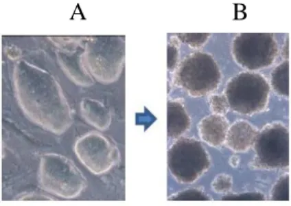
Figure 2 The various aspects of ES cells and Embryoid Bodies. A) Mouse ES cells colonies cultured on feeder cells. B) The differentiation of mouse ES cells. Embryoid body formation (Differentiation day 7)
11
2. Important signaling pathways that control the mouse ES cells state
The three major signaling pathways that control the ES cell state comprise: The
LIF-dependent (Leukemia Inhibiting Factor LIF-dependent) signaling pathways, the Wnt pathway and
BMP (Bone Morphogenetic Protein) signaling pathway that act with different mechanisms to
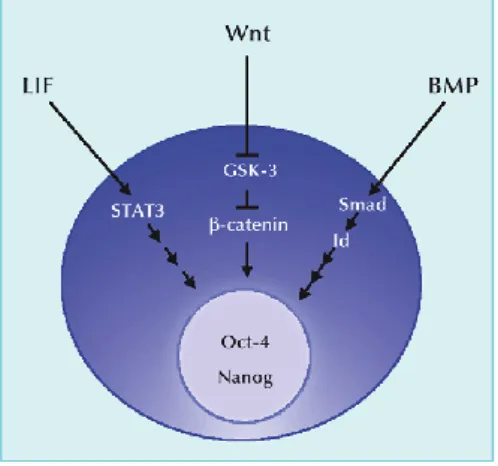
assure the correct transcriptional profile for ES cell maintenance (Figure 3).
Figure 3 Pathways known to contribute to the maintenance of embryonic stem cell pluripotency (Nakashima et al., 2004)
a. The LIF-dependent signaling pathways
LIF belongs to the interleukin-6 cytokine family. It binds a heterodimeric receptor consisting
of the low-affinity LIF receptor and gp130, with downstream signals being transmitted
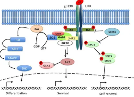
through gp130. The gp130 downstream signaling pathways include: the STAT3,
phosphatidylinositol 3-kinase (PI3K) and Ras/Erk pathways (Figure 4). Maintaining the balance among the three pathways allows fine-tuning of the LIF-dependent maintenance of
12 Figure 4 A schematic representation of LIF-dependent pathways (Graf et al., 2011)
The LIF/STAT3 pathway
Signaling through gp130 leads to activation of Janus-associated tyrosine kinases (JAKs),
which in turn phosphorylate STAT3. Phosphorylated STAT3 homodimerizes and moves to
the nucleus, where it functions as a transcription factor. In mouse ES cells, the STAT3
pathway plays a critical role in the maintenance of self-renewal. When STAT3 is
down-regulated, ES cells undergo differentiation (Boeuf et al., 1997; Niwa et al., 1998; Ying et al.,
2008). Artificial activation of the STAT3 pathway can maintain ES cell self-renewal even in
the absence of LIF (Matsuda et al., 1999).
STAT3 was shown to bind to the regulatory regions of several self-renewal genes in ES cells
(Chen et al., 2008a; Kidder et al., 2008). One major role of the LIF/STAT3 pathway is to
form transcriptional networks with other key TFs such as Oct3/4, Sox2, Nanog, c-Myc, Klf4
and Esrrb (Transcriptional regulatory networks will be developed in Chapter II). These
13 their expression. In this way, the LIF/STAT3 pathway participates in the formation of
self-renewal transcriptional networks.
The LIF/PI3K pathway
PI3K phosphorylates phosphoinositides leading to activation of the serine/threonine protein
kinase Akt, a serine/threonine kinase implicated in the regulation of cell cycle progression,
cell death, adhesion, migration, metabolism and tumorigenesis (Brazil et al., 2004). Activated
Akt then phosphorylates its target molecules, such as glycogen synthase kinase (GSK)-3 and
pro-apoptotic BCL2-antagonist of death (BAD) protein, blocking their activity. Similarly to
STAT3, the PI3K pathway positively regulates self-renewal as inhibition of PI3K and Akt
induces differentiation of mouse and human ES cells, suggesting that PI3K/Akt signaling is
necessary for the maintenance of ES cell pluripotency (Paling et al., 2004; Watanabe et al.,
2006).
The Ras/Erk (MAPK) pathway
Ras is capable of the sequential activation of the Raf/MEK/Erk kinase cascade, leading to
phosphorylation of Erk target molecules, including the transcription factor Elk-1, proapoptotic
protein caspase-9, and p90 ribosomal S6 protein kinase (RSK). The activation of the Ras/Erk
pathway leads to ES cell differentiation into the endoderm lineage (Yoshida-Koide et al.,
2004) while suppressing the Ras/Erk signaling promotes self-renewal (Burdon et al., 1999).
b. Other signaling pathways The Wnt pathway
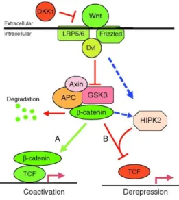
Wnt signaling pathways play an important role in the lineage specification of the vertebrate
embryo (Amerongen and Nusse, 2009; Clevers, 2006) and regulate pluripotency in ES cells
14 Frizzled receptors and LRP5/6 co-receptors to trigger downstream events that eventually cause the inactivation of the β-catenin-degradation complex. By this manner, ES cell
pluripotency is facilitated by the inhibition of GSK3 (Aubert et al., 2002; Doble et al., 2007),
a protein kinase that phosphorylates β-catenin, marking it for ubiquitin-dependent degradation
(Clevers, 2006; MacDonald et al., 2009). Further on, β-catenin interacts with TcF (T-cell
Factor) proteins, the terminal nuclear effectors of the Wnt pathway, to regulate the
transcription of specific target genes (Figure 5). In the absence of Wnt signaling, TcF proteins act as transcriptional repressors (Behrens et al., 1996; Cadigan, 2002).
Figure 5 Schematic of the canonical vertebrate Wnt signaling pathway (Sokol, 2011)
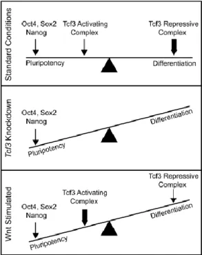
TcF3, a member of the TcF family is highly expressed in mouse ES cells, and is critical in
early mouse development (Korinek et al., 1998; Merrill et al., 2001; Pereira et al., 2006). It
was shown that TcF3 acts to repress Nanog gene in ES cells (Pereira et al., 2006).
Interestingly, a study highlights the importance of TcF3 in pluripotency, as it was
demonstrated that TcF3 co-occupies the ES cell genome with the pluripotency factors Oct4
and Nanog (Cole et al., 2008). This study suggests that TcF3 affects the balance between
pluripotency and differentiation in ES cells (Figure 6). At standard conditions there is a balance between the core TFs, the TcF3 activating complex and the TcF3 repressing complex.
15 TFs increases, pluripotency is favored and differentiation is inhibited. Upon Wnt stimulation,
TcF3 becomes more of an activating complex of pluripotency, assuring the maintenance of
the ES cell state. Upon loss of pluripotency gene expression, the Tcf3 repressive complex
takes hold of the expression of pluripotency genes and differentiation is favored.
Figure 6 Model depicting the influence of Wnt pathway components on pluripotency and differentiation in ES cells (Cole et al., 2008)
The BMP pathway
BMPs or Bone Morphogenetic Proteins belong to the transformation growth factor beta (TGFβ) superfamily. They were shown to play important roles cell proliferation,
differentiation, and apoptosis making them essential actors during embryonic development
and pattern formation (Massagué, 1998). It was long known that BMPs protect the
pluripotency state of ES cells, inhibit the differentiation into neural lineages (Di-Gregorio et
al., 2007; Ying et al., 2003a) and prime ES cells for mesoderm formation (Davis et al., 2004;
Lawson et al., 1999; Mishina et al., 1995). BMP activates Smad proteins that in turn act
16 al., 2003a; Zhang et al., 2010), where these factors inhibit bHLH neurogenesis TFs, thus
inhibiting neurogenesis (Norton, 2000). Furthermore, the BMP/Id complex was shown to
protect pluripotent stem cells from differentiating especially into neural lineages through the
maintenance of the expression of E-cadherin (Malaguti et al., 2013). Moreover, the
maintenance of the high expression of E-cadherin by BMP/Id was shown to impose a
proximal posterior identity on epiblast cells priming them for mesodermal lineages (Malaguti
et al., 2013).
The capacity of the BMP signaling pathway to maintain pluripotency is assured by the
coordination with LIF signaling pathways. On one hand, BMP/Smad signaling inhibits
neural lineages (Tropepe et al., 2001; Ying et al., 2003b) and induces other lineages, on the
other hand; LIF/STAT3 signaling inhibits non-neural lineages and possibly regulates Smad
function (Ying et al., 2003a) . The balance between LIF and BMP signaling pathways is
essential to assure pluripotency of ES cells, as it was shown that the constitutive expression of
Smad over rides the effect of LIF/STAT3 and differentiates ES cells into non-neural lineages
(Figure 7).
17
B. Naïve and primed pluripotency
1. Mouse ES cell in serum versus 2i medium
For a long time, the derivation and culture of mouse ES cells was poorly understood. Most of
the derived mouse ES cells were from a specific mice strain which is the 129 strain. This
strain was thought to have genetic and/or epigenetic profiles permissive for ES cell
establishment (Brook and Gardner, 1997). Back then, the mouse ES cell culture conditions
used were on feeder cells in serum-supplemented medium, these conditions supplemented the
cells with the necessary LIF and BMP required for self-renewal through the activation of
STAT pathway and the inhibition of differentiation pathways. However, such culture
conditions restrained the derivation of mouse ES cells from most of the other mouse strains.
This marked variability between the strains was later shown to be due to the level of ERK
signaling (Batlle-Morera et al., 2008), a pathway important to promote differentiation and
inhibit self-renewal. For the inhibition of ERK pathway was shown to improve the derivation
of mouse ES cells from mouse strains other the 129 strain (Batlle-Morera et al., 2008; Buehr
and Smith, 2003).
Later on, this strain specificity was eliminated by the discovery of a new approach where
certain kinase inhibitors were used. This novel approach, called the 2i culture approach,
makes it possible to culture mouse ES cells without serum by using two small molecules that
inhibit kinases in combination with LIF (Ying et al., 2008). The two inhibitors are
PD0325901 and CHIR99021 that respectively target the mitogen-activated protein kinase
(MEK) and the glycogen synthase kinase-3 (GSK3).
The 2i medium provides a more-tuned environment for mouse ES cells, as a mosaic
expression of some of the pluripotency factors (Chambers et al., 2007; Niwa et al., 2009;
18 et al., 2010), establishing a more naïve ground state. However, several studies show that this
heterogeneous expression of some of the core TFs is not an only characteristic of ES cells
cultured in serum, rather it is also observed in 2i-grown ES cells (Abranches et al., 2014,
2014; Morgani et al., 2013). In these studies the expression of Nanog was observed, were it
was shown to be in a continuous fluctuation in individual cells, suggesting a certain role for
such fluctuations in the capacity of ES cells to maintain a state where different differentiation
opportunities are considered. Another study showed that Oct4 and Sox2 transcriptional
heterogeneities are also observed in ES cells under the 2 different culture conditions (Faddah
et al., 2013); however, these variations were not as prominent as Nanog fluctuations. This
observation supports the hypothesis that the permissive nature of the chromatin and the noisy
mRNA transcription observed (Gaspar-Maia et al., 2011) might cause the variable
transcription of Oct4 and Sox2, as their expression contrary to that of Nanog seems to be quite
homogeneous.
2. EpiS cells versus ES cells
ES cells are derived from the inner cell mass (ICM) of the mature blastocyst (the
preimplantation epiblast). After implantation, the epiblast becomes primed for lineage
specification and commitment in response to stimuli from the extraembryonic tissues. EpiS
cells could be experimentally derived from the epiblast at this stage of development. Mouse
ES cells grow in culture as compact domed colonies, while EpiS cells are larger and grow as a
monolayer (Figure 8). Several differences demarcate ES cells from EpiS cells, as these two present different developmental stages. Although both cell types retain their pluripotency
capacity and express the core TFs, EpiS cells are not able to form chimera when injected to
19 Moreover, EpiS cells have different signaling mechanisms that control their pluripotency and
self-renewal states. Therefore, culture requirements differ between the two cell states, while
ES cells need a LIF containing medium to survive, EpiS cells have different requirements
including Fgf2 and Activin (Brons et al., 2007; Nichols and Smith, 2009; Tesar et al., 2007).
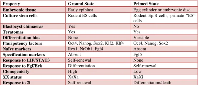
The various differences that characterize ES cells and EpiS cells are listed in Table 1. Compared to ES cells, EpiS cells are defined by a more restricted, primed pluripotency state.
Figure 8 The two phases of pluripotency (Nichols and Smith, 2009)
Table 1 Differences between the ground and primed cell states. Adapted from (Nichols and Smith, 2009)
Property Ground State Primed State
Embryonic tissue Early epiblast Egg cylinder or embryonic disc Culture stem cells Rodent ES cells Rodent EpiS cells; primate “ES”
cells
Blastocyst chimaeras Yes No
Teratomas Yes Yes
Differentiation bias None Variable
Pluripotency factors Oct4, Nanog, Sox2, Klf2, Klf4 Oct4, Nanog, Sox2
Naïve markers Rex1, NrOb1, Fgf4 Absent
Specification markers Absent Fgf5
Response to LIF/STAT3 Self-renewal None Response to Fgf/Erk Differentiation Self-renewal
Clonogenicity High Low
XX status XaXa XaXi
20
C. Human ES cells
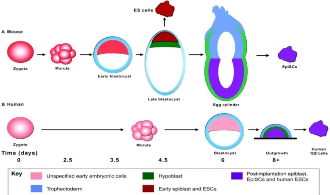
Human ES cells were first isolated by Thomson and collegues in 1998. These cells were
capable to differentiate to the different cell lineages (Thomson et al., 1998). Although the core
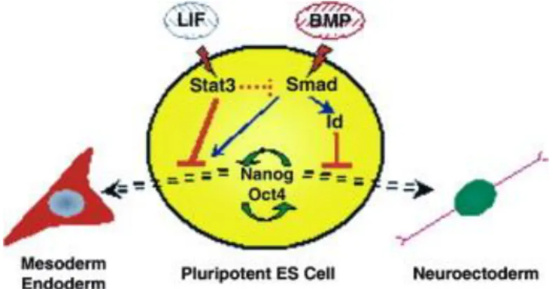
transcription regulatory network is conserved; however, these cells require different culture conditions. Human “ES” cells do not respond to LIF nor are they sustained by Erk inhibition;
on the contrary, they depend on Erk signaling for continued proliferation. This signaling is
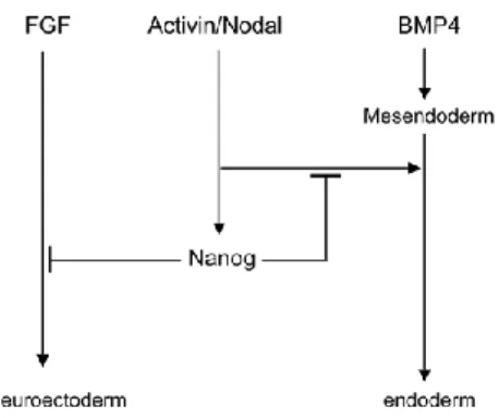
assured in hESCs by the Activin/Nodal pathway (Figure 9) through Smad2/3 that control the
expression of the pluripotency factor, Nanog, which in turn blocks the expression of
neuroectoderm genes induced by FGF (Vallier et al., 2009). The most probable explanation
for this difference is that human ES cells correspond to a more advanced developmental state,
more similar to the mouse EpiS cells (Brons et al., 2007; Tesar et al., 2007).
Figure 9 Model explaining the regulation of Nanog in hESCs/mEpiSCs and its function in both cell types (Vallier et al., 2009)
This EpiS cell state might be due to the longer period of culture required for the appearance of
human ES cells, that would allow these cells to progress in vitro to a state equivalent to post
21 Figure 10 Comparison of pluripotent cell line derivation protocols for (A) mouse and (B) human embryos. This figure highlights the difference in timing of mouse and human development in vivo (in days), and the appearance of pluripotent cell lines in vitro (Nichols and Smith, 2011).
22
Chapter II.
The Transcriptional and Epigenetic Control of the
Embryonic Stem Cell State
The genome of ES cells is tightly controlled at the transcriptional level. This control is exerted
by a large number of actors, including transcription factors (TFs) and elements of the
transcriptional machinery, as well as chromatin regulators that determine the epigenetic state of DNA. It has been suggested that ES cells possess a more ‘open’ chromatin, where
chromatin proteins are in a hyper-dynamic state (Meshorer et al., 2006a). This chromatin state
would work in parallel with a specific gene expression program that favors the expression of
self-renewal genes and poises the differentiation genes. So how do TFs and epigenetic
modifications operate in order to maintain pluripotency in ES cells and start differentiation
under the appropriate cues?
A. The Transcription Factor Network for ES state maintenance
1. The Core Transcriptional Regulatory Network
Pluripotency maintenance was shown to be mainly controlled and assured by three major TF
that form the core regulatory network. These factors are Oct4, Sox2 and Nanog (Chambers
and Smith, 2004; Niwa, 2007; Silva and Smith, 2008).
a. Oct4
Oct4 or the octamer-binding transcription factor 4 belongs to the POU family and is encoded
by the gene Pou5f1. The POU family is characterized by the presence of the POU domain: a 75 amino acid amino-terminal POU specific (POUs) region and a 60 amino-acid
carboxyl-23 terminal homeodomain (POUh). This domain binds the octamer consensus sequence
ATGCAAAT on DNA (Pan et al., 2002).
It has been shown that Oct4 is a pivotal factor and a gatekeeper that prevents ES cells from
differentiation (Nichols et al., 1998). Moreover, Oct4 is almost exclusively expressed in ES
cells (Nichols et al., 1998; Pesce and Scholer, 2000; Pesce and Schöler, 2001). Studies
indicate that during embryonic development, Oct4 is first expressed in all blastomeres. Later
on, it becomes only expressed in the ICM; however; at maturity, Oct4 expression becomes
restricted to the developing germ cells (Pesce and Scholer, 2000; Pesce and Schöler, 2001).
When Oct4 was disrupted in mice, the embryos lacked a pluripotent ICM (Nichols et al.,
1998), further emphasizing its role in pluripotency.
More interestingly, the correct level of expression of Oct4 is essential in the determination of
the cell state, as quantitative analysis of Oct4 expression showed that a high level of Oct4
expression drives ES cells towards mesoderm or endoderm lineages, while low levels of Oct4
drive trophectodermal lineages. To maintain the ES cells pluripotency state, a normal level of
Oct4 is required(Niwa et al., 2000).
Oct4 is considered to be a pioneer transcription factor, where it was shown not only to bind
nucleosome depleted regions and adopt different configurations depending on whether it
binds alone or in cooperation with other factors from the core transcriptional regulatory
network, but also can bind compacted chromatin structures at the beginning of
reprogramming (Soufi et al., 2012). Oct4 was shown to bind the regulatory elements, in
particular enhancers, of both pluripotency-related genes and differentiation genes, conferring
its role in both activation and repression in order to maintain the ES cell state (Chen et al.,
2008b). In addition, Oct4 works mostly in association with the other two core TFs Sox2 and
24 Proteomic analyses of Oct4 interactome revealed a wide range of actors that interact in a
direct or indirect manner with Oct4 (van den Berg et al., 2010a; Pardo et al., 2010). This
interactome revealed various actors including TFs such as Esrrb and Klf4, chromatin
remodeling complexes such as members of NuRD, esBAF and Tip60 and cofactors most of
them known to play an important role in pluripotency maintenance.
b. Sox2
Sox2 or the Sex determining region Y-box 2 is a member of the Sox family of TFs. This
family is characterized by the presence of a conserved high-mobility-group (HMG) that binds
DNA on an CTTTG(T/A)(T/A) motif (Chambers and Smith, 2004). Sox2 has distinct
biological functions and is essential during development. This distinct functionality is due to
the interaction of Sox2 with various cofactors during development. Many factors have been
shown to influence binding of Sox proteins to their target genes (Wegner, 2010). Sox2
expression is first detected at the morula stage, later on , it becomes located in the ICM of the
developing blastocyst and the epiblast (Avilion et al., 2003). The deletion of Sox2 at the
zygotic level cause embryonic lethality due the incapacity of the formation of a pluripotent epiblast; however, Sox2 doesn’t seem to affect the formation of the trophoectoderm (Wegner,
2010).
Interestingly, during development, Sox2 continues to be expressed, majorly in the central
nervous system after gastrulation, conferring its possible role in neural differentiation
(Wegner and Stolt, 2005). Sox2 induces neural differentiation by repressing key regulators of
other lineage-linked genes (Thomson et al., 2011; Wang et al., 2012; Zhao et al., 2004). This
ES cell specification control was not only observed for Sox2 but also for the other two TFs
Oct4 and Nanog where they rather promote the differentiation to mesendoderm lineages
25
c. Oct4/Sox2 complex
Oct4 and Sox2 were found to be co-expressed in several pluripotent cells such as the cells of
the morula, ICM, epiblast, and germ cells. It was demonstrated that the Oct4/Sox2 complex
works in a synergic way to control transcription, where a physical interaction was detected
between the two proteins (Ambrosetti et al., 1997). It was further shown that the octamer
elements within the enhancers of Oct4 controlled genes were found in proximity to
Sox2-binding sox elements (Avilion et al., 2003) creating a juxtaposed oct-sox Sox2-binding motif.
Moreover, a cooperative Oct4/Sox2 mechanism was observed at Oct4 target genes, where
oct-sox binding motifs were present (Loh et al., 2006). Indeed, Sox2 CHIP showed its binding at
the majority of Oct4-occupied loci (especially at key regulatory regions of Pou5f1, Sox2,
Nanog, Fgf4 and other pluripotency related genes) emphasizing the cooperation of these two
factors to control the gene expression of their targets (Loh et al., 2006), and showing that not
only does this Oct4/Sox2 complex control other genes, but also they auto regulate their own
enhancer elements. However, Masui et al. demonstrated that Sox2 is not essential for the
activation of these oct-sox enhancer motifs, as the deletion effect of Sox2 can be saved by a
forced expression of Oct4, conferring its probable role as rather an Oct4 stabilizer at its motifs
(Masui et al., 2007).
d. Nanog
Nanog is the last discovered transcription factor of the three core TFs (Chambers et al., 2003;
Mitsui et al., 2003). It is a homeobox transcription factor that recognizes the consensus
sequences ATTAT (Mitsui et al., 2003). In Nanog null mice, the ICM has impaired
development. Nanog transcripts were shown to appear at maximal levels between the late
morula and the mid-blastocyst and down regulated just prior to implantation (Chambers et al.,
2003). It was proposed that Nanog interferes at the blastocyst stage, after the initial action of
26 or become a trophoectoderm. At the later blastocyst stage, Nanog interferes and determines
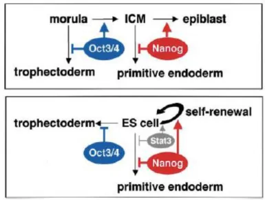
whether cells of the ICM remain pluripotent or differentiate into primitive endoderm (Figure 11) (Mitsui et al., 2003).
Figure 11 The proposed function of Oct4 and Nanog in preimplantation embryos (upper) and in ES cells (lower) (Mitsui et al., 2003).
Nanog discovery by Mitsui and Chambers have opened the gate to the characterization of
LIF/STAT3 independent pathways that control the pluripotency of ES cells. They further
demonstrate that the over expression of Nanog is enough to maintain the pluripotency of ES
cells; where this was not the case of the over expression of the other core transcription factor
Oct4, where ES cells differentiate into endoderm (Niwa et al., 2000). Therefore, it was
proposed that Nanog has an essential role in pluripotency maintenance independently from
the LIF/STAT3 pathway and the over expression of Nanog is sufficient to maintain the ES
cell state in the absence of LIF.
The importance of Nanog in the maintenance of pluripotency was debated in later studies. A
study has shown that Nanog null ES cells conserve their pluripotency characteristics and are
capable to differentiate correctly confirming that LIF pathways can maintain pluripotency
27 be hindered (Chambers et al., 2007). Indeed, Chambers demonstrates that Nanog is required
for primordial germ cells to prosecute the germ-cell development program beyond E11.5.
Moreover, it was shown that Nanog has different expression levels across ES colonies and can
vary in each ES cell itself when in culture. This notion of a heterogeneous expression of
Nanog reflects that ES cells are maintained in a condition where high levels of Nanog
preserve pluripotency and lower levels give a window of opportunity for lineage commitment
(Chambers et al., 2007). Indeed, an exogeneous control of Nanog expression can help produce
a more homogeneous ES cell population (MacArthur et al., 2012). Furthermore, Abranches
emphasizes more the role of Nanog and speculates that not only does it allow ES cells to
explore their commitment opportunities, but also acts as an autonomous component of the
pluripotency gene regulatory network, where it buffers the molecular heterogeneity and
controls cellular decision-making in ES cells (Abranches et al., 2013, 2014).
At the transcriptional level, Nanog doesn’t work alone (even though it might belong to
differential signaling pathways), it rather cooperates with Oct4 in order to govern the ES cell
state (Liang et al., 2008a; Loh et al., 2006). Loh et al. show that Nanog and Oct4 binding
patterns overlap and depletion of either one of these factors influences a common important
target of genes; conferring the interconnection between the two factors in order to control
pluripotency, self-renewal and fate determination of ES cells.
e. Interplay between the core regulatory factors to regulate transcription
The role of the Oct4/Sox2/Nanog trio in the control of ES cell state resides in two
mechanisms (Figure12): First, the three TFs co-occupy enhancer regions and positively control the transcription of genes necessary for pluripotency maintenance (Chen et al., 2008b;
Loh et al., 2006); on the other hand they occupy repressed genes encoding for differentiation
28 2009; Loh et al., 2006; Pasini et al., 2008). Second, the core TFs are capable to regulate their
own promoters forming an interconnected autoregulatory loop that provides positive feedback
expression for pluripotency maintenance. The deregulation of one of the core TFs alters this
loop and causes the cell entrance in the different differentiation programs (Boyer et al., 2006;
Loh et al., 2006).
Figure 12 Oct4, Sox2 and Nanog collaborate to control their own promoters forming an autoregulatory loop. Together these core transcription factors function to activate pluripotency genes and inhibit developmental genes. Adapted
from (Young, 2011).
2. The Expanded Transcriptional Regulatory Network
The core TFs Oct4, Sox2 and Nanog form the central unit of transcriptional control in ES
cells. However, these factors are a part of a much bigger transcriptional regulatory network,
composed of a large number of additional TFs, cofactors, chromatin regulators and
non-coding RNAs.
a. Important transcription factors in ES cells
As mentioned in Chapter I, the main signaling pathways that control the ES cell state are the
LIF, Wnt and BMP pathways. These external signaling cues have downstream internal
transcription responses. Three main TFs play essential roles in communicating the various
signaling cues into the core transcription network. They include, STAT3 (LIF signaling),
29 in Chapter I. Several studies have expanded gradually the transcription regulatory network
responsible for the control of the ES cell state.
c-Myc:
Myc (Myelocytomatosis oncogene) belongs to a family of helix-loop-helix/leucine zipper TFs. It’s potential role in ES cells was speculated through studies on human tumors where it
was shown to delay differentiation and promote cell proliferation (Knoepfler et al., 2002;
MacLean-Hunter et al., 1994; Pelengaris et al., 1999; Schreiner et al., 2001). Further studies
showed that high c-Myc expression is required for the ES cell self-renewal through
downstream the LIF/STAT3 signaling pathway (the overexpression of c-Myc replaces the
need for LIF supplement) and inhibiting c-Myc causes ES cell differentiation (Cartwright et
al., 2005). c-Myc was shown to control the transcription in ES cells through its capacity to
bind E box sequences at core promoters and stimulating RNA polymerase II pause release
(Rahl et al., 2010). Moreover, c-Myc was shown to be mostly bound on the regulatory
elements of transcribed genes along with the core TFs (Young, 2011). c-Myc is one of the
TFs essential in reprogramming into induced pluripotent stem cells (Takahashi and
Yamanaka, 2006).
Esrrb:
The estrogen related receptor beta is an orphan nuclear receptor shown to be a part of the core
pluripotency network, where it was shown to interact with Oct4 (van den Berg et al., 2010a;
Chen et al., 2008b; Loh et al., 2006). The importance of Essrb in self-renewal was revealed by
RNA interference studies where it was shown to be essential in ES cell self-renewal and its
absence triggers ES cell differentiation (Ivanova et al., 2006; Loh et al., 2006). Interestingly,
Essrb overexpression was shown to substitute for a short-term LIF requirement (Zhang et al.,
30 Furthermore, Essrb was shown to regulate Nanog’s activity where it mediates the positive
regulatory effect of Oct4 (van den Berg et al., 2008). Moreover, recent studies have shown
that Essrb overexpression can even substitute Nanog function in ES cells and its deletion
causes severe impaired self-renewal (Festuccia et al., 2012). However, the derivation of Essrb
null ES is possible (like Nanog null ES cells), in contrast to the absolute requirement of Oct4,
this reflects the relative importance of such factors, where Essrb, like Nanog seem to help
more in the fine tuning of the gene expression in ES cells.
Sall4:
Sall4 belongs to the Spalt family of zinc-finger TFs, mutations in the Sall4 gene cause
numerous developmental defects (Al-Baradie et al., 2002; Kohlhase et al., 2002). Sall4 null
mice die shortly after implantation; however these mice do not present ICM defects and ES
cells isolated from such ICM retain their pluripotency with a slower proliferation rate
(Sakaki-Yumoto et al., 2006).
The role of Sall4 was studied in several reports. Sall4 RNA interference experiments have
demonstrated that Sall4 depleted ES cells differentiate and fail to maintain their self-renewal
on feeder free culture conditions (Zhang et al., 2006). Furthermore, Zhang et al show that
Sall4 binds to Pou5f1 regulatory elements where it affects Oct4 expression in a
dosage-dependent manner. Additional studies also show that Sall4 binds Nanog regulatory elements
and Nanog gene targets (Wu et al., 2006), conferring its involvement in the core transcription
circuitry. Nevertheless, the absolute necessity of Sall4 in ES cell pluripotency is debated. As a
study shows that Sall4 null ES cells retain their pluripotency even on feeder-free culture
conditions (Yuri et al., 2009). This study suggests that Sall4 acts as a stabilizer of the ES cell
31
Tbx3:
The T-box transcription factor family was shown to be important in a variety of
developmental processes (Miller et al., 2008). Tbx3 is expressed early in the mouse inner cell
mass, and it is later expressed in extra embryonic endoderm cells (Chapman et al., 1996). In
ES cells, Tbx3 was reported to be essential for ES cell self-renewal (Ivanova et al., 2006), and
the continuous expression of Tbx3 allows the maintenance of ES cell in an undifferentiated
state in the absence of LIF (Niwa et al., 2009). Interestingly, Niwa et al. show that Tbx3 is an
upstream regulator of Nanog gene along with Klf4 where it boosts the expression of core
pluripotency genes (Figure 13) without being essential in pluripotency maintenance. Indeed, Tbx3 was reported to improve the germ-line competency of induced pluripotent stem cells
(Han et al., 2010).
Klf4:
The Krüppel-like factor (Klf) family is an evolutionarily conserved family of zinc finger TFs
that play a role in different biological processes, including proliferation, differentiation,
development and apoptosis (McConnell et al., 2007). Klf4 is a member of the Klf family,
studies have shown to interact with Oct4 in order to activate Oct4/Sox2 target genes
(Nakatake et al., 2006). Klf4 is one of the TFs used in reprogramming into induced
pluripotent stem cells (Takahashi and Yamanaka, 2006).
The overexpression of Klf4 prevents differentiation of ES cells, emphasizing more its role in
self-renewal (Li et al., 2005). However, Klf4 depletion does not seem to effect on ES cell
morphology or self-renewal (Jiang et al., 2008; Nakatake et al., 2006), due to the presence of
other Klf proteins, mainly Klf2 and Klf5 with redundant functions in ES cells. Indeed, the
32 Klf2/4/5 TFs were also shown to be bound to regulatory elements of key pluripotency genes
such as Pou5f1, Sox2, Nanog and Essrb.
Figure 13 Transcription control by Klf4 and Tbx3. Klf4 and Tbx3 mainly activate Sox2 and Nanog, respectively, and maintain expression of Oct3/4. Transcription of all these transcription factors is positively regulated by Oct3/4, Sox2
and Nanog (Niwa et al., 2009). Foxd3:
Foxd3 belongs to the family of fork head box TFs. Initially, it was solely identified in ES cell
and their malignant progenitors (Sutton et al., 1996). Later on, it was demonstrated that Foxd3
is also expressed during embryogenesis in the epiblast and even the neural crest (Dottori et al.,
2001; Hromas et al., 1999; Labosky and Kaestner, 1998). Foxd3 knockdown mice die early
during embryogenesis with a loss of the epiblast; moreover Foxd3 knockdown cells fail to
proliferate and give a normal ICM (Hanna et al., 2002).
Zfx:
Zfx is a zinc finger TFs of the Zfy family, a family highly conserved in vertebrates
(Schneider-Gädicke et al., 1989). It was shown to be required in ES cell renewal but not
essential for pluripotency, where Zfx deletion caused impaired cell proliferation and increase
apoptosis but did not influence the differentiation potential (Galan-Caridad et al., 2007).
Interestingly, Zfx was shown to upregulate the expression of c-Myc in order to enhance
33
Ronin:
Ronin, referring to the master-less Japanese samurai, is a zinc finger transcription factor. This
factor was shown not to have a direct relationship with the core TFs and it seemed to be able
to partially override the necessity for Oct4 (Dejosez et al., 2008). Moreover, Ronin
independency from the core transcription network was further demonstrated with its persistent
expression upon knockdown of Oct4, Sox2 and Nanog.
Ronin knockdown causes ES cell death, probably due to the activation of repressed genes
simultaneously or due to an unknown effect on apoptosis pathways. Moreover, Ronin knock
down during embryonic development show a common phenotype with Oct4 deletion, where
there is failure of ICM formation and embryo lethality (Dejosez et al., 2008).
Dax1:
Dax1 (DSS-AHC on X chromosome gene) is an atypical orphan nuclear receptor that has
been shown to play a role in ES cell pluripotency (Kelly et al., 2010; Niakan et al., 2006; Sun
et al., 2009). DAX1 gene knockdown caused mouse ES cell differentiation (Niakan et al.,
2006). It was demonstrated that Dax1 interacts with Esrrb and represses its function (Uranishi
et al., 2013). Moreover, Dax1 was shown to regulate in a negative way the expression of
Oct4, where it seems to fine tune the expression of Oct4 in order to maintain ES cell
pluripotency (Sun et al., 2009).
Other transcription factors with potential roles in ES cells:
Several core pluripotency factors interactome studies revealed a large number of involved TFs
with potential roles in the regulation of the ES cell state (van den Berg et al., 2010a; Pardo et
34 and Zfp281 shown to be a part of the ES cell transcriptional regulatory network with potential
roles in the optimal maintenance of the ES cell state.
b. Additional actors important in ES cell self-renewal Transcription Cofactors:
Cofactors are capable to interact selectively and non-covalently with TFs and the basal
transcription machinery in order to regulate transcription. They can either activate
(coactivators) or repress (corepressors) gene transcription. Cofactors generally do not bind
DNA, but rather assure the protein-protein interactions between TFs and the transcription
machinery. They are expressed in all cell types, but ES cells seem to be quite sensitive to
reduced levels of such cofactors (Fazzio and Panning, 2010; Kagey et al., 2010).
Mediator is one of the most important coactivators of transcription. In ES cells, the mediator
was shown to physically link Oct4/Sox2/Nanog bound enhancers to the promoters of active
genes (Kagey et al., 2010). This physical link is assured by a second cofactor, the cohesin
(Figure 14). Cohesin was shown to assure the DNA looping needed to approach regulatory
elements and activate transcription in ES cells (Kagey et al., 2010). ChIP-seq data
demonstrate the co-occupancy of mediator and cohesion with the core TFs at regulatory
elements (Young, 2011).
35 The proper chromatin structure and condensation is assured by an additional cofactor, the
condensin. Condensin complexes were shown to be required in ES cells where the deletion of
such complexes causes various phenotypic alterations at the level of the chromatin structure
(Fazzio and Panning, 2010).
Corepressors were also found to play a role in the maintenance of ES cells. Cnot3 (Ccr4-Not)
and Trim28 (Tripartite motif-containing 28) are two corepressors shown to co-occupy
promoter regions with important TFs in ES cells such as c-Myc and Zfx (Hu et al., 2009a).
The down-regulation of Cnot3 and Trim28 causes ES cell differentiation into trophectoderm and primitive ectoderm respectively, conferring their role in self-renewal. Moreover, Hu et al. demonstrate that c-Myc/Zfx/Cnot3/Trim28 form a distinct pluripotency module from the Oct4/Sox2/Nanog module, indicating the increased transcriptional regulatory network complexity in ES cells.
Chromatin regulators:
Many chromatin regulators were shown to play important roles in ES cells (Fazzio and
Panning, 2010; Kagey et al., 2010; Leeb et al., 2010; Meissner, 2010; Niwa, 2007).
Chromatin regulators can be divided into four groups: Cohesin/condensing complexes
(discussed earlier), histone-modifying enzymes, ATP-dependent chromatin remodeling
complexes and DNA methyltransferases (Young, 2011).
Histone-modifying enzymes encompass a significant number of different complexes that
participate in either the activation or repression of transcription in ES cells. They include
Polycomb complexes, SetDB1, Tip60 and TrxG proteins, the function of these proteins will
be detailed further in this chapter.
ATP-dependent chromatin remodeling factors were shown to regulate transcription,
36 and Cairns, 2009a; Ho and Crabtree, 2010). Several remodeling factors were shown to be
involved in the control of the ES cell state. The different remodeling complexes involved in
ES cell transcriptional control will be discussed later in Chapter II and more widely in
Chapter III.
DNA methylation does not seem to be essential in ES cell maintenance. Indeed, although
DNA methyltransferases (Dnmts) are expressed in ES cells and about 60-80% of the CpG
islands are methylated, ES cells can be established and maintained in the absence of Dnmts
and DNA methylation (Meissner, 2010). However, DNA methylation becomes required
during differentiation, as Dnmt-deficient ES cells fail to properly differentiate (Jackson et al.,
2004).
Noncoding RNAs:
miRNAs
Evidence have shown that several microRNAs (miRNAs) play essential roles during
developmental, where they were demonstrated to regulate the expression of a large number of
genes (Farh et al., 2005). A group of miRNAs was shown to play a role in the control of the
ES cell identity (Kanellopoulou et al., 2005; Murchison et al., 2005; Wang et al., 2008).
Furthermore, these miRNA polycistrons where shown to be controlled by the core TFs
Oct4/Sox2/Nanog (Marson et al., 2008a). Marson et al. demonstrate that the core TFs regulate
the transcription of miRNAs in ES cells. miRNA genes involved in ES cell maintenance and
proliferation are activated, where these miRNAs (mainly the most abundant mir 290-295
cluster) fine-tune the expression of the ES TFs. On the other hand, Oct4/Sox2/Nanog occupy
the genes of developmental miRNAs along with repressing complexes poising their
expression (Marson et al., 2008b).
37 Recent studied have revealed the role of long non-coding RNAs (lncRNAs) in the control of
the ES cell state (Guttman et al., 2009; Khalil et al., 2009). It was shown that a set of lncRNA
genes are bound by the core TFs, and the downregulation of such lncRNAs results in dramatic
effects on the expression of the core circuitry indicating a potential feedback loop created by
lncRNAs (Sheik Mohamed et al., 2010). On the other hand; lncRNAs were also shown to be
involved during differentiation. A lncRNA called Xist is responsible for X chromosome
imprinting during development and ES cell lineage commitment (Navarro and Avner, 2009).
Moreover, lncRNAs where shown to be involved in later developmental steps, inducing the
expression of lineage associated genes ( Ng et al., 2012). Interestingly, the expression of
lncRNAs was shown to be correlated with the development potential of induced pluripotent
stem cells (Liu et al., 2010).
3. Pluripotency transcription factors and iPS cell generation
a. Scientific milestones leading to induced pluripotent stem cell generation
Reprogramming of somatic cells into pluripotent embryonic stem cells first started using
nuclear transfer (NT) approaches. The early experiments in amphibians and later in mammals
showed that terminally differentiated somatic cells were able to generate cloned animals
(Briggs and King, 1952; Gurdon, 1962; Wilmut et al., 1997). This ability of a somatic cell to
revert to an early embryonic state rose questions about the nature of genome changes upon
differentiation, introducing the probable role of epigenetics (Hochedlinger and Jaenisch,
2002). Moreover, this capacity of somatic cells to dedifferentiate in oocyte conditions led to
the speculation of the presence of specific factors in the oocyte that are capable to induce
38 Another strategy to induce the reprogramming is fusing adult somatic cells with ES cells in
culture generating tetraploid hybrid cells (Cowan et al., 2005; Tada et al., 1997, 2001). These
observations indicated that there are some nuclear factors present in ES cells (as in oocytes)
that are responsible for this reprogramming mechanism (Do and Schöler, 2004; Egli et al.,
2007). More recent studies have demonstrated that the overexpression of pluripotency factors
such as Nanog enhances the formation of the reprogrammed hybrids about 200 folds (Silva et
al., 2006). Indeed, previous work have demonstrated that the expression of certain TFs in
certain mature terminally differentiated cells is sufficient to reprogram them into certain adult
stem cell progenitors; by the same manner, the deletion of certain TFs can lead to
reprogramming to multipotent progenitors (Davis et al., 1987; Laiosa et al., 2006; Nutt et al.,
1999). These observations have led to the rational thinking that the overexpression of ES cells
TFs in somatic cells could lead to their stable reprogramming.
b. Induced pluripotent stem cell generation from mouse and human somatic cells
The first attempt to generate induced pluripotent stem cells (iPS cells) from somatic mouse
cells was conducted by Takahashi and Yamanaka. Experiments were conducted on mouse
fibroblasts, where 24 ES cell TFs were introduced with retroviral vectors and reprogrammed
cells were drug selected for the ES cell-specific gene Fbx15 (Takahashi and Yamanaka,
2006). In those experiments, Takahashi and Yamanaka were capable of identifying the 4 TFs
Oct4, Sox2, Klf4 and c-Myc (the OSKM factors) sufficient to reprogram mouse fibroblasts
into iPS cells. This first generation of iPS cells was similar to ES cells but not identical, as
transcriptional and epigenetic patterns appeared to be only partially reset and such cells could
not form mouse chimeras when injected into blastocysts (partially reprogrammed cells).
Later studies, gave rise to the second generation of iPS cells from mouse fibroblasts. The iPS
cell drug selection was based this time on the usage of essential pluripotency genes such as
39 second generation of ES cells with transcriptional and epigenetic patterns highly similar to
those in ES cells and can form chimeric mice. The different steps and the molecular changes
involved in the direct reprogramming to iPS cells are demonstrated in Figure 15.
Figure 15 Steps involved in direct reprogramming to pluripotency. The starting, intermediate and end stages of reprogramming to pluripotency that can be identified during the generation of iPS cells (Hochedlinger and Plath,
2009)
Since the iPS cell derivation from mouse fibroblasts, a dozen of studies have been capable to
derivate iPS cells from various cell types including the blood, liver, stomach, pancreas, brain,
intestine and adrenals. Furthermore, the generation of human iPS cells was also successful
from fibroblasts (Lowry et al., 2008; Park et al., 2008; Takahashi et al., 2007) and
keratinocytes (Aasen et al., 2008; Maherali et al., 2007) using the OSKM cocktail or a variant
combination of factors such as the core TFs Oct4/Sox2/Nanog with a miRNA called LIN28