HAL Id: cea-02915659
https://hal-cea.archives-ouvertes.fr/cea-02915659
Submitted on 15 Aug 2020
HAL is a multi-disciplinary open access archive for the deposit and dissemination of sci-entific research documents, whether they are pub-lished or not. The documents may come from teaching and research institutions in France or abroad, or from public or private research centers.
L’archive ouverte pluridisciplinaire HAL, est destinée au dépôt et à la diffusion de documents scientifiques de niveau recherche, publiés ou non, émanant des établissements d’enseignement et de recherche français ou étrangers, des laboratoires publics ou privés.
Polypeptides derived from α-Synuclein binding partners
to prevent α-Synuclein fibrils interaction with and
take-up by cells
Elodie Monsellier, Maya Bendifallah, Virginie Redeker, Ronald Melki
To cite this version:
Elodie Monsellier, Maya Bendifallah, Virginie Redeker, Ronald Melki. Polypeptides derived from α-Synuclein binding partners to prevent α-α-Synuclein fibrils interaction with and take-up by cells. PLoS ONE, Public Library of Science, 2020, �10.1371/journal.pone.0237328�. �cea-02915659�
RESEARCH ARTICLE
Polypeptides derived from
α-Synuclein
binding partners to prevent
α-Synuclein fibrils
interaction with and take-up by cells
Elodie Monsellier, Maya Bendifallah, Virginie Redeker, Ronald MelkiID*
CEA, Institut Franc¸ois Jacob (MIRcen) and CNRS, Laboratory of Neurodegenerative Diseases (UMR9199), Fontenay-aux-Roses, France
*ronald.melki@cnrs.fr
Abstract
α-Synuclein (αSyn) fibrils spread from one neuronal cell to another. This prion-like phenom-enon is believed to contribute to the progression of the pathology in Parkinson’s disease and other synucleinopathies. The binding ofαSyn fibrils originating from affected cells to the plasma membrane of naïve cells is key in their prion-like propagation propensity. To inter-fere with this process, we designed polypeptides derived from proteins we previously showed to interact withαSyn fibrils, namely the molecular chaperone Hsc70 and the sodium/potassium pump NaK-ATPase and assessed their capacity to bindαSyn fibrils and/ or interfere with their take-up by cells of neuronal origin. We demonstrate here that polypep-tides that coatαSyn fibrils surfaces in such a way that they are changed affectαSyn fibrils binding to the plasma membrane components and/or their take-up by cells. Altogether our observations suggest that the rationale design ofαSyn fibrils polypeptide binders that inter-fere with their propagation between neuronal cells holds therapeutic potential.
Introduction
The aggregation of proteins into fibrillar high molecular-weight species is involved in human degenerative diseases, including Alzheimer’s, Parkinson’s, or Huntington’s [1]. During the last decade, it has become evident that those protein aggregates traffic between neuronal cells and amplify by seeding the aggregation of their constituting proteins [2–5]. This prion-like phe-nomenon is thought to be responsible for the stereotypic progression of the pathology in the brain [2,5]. Impeding this phenomenon would be valuable to slow down the progression of disease [6,7].
The spread of amyloid fibrils is a vicious circle involving different steps. First, protein aggre-gates form with time within neuronal cells [8]. They next escape actively, through export, or passively, upon cell death, the cells where they form [9–12]. The extracellular aggregates dock next to the membrane of naïve neuronal cells [13,14]. This membrane binding steps is fol-lowed by the internalization of the fibrils, mainly through endocytosis [15,16]. Once in the cells the aggregates reach the cytoplasm, where they recruit the otherwise soluble endogenous protein they are made of [17], after compromising endo-lysosomal integrity [18].
a1111111111 a1111111111 a1111111111 a1111111111 a1111111111 OPEN ACCESS
Citation: Monsellier E, Bendifallah M, Redeker V,
Melki R (2020) Polypeptides derived from α-Synuclein binding partners to preventα-Synuclein fibrils interaction with and take-up by cells. PLoS ONE 15(8): e0237328.https://doi.org/10.1371/ journal.pone.0237328
Editor: Stephan N. Witt, Louisiana State University
Health Sciences Center, UNITED STATES
Received: December 20, 2019 Accepted: July 23, 2020 Published: August 13, 2020
Peer Review History: PLOS recognizes the
benefits of transparency in the peer review process; therefore, we enable the publication of all of the content of peer review and author responses alongside final, published articles. The editorial history of this article is available here:
https://doi.org/10.1371/journal.pone.0237328 Copyright:© 2020 Monsellier et al. This is an open access article distributed under the terms of the
Creative Commons Attribution License, which permits unrestricted use, distribution, and reproduction in any medium, provided the original author and source are credited.
Data Availability Statement: All relevant data are
within the manuscript and its Supporting Information files.
Alternatively, they imbalance neuronal proteostasis and trigger the de-novo aggregation of other aggregation prone proteins involved in neurodegenerative diseases [19]. The circle com-pletes when amplified aggregates escape into the extracellular media, targeting new cells.
Every single step of the prion-like propagation process is a potential target for the development of new drugs that would delay the progression of disease. The binding of the extracellular aggre-gates to the membrane is especially attractive for different reasons [7]. As it takes place in the extracellular environment, it is more easily targetable than intracellular mechanisms [20]. Its underlying molecular mechanisms have been particularly well studied over the past few years [21]. The fibrils bind laterally to the plasma membrane [13]. The binding is mediated by interac-tion with the plasma membrane lipids [22], with different proteins partners [23–26] and with the extra cellular matrix components [27,28]. The efficiency of the binding depends both on the aggregates characteristics, such as their primary sequence [29,30], their net charge [22], their size [13] or their conformation [17], and on the properties of the membrane, with an emphasis on the role of the membrane curvature [31] and a specific lipid [32] and protein [24,25] composition.
As different membrane components are involved in the interaction with extracellular aggregates in their prion-like propagation process, it seems unlikely that targeting one of them would exert an effect. We therefore decided to target the fibrils themselves, coating them with peptide ligands so that their surface properties are changed and their interaction with mem-brane partners is compromised. We decided to develop polypeptide binders of fibrils as from a clinical point of view such binders are specific and safe, and their poor pharmacokinetics prop-erties are amenable to optimization [33,34]. Incidentally over 60 peptide drugs have now reached the market [35]. Using a cross-linking and mass spectrometry strategy, we previously mapped the surface interfaces betweenαSyn monomers or fibrils and two protein partners, namely, the molecular chaperone Hsc70 [36–38] and the sodium/potassium pump Na+/K +-ATPase (NKA) [25]. Here, we designed a set of polypeptides based on the Hsc70 and NKA surface areas we identified to interact withαSyn. We assessed the interaction of the polypep-tides derived from Hsc70 and NKA with fibrillarαSyn in vitro. We identify Hsc70-derived polypeptides that bind bestαSyn fibrils. We also show that an NKA-derived peptide affect fibrils binding to Neuro-2a cells. Overall, our results lay the basis for developing further such polypeptides and improving their affinity forαSyn fibrils, so that their interactions with and uptake by Neuro-2a neuronal cells are affected.
Results
Hsc70 binds to
αSyn fibrils with a high affinity, preventing their
interaction with the plasma membrane and their take-up by cultured cells
We previously brought evidence for Hsc70 interaction with fibrillarαSyn using a sedimenta-tion assay [36]. The dissociation constant we measured was 0.1μM. Here we confirmed the interaction between Hsc70 and fibrillarαSyn using the same sedimentation assay followed by quantitative analysis of the proteins in the pellet and the supernatant fractions by SDS-PAGE (Fig 1A). Hsc70 alone remains in the supernatant, whereas it is pulled into the pellet when incubated for 1h at room temperature with pre-formedαSyn fibrils. To determine the affinity of Hsc70 for fibrillarαSyn we incubated preformed αSyn fibrils (1 μM) with increasing amount (0–2μM) of a mix of unlabelled and ATTO488-labeled Hsc70 (labelled:unlabelled molar ratio of 1:50) for 1h at room temperature. The samples were then filtered through cellu-lose acetate membranes that retains fibrillarαSyn along with their binders, and the amount of ATTO488-Hsc70 was quantified by fluorescence measurements (Fig 1B). We measured a dis-sociation constant (KD) between Hsc70-ATTO488 andαSyn fibrils of 0.49 ± 0.02 μM,
consis-tent with previously published values [36,39]. We demonstrated that the binding between the
PLOS ONE Polypeptide binders of alpha-Synuclein fibrils
Funding: MB, Re´gion Ile de France through DIM
Cerveau et Pense´e; RM, Institut de France-Fondation Simone et Cino Del Duca; RM, France-Fondation Pour La Recherche Me´dicale (contract DEQ. 20160334896); RM, EC Joint Programme on Neurodegenerative Diseases and Agence Nationale pour la Recherche (TransPathND, ANR-17-JPCD-0002-02 and Protest-70, ANR-17-JPCD-0005-01). Nothing to disclose. The funders had no role in study design, data collection and analysis, decision to publish, or preparation of the manuscript.
Competing interests: The authors have declared
that no competing interests exist.
Abbreviations:αSyn, α-Synuclein; NKA, Na+/K+ -ATPase; NBD, Nucleotide Binding Domain; SBD, Substrate Binding Domain; TEV, tobacco etch virus; ThT, Thioflavin T.
two partners was not affected by Hsc70 labelling. Indeed, unlabeled Hsc70 competed in a dose-dependent way with the binding of labeled Hsc70 toαSyn fibrils (Fig 1C), and theKD
between Hsc70 andαSyn fibrils was identical to the KDbetween Hsc70-ATTO488 andαSyn
fibrils (0.45± 0.08 μM).
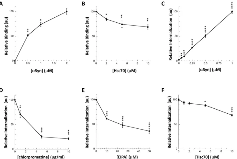
We next assessed the consequences of Hsc70 interaction withαSyn fibrils on fibrils binding to the cell membrane and subsequent internalization. We set-up two different assays to assess separatelyαSyn fibrils binding and internalization (Fig 2). Preformed Alexa488-labeledαSyn fibrils (S1 Fig) bound to cultured Neuro-2a cells within 30 min incubation in a dose-depen-dent manner as assessed by quantification of fluorescent foci at cell membranes (Figs2Aand
S2A). The addition of Trypan blue quenched all the fluorescence, indicating that the fibrils are located at the plasma membranes. We previously demonstrated that monomericαSyn does not bind to cells in such a way [40,41]. This robust cellular binding assay was next used to monitor the effect Hsc70-fibrillarαSyn interaction on fibrils binding to Neuro-2a cells. αSyn fibrils (1μM) were pre-incubated with increasing amounts of Hsc70 (0–10 μM). Neuro-2a Fig 1. Hsc70 binds toαSyn fibrils with high affinity. A, Hsc70 binds to αSyn fibrils in vitro. SDS-PAGE analysis of the pellet (P) and supernatant (S) fractions of
Hsc70 (10μM), fibrillar αSyn (100 μM), and fibrillar αSyn (100 μM) incubated with Hsc70 (10 μM) for 1 h at RT. B, Quantification of Hsc70-ATTO488 binding to αSyn fibrils using the cellulose acetate filter trap assay. Hsc70-ATTO488 was diluted with unlabelled Hsc70 (labelled:unlabelled molar ratio of 1:50) to different final concentrations (0–2μM) and incubated with or without αSyn fibrils (1 μM) for 1h at RT. Each sample was filtered in triplicate through a cellulose acetate membrane and the amount of Hsc70-ATTO488 trapped onto the membrane was quantified. The mean amount of Hsc70-ATTO488 bound to theαSyn fibrils normalized to the amount of Hsc70-ATTO488 bound at the maximal concentration used (“% of maximal binding”) and the associated standard error values were calculated from 2 to 3 independent experiments. A filter trap membrane from one representative experiment is shown. C, Unlabeled Hsc70 compete with Hsc70-ATTO488 for binding to αSyn fibrils. A fixed concentration of Hsc70-ATTO488 (0.2 μM) was incubated with increasing concentrations of unlabeled Hsc70 (0–10 μM) and with or without αSyn fibrils (1μM). Each sample was then filtered in triplicate through a cellulose acetate membrane. The mean amount of Hsc70-ATTO488 bound to the αSyn fibrils and the associated standard error values were calculated from these triplicates.
cultured cells were then incubated for 30 min with this mix. The data presented in Figs2Band
S2Bclearly demonstrate that Hsc70 affectsαSyn fibrils binding to Neuro-2a cells in a dose-dependent manner.
Fig 2. Hsc70 binding toαSyn fibrils interferes with their interaction with the plasma membrane and their subsequent internalization. A, Dose-dependent binding
ofαSyn fibrils to the plasma membrane of Neuro-2a cells. Neuro-2a cells were exposed for 30 min to αSyn-Alexa488 fibrils (0–2 μM). The cells were extensively washed and the fluorescence quantified. Representative images are shown inS2A Fig. For each concentration the mean percentage of Neuro-2a cells bound with at least 1 αSyn-Alexa488 fibrils foci and its associated standard error value was calculated from 3 independent experiments. The results and the associated significances are expressed relative to maximum binding. B, Hsc70 preventsαSyn fibrils binding to the plasma membrane. αSyn-Alexa488 fibrils (1 μM) were incubated with increasing concentrations of Hsc70 (0–10μM) in DMEM for 30 min at 37˚C. Neuro-2a cells were next exposed to the mixture for 30 min. Fluorescence was quantified after extensive washing. Representative images are shown inS2B Fig. For each Hsc70 concentration, the mean percentage of Neuro-2a cells with at least oneαSyn-Alexa488 fibrils foci and its associated standard error value was calculated from 3 to 5 independent experiments. The results and the associated significances are expressed relative to fibrils binding in the absence of Hsc70. C,αSyn fibrils take-up by Neuro-2a cells. Neuro-2a cells in 96-wells plates were exposed for 4 hours to increasing
concentrations ofαSyn-Alexa488 fibrils. Trypan blue was added after extensive washing to quench the fluorescence of plasma membrane-bound αSyn fibrils. The amount of internalizedαSyn-Alexa488 was measured on a fluorescence plate reader. Means and their associated standard error values were calculated from 5 independent wells. The results are expressed relative to maximum internalization (1μM αSyn fibrils). Significances are calculated in comparison to the absence of internalization (noαSyn). D,E, Chlorpromazine (D) and l’5-N-ethyl-isopropyl-amiloride (EIPA; E) prevent αSyn fibrils internalization by Neuro-2a cells. Neuro-2a cells in 96-wells plates were exposed for 1 hour to increasing concentrations of chlorpromazine (0–10μg/ml) or EIPA (0–50 μM) before addition of αSyn-Alexa488 fibrils (0.5μM). After 4 hours of incubation and extensive washing, Trypan blue was added to quench the fluorescence of plasma membrane-bound αSyn fibrils. The amount of internalizedαSyn-Alexa488 was measured on a fluorescence plate reader. Means and their associated standard error values were calculated from 5 independent wells. The results and the associated significances are expressed relative to the absence of inhibitors. F, Hsc70 preventsαSyn fibrils internalization by Neuro-2a cells. αSyn-Alexa488 fibrils (0.5μM) were incubated with increasing concentrations of Hsc70 (0–10 μM) in DMEM for 30 min at 37˚C. Neuro-2a cells in 96-wells plates were exposed for 4 hours to the mixture. Trypan blue was added after extensive washing to quench the fluorescence of plasma membrane-boundαSyn fibrils. The amount of internalizedαSyn-Alexa488 was measured on a fluorescence plate reader. Means and their associated standard error values were calculated from 3 independent experiments. The results and the associated significances are expressed relative to internalization in the absence of Hsc70.
https://doi.org/10.1371/journal.pone.0237328.g002
FibrillarαSyn uptake by cells can be assessed quantitatively by fluorescence microscopy after quenching of the fluorescence at cells plasma membrane by Trypan blue (S3 Fig). To increase statistical power we set-up a robust 96-wells plate assay [42]. Neuro-2a cells, in 96-wells plate, were exposed for 4h to Alexa488-labeledαSyn fibrils pre-incubated with Hsc70 or not, prior to Trypan blue addition and quantification of Alexa488 fluorescence in a plate-reader. The amount of internalized fibrils was determined in a dose- (Fig 2C) and time-depen-dent manner. FibrillarαSyn take-up was inhibited in a dose-dependent manner by chlorprom-azine and l’5-N-ethyl-isopropyl-amiloride (EIPA) that inhibits clathrin-mediated endocytosis [43] and macropinocytosis [44], respectively, suggesting that the fibrils are taken up by endo-cytosis (Fig 2D and 2E). We and others previously demonstrated that endocytically internal-izedαSyn fibrils are then able to escape the endocytic pathway and reach the cytosol by endosomal rupture [45–47]. Preincubation of Alexa488-labeledαSyn fibrils with Hsc70 signif-icantly affected their take-up (Fig 2F). We used Hoechst staining to ascertain that the number of cells remained constant (seeMaterial & Methods).
We conclude from these observations that Hsc70 binding toαSyn fibrils affects their bind-ing and take-up by neuronal Neuro-2a cells in a dose-dependent manner. The use of full-length Hsc70 for therapeutic purposes has drawbacks because of its pleiotropic effects within cells. We thus aimed at generating fragments of Hsc70 that retainαSyn fibrils binding capacity.
Hsc70 Substrate Binding Domain and sub-domains retain
αSyn fibrils
binding capacity
In a first step toward the design of Hsc70-derived peptides that would potentially retain their ability to bindαSyn fibrils, we assessed the affinity of different Hsc70 sub-domains for αSyn fibrils (Figs3and4). Hsc70 is composed of two domains, a Nucleotide Binding Domain (NBD), responsible for the chaperone ATPase activity, and a Substrate Binding Domain (SBD), that binds Hsc70 clients. We previously used lysine-reactive chemical cross-linkers and mass-spectrometry to map the surface areas within Hsc70 that interact with monomericαSyn; all the identified areas were within the SBD (Fig 3A) [37,38]. To determine whether Hsc70 SBD retains the ability to bindαSyn fibrils we expressed and purified it. Hsc70 SBD (Fig 3B, left) can be subdivided in 2 sub-domains, aβ-strands/sheet rich (SBDβ;Fig 3B, middle) and
anα-helical domain, named “SBD-lid” (Fig 3B, right). Lysine residues from both of these
sub-domains are located within the Hsc70-αSyn interaction interface suggesting that they both contribute toαSyn binding. We therefore expressed and purified SBDβ and SBD-lid.
The secondary structure content of Hsc70 SBD, SBDβ and SBD-lid was assessed by circular dichroism measurements. The data suggest that the polypeptide conformation is retained (Figs3B,4AandS4A Fig). We next assessed SBD, SBDβ and SBD-lid binding to αSyn fibrils
as described for full-length Hsc70 and derived dissociation constants from those measure-ments (Figs4BandS5 Fig). All three domains bindαSyn fibrils. Moreover, the KDwere
simi-lar to that we determined for full-length Hsc70 (Fig 4B). Thus, both Hsc70 SBDβ and SBD-lid contribute to fibrillarαSyn binding as for monomeric αSyn [37,38].
αSyn fibrils Hsc70-derived peptides binders
To identify peptides derived from Hsc70 that have all that is necessary and sufficient to bind αSyn fibrils, we synthesized ten 11 to 24 residues Hsc70-derived polypeptides (Table 1;Fig 3C) based on the regions that contribute toαSyn binding and/or participate to the canonical substrate groove (Fig 3A). Some peptides were overlapping to maximise their chances of adopting the right conformation and binding (i.e. Hsc-2 and Hsc-3). Hsc-1, 2, 3, 9 and 10
encompass Hsc70-αSyn interaction surface interfaces [37,38]. Hsc-4, 5 and 6 reproduce Hsc70 canonical client binding cavity [40]. Hsc-7 and 8 decal the rest of Hsc70 SBDβ loops. Hsc-4
and 9 were found insoluble in PBS. Their interaction withαSyn was not further studied. The secondary structure content of the 8 remaining peptides was assessed by circular dichroism measurements (Table 1andS4B Fig). The peptides were predominantly unstructured, with the exception of Hsc-1 and 10 (52 and 46%α-helical, respectively). The presence of an α-heli-cal conformation in the Hsc-1 peptide is coherent with the structure of this peptide within Hsc70 while Hsc-10 was expected to adopt a hairpin structure (Fig 3C).
Hsc70 binding to monomericαSyn affects assembly into fibrils [36]. We therefore first assessed Hsc70-derived peptide capacity to interact with monomericαSyn through their abil-ity to affect assembly into fibrils (Fig 5). MonomericαSyn assembly into fibrils was monitored Fig 3. Hsc70 domains and peptides used throughout this study. A,αSyn-binding sites on Hsc70. The binding sites were determined by cross-linking Hsc70 to monomericαSyn with chemical cross-linkers and identifying the surface interfaces by mass-spectrometry [37,38]. Only the substrate-binding domain (SBD) of Hsc70 is shown. Cross-linked lysines are depicted in yellow (space fill). Hsc70 model was built as described in [37]. B, Hsc70 SBD sub-domains. SBD β-sandwich (SBDβ) and lid (SBD-Lid) sub-domain are coloured. C, Hsc70-derived peptides. 10 peptides, which primary structure is given inTable 1, reproducing Hsc70 amino acid stretches involved inαSyn binding and the canonical Hsc70 client proteins binding sites [48], were synthesized.
https://doi.org/10.1371/journal.pone.0237328.g003
using Thioflavin T (ThT) binding at 37˚C in the absence or the presence of equimolar amounts of each peptide. Hsc-6 significantly slowed downαSyn assembly into fibrils while Hsc-10 accelerated aggregation (Fig 5A and 5B). The fibrillar nature of the assemblies obtained at the end of the reactions were assessed by transmission electron microscopy (Fig 5C). We conclude from these observations that 2 out of the 8 Hsc70-derived peptides we tested (Hsc-6 and 10) interact with monomericαSyn in such a way that the time course of assembly into fibrils is sig-nificantly affected.
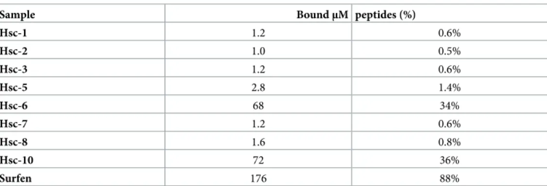
We next assessed Hsc70-derived peptides binding to fibrillarαSyn. Fibrillar αSyn (100 μM) was incubated with each peptide (200μM). The fibrils were sedimented and resuspended and the bound Hsc70-derived peptides were quantified by reversed phase chromatography. The results are presented inTable 2andS6A–S6F Fig. Hsc-1, 2, 3, 5, 7 and 8, did not bind toαSyn fibrils. Hsc-6 and 10 did bind to the fibrils. As a positive control we used the aromatic molecule Surfen, which is known to bind to the SEVI amyloid fibrils and to prevent their interaction with cells [49]. Surfen was found to bind toαSyn fibrils (Table 2).
The affinities of Hsc-6 and 10 forαSyn fibrils were determined and the KDwere over
100μM (S6G and S6H Fig). Nonetheless, to determine whether Hsc70-derived peptides affect fibrillarαSyn uptake by cells, Alexa488-labeled αSyn fibrils were pre-incubated with up to 10 Fig 4. Hsc70 domains and their binding toαSyn fibrils. A, Secondary structure of Hsc70 SBD and sub-domains. The CD spectra used for deconvolution are shown in S4A Fig. B, Determination of Hsc70 domains–αSyn interactions KD. The experiments were performed as inFig 1B. In each case the normalized mean amount of
labelled Hsc70 domain bound to the fibrils (% of maximal binding) and the associated standard error values were calculated from 2 to 3 independent experiments. Representative raw data are shown inS5 Fig.
https://doi.org/10.1371/journal.pone.0237328.g004
Table 1. Hsc70-derived peptides primary and secondary structures. The CD spectra used for deconvolution are shown inS4B Fig.
Peptide Hsc70 sequence Sequence Secondary structure compositionα-helix β-strand other Hsc-1 510–525 LSKEDIERMVQEAEKY 52% 0% 48% Hsc-2 553–566 VEDEKLQGKINDED 6% 13% 81% Hsc-3 548–571 NMKATVEDEKLQGKINDEDKQKIL 9% 7% 84% Hsc-4 400–415 SLGIETAGGVMTVLIK Hsc-5 428–439 FTTYSDNQPGV 0% 15% 85% Hsc-6 422–444 TKQTQTFTTYSDNQPGVLIQVYE 0% 20% 80% Hsc-7 461–475 LTGIPPAPRGVPQIE 10% 17% 73% Hsc-8 457–477 GKFELTGIPPAPRGVPQIEVT 7% 24% 69% Hsc-9 489–500 SAVDKSTGKENK Hsc-10 484–505 GILNVSAVDKSTGKENKITITN 46% 13% 41% https://doi.org/10.1371/journal.pone.0237328.t001
molar excess of the different Hsc70-derived peptides and fibrils uptake by Neuro-2a cells was quantified. None of the Hsc70-derived peptides had an effect onαSyn fibrils take-up (Fig 6). This is consistent with the poor affinity of the best fibrillarαSyn peptide binders. In contrast, Fig 5. Effect of Hsc70 SBD-derived peptides onαSyn aggregation. A, Time-course of αSyn aggregation in the absence or presence of Hsc70 SBD-derived
peptides. SolubleαSyn (50 μM) was incubated with or without Hsc70-derived peptides (50 μM) at 37˚C and 600 rpm in PBS. The assembly reactions were monitored by Thioflavin T binding. Means and their associated standard errors values were calculated from 4 independent experiments. The lines through the data points represent the best fits to a sigmoid function. B, Effect of Hsc70-derived peptides on the half-time (t1/2) ofαSyn aggregation. For each independent
experiment, thet1/2parameter was extracted from the best fit to a sigmoid function. The means and their associated standard error values were calculated from
4 independent experiments. C, Negative stained electron micrographs ofαSyn assembled alone (left) or in the presence of equimolar concentration of Hsc-6 (right). Scale bar, 200 nm.
https://doi.org/10.1371/journal.pone.0237328.g005
preincubation ofαSyn fibrils with Surfen affected, in a dose-dependent way, their take-up by Neuro-2a cells (Fig 6).
Peptide derived from an
αSyn fibrils membrane partner, the α3 subunit of
the Na
+/K
+-ATPase (NKA)
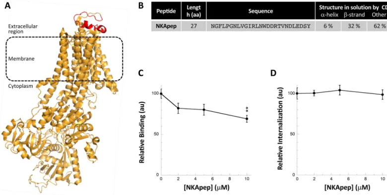
We previously brought evidence for interaction of fibrillarαSyn with the α3-subunit of the membrane ion pump NKA by pull-down [25].α3NKA amino acid stretch that interacts with αSyn was identified by cross-linking and mass spectrometry. It consists of the extracellular loop connecting the transmembrane helices 7 and 8 [25]. Interaction of fibrillarαSyn with this extracellular loop ofα3NKA was further confirmed by mutagenesis studies [25]. To determine whether NKA derived peptides affectαSyn fibrils binding to and take-up by Neuro-2a cells, we synthesized a 27 amino acid residues long peptide (NKApep) that reproduces this loop within α3NKA (Fig 7A). NKApep is soluble in PBS; it is disordered with someβ-strand content, as assessed by circular dichroism (Figs7BandS4C).
NKApep neither affectedαSyn aggregation (not shown) nor bound to preformed αSyn fibrils (S6D and S6F Fig). Preincubation of preformedαSyn fibrils with NKApep resulted in a dose-dependent reduction in fibrillarαSyn binding to Neuro-2a cells (Figs7CandS2C) but did not affect fibrils take-up (Fig 7D). Altogether, although designed to affect fibrillarαSyn binding to cells, NKApep acts somewhat differently, possibly through interactions with other membranous components.
Discussion
αSyn fibrils are able to spread from one neuronal cell to another [2–4]. This process is believed to contribute to the spatiotemporal progression of pathology in the central nervous system [2,5]. The binding ofαSyn fibrils to naïve cells, after their formation and release from affected counterparts, is key and has been actively documented as it constitutes a potential target for therapeutic interventions [7,13]. We hypothesized that ligands that change the surface proper-ties ofαSyn fibrils should affect binding to cell membranes. We previously showed that Hsc70 binding toαSyn fibrils affects the viability of cultured cells of neuronal origin [36]. We demon-strate here that Hsc70 interaction withαSyn fibrils compromises their binding and take-up by cells. The pleiotropic functions of full-length Hsc70 limit its therapeutic potential [50,51]. We therefore generated polypeptides reproducing Hsc70 sub-domains and surfaces that we previ-ously showed to interact withαSyn through cross-linking studies [37,38] and assessed their effect onαSyn fibrils binding to and take-up by cells of neuronal nature. We show here that Table 2. Binding of the Hsc70-derived peptides toαSyn fibrils assessed by phase reverse chromatography analysis.
Sample BoundμM peptides (%)
Hsc-1 1.2 0.6% Hsc-2 1.0 0.5% Hsc-3 1.2 0.6% Hsc-5 2.8 1.4% Hsc-6 68 34% Hsc-7 1.2 0.6% Hsc-8 1.6 0.8% Hsc-10 72 36% Surfen 176 88% https://doi.org/10.1371/journal.pone.0237328.t002
two peptides derived from Hsc70 SBD interact withαSyn without affecting, most probably because of their limited affinity, their take-up by Neuro-2a cells [52,53].
We previously identified through unbiased pull-down and cross-linking strategies a fibrillar αSyn neuron membrane proteins interactome [25]. Polypeptides reproducingαSyn protein partners may interfere with fibrils binding to their targets. We therefore assessed the shielding propensity of NKApep, an NKA-derived peptide that encompasses a region we showed to interact with fibrillarαSyn [25]. NKApep did not bind toαSyn fibrils under our experimental conditions, nonetheless, we show here that it interferes with fibrillarαSyn binding to cells. This suggests that NKApep affectsαSyn fibrils binding to the cell indirectly, possibly through the redistribution of otherαSyn fibrils target proteins [21].
Fig 6. Effect of Hsc70 SBD-derived peptides onαSyn fibrils take-up by Neuro-2a cells. Each Hsc70 SBD-derived peptide (2 or 10 μM) was incubated with
Alexa488-labeledαSyn fibrils (1 μM) in DMEM for 30 min at 37˚C. Neuro-2a cells grown in 96-wells plates were exposed to the mixture for 2 hours. After extensive washing trypan blue was added to quench the fluorescence of plasma membrane-boundαSyn fibrils. The amount of internalized αSyn-Alexa488 was measured using a fluorescence plate reader. Means and their associated standard error values were calculated from 3 independent experiments. The same experiment was carried out with Surfen.
https://doi.org/10.1371/journal.pone.0237328.g006
Fig 7. Effect of a peptide derived from theα3 subunit of the NKA on αSyn fibrils binding and take-up by Neuro-2a cells. A, Structure of the α3 subunit of the NKA
Bos taurus (PDB 4xe5) where the 27 amino acid residues long peptide NKApep corresponding to the extracellular loop previously shown to interact with αSyn fibrils [25] is coloured in red. B, Secondary structure content of NKApep determined by circular dichroism. The CD spectra used for deconvolution is shown inS4C Fig. C, Effect of NKApep onαSyn fibrils binding to the plasma membrane of Neuro-2a cells. αSyn-Alexa488 fibrils (1 μM) were incubated without or with increasing concentrations of NKApep in DMEM for 30 min at 37˚C. Neuro-2a cells were next exposed to the mixture for 30 min. Fluorescence was quantified after extensive washing. Representative images are shown inS2B Fig. For each peptide concentration, the mean percentage of Neuro-2a cells with at least 1αSyn-Alexa488 fibrils foci and its associated standard error value was calculated from 3 independent experiments. The results and the associated significances are expressed relative to fibrils binding in the absence of peptide. D, Effect of NKApep onαSyn fibrils take-up by Neuro-2a cells. αSyn-Alexa488 fibrils (0.5 μM) were incubated with increasing concentrations of NKApep (0–10μM) in DMEM for 30 min at 37˚C. Neuro-2a cells grown in 96-wells plates were exposed to the mixture for 4 hours. After extensive washing trypan blue was added to quench the fluorescence of plasma membrane-boundαSyn fibrils. The amount of internalized αSyn-Alexa488 was measured using a fluorescence plate reader. Means and their associated standard error values were calculated from 4 independent experiments. The results are expressed relative to the internalization in the absence of peptide.
Overall, our results suggest that polypeptides that bindαSyn fibrils must have a very high affinity to affect fibrils uptake by cells and hold therapeutic potential. Advantageously, the affinity of polypeptides is amenable to improvements. They can be trimmed and modified by replacing a number of amino acid residues and reassessing affinity in an iterative manner [54]. To limit their folding landscape, they can be stapled using unnatural amino acids bearing alkyl tethers of various lengths at either one or two helix turns [55] or compatible with covalent crosslinking via click chemistry [56], or fused to a scaffolding protein such as thioredoxin [57]. Alternatively, their avidity could be increased by generating tandem repeats of the same or dif-ferent peptides that bindαSyn fibrils. Many other modifications can be made so that pharma-cokinetic properties of polypeptides that interfere withαSyn fibril binding and take-up by cells are improved. Thus, such peptide could yield new therapeutic tools to slow down the progres-sion of synucleinopathies and other neurodegenerative diseases.
Materials and methods
Expression and purification of
αSyn, Hsc70 and Hsc70 subdomains
Recombinant human wild-typeαSyn was purified as described [58]. Recombinant His6-tagged
Hsc70 was purified as described [36]. The activity of the purified Hsc70 was assessed using a luciferase refolding assay, as described in [36].
Genes encoding the Hsc70 domain and subdomains SBD, SBDβ and SBD-lid were ampli-fied from the pPRO-EXHTb (Invitrogen) Hsc70 vector [36] and inserted into a pET-M11 vec-tor with an N-terminal 6xHis tag followed by a tobacco etch virus (TEV) protease cleavage site. Recombinant His-tagged proteins were expressed at 37˚C in E.coli strain BL21(DE3) (Stratagene, Santa Clara, CA) and purified as follow. Cells were harvested and resuspended in buffer A (30 mM HEPES pH 7.5, 300 mM NaCl, 10% glycerol, 20 mM imidazole). After soni-cation and centrifugation, lysate supernatants were filtered and loaded onto a 5 mL Talon metal affinity resin column (Clontech, Saint-Germain-en-Laye, France), equilibrated in buffer A. His tagged proteins were eluted with buffer A supplemented with 500 mM imidazole, and then dialysed in PBS. The His tags were cleaved with addition of His-TEV protease, produced using the plasmid pRK1043 (Addgene, Cambridge, MA), at a 1:25 His-TEV:chaperone molar ratio. 100% cleavage, as assessed by SDS-PAGE, was achieved upon incubating the mixtures for 1h at 37˚C. The untagged proteins were purified by collecting the flow through of a 5 mL HisTrap FF column.
Proteins concentrations were determined spectrophotometrically using the following extinction coefficients at 280 nm (M-1.cm-1): 5960 forαSyn; 39310 for Hsc70; 12950 for SBD; 2980 for SBDβ; and 9970 for SBD-lid. Pure proteins in PBS were filtered through sterile 0.22-μm filters, aliquoted and stored at -80˚C.
Synthetic peptides and Surfen
All the peptides we designed were purchased from GL Biochem Ltd. (Shanghai, China). Pep-tides were dissolved in PBS at 0.5 mM, aliquoted, and stored at -20˚C. Surfen (S6951) was pur-chased from Sigma, dissolved in DMSO at 5 mM, aliquoted, and stored at -20˚C.
Circular dichroism
Far-UV CD spectra were recorded at 20˚C using a JASCO J-810 dichrograph equipped with a thermostated cell holder using a 0.01-cm path length quartz cuvette. Each spectrum was the average of 10 acquisitions recorded in the 260–195 nm range with 0.5-nm steps, a bandwidth
of 2 nm, and at a speed of 50 nm/min. All spectra were buffer corrected. The spectra were deconvoluted with the Dichroweb software [59].
Assembly of
αSyn into fibrils and labelling
For fibril formation,αSyn was incubated at 200 μM in PBS at 37˚C under continuous shaking in an Eppendorf Thermomixer set at 600 rpm for 2 weeks to allow completion of the aggrega-tion reacaggrega-tion. The compleaggrega-tion of the aggregaaggrega-tion reacaggrega-tion was monitored by withdrawing an aliquot (100μL), subjecting it to centrifugation in a 5415R tabletop centrifuge (Eppendorf) at 20,000g and 20˚C for 30 min and assessing spectrophotometrically the amount ofαSyn remaining in the supernatant. The proportion of solubleαSyn was systematically less than 10% (S1A Fig). The fibrillar nature of the aggregates obtained at the end of the aggregation reaction (S1B Fig) was assessed using a Jeol 1400 transmission electron microscope (Jeol Ltd.)
following adsorption of the samples onto carbon-coated 200-mesh grids and negative staining with 1% uranyl acetate. The images were recorded with a Gatan Orius CCD camera (Gatan).
For cellular binding and internalization experiments, the fibrils were labeled by addition of the aminoreactive fluorescent dye Alexa 488 (Invitrogen, Carlsbad, CA, USA) using a protein: dye molar ratio of 10:1 based on initial monomer concentration. Labelling was performed fol-lowing the manufacturer’s recommendation. The reaction was stopped by adding Tris-HCl pH 7.5 (20 mM final concentration). Finally, the fibrils were sonicated with an ultrasound sonicator (Hielscher Ultrasonic, Teltow, Germany) set at an amplitude of 75 and 0.5 s cycles for 1 min.
Binding of Hsc70, SBD, SBDβ and SBD-lid to preformed αSyn fibrils and
K
Ddetermination
For binding assay,αSyn fibrils (100 μM) alone, Hsc70 alone (10 μM) or αSyn fibrils and Hsc70 (100 and 10μM, respectively) were incubated for 1h at RT in PBS. Samples were spun for 30 min at 50,000g and 20˚C in a TL100 tabletop ultracentrifuge (Beckman) and the proportion of Hsc70 present in the pelletvs the supernatant was analysed by SDS-PAGE.
TheKDfor Hsc70, SBD, SBDβ and SBD-lid interaction with αSyn fibrils were measured as
follow. Hsc70 and its subdomains were first labeled by addition of the aminoreactive fluores-cent dye ATTO488 (Invitrogen, Carlsbad, CA, USA) using a protein:dye molar ratio of 1:5. The reaction was stopped by adding Tris-HCl pH 7.5 (20 mM final concentration). The unreacted fluorophore was removed by NAP5 desalting column. Under these conditions the majority of primary amines unaffected by the labelling as 0.05 to 0.08 dye molecules were incorporated on average within Hsc70 or its subdomains, as assessed by absorbance spectros-copy. Binding of ATTO488-labeled polypeptides to fibrillarαSyn was then followed by a filter retardation assay where fibrils and associated proteins are retained on a membrane [60]. The different ATTO488-labeled polypeptides were diluted with their unlabeled counterpart (labeled:unlabeled polypeptides ratio of 1:50) at different final concentrations (0–2μM) and incubated with or withoutαSyn fibrils (1 μM) in PBS for 1h at RT. 200 μl of each sample were filtered in triplicate through cellulose acetate membranes (0.2μm pore size, Millipore Corp., Bedford, MA) using a 48-slot slot-blot filtration apparatus (GE Healthcare). The amount of labeled polypeptide retained on the membrane was visualized using a ChemiDocTM MP (BioRad). Images were processed and quantified using Image Lab.
Alternatively, to ensure that the labelling did not affect the binding properties of Hsc70 to αSyn fibrils, a fixed concentration of Hsc70-ATTO488 (0.2 μM) was incubated with increasing concentrations of unlabeled Hsc70 (0–10μM) and with or without αSyn fibrils (1 μM) in PBS for 1h at RT. The experiment was then performed as above.
Assessment of synthetic peptides effect on
αSyn assembly
αSyn (50 μM monomer concentration) was incubated in the absence or in the presence of pep-tides (50μM) in PBS at 37˚C under continuous shaking in an Eppendorf Thermomixer set at 600 rpm. Aliquots (10μL) were withdrawn at different time intervals from the assembly reac-tion and mixed to a Thioflavin T solureac-tion (10μM; 400 μL). Thioflavin T fluorescence was recorded with a Cary Eclipse spectrofluorimeter (Varian Medical Systems Inc.) using excita-tion and emission wavelengths set at 440 and 480 nm, respectively. The nature of the fibrils obtained at the end of the aggregation reaction was assessed by electron microscopy as described above. The proportion ofαSyn assembled into fibrils was assessed by ultracentifuga-tion in a TL100 tabletop centrifuge (Beckman) at 50,000g and 20˚C for 30 min and analyse of the supernatant and pellet fractions by SDS-PAGE. Following Coomassie staining / destaining the gels were visualized using a ChemiDocTM MP (BioRad). Images were processed and quantified using Image Lab.
Binding of peptides derived from Hsc70 and NKA and Surfen to preformed
αSyn fibrils and K
Ddetermination
Hsc70-derived peptides, the NKApep peptide or the Surfen molecule (0 or 200μM) were incu-bated with or withoutαSyn fibrils (100 μM) for 1h at RT in PBS. The samples were centrifuged for 30 min in a 5415R tabletop centrifuge (Eppendorf) at 20,000g and 20˚C. The pellets were first washed by 100μL of 0.1% TFA and then dissolved for 30 min in 30 μL of pure TFA. After TFA evaporation, the samples were resuspended in 0.1% TFA and stored at -20˚C. The com-position of each sample was assessed by phase reverse chromatography on a C18 column (Jupi-ter C18 300A from Phenomenex, Torrance, CA, USA). The solvant composition was 0.1% TFA for solvent A and 80% acetonitrile, 0.09% TFA for solvent B, and the flow was set at 200μl/min. The column was equilibrated in 5% B. The peptides were eluted by a gradient from 5% to 80% of solvent B. The amount ofαSyn-associated ligand present in each sample was determined by comparing their respective absorbance at 215 nm (peptides) or 260 nm (Surfen) to the absorbance of a known amount of the same ligand. ForKDmeasurements the
same experiment was performed using a range of peptide concentrations (0–200μM).
Cell culture
Murine neuroblastoma Neuro-2a cells (ATCC, Manassas, VA) were culture at 37˚C in humidi-fied air with 5% CO2 in Dulbecco’s modihumidi-fied Eagle’s medium (DMEM) containing 10% foetal bovine serum, 2 mM glutamine, 100 units.ml-1penicillin and 100μg.ml-1streptomycin. All materials used for cell culture were from PAA Laboratories GmbH (Pasching, Austria).
Binding of
αSyn fibrils to Neuro-2a cells
Alexa488-labeledαSyn fibrils (1 μM equivalent monomer concentration) were first incubated for 30 min at 37˚C in DMEM without or with the ligands (Hsc70 or the NKApep peptide) at different concentrations. Neuro-2a cells cultured on ibidi-μ-Dishes (ibidi, Martinsried, Germany) were then incubated for 30 min with this mix. Then, the cells were washed and immediately imaged in serum-free, phenol red-free DMEM on a Zeiss Axio Observer Z1 epi-fluorescence microscope equipped with an Incubator XLmulti S2 RED LS (Carl Zeiss) and an Orca-R2 camera (Hamamatsu) at a 20x magnification. The percentage of cells with bound Alexa488 foci was estimated by randomly counting at least 500 cells in 10–15 fields and the experiments were reproduced independently 3 times. For each field the number of foci was automatically assessed using the software Fiji [61,62] and an in-house built plugin.
Internalization of
αSyn fibrils by Neuro-2a cells
Neuro-2a cells cultured on ibidi-μ-Dishes (ibidi, Martinsried, Germany) were exposed for 4h to Alexa488-labeledαSyn fibrils (1 μM equivalent monomer concentration) at 37˚C in DMEM. The cells were washed twice with serum-free, phenol red-free DMEM and 0.1% Try-pan Blue (Sigma-Aldrich) was added to quench Alexa488 fluorescence at the plasma mem-brane. The cells were then imaged and the percentage of cells with internalized Alexa488 foci was estimated as described above.
The uptake of Alexa488-labeledαSyn fibrils (0.5 μM equivalent monomer concentration) pre-incubated or not for 30 min at 37˚C in DMEM with the ligands (Hsc70, peptides or Sur-fen) at different concentrations to Neuro-2A cells was also assessed using a 96-well plate assay. The cells cultured on 96-wells plates were incubated with the fibrils, preincubated or not with the ligands for 30 min at 37˚C in DMEM, in 5 independent wells. After 4 hours the media was removed and Hoechst (Sigma-Aldrich) diluted at 0.2μg/ml in serum-free, phenol red-free DMEM was added for 30 min. The cells were washed twice with serum-free, phenol red-free DMEM and 0.1% Trypan Blue (Sigma-Aldrich) was added to quench the fluorescence of plasma membrane-bound Alexa488-labeledαSyn fibrils. For each wells Alexa488 and Hoechst fluorescences were recorded on a Clariostar microplate reader (BMG LABTECH GmbH, Ger-many). For each condition Alexa488 fluorescence value was considered and averaged over the 5 wells only if the Hoescht value was not significantly different from the one of untreated cells. To assess to role of endocytosis inαSyn fibrils internalization, Neuro-2a cells cultured on 96-wells plates were first incubated with increasing concentrations of chlorpromazine or l’5-N-ethyl-isopropyl-amiloride (EIPA). After 1 hour Alexa488-labeledαSyn fibrils (0.5 μM equivalent monomer concentration) was added. The experiment was then performed as above.
Statistical significance
Statistical significance was determined through an unpaired student’s t-test. Annotations used throughout the manuscript to indicate level of significance are as follows:�p<0.05;��p<0.01; ���p<0.001;����p<0.0001.
Supporting information
S1 Fig.αSyn fibrils used for the binding and internalization studies. Monomeric αSyn was
assembled for 2 weeks at 200μM (equivalent monomer concentration), labelled with Alexa488 and sonicated for 1 min, as described in Material & Methods. A, The completion of the aggre-gation reaction was assessed by measuring the concentration ofαSyn in the supernatant at t = 0 and t = 2 weeks. The mean and associated standard deviation values were calculated from 5 independent experiments. B. The fibrillar nature of the resulting aggregates assessed by transmission electron microscopy after negative staining. Scale bar, 200 nm.
(PDF)
S2 Fig. Representative epifluorescence and phase contrast images for the binding ofαSyn fibrils to Neuro-2a cells. A, Dose-dependent binding ofαSyn fibrils to the plasma membrane
of Neuro-2a cells. Neuro-2a cells were imaged after exposure for 30 min toαSyn-Alexa488 fibrils (0–2μM equivalent monomer concentration) and extensive washing. B, αSyn fibrils binding to the plasma membrane of Neuro-2a cells in the presence or the absence of Hsp70 and NKApep.αSyn-Alexa488 fibrils (1 μM equivalent monomer concentration) were incu-bated in the absence (top pannels) or in the presence of Hsc70 (10μM; middle panels) or NKApep (10μM; bottom panels) in DMEM for 30 min at 37˚C. Neuro-2a cells were imaged
after exposure to the mixture for 30 min and extensive washing. Scale bars, 20μM. (PDF)
S3 Fig. Internalization ofαSyn fibrils assessed by fluorescence microscopy. Neuro-2a cells
were exposed for 4 hours toαSyn-Alexa488 fibrils (1 μM equivalent monomer concentration). The cells were washed twice with serum-free, phenol red-free DMEM then 0.1% Trypan Blue was added to quench the fluorescence of plasma membrane-bound Alexa488-labeledαSyn fibrils. Scale bars, 20μM.
(PDF)
S4 Fig. CD spectra of domains and peptides used throughout this study. A, Hsc70 domains
SBD, SBDβ and SBD-lid. B, Hsc70 peptides. C, NKApep. (PDF)
S5 Fig. Quantification of SBD-ATTO488 (A), SBDβ-ATTO488 (B) and SBD-lid-ATTO488 (C) binding toαSyn fibrils. ATTO488-labelled Hsc70 SBD domain and sub-domains were
diluted with the corresponding unlabelled proteins (at a molar ratio 1:50) to different final concentrations (0–5μM) and incubated with or without αSyn fibrils (1 μM) for 1h at RT. Each sample was then filtered in triplicate through a cellulose acetate membrane and the amount of ATTO488-labelled Hsc70 domain trapped onto the membrane was quantified. In each case a representative experiment is shown.KDvalues presented inFig 4Bwere derived from 2 to 3
independent experiments. (PDF)
S6 Fig. Binding of the peptides Hsc-6, Hsc-7, Hsc-10 and NKApep toαSyn fibrils and KD
determination. A-F,αSyn fibrils alone (100 μM), αSyn fibrils (100 μM) and peptides
(200μM) (panels A-D) and Peptides alone (100 μM; panels E and F), were incubated 1h at RT. The samples were centrifuged for 30 min at 20.000g and 20˚C. The pellets were dissolved in TFA 100%. After evaporation, the samples were resuspended in TFA 0.1%, and analysed by reversed phase chromatography on a C18 column. The retention time of each peptide was determined by a separate injection of 1 nmole of the peptide and is indicated by an arrow; for Hsc-7 (B) and NKApep (D), the arrow indicating the putative position of the peptide is in dot-ted line since no peptide was found to be associadot-ted with theαSyn pellet. Hsc-6 and Hsc-10 co-sediment withαSyn fibrils, whereas Hsc-7 and NKApep do not. G, H, Determination of Hsc-6 and Hsc-10 -αSyn fibrils KD. Measurements as described above were performed for
increasing peptide concentrations (0–200μM). The amount of αSyn fibrils-bound Hsc-6 and Hsc-10 is plotted against the total peptide concentration. The lines through the data points represent the best fits to a linear function and are drawn for visual guidance only.
(PDF)
S1 Raw images.
(PDF)
Acknowledgments
We thank Mrs Tracy Bellande for expert technical assistance. This work was supported by the Centre National de la Recherche Scientifique, the Institut National de la Sante´ et de la Recherche Me´dicale, the Re´gion Ile de France through DIM Cerveau et Pense´e, the Institut de France-Fonda-tion Simone et Cino Del Duca, the FondaFrance-Fonda-tion Pour La Recherche Me´dicale (contract DEQ. 20160334896), the EC Joint Programme on Neurodegenerative Diseases and Agence Nationale pour la Recherche (TransPathND, ANR-17-JPCD-0002-02 and Protest-70, ANR-17-JPCD-0005-01). This work benefited from the electron microscopy facility Imagerie-Gif.
Author Contributions
Conceptualization: Elodie Monsellier, Ronald Melki.
Data curation: Elodie Monsellier, Maya Bendifallah, Virginie Redeker.
Formal analysis: Elodie Monsellier, Maya Bendifallah, Virginie Redeker, Ronald Melki. Funding acquisition: Ronald Melki.
Investigation: Elodie Monsellier, Maya Bendifallah, Virginie Redeker, Ronald Melki. Methodology: Ronald Melki.
Project administration: Ronald Melki. Resources: Ronald Melki.
Supervision: Ronald Melki.
Writing – original draft: Elodie Monsellier, Ronald Melki.
References
1. Knowles TPJ, Vendruscolo M, Dobson CM. The amyloid state and its association with protein misfold-ing diseases. Nat Rev Mol Cell Biol. 2014; 15: 384–396.https://doi.org/10.1038/nrm3810PMID:
24854788
2. Brundin P, Melki R, Kopito R. Prion-like transmission of protein aggregates in neurodegenerative dis-eases. Nat Rev Mol Cell Biol. 2010; 11: 301–307.https://doi.org/10.1038/nrm2873PMID:20308987
3. Brundin P, Melki R. Prying into the Prion Hypothesis for Parkinson’s Disease. J Neurosci. 2017; 37: 9808–9818.https://doi.org/10.1523/JNEUROSCI.1788-16.2017PMID:29021298
4. Vargas JY, Grudina C, Zurzolo C. The prion-like spreading ofα-synuclein: From in vitro to in vivo mod-els of Parkinson’s disease. Ageing Res Rev. 2019; 50: 89–101.https://doi.org/10.1016/j.arr.2019.01. 012PMID:30690184
5. Jucker M, Walker LC. Propagation and spread of pathogenic protein assemblies in neurodegenerative diseases. Nat Neurosci. 2018; 21: 1341–1349.https://doi.org/10.1038/s41593-018-0238-6PMID:
30258241
6. Eisele YS, Monteiro C, Fearns C, Encalada SE, Wiseman RL, Powers ET, et al. Targeting protein aggregation for the treatment of degenerative diseases. Nat Rev Drug Discov. 2015; 14: 759–780.
https://doi.org/10.1038/nrd4593PMID:26338154
7. Pemberton S, Melki R. The interaction of Hsc70 protein with fibrillarαSynuclein and its therapeutic potential in Parkinson disease. Commun Integr Biol. 2012; 5: 94–95.https://doi.org/10.4161/cib.18483
PMID:22482021
8. Xilouri M, Brekk OR, Stefanis L.α-Synuclein and protein degradation systems: a reciprocal relationship. Mol Neurobiol. 2013; 47: 537–551.https://doi.org/10.1007/s12035-012-8341-2PMID:22941029
9. Abounit S, Bousset L, Loria F, Zhu S, de Chaumont F, Pieri L, et al. Tunneling nanotubes spread fibrillar
α-synuclein by intercellular trafficking of lysosomes. EMBO J. 2016; 35: 2120–2138.https://doi.org/10. 15252/embj.201593411PMID:27550960
10. Deng J, Koutras C, Donnelier J, Alshehri M, Fotouhi M, Girard M, et al. Neurons Export Extracellular Vesicles Enriched in Cysteine String Protein and Misfolded Protein Cargo. Sci Rep. 2017; 7: 956.
https://doi.org/10.1038/s41598-017-01115-6PMID:28424476
11. Katsinelos T, Zeitler M, Dimou E, Karakatsani A, Mu¨ller HM, Nachman E, et al. Unconventional Secre-tion Mediates the Trans-cellular Spreading of Tau. Cell Rep. 2018; 23: 2039–2055.https://doi.org/10. 1016/j.celrep.2018.04.056PMID:29768203
12. Ren PH, Lauckner JE, Kachirskaia I, Heuser JE, Melki R, Kopito RR. Cytoplasmic penetration and per-sistent infection of mammalian cells by polyglutamine aggregates. Nat Cell Biol. 2009; 11: 219–225.
https://doi.org/10.1038/ncb1830PMID:19151706
13. Monsellier E, Bousset L, Melki R.α-Synuclein and huntingtin exon 1 amyloid fibrils bind laterally to the cellular membrane. Sci Rep. 2016; 6: 19180.https://doi.org/10.1038/srep19180PMID:26757959
14. Han S, Kollmer M, Markx D, Claus S, Walther P, Fa¨ndrich M. Amyloid plaque structure and cell surface interactions ofβ-amyloid fibrils revealed by electron tomography. Sci Rep. 2017; 7: 43577.https://doi. org/10.1038/srep43577PMID:28240273
15. Angot E, Steiner JA, Tome´ CM, Ekstro¨ m P, Mattsson B, Bjo¨rklund A, et al. Alpha-synuclein cell-to-cell transfer and seeding in grafted dopaminergic neurons in vivo. PLoS One. 2012; 7: e39465.https://doi. org/10.1371/journal.pone.0039465PMID:22737239
16. Mu¨nch C, O’Brien J, Bertolotti A. Prion-like propagation of mutant superoxide dismutase-1 misfolding in neuronal cells. Proc Natl Acad Sci. 2011; 108: 3548–3553.https://doi.org/10.1073/pnas.1017275108
PMID:21321227
17. Bousset L, Pieri L, Ruiz-Arlandis G, Gath J, Jensen PH, Habenstein B, et al. Structural and functional characterization of two alpha-synuclein strains. Nat Commun. 2013; 4: 2575.https://doi.org/10.1038/ ncomms3575PMID:24108358
18. Flavin WP, Bousset L, Green ZC, Chu Y, Skarpathiotis S, Chaney MJ, et al. Endocytic vesicle rupture is a conserved mechanism of cellular invasion by amyloid proteins. Acta Neuropathol. 2017; 134: 629– 653.https://doi.org/10.1007/s00401-017-1722-xPMID:28527044
19. Gidalevitz T, Ben-Zvi A, Ho KH, Brignull HR, Morimoto RI. Progressive disruption of cellular protein fold-ing in models of polyglutamine diseases. Science (80-). 2006; 311: 1471–1474.https://doi.org/10.1126/ science.1124514PMID:16469881
20. Holmes BB, Diamond MI. Prion-like properties of Tau protein: The importance of extracellular Tau as a therapeutic target. J Biol Chem. 2014; 289: 19855–19861.https://doi.org/10.1074/jbc.R114.549295
PMID:24860099
21. Shrivastava AN, Aperia A, Melki R, Triller A. Physico-Pathologic Mechanisms Involved in Neurodegen-eration: Misfolded Protein-Plasma Membrane Interactions. Neuron. 2017; 95: 33–50.https://doi.org/10. 1016/j.neuron.2017.05.026PMID:28683268
22. Trevino RS, Lauckner JE, Sourigues Y, Pearce MM, Bousset L, Melki R, et al. Fibrillar structure and charge determine the interaction of polyglutamine protein aggregates with the cell surface. J Biol Chem. 2012; 287: 29722–29728.https://doi.org/10.1074/jbc.M112.372474PMID:22753412
23. Hamilton A, Zamponi GW, Ferguson SSG. Glutamate receptors function as scaffolds for the regulation ofβ-amyloid and cellular prion protein signaling complexes. Mol Brain. 2015; 8: 18.https://doi.org/10. 1186/s13041-015-0107-0PMID:25888324
24. Shrivastava AN, Redeker V, Pieri L, Bousset L, Renner M, Madiona K, et al. Clustering of Tau fibrils impairs the synaptic composition ofα3-Na + /K + -ATPase and AMPA receptors. EMBO J. 2019; 38: e99871.https://doi.org/10.15252/embj.201899871PMID:30630857
25. Shrivastava AN, Redeker V, Fritz N, Pieri L, Almeida LG, Spolidoro M, et al.α-synuclein assemblies sequester neuronalα3-Na+/K+-ATPase and impair Na+ gradient. EMBO J. 2015; 34: 2408–2423.
https://doi.org/10.15252/embj.201591397PMID:26323479
26. Mao X, Ou MT, Karuppagounder SS, Kam TI, Yin X, Xiong Y, et al. Pathologicalα-synuclein transmis-sion initiated by binding lymphocyte-activation gene 3. Science (80-). 2016; 353: aah3374.https://doi. org/10.1126/science.aah3374PMID:27708076
27. Holmes BB, DeVos SL, Kfoury N, Li M, Jacks R, Yanamandra K, et al. Heparan sulfate proteoglycans mediate internalization and propagation of specific proteopathic seeds. Proc Natl Acad Sci. 2013; 110: E3138–E3147.https://doi.org/10.1073/pnas.1301440110PMID:23898162
28. Jacob RS, George E, Singh PK, Salot S, Anoop A, Jha NN, et al. Cell adhesion on amyloid fibrils lacking integrin recognition motif. J Biol Chem. 2016; 291: 5278–5298.https://doi.org/10.1074/jbc.M115. 678177PMID:26742841
29. Kegel KB, Sapp E, Alexander J, Valencia A, Reeves P, Li X, et al. Polyglutamine expansion in huntingtin alters its interaction with phospholipids. J Neurochem. 2009; 110: 1585–1597.https://doi.org/10.1111/j. 1471-4159.2009.06255.xPMID:19566678
30. Burke KA, Kauffman KJ, Umbaugh CS, Frey SL, Legleiter J. The interaction of polyglutamine peptides with lipid membranes is regulated by flanking sequences associated with huntingtin. J Biol Chem. 2013; 288: 14993–15005.https://doi.org/10.1074/jbc.M112.446237PMID:23572526
31. Garten M, Pre´vost C, Cadart C, Gautier R, Bousset L, Melki R, et al. Methyl-branched lipids promote the membrane adsorption ofα-synuclein by enhancing shallow lipid-packing defects. Phys Chem Chem Phys. 2015; 17: 15589–15597.https://doi.org/10.1039/c5cp00244cPMID:25824255
32. Evangelisti E, Cecchi C, Cascella R, Sgromo C, Becatti M, Dobson CM, et al. Membrane lipid composi-tion and its physicochemical properties define cell vulnerability to aberrant protein oligomers. J Cell Sci. 2012; 125: 2416–2427.https://doi.org/10.1242/jcs.098434PMID:22344258
33. Bock JE, Gavenonis J, Kritzer JA. Getting in shape: Controlling peptide bioactivity and bioavailability using conformational constraints. ACS Chemical Biology. 2013. pp. 488–499.https://doi.org/10.1021/ cb300515uPMID:23170954
34. Tsomaia N. Peptide therapeutics: Targeting the undruggable space. Eur J Med Chem. 2015; 94: 459– 470.https://doi.org/10.1016/j.ejmech.2015.01.014PMID:25591543
35. Usmani SS, Bedi G, Samuel JS, Singh S, Kalra S, Kumar P, et al. THPdb: Database of FDA-approved peptide and protein therapeutics. PLoS One. 2017; 12: e0181748.https://doi.org/10.1371/journal.pone. 0181748PMID:28759605
36. Pemberton S, Madiona K, Pieri L, Kabani M, Bousset L, Melki R. Hsc70 protein interaction with soluble and fibrillarα-synuclein. J Biol Chem. 2011; 286: 34690–34699.https://doi.org/10.1074/jbc.M111. 261321PMID:21832061
37. Redeker V, Pemberton S, Bienvenut W, Bousset L, Melki R. Identification of protein interfaces between
α-synuclein, the principal component of Lewy bodies in Parkinson disease, and the molecular chaper-ones human Hsc70 and the yeast Ssa1p. J Biol Chem. 2012; 287: 32630–32639.https://doi.org/10. 1074/jbc.M112.387530PMID:22843682
38. Nury C, Redeker V, Dautrey S, Romieu A, Van Der Rest G, Renard PY, et al. A novel bio-orthogonal cross-linker for improved protein/protein interaction analysis. Anal Chem. 2015; 87: 1853–1860.https:// doi.org/10.1021/ac503892cPMID:25522193
39. Gao X, Carroni M, Nussbaum-Krammer C, Mogk A, Nillegoda NB, Szlachcic A, et al. Human Hsp70 Disaggregase Reverses Parkinson’s-Linkedα-Synuclein Amyloid Fibrils. Mol Cell. 2015; 59: 781–793.
https://doi.org/10.1016/j.molcel.2015.07.012PMID:26300264
40. Pieri L, Madiona K, Bousset L, Melki R. Fibrillarα-synuclein and huntingtin exon 1 assemblies are toxic to the cells.Biophys J. 2012 Jun 20; 102(12):2894–905.https://doi.org/10.1016/j.bpj.2012.04.050
PMID:22735540
41. Pieri L, Madiona K, Melki R. Structural and functional properties of prefibrillarα-synuclein oligomers. Sci Rep. 2016 Apr 14; 6:24526.https://doi.org/10.1038/srep24526PMID:27075649
42. Bendifallah M, Redeker V, Monsellier E, Bousset L, Bellande T, Melki R. Interaction of the chaperones alpha B-crystallin and CHIP with fibrillar alpha-synuclein: Effects on internalization by cells and identifi-cation of interacting interfaces.Biochem Biophys Res Commun. 2020 Jun 30; 527(3):760–769.https:// doi.org/10.1016/j.bbrc.2020.04.091PMID:32430178
43. Wang LH, Rothberg KG, Anderson RG. Mis-assembly of clathrin lattices on endosomes reveals a regu-latory switch for coated pit formation.J Cell Biol. 1993 Dec; 123(5):1107–17.https://doi.org/10.1083/jcb. 123.5.1107PMID:8245121
44. West MA, Bretscher MS, Watts C. Distinct endocytotic pathways in epidermal growth factor-stimulated human carcinoma A431 cells. J Cell Biol. 1989 Dec; 109(6 Pt 1):2731–9.https://doi.org/10.1083/jcb. 109.6.2731PMID:2556406
45. Freeman D, Cedillos R, Choyke S, Lukic Z, McGuire K, Marvin S, et al. Alpha-synuclein induces lyso-somal rupture and cathepsin dependent reactive oxygen species following endocytosis. PLoS One. 2013 Apr 25; 8(4):e62143.https://doi.org/10.1371/journal.pone.0062143PMID:23634225
46. Jiang P, Gan M, Yen SH, McLean PJ, Dickson DW. Impaired endo-lysosomal membrane integrity accelerates the seeding progression ofα-synuclein aggregates. Sci Rep. 2017 Aug 9; 7(1):7690.
https://doi.org/10.1038/s41598-017-08149-wPMID:28794446
47. Flavin WP, Bousset L, Green ZC, Chu Y, Skarpathiotis S, Chaney MJ, et al. Endocytic vesicle rupture is a conserved mechanism of cellular invasion by amyloid proteins.Acta Neuropathol. 2017 Oct; 134 (4):629–653.https://doi.org/10.1007/s00401-017-1722-xPMID:28527044
48. Zhu X, Zhao X, Burkholder WF, Gragerov A, Ogata CM, Gottesman ME, et al. Structural Analysis of Substrate Binding by the Molecular Chaperone DnaK. Science (80-). 1996; 272: 1606–1614.https://doi. org/10.1126/science.272.5268.1606PMID:8658133
49. Roan NR, Sowinski S, Mu¨nch J, Kirchhoff F, Greene WC. Aminoquinoline surfen inhibits the action of SEVI (Semen-derived Enhancer of Viral Infection). J Biol Chem. 2010; 285: 1861–1869.https://doi.org/ 10.1074/jbc.M109.066167PMID:19897482
50. Freilich R, Arhar T, Abrams JL, Gestwicki JE. Protein-Protein Interactions in the Molecular Chaperone Network. Acc Chem Res. 2018; 51: 940–949.https://doi.org/10.1021/acs.accounts.8b00036PMID:
29613769
51. Rosenzweig R, Nillegoda NB, Mayer MP, Bukau B. The Hsp70 chaperone network. Nat Rev Mol Cell Biol. 2019; 20: 665–680.https://doi.org/10.1038/s41580-019-0133-3PMID:31253954
52. Arispe N, De Maio A. ATP and ADP modulate a cation channel formed by Hsc70 in acidic phospholipid membranes. J Biol Chem. 2000; 275: 30839–30843.https://doi.org/10.1074/jbc.M005226200PMID:
10899168
53. Calderwood SK, Theriault J, Gray PJ, Gong J. Cell surface receptors for molecular chaperones. Meth-ods. 2007; 43: 199–206.https://doi.org/10.1016/j.ymeth.2007.06.008PMID:17920516
54. Araghi RR, Bird GH, Ryan JA, Jenson JM, Godes M, Pritz JR, et al. Iterative optimization yields Mcl-1– targeting stapled peptides with selective cytotoxicity to Mcl-1–dependent cancer cells. Proc Natl Acad Sci U S A. 2018; 115: E886–E895.https://doi.org/10.1073/pnas.1712952115PMID:29339518
55. Bernal F, Tyler AF, Korsmeyer SJ, Walensky LD, Verdine GL. Reactivation of the p53 tumor suppressor pathway by a stapled p53 peptide. J Am Chem Soc. 2007; 129: 2456–2457.https://doi.org/10.1021/ ja0693587PMID:17284038
56. JacobsenØ, Maekawa H, Ge NH, Go¨rbitz CH, Rongved P, Ottersen OP, et al. Stapling of a 310-helix with click chemistry. J Org Chem. 2011; 76: 1228–1238.https://doi.org/10.1021/jo101670aPMID:
21275402
57. Reverdatto S, Burz D, Shekhtman A. Peptide Aptamers: Development and Applications. Curr Top Med Chem. 2015; 15: 1082–1101.https://doi.org/10.2174/1568026615666150413153143PMID:25866267
58. Ghee M, Melki R, Michot N, Mallet J. PA700, the regulatory complex of the 26S proteasome, interferes withα-synuclein assembly. FEBS J. 2005; 272: 4023–4033.https://doi.org/10.1111/j.1742-4658.2005. 04776.xPMID:16098186
59. Whitmore L, Wallace BA. DICHROWEB, an online server for protein secondary structure analyses from circular dichroism spectroscopic data. Nucleic Acids Res. 2004; 32: W668–W673.https://doi.org/10. 1093/nar/gkh371PMID:15215473
60. Wanker EE, Scherzinger E, Volker H, Sittler A, Eickhoff H, Lehrach H. Membrane Filter Assay for Detection of Amyloid-like Polyglutamine-Containing Protein Aggregates. Methods Enzymol. 1999; 309: 375–386.https://doi.org/10.1016/s0076-6879(99)09026-6PMID:10507036
61. Schindelin J, Arganda-Carreras I, Frise E, Kaynig V, Longair M, Pietzsch T, et al. Fiji: An open-source platform for biological-image analysis. Nature Methods. 2012. pp. 676–682.https://doi.org/10.1038/ nmeth.2019PMID:22743772
62. Rueden CT, Schindelin J, Hiner MC, DeZonia BE, Walter AE, Arena ET, et al. ImageJ2: ImageJ for the next generation of scientific image data. BMC Bioinformatics. 2017; 18: 529.https://doi.org/10.1186/ s12859-017-1934-zPMID:29187165