HAL Id: hal-00287790
https://hal.archives-ouvertes.fr/hal-00287790
Submitted on 8 Jun 2021
HAL is a multi-disciplinary open access
archive for the deposit and dissemination of
sci-entific research documents, whether they are
pub-lished or not. The documents may come from
teaching and research institutions in France or
abroad, or from public or private research centers.
L’archive ouverte pluridisciplinaire HAL, est
destinée au dépôt et à la diffusion de documents
scientifiques de niveau recherche, publiés ou non,
émanant des établissements d’enseignement et de
recherche français ou étrangers, des laboratoires
publics ou privés.
respiratory systems in mice.
Jean-Charles Viemari, Jean-Christophe Roux, Andrew K Tryba, Véronique
Saywell, Henri Burnet, Fernando Peña, Sébastien Zanella, Michelle Bévengut,
Magali Barthelemy-Requin, Laura B K Herzing, et al.
To cite this version:
Jean-Charles Viemari, Jean-Christophe Roux, Andrew K Tryba, Véronique Saywell, Henri Burnet, et
al.. Mecp2 deficiency disrupts norepinephrine and respiratory systems in mice.. Journal of
Neuro-science, Society for NeuroNeuro-science, 2005, 25 (50), pp.11521-30. �10.1523/JNEUROSCI.4373-05.2005�.
�hal-00287790�
Neurobiology of Disease
Mecp2 Deficiency Disrupts Norepinephrine and Respiratory
Systems in Mice
Jean-Charles Viemari,
1* Jean-Christophe Roux,
2* Andrew K. Tryba,
1,7Ve´ronique Saywell,
2Henri Burnet,
3Fernando Pen˜a,
1,4Se´bastien Zanella,
3Michelle Be´vengut,
3Magali Barthelemy-Requin,
3Laura B. K. Herzing,
5Anne Moncla,
2Josette Mancini,
6Jan-Marino Ramirez,
1Laurent Villard,
2and Ge´rard Hilaire
31Department of Organismal Biology and Anatomy, The University of Chicago, Chicago, Illinois 60637,2Institut National de la Sante´ et de la Recherche Me´dicale, Unité 491, Faculte´ de Me´decine de la Timone, Universite´ de la Me´diterranne´e, 13385 Marseille Cedex 5, France,3Centre National de la Recherche Scientifique, Formation de Recherche en Evolution 2722, Groupe d’Etude des Re´seaux Moteurs, Universite´ de la Me´diterranne´e, 13009 Marseille, France, 4Departamento de Farmacobiologı´a, Centro de Investigacio´n y Estudios Avanzados, 14330 Mexico Distrito Federal, Mexico,5Northwestern University Feinberg School of Medicine, Program in Human and Molecular Genetics, Children’s Memorial Institute for Education and Research, Chicago, Illinois 60614,6De´partement de Neurologie Pe´diatrique, Hoˆpital d’Enfants de La Timone, 13385 Marseille Cedex 5, France, and7Department of Physiology, Medical College of Wisconsin, Milwaukee, Wisconsin 53226
Rett syndrome is a severe X-linked neurological disorder in which most patients have mutations in the methyl-CpG binding protein 2
(
MECP2) gene and suffer from bioaminergic deficiencies and life-threatening breathing disturbances. We used in vivo plethysmography,
in vitro electrophysiology, neuropharmacology, immunohistochemistry, and biochemistry to characterize the consequences of the
MECP2 mutation on breathing in wild-type (wt) and Mecp2-deficient (Mecp2-/y) mice. At birth, Mecp2-/y mice showed normal breathing
and a normal number of medullary neurons that express tyrosine hydroxylase (TH neurons). At
⬃1 month of age, most Mecp2-/y mice
showed respiratory cycles of variable duration; meanwhile, their medulla contained a significantly reduced number of TH neurons and
norepinephrine (NE) content, even in
Mecp2-/y mice that showed a normal breathing pattern. Between 1 and 2 months of age, all
unanesthetized
Mecp2-/y mice showed breathing disturbances that worsened until fatal respiratory arrest at
⬃2 months of age. During
their last week of life,
Mecp2-/y mice had a slow and erratic breathing pattern with a highly variable cycle period and frequent apneas. In
addition, their medulla had a drastically reduced number of TH neurons, NE content, and serotonin (5-HT) content.
In vitro experiments
using transverse brainstem slices of mice between 2 and 3 weeks of age revealed that the rhythm produced by the isolated respiratory
network was irregular in
Mecp2-/y mice but could be stabilized with exogenous NE. We hypothesize that breathing disturbances in
Mecp2-/y mice, and probably Rett patients, originate in part from a deficiency in noradrenergic and serotonergic modulation of the
medullary respiratory network.
Key words: Mecp2 gene; respiration; norepinephrine; A1/C1 neurons; A2/C2 neurons; Rett syndrome
Introduction
Rett syndrome is a severe neurological disorder, which may
ac-count for up to 10% of the cases of severe mental retardation of
genetic origin in women (Armstrong, 1997). Although a few
fa-milial cases have been reported, most of the cases are sporadic
and are frequently (80 –90%) associated with mutations in the
methyl-CpG binding protein 2 (MECP2) gene (Van den Veyver
and Zoghbi, 2000; Ravn et al., 2005). Rett patients develop
nor-mally until 6 –18 months of age. Thereafter, they suffer from a
number of neurological symptoms, including regression of
ac-quisitions, behavioral disturbances with stereotypic hand
move-ments (Hagberg et al., 1983), and severe breathing irregularities
(Elian and Rudolf, 1991; Kerr, 1992; Woodyatt and Murdoch,
1996; Morton et al., 1997; Cooper et al., 1998; Kerr and Julu,
1999; Julu et al., 2001). Twenty-six percent of deaths in girls with
Rett syndrome are attributed to sudden respiratory arrhythmia
(Kerr et al., 1997). This breathing arrhythmia has puzzled
clini-cians because of its state dependency. Breathing is regular during
sleep but can switch from highly irregular to regular during
wake-fulness (Marcus et al., 1994; Julu, 2001). Because breathing can at
times be regular, many clinicians believe that these breathing
problems are a consequence of disturbed cortical rather than
Received April 6, 2005; revised Oct. 13, 2005; accepted Oct. 22, 2005.
This study was supported by Centre National de la Recherche Scientifique Grant Formation de Recherche en Evolution 2722, French Ministry for Research Grant Action Concertée Incitative NIC0054, the Association Franc¸aise du Syndrome de Rett (AFSR), the University of Me´diterrane´e and Rett Syndrome Research Foundation (RSRF), the Institut National de la Sante´ et de la Recherche Me´dicale and Region Provence-Alpes-Coˆte d’Azur (V.S.), AFSR and RSRF (J.-C.R., S.Z.), International Rett Syndrome Association (L.B.K.H.), National Institutes of Health Grants HL 68860 and 60120 (J.-M.R.), the Parker B. Francis Fellowship (A.K.T.), a Pew Postdoctoral Fellowship (F.P.), and the Philippe Foundation (J.-C.V.). We thank Marie Gardette for figure assistance and Celine Beccaris for genotyping and animal care. We also thank Dr. Andrew A. Hill and Michael S. Carroll for critically reading this manuscript and carefully checking for errors. We also thank Monica Coenraads and RSRF for continued help and enthusiastic support through-out this study, and we dedicate this work to all Rett syndrome children that continue to suffer from major breathing problems.
*J.-C.V. and J.-C.R. contributed equally to this work.
Correspondence should be addressed to Jan-Marino Ramirez, Department of Organismal Biology and Anatomy, University of Chicago, 1027 East 57th Street, Chicago, IL 60637. E-mail: [email protected].
DOI:10.1523/JNEUROSCI.4373-05.2005
Copyright © 2005 Society for Neuroscience 0270-6474/05/2511521-10$15.00/0
brainstem mechanisms (Marcus et al., 1994) and may thus be
behaviorally determined (Elian and Rudolf, 1991). Here, we
pro-pose that these breathing irregularities are caused by a lack of
neuromodulators required for generating regular respiratory
rhythms in the brainstem. Indeed, disturbances in
neuromodu-lators have in the past been proposed to explain Rett syndrome,
but no definitive conclusions have been reached (Nomura et al.,
1985; Zoghbi et al., 1985, 1989; Riederer et al., 1986; Lekman et
al., 1990; Nielsen et al., 1990; Segawa, 1997; Kerr et al., 1998;
Dunn, 2001; Dunn and MacLeod, 2001). Considering the key
role that norepinephrine (NE) plays in the maturation and
mod-ulation of the respiratory network (Hilaire et al., 2004; Viemari et
al., 2004, 2005), the fact that metabolites of NE are found at
reduced levels in Rett patients (Zoghbi et al., 1985, 1989) is a
particularly important finding.
To address this neuromodulator hypothesis in more detail, we
studied the relationship between NE and the respiratory system
in mice in which the Mecp2 gene had been inactivated (Guy et al.,
2001). Using a multilevel approach, we found that Mecp2-/y mice
have a deficiency in NE and 5-HT content in the medulla and a
drastic reduction of medullary TH neurons. In vivo experiments
revealed breathing disturbances that began after 1 month of age
and were characterized by a highly variable cycle period and
fre-quent apneas that worsened over time. Parallel in vitro
experi-ments showed that animals
⬍1 month of age already express an
irregular fictive respiratory rhythm that could be stabilized by the
addition of exogenous NE. We propose that MECP2 deficiency
leads to severe respiratory disturbances that may be mediated by
neuromodulatory disturbances, which include a disruption of
the noradrenergic and serotonergic systems.
Materials and Methods
Animals breeding and genotyping. Experiments were performed on mice
using the mouse model [strain B6.129P2(C)-Mecp2tm1–1Bird] for Rett
syndrome developed by Prof. Adrian Bird (Wellcome Trust Centre for Cell Biology, Institute of Cell and Molecular Biology, University of Ed-inburgh, EdEd-inburgh, UK) (Guy et al., 2001). The mice were obtained from The Jackson Laboratory (Bar Harbor, ME) and maintained on a C57BL/6 background. Hemizygous mutant Mecp2 males were generated by crossing heterozygous knock-out females with C57BL/6 males. All of our experiments were performed in hemizygous Mecp2 males. Although Rett syndrome in humans affects female patients, most researchers use
Mecp2-/y male mice for their studies. This choice is dictated by the fact
that the Mecp2 gene is X-linked in mice and humans, and females will thus have a different amount of normally Mecp2-expressing cells de-pending on their X-chromosome inactivation profile. Because this pro-duces an unpredictable and heterogenous phenotype in female mutants, we decided to use Mecp2-/y male mice, assuring the complete absence of the Mecp2 gene product in all cells (i.e., a real null phenotype). Genotyp-ing was performed by routine PCR technique accordGenotyp-ing to The Jackson Laboratory protocols. The experimental procedures were performed in keeping with European guidelines for the care and use of laboratory animals (Council Directive 86/6009/EEC) and with The Institutional Animal Care and Use Committee at The University of Chicago. Unless otherwise stated, all of the chemical compounds were obtained from Sigma (St. Louis, MO and St. Quentin, France).
Plethysmographic recording of mouse breathing patterns. As reported
previously in detail (Burnet et al., 2001; Viemari et al., 2004), the breath-ing patterns were recorded from unrestrained mice by whole-body baro-metric plethysmography. The animal and reference chambers (200 and 25 ml for adult and young mice, respectively) were immersed in a temperature-regulated water bath and maintained at 26 –28°C (temper-ature sensor Checktemp 1; Hanna Instruments, Lingolsheim, France). The spirogram was obtained by recording the pressure difference be-tween the two chambers (Validyne CD 15; frequency response, DC to 1000 Hz; Validyne, Northbridge, CA). The signal was amplified, filtered
(DC-50 Hz), fed to an analog-to-digital converter (sampling frequency, 1 kHz), and stored on a personal computer disk via the Spike 2 interface and software (Cambridge Electronic Design, Cambridge, UK). For each mouse, successive 3 min plethysmographic measurements were per-formed until the animal was quiet (i.e., without limb, body, and head movements). Only the recording periods during which the animals were quiet were analyzed. The total respiratory cycle duration (TTOT) and the
tidal volume (VT) were measured for each cycle from a recording of a 100 consecutive respiratory cycles. In the figures, VTvalues were normalized as the ratio of the VTdivided by the body weight (VT/B). The measure-ments were interrupted every 3 min to flush the animal chamber with air for 1 min. Controls performed in some experiments showed that oxygen and carbon dioxide fractions were normal in the animal chamber (Por-table Gaz Analyzer KG850; Hitech Instruments, Luton, UK). During ongoing experiments, breathing was recorded in a whole-body flow ple-thysmograph (EMKA Technologies, Paris, France) in which a constant flow pump connected to the animal chamber ensured proper and con-tinuous inflow of fresh air, avoiding interruption of recording to flush air in the animal chamber. Similar results were obtained in Mecp2-/y mice with both types of plethysmograph. To test the effect of anesthesia on breathing of Mecp2-/y mice, some plethysmographic recordings were performed in lightly anesthetized Mecp2-/y mice, which received half surgical doses of sodium pentobarbitone (30 mg kg⫺1i.p.; Sanofi, Li-bourne, France).
In vitro electrophysiological study of the medullary respiratory network in
young mice. Experiments were performed on wt and Mecp2-/y mice at
postnatal day 14 (P14) to P21 using a slice preparation technique previ-ously described in detail (Pen˜a and Ramirez, 2002). Throughout the experiments, the experimenter was blind to the mouse genotype. Briefly, the animals were decapitated under ether anesthesia, and the isolated brainstem was placed in ice-cold artificial CSF (ACSF) bubbled with carbogen (95% O2and 5% CO2). The ACSF contained the following (in
mM): 128 NaCl, 3 KCl, 1.5 CaCl2, 1 MgCl2, 24 NaHCO3, 0.5 NaH2PO4,
and 30D-glucose, pH 7.4. The brainstem was glued rostral end up onto an agar block, mounted into a vibratome (Leica Microsystems, Waukegan, IL), and serially sliced until disappearance of the facial nucleus and ap-pearance of the inferior olive, the nucleus ambiguus, and the hypoglossal nucleus. A single 650-m-thick slice was then taken and used for study. We refer to the area encompassed in the slice as the ventral respiratory group (VRG). Slices were transferred into a recording chamber, contin-uously superfused with oxygenated ACSF, and maintained at a temper-ature of 29⫾ 0.5°C. To initiate and maintain fictive respiratory rhythmic activity, the potassium concentration of the perfusate was raised from 3 to 8 mMover 30 min (Tryba et al., 2003). The population activity from the
VRG neurons was recorded with suction electrodes positioned on the surface of the slice and was used as a marker for inspiratory activity. The signals were amplified, filtered (low pass, 1.5 kHz; high pass, 250 Hz), rectified, and integrated (time constant, 60 ms). All recordings were stored on a computer using AxoTape (version 2.0; Molecular Devices, Union City, CA) and analyzed off-line using customized analysis soft-ware written with IGOR Pro (Wavemetrics, Lake Oswego, OR). In some experiments, 20MNE was added to carbogenated ACSF. The normal ACSF was changed to ACSF containing 20MNE for 5–10 min, and the resulting alterations in frequency and stability of VRG bursts were ana-lyzed as reported below.
Biochemical analysis. Twelve Mecp2-/y mice and 18 wt mice were killed
with a lethal pentobarbitone injection (300 mg kg⫺1, i.p.), and their brains were dissected out within 5 min of their last breaths. The fore-brain, pons, and medulla were separated, weighed, and kept at⫺80°C until biochemical analysis. Each sample was homogenized in cold tri-chloroacetic acid (5% in H2O; 200l for pons and medulla; 1000 l for
forebrain) with a micropotter. The cellular suspension was then centri-fuged (10 min, 600⫻ g, 5°C), and the supernatant was collected and diluted by adding a volume of an antioxidant solution (0.65 mMascorbic
acid and 0.35 mMEDTA in H2O) corresponding to one-fifth of the
su-pernatant volume. HPLC (UVK Laboratory, Paris, France) coupled with electrochemical detection was used to measure the endogenous concen-trations of NE, 5-HT, dopamine, and the main 5-HT metabolite 5-hydroxyindole-3-acetic acid (5HIAA). The carbon electrode was at a
potential of⫹650 mV against the Ag/AgCl reference electrode of the electrochemical detector (model 105; Precision Instruments, Sarasota, FL), and the sensitivity of the detection was set to 0.05 nA V⫺1. The compound concentrations were also measured in 1l standard samples injected by a Biotek 565 Autosampler (UVK Laboratory) into an hypersil ODS Column (200⫻ 3 mm; 3m; Phymep, Paris, France) in which the polar mobile phase (in mM: 120 citric acid, 430 potassium hydrogeno-phosphate, 4.2 heptane sulfonic acid, 1.7 EDTA, and 10% methanol in H2O) was delivered at a rate of 0.2 ml min⫺1. The endogenous
concen-trations were expressed in nanomol per liter per milligram of brain sample.
Immunohistofluorescence. One- and 2-month-old mice were
anesthe-tized with a lethal pentobarbitone injection (300 mg kg⫺1, i.p.) and transcardially perfused (chilled saline for 1 min followed by 0.1MPBS containing 4% paraformaldehyde for 10 min). Brains were postfixed for 5 h and placed overnight in PBS containing 20% sucrose. For neonatal mice, brains were dissected and fixed by immersion for 12 h and placed overnight in PBS containing 20% sucrose. Medullary coronal sections were cut on a cryostat (20m), and one of every successive five slices was arranged serially on a slide. Sections were permeabilized (0.15% Triton X-100), blocked with 7% normal goat serum, and incubated overnight at 4°C with primary antibody in PBS containing 3.5% serum and 0.15% Triton X-100. Sections were washed, incubated with secondary antibody in PBS containing 3.5% serum and 5% Triton X-100, and rewashed. The sections were subsequently mounted in ProLong Antifade (Molecular Devices, Eugene, OR). Tyrosine hydroxylase (TH) (1:1000; Institut J. Boy, Reims, France), 5-HT (1:1000; Sigma-Aldrich), and choline acetyl transferase (ChAT) (1:500; Chemicon, Temecula, CA) were probed with affinity-purified rabbit polyclonal antibodies. Goat anti-rabbit (Alexa 488; 1:200; Molecular Devices) was used as a secondary antibody. Each TH, 5-HT, and ChAT antibody was applied to only one of every five successive sections. The nuclei of immunolabeled cell bodies were counted with an Olympus (Tokyo, Japan) BX50 microscope equipped with a high-resolution digital camera (excitation, 488 nm; detection, 515–540 nm bandpass filter). The number of TH neurons in the ventral A1/C1 and dorsal A2/C2 groups, 5-HT-positive neurons in the median B1-B2, and lateral B3 5-HT groups and ChAT-positive neurons in the X and XII motor nuclei were determined in every immunolabeled section. For neonatal and 1-month-old mice, only the TH analysis was per-formed. The number of TH neurons is expressed as the mean⫾ SE.
Statistical analysis. The data were analyzed with SPSS software (SPSS
Science, Erkrath, Germany). For all tests, the statistical significance was taken at pⱕ 0.05.
Variability of respiratory cycle period. To analyze the variability of the
respiratory cycle period, we used several statistical tests depending on experimental conditions. For in vivo data, we used the one-tailed Moses rank-like test for scale differences (Siegel and Catellan, 1989) to compare the dispersion of TTOTor VTdistributions (100 respiratory cycles)
be-tween paired animals (one Mecp2-/y mouse and its wt littermate
re-corded on the same day) or between paired conditions (unanesthetized and anesthetized
Mecp2-/y mouse). Then, the p values obtained
for each pair were combined by the Edgington procedure as described by Krauth (1990). Briefly, if k-independent comparisons give k the
p values, respectively, p1, p2. . . pk, the p value
for the combined test, pT, is calculated
accord-ing to the equation: pT⫽ sk/k!, in which s⫽ p1
⫹ p2⫹ . . . ⫹ pk. The pTvalue gives the
proba-bility that the variaproba-bility was higher in one con-dition than the other. For in vitro data, we cal-culated both the coefficient of variation (CVd) and the irregularity score (IS) of cycle period of VRG bursts produced in slice preparations. The CVd was defined here as the ratio between of the SD and the mean cycle period measured during 80 successive respiratory cycles (Vi-emari et al., 2004); mean CVd values are given in the text but are not statistically compared because we lack adequate tests. The IS was de-fined for each cycle by applying the formula for consecutive cycle period values, 100⫻ ABS(Pn⫺ Pn-1)/Pn-1, with P being the period of the nth
respiratory cycle (Telgkamp et al., 2002); mean IS values for wt and
Mecp2-/y mice were compared by Student’s t test. In addition, we also
used the one-tailed Moses rank-like test for scale differences followed by the Edgington procedure to compare the distribution of respiratory cycle period between paired wt-Mecp2-/y slices (mice from the same litter).
Other data. For biochemical data, results are given as medians⫾
quar-tile deviation (i.e., half of the difference between the 75th and the 25th percentile), and the statistical differences between the wt and Mecp2-/y mice were analyzed by the nonparametric Mann–Whitney U test. For pharmacological data, the frequency changes induced by ACSF contain-ing NE were analyzed by one-way ANOVA (experimental conditions: control and NE application) for repeated measures in the same subjects, followed by Tukey’s tests as multiple-comparisons procedure. The effect of NE application on the IS was analyzed by a two-way ANOVA for repeated measurements in the same subjects with only one repeated factor (experimental conditions), with the factors strain (wt or Mecp2-/y mice) and experimental condition (control, NE application and recov-ery). This was followed by Tukey’s tests as a multiple-comparisons procedure.
Results
Altered breathing pattern in adult Mecp2-/y mice
Plethysmography recordings were performed in 25 unrestrained
Mecp2-/y mice. Fifteen young mice were recorded at least once
before 1 month of age and thereafter were killed for other
analy-ses. Additionally, 10 adult mice were recorded several times
be-tween 1 and 2 months of age.
None of the 15 young Mecp2-/y mice presented severe
breath-ing disturbances when studied at postnatal day 4 (P4) to P5 (n
⫽
2), P10 –P14 (n
⫽ 8), and P21 (n ⫽ 5). Most of them had normal
breathing patterns with stable cycle periods, although short
ap-neas lasting 1–2 s were occasionally observed in one of the eight
P10 –P14 mice and in three of the five P21 mice (median, three
apneas during 15 min recording sessions; range, 2–5). These
ap-neic episodes were interleaved with respiratory cycles of variable
period.
At 4 weeks of age, the 10 adult Mecp2-/y mice also had
breath-ing patterns that were not obviously different from that of their
wt littermates (Figs. 1 A, 2 A). However, in the following weeks,
they began to develop breathing difficulties that worsened until
death (Fig. 1 B–D). The appearance and progression of these
breathing disturbances were highly variable from individual to
individual. As reported previously (Guy et al., 2001), Mecp2-/y
mice fail to thrive, and their lifespan is typically short (averaging
Figure 1. Pattern of breathing in Mecp2-/y adult mouse at different ages. A–D, The traces show typical plethysmographic recordings of breathing (inspiration upward) performed in the same unanesthetized, quiet Mecp2-/y adult mice at different ages: 30 d (A), 45 d (B), 55 d (C), and 59 d (D). This animal died at 60 d of age; (d-30), (d-15), (d-5), and (d-1) refer to the number of days before death. E, Sequential plot of cycle period values (TTOTin seconds) of 80 consecutive respiratory cycles recorded in the
unanesthetized Mecp2-/y mouse shown in A–D at 30 d (lozenges), 5 d (open squares), and 1 d (triangles) before death.
54 d), although some animals survive until 3 months of age. In
our sample, the adult Mecp2-/y mice had a significantly reduced
body weight (23
⫾ 1 and 15 ⫾ 2 g for wt and Mecp2-/y mice) and
a shortened lifespan. Eight Mecp2-/y mice died before 2 months
of age (median, 54 d; range, 32– 60 d), two survived until 67 and
89 d and all presented breathing disturbances that developed
during the studied period. Data from recordings taken
⬃15 d
before death (i.e., typically
⬃6 weeks of age) indicated that the
mean breathing frequency was not significantly different in wt
and Mecp2-/y mice (3.25
⫾ 0.28 vs 2.95 ⫾ 0.50 Hz, respectively),
but Mecp2-/y mice displayed alternating periods of fast and slow
respiratory frequencies (Fig. 2 B) and apneas of variable duration
(Figs. 1 B, 2C,D) (median, six apneas lasting
⬎1 s during 15 min
recording sessions; range, 3–25). Indeed, the respiratory cycle
period was more variable in Mecp2-/y than wt mice (Fig. 2 E, F ).
In Mecp2-/y mice, the apneas were sometimes preceded by an
increase in the breathing frequency or by a large inspiration (Fig.
2 D). However, this was not always the case (Fig. 1 B); thus, the
occurrence of apneas was therefore unpredictable. In addition, a
given Mecp2-/y mouse that displayed breathing disturbances
during two to three consecutive recording sessions of 15 min
might transiently show an apparently normal breathing during
the next recording session, only to return to disordered breathing
in subsequent recordings. One week later (i.e.,
⬃7 d before
death), breathing disturbances were nearly unremitting and very
severe (Fig. 1C,D), with a significantly reduced mean breathing
frequency (3.33
⫾ 0.23 vs 1.55 ⫾ 0.38 Hz for wt and Mecp2-/y
mice, respectively) and very frequent long-lasting apneas
(me-dian, 10 apneas lasting
⬎1 s during 15 min recording sessions;
range, 5–75).
The variability of the lifespan as well as the onset and
pro-gression of breathing disturbances in Mecp2-/y mice
pre-cluded a statistical study on the whole sample of 10 Mecp2-/y
mice. For this reason, we focused our statistical analysis
in-stead on five pairs of Mecp2-/y and wt littermates, the
breath-ing patterns of which were recorded on the same day at least
once every week from 1 month of age up to the spontaneous
death of the Mecp2-/y mouse. Specifically, we compared the
stability of V
T(divided by the body weight, V
T/B) and T
TOTin
these mice. Although T
TOTvaried from individual to
individ-ual, it was very regular in a given wt mouse (Fig. 2 A, E) but
very irregular in a given Mecp2-/y mouse (Fig. 2 B, F ). As
illus-trated in the frequency histograms (Fig. 3), the distribution of
the V
T/B and T
TOTvalues was less dispersed in wt than in
Mecp2-/y mice. At
⬃15 d before the Mecp2-/y death, statistical
analysis of the raw values with the Moses rank-like test for
scale differences revealed a significant difference between the
members of each pair of T
TOTand V
T/B values. The Edgington
procedure, which allows generalization of the statistical
anal-ysis to the tested populations, confirmed a significant
differ-ence between Mecp2-/y and wt mouse populations as a whole.
Thus, Mecp2 deficiency significantly alters the breathing
pat-terns in adult mice, inducing a highly variable cycle period.
In-terestingly, Mecp2-/y mice were, however, capable of generating
regular breathing in the presence of light anesthesia. This effect
was analyzed in five Mecp2-/y adult mice that displayed breathing
disturbances (Fig. 4 A1). Ten minutes after anesthesia onset,
breathing activity became regular and stable with no apneas (Fig.
4 A2). The distributions of T
TOTand V
Twere significantly
differ-ent in unanesthetized (Fig. 4 B1,C1) and anesthetized conditions
(Fig. 4 B2,C2), and the significance was confirmed for the
popu-lation with the Edgington procedure.
Altered in vitro respiratory rhythmogenesis in young
Mecp2-/y mice
In young mice, the numerous peripheral inputs that converge on
the respiratory rhythm generating (RRG) network and regulate
its activity in vivo may have masked or compensated for some
central respiratory deficits. Therefore, we isolated a critical
por-Figure 2. Breathing disturbances in Mecp2-/y adult mice. A–D, Plethysmographic record-ings of breathing (inspiration upward) at⬃6weeksofageinunanesthetized,quietwt(A),and
Mecp2-/y (B–D) adult mice. Mecp2-/y mice show a mixture of slow and fast respiratory rhythm
(B) and periods of short-lasting (C) and long-lasting (D) apneas. E, F, Sequential plot of TTOT
values (in seconds) of 80 consecutive respiratory cycles recorded in wt (E) and Mecp2-/y (F ) adult mice reveal that TTOTvalues are regular in the wt mouse but scattered in the Mecp2-/y
mouse.
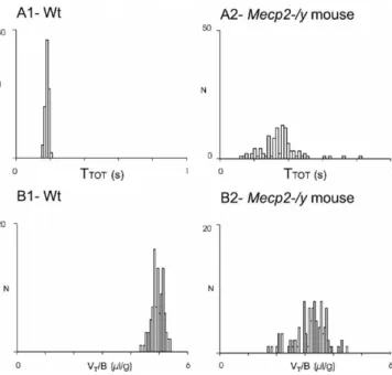
Figure 3. Comparison of breathing parameters in wt and Mecp2-/y adult mice. Distribution of TTOTand VT/B values recorded in unanesthetized paired wt and Mecp2-/y mice from the same
litter (recording performed the same day). Frequency histograms represent the number of occurrences (ordinate) of TTOT(A1, A2, abscissa) and VT/B (B1, B2, abscissa) values on 100
consecutive respiratory cycles during quiet breathing in the wt mouse (A1, B1) and the
tion of the RRG network from these peripheral inputs by using a
transverse brainstem slice preparation. This slice contains neurons
of the VRG (Fig. 5A) that are thought to form a network that
gener-ates the basic respiratory rhythm. Spontaneously generated bursts of
population activity recorded from this region in vitro have been
shown to correspond to in vivo inspiratory activity and are thus
termed fictive inspiration (Telgkamp and Ramirez, 1999).
Abnormal respiratory rhythm in brainstem slices from
Mecp2-/y mice
The in vitro generation of rhythmic VRG bursts was compared
between slices from 14 wt and 9 Mecp2-/y mice (P14 –P21)
(i.e., an age when plethysmographic recordings did not yet
reveal drastic breathing alterations in vivo). Neither the shape
of the VRG burst, not the mean duration of VRG bursts
(0.42
⫾ 0.02 and 0.43 ⫾ 0.03 s for wt and Mecp2-/y mice), nor
their mean frequency (0.22
⫾ 0.02 and 0.29 ⫾ 0.05 Hz for wt
and Mecp2-/y mice) was statistically different. However, the
cycle period of VRG bursts was highly irregular in Mecp2-/y
slices when compared with wt slices (Fig. 5B1,B2) as illustrated
by sequential cycle period scatter plots (Fig. 5C1,C2). The
irregularity of the cycle period was quantified by calculating
both the CVd and the IS. Both values were approximately
twofold higher in Mecp2-/y (0.56
⫾ 0.07 and 54 ⫾ 6 for CVd
and IS, respectively) than in wt slices (0.27
⫾ 0.03 and 28 ⫾ 2
for CVd and IS, respectively). In addition, in five paired slices
from wt and Mecp2-/y littermates, the Moses rank-like test for
scale differences confirmed that the distribution of the VRG
cycle period was less dispersed in wt (Fig. 5D1) than in
Mecp2-/y slices (Fig. 5D2), and the Edgington procedure
re-vealed a significant difference between the two populations.
NE stabilizes the respiratory rhythm in brainstem slices of
Mecp2-/y mice
Because NE is known to play a role in respiratory rhythm
regula-tion (Hilaire et al., 2004; Viemari et al., 2004, 2005), we examined
whether the irregularity of the cycle period in slices from
Mecp2-/y mice may result from a disruption of noradrenergic
mechanisms. Exogenously applied NE significantly increased the
VRG burst frequency in both wt and Mecp2-/y slices by 79
⫾ 17%
(n
⫽ 5) (Fig. 6A) and 123 ⫾ 53% (n ⫽ 5) (Fig. 6B), respectively.
In the presence of NE, the mean VRG burst frequency did not
differ between wt and Mecp2-/y slices (0.39
⫾ 0.01 and 0.49 ⫾
0.11 Hz, respectively). Moreover, NE application eliminated the
rhythm irregularity in all examined slices from Mecp2-/y mice
(Fig. 6 B2,C2,C3), halving the CVd and IS values (0.29
⫾ 0.1 and
31
⫾ 1, n ⫽ 5, respectively). In contrast, NE application in slices
from wt mice had no effect on CVd and IS values. Thus, in the
presence of NE, slices from wt and Mecp2-/y mice produced
rhythmic VRG bursts with a statistically similar frequency and
regularity. Furthermore, the cycle period of VRG bursts
re-mained regular in Mecp2-/y mice for at least 5–10 min after
wash-out of NE (0.33
⫾ 0.04 and 33 ⫾ 3, n ⫽ 3, for CVd and IS,
respectively), whereas the VRG burst frequency returned to
con-trol values (0.18
⫾ 0.03 Hz, n ⫽ 3).
Thus, these in vitro results in slices from wt and Mecp2-/y mice
reveal that Mecp2 deficiency alters the cycle period stability and
that application of exogenous NE restores the cycle period
stability.
NE alterations in Mecp2-/y mice
We examined whether Mecp2-/y adult and young mice have a
deficiency in NE systems that could contribute to their breathing
disturbances.
Two-month-old mice
We assessed the endogenous concentrations of NE, 5-HT,
5HIAA, and dopamine using HPLC analysis of the medulla, pons,
and forebrain of 2-month-old wt (n
⫽ 12) and Mecp2-/y mice
(n
⫽ 7), which had severe respiratory problems similar to those
illustrated in Figure 1, C and D. In the medulla, NE and 5-HT
concentrations were significantly lower in Mecp2-/y than in wt
mice (7.0
⫾ 4.3 vs 13.5 ⫾ 2.3 n
M/mg for NE and 6.4
⫾ 2.6 vs
10.1
⫾ 1.6 n
M/mg for 5-HT) (see Table 1). Despite the decreased
5-HT concentration in Mecp2-/y mice, the 5HIAA
concentra-tions were not significantly different in wt and Mecp2-/y mice
(8.7
⫾ 0.8 vs 8.5 ⫾ 0.7 n
M/mg, respectively), leading to a
signif-icantly increased 5HIAA/5-HT ratio in Mecp2-/y mice. In
con-trast, the dopamine concentration was not different. In the pons
and forebrain, NE, dopamine, and 5-HT concentrations did not
differ between wt and Mecp2-/y mice (data not shown).
We immunolabeled medullary TH neurons in five paired wt
and Mecp2-/y littermates at 2 months of age. TH neurons were
found in the ventral A1/C1 (Fig. 7D) and dorsal A2/C2 (Fig.
7 B, B’) catecholaminergic groups of wt and Mecp2-/y mice.
Neu-ron counts revealed a significantly reduced number of TH
neu-rons in Mecp2-/y mice. The number of TH neuneu-rons was
signifi-cantly reduced in the A1/C1 group of Mecp2-/y mice (302
⫾ 70
and 211
⫾ 48 neurons for wt and Mecp2-/y mice, respectively). In
the entire A2/C2 group (from 2 mm caudal to 2 mm rostral to the
obex), the number of TH neurons was also significantly reduced
in Mecp2-/y mice (325
⫾ 19 and 204 ⫾ 9 TH neurons for wt and
Mecp2-/y mice, respectively). The number of immunolabeled
neurons expressing 5-HT in the median B1-B2 groups and the
lateral B3 group was not significantly different between wt and
Figure 4. Slight anesthesia stabilizes the breathing parameters of Mecp2-/y adult mice. A, Plethysmographic recordings obtained from the same Mecp2-/y mouse when unanesthetized (A1) and anesthetized (A2). B, C, Frequency histograms present the number of occurrences of
TTOT(B1, B2) and VT/B (C1, C2) values on 100 consecutive respiratory cycles during awake (B1,
C1) and anesthetized (B2, C2) conditions in the same Mecp2-/y mouse.
Mecp2-/y mice (244
⫾ 26 and 228 ⫾ 26,
respectively) (Fig. 7C). The number of
im-munolabeled neurons expressing ChAT in
the X and XII motor nuclei (Fig. 7A) was
not significantly different between wt and
Mecp2-/y mice (235
⫾ 62 and 213 ⫾ 90 in
the X motor nucleus and 280
⫾ 63 and
295
⫾ 79 in the XII motor nucleus, for wt
and Mecp2-/y mice, respectively).
One-month-old mice and neonatal mice
We assessed the endogenous
concentra-tions of endogenous bioamines by HPLC
analysis of eight wt and seven Mecp2-/y
mice at 1 month of age. At this early age,
these Mecp2-/y mice presented no (n
⫽ 2)
or only minor alterations (n
⫽ 5) of the
respiratory rhythm stability and few
ap-neas (three to six apap-neas lasting
⬎1 s
dur-ing 15 min recorddur-ing sessions). However,
NE concentrations were already
signifi-cantly lower in Mecp2-/y mice than in wt
mice (40%). This reduction was observed
in all five studied Mecp2-/y mice (i.e., three
with slight breathing alterations and two
with unaltered breathing). In contrast,
medullary 5-HT and medullary dopamine
concentrations were normal in all
exam-ined mice (Table 1). At 1 month of age,
neuron counts in two paired wt and
Mecp2-/y young littermates revealed that
the number of TH neurons in the dorsal
A2/C2 area of the Mecp2-/y mice was
⬃60% of the wt values (345 vs 185 and 393
vs 269 neurons for the two pairs of wt and
Mecp2-/y mice, respectively). In the
ven-tral A1/C1 area, the Mecp2-/y cell count
was low in the Mecp2-/y mouse from one
pair (236 vs 144) but within the same range
in the Mecp2-/y mouse and the wt mouse
from the other pair (280 vs 260).
Finally, we counted the number of TH
neurons in the medulla of three paired wt
and Mecp2-/y neonates (3 d of age) that
displayed a normal breathing. Although a
larger number of TH neurons was found
in neonates than in adults, no significant
differences were observed between wt and
Mecp2-/y
neonates
(A1/C1
neurons,
414
⫾ 63 vs 382 ⫾ 13 neurons for wt and
Mecp2-/y neonates; A2/C2 neurons, 485
⫾ 61 vs 458 ⫾ 27
neu-rons for wt and Mecp2-/y neonates).
These findings suggest that Mecp2 deficiency induces a
spe-cific reduction of the medullary NE contents in adult mice
con-comitant with a reduction of the number of medullary TH
neu-rons that occurs postnatally around the first month of age and
precedes the occurrence of drastic breathing alterations.
Discussion
Here, we show that Mecp2 deficiency in mice leads to slow,
irreg-ular respiratory rhythmic activity, a reduction in medullary NE
and 5-HT content, and a decrease in the number of medullary TH
neurons. We hypothesize that breathing disturbances in
Mecp2-/y mice, and possibly also in Rett patients, involve a
dis-turbance in the bioaminergic modulation of the respiratory
sys-tem. The neurological disorders associated with Rett syndrome
appear after 6 –18 months of age and are caused by mutations in
the MECP2 gene (Van den Veyver and Zoghbi, 2000; Ravn et al.,
2005). However, it remains unknown how a mutation in the
MECP2 gene leads to the observed neurological phenotype.
Among the hypotheses proposed to explain the mechanisms that
lead to the neurological symptoms of Rett syndrome, several
sug-gest an alteration in bioaminergic function. However, existing
evidence is not conclusive (Nomura et al., 1985; Zoghbi et al.,
1985, 1989; Riederer et al., 1986; Lekman et al., 1990; Nielsen et
al., 1990; Segawa, 1997; Dunn, 2001).
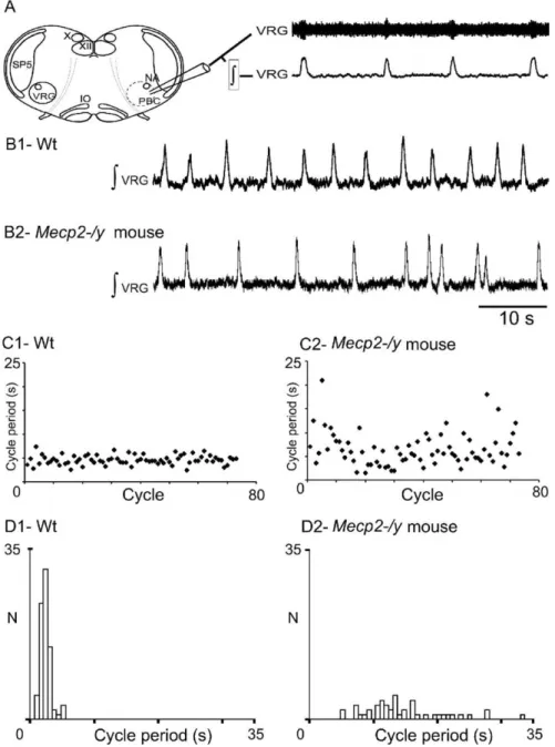
Figure 5. Central respiratory rhythm is disturbed in young Mecp2-/y mice. A, Schematic of a transverse brainstem slice that spontaneously generates respiratory rhythmic activity. Top trace, Extracellular population activity recorded from the VRG. Bottom trace, Integrated activity obtained from the extracellular population activity. X, Vagus nuclei; XII, hypoglossal nuclei; PBC, pre-Bo¨tzinger complex area; SP5, spinal trigeminal nucleus; VRG, ventral respiratory group; IO, inferior olive. B1, Integrated regular respiratory activity from a wt mouse. B2, Mecp2-/y mouse with an irregular respiratory rhythm. C, D, Sequential plot (C) and frequency histograms (D) of cycle period (s) obtained for 80 consecutive VRG bursts recorded in brainstem slices from wt (C1, D1) and Mecp2-/y (C2, D2) mice. Note the cycle period variability in the Mecp2-/y mouse.
Our data suggest that Mecp2 deletion produced altered
bio-aminergic function of Mecp2-/y mice during the postnatal
pe-riod. The time courses of these postnatal changes in bioaminergic
function were diverse and differed for different portions of the
bioaminergic system. In Mecp2-/y mice, an initial decrease in NE
content at 1 month of age was followed by a secondary decrease in
5-HT content at 2 months of age. These changes in NE and 5-HT
content were specific to the medulla, whereas bioaminergic
con-tent in pons and forebrain remained normal. Our HPLC results
confirm HPLC measurements performed in the whole brain of
Mecp2-/y mice (Ide et al., 2005). Ide et al. (2005) found no
bioaminergic alteration at birth in Mecp2-/y mice, and NE
con-tent tended to be reduced at P14. Significant reduction in NE
content was measured at P28, and a significant reduction in 5-HT
content was measured at P42 (Ide et al., 2005).
Our immunohistological approach shows that the number of
TH neurons in Mecp2-/y mice was normal at birth. At 1 month of
age, the number of A2/C2 neurons was decreased, and at 2
months of age, the number of A1/C1 as well as A2/C2 neurons
was decreased in Mecp2-/y mice. Although counts of TH neurons
cannot discriminate between NE or
epi-nephrine neurons located within A1/C1
and A2/C2, HPLC measurements in
Mecp2-/y mice indicate a reduction in NE
content. The mechanisms through which
Mecp2 deficiency results in a progressive
neuronal death of TH neurons and/or a
loss of the ability to synthesize NE remain
unknown and were not addressed in the
present study.
Here, we demonstrate that the decrease
in 5-HT content measured with the HPLC
approach was not accompanied by a
change in the number of 5-HT neurons in
the regions we studied. Our finding that
the 5-HT content is decreased without a
measurable change in the number of 5-HT
neurons may be surprising. Our HPLC
measurements and those performed by Ide
et al. (2005) also revealed that the decrease
in 5-HT concentration was not
accompa-nied by a decreased 5HIAA concentration.
We measured a significantly increased
5HIAA/5-HT ratio in Mecp2-/y mice that
suggests changes in their 5-HT turnover. It
is well established that raphe 5-HT
neu-rons receive numerous inputs from most
NE groups (Peyron et al., 1996). NE has
been shown to regulate central 5-HT
activ-ity at the cell body level via stimulatory
adrenoceptors (Svensson et al., 1975; Baraban and Aghajanian,
1980, 1981; Hopwood and Stamford, 2001; Bortolozzi and
Arti-gas, 2003; Pudovkina et al., 2003; Linner et al., 2004). Based on
these published observations, it can be assumed that NE inputs
modulate 5-HT metabolism. One parsimonious explanation for
the decreased 5-HT content is that the progressive loss of A1/C1
and A2/C2 neurons and the reduced NE content caused a
pro-gressive reduction of NE inputs to 5-HT neurons. The initial
effects on the NE modulatory system may not be sufficient to
significantly affect the serotonergic system. The 5-HT effects may
become only apparent at a time when the NE effects are fully
manifested. A decreased activation of 5-HT neurons could
indi-rectly alter 5-HT metabolism, thereby increasing the 5HIIAA/
5-HT ratio.
Although the breathing of unanesthetized Mecp2-/y mice was
normal at birth, obvious breathing irregularities were displayed
at 1 month of age. These breathing irregularities were
character-ized by consecutive short and long respiratory cycles and
recur-rent respiratory arrests. NE deficits in Mecp2-/y mice may
pre-cede the measurable breathing disturbances in vivo. In vitro
recordings in medullary P14 –P21 slices obtained from Mecp2-/y
mice revealed an increased variability in respiratory cycle
dura-tion that was abolished by exogenous NE applicadura-tion. Although
pontine mechanisms contribute to normal breathing (St-John
and Paton, 2004), compelling evidence suggests that the medulla
is critical for generating normal breathing in vivo as well as
respiratory-like rhythm in vitro. Lesioning of the pre-Bo¨tzinger
complex, a medullary region crucial for respiratory
rhythmogen-esis, severely disrupts breathing in vivo (Ramirez et al., 1998; Gray
et al., 2001; Solomon 2002, 2003; Wenninger et al., 2004a,b) and
fictive respiration in vitro (Onimaru et al., 1988; Viemari et al.,
2003). Thus, the irregular respiratory rhythm measured in P14 –
P21 Mecp2-/y slices suggests that central respiratory deficits in
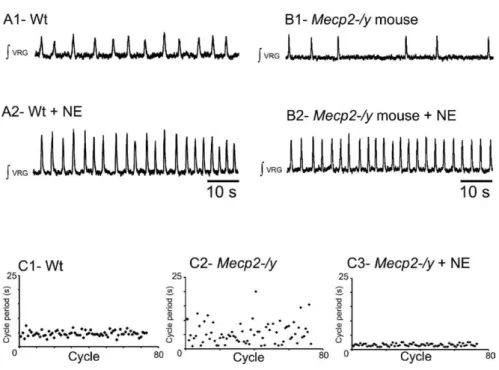
Figure 6. Effect of NE application on VRG rhythm produced in slices from wt and Mecp2-/y mice. A1, Integrated population activity from a wt slice preparation before application of NE. A2, In the same animal, exogenous application of NE (20M) increases the respiratory rhythmic frequency. B1, Integrated population activity from a Mecp2-/y mouse before application of NE.
B2, Exogenously applied NE (20M) significantly increases the regularity and frequency of respiratory rhythmic activity. C, Examples of sequential cycle period histograms for a wt (C1), a Mecp2-/y mouse before NE application (C2), and the same
Mecp2-/y mouse in the presence of NE (C3). Each histogram represents cycle period values (ordinate; seconds) calculated for 80
consecutive cycles (abscissa). Note that the cycles period values become more regular after adding NE.
Table 1. Mecp2 deficiency alters NE systems in the medulla
wt p Mecp2-/y 2 months NE 13.5⫾ 2.3 (n ⫽ 12) * 7.0⫾ 4.3 (n ⫽ 7) 5-HT 10.1⫾ 1.6 (n ⫽ 12) * 6.4⫾ 2.6 (n ⫽ 7) DA 0.6⫾ 0.2 (n ⫽ 12) ns 0.5⫾ 0.2 (n ⫽ 7) 1 month NE 15.0⫾ 1.6 (n ⫽ 6) * 9.2⫾ 0.8 (n ⫽ 5) 5-HT 10.9⫾ 1.8 (n ⫽ 6) ns 8.1⫾ 1.5 (n ⫽ 5) DA 0.8⫾ 0.2 (n ⫽ 6) ns 0.6⫾ 0.3 (n ⫽ 5)
The results are expressed as medians⫾ quartile deviation in nM/mg. Asterisks and ns indicate significant and nonsignificant differences, respectively, between wt and Mecp2-/y mice of the same age. DA, Dopamine.
Mecp2-/y mice may arise early during
postnatal development. The slices
ob-tained from Mecp2-/y mice contain
med-ullary nuclei that are strongly affected by
the Mecp2 deficiency, the A1/C1 and
A2/C2 regions. In wt mice, endogenous
NE modulates the respiratory rhythm
gen-erator via A5 and A6 pontine neurons
(Dawid-Milner et al., 2001; Viemari et al.,
2003, 2004; Hilaire et al., 2004), as well as
via A1/C1 and A2/C2 medullary neurons
(Zanella et al., 2005). Although pontine
NE content was normal in Mecp2-/y mice,
the number of medullary TH neurons and
the medullary NE content was
signifi-cantly reduced. Because the number of TH
neurons and the medullary content were
reduced, we hypothesize that the variable
respiratory rhythm in P14 –P21 Mecp2-/y
medullary slices is caused by the
progres-sive loss of A2/C2 TH neurons.
At this early age, plethysmography
re-cordings obtained from Mecp2-/y mice
showed an apparently normal breathing
pattern in vivo, although some pups
occa-sionally displayed apneic episodes
inter-leaved with respiratory cycles of variable
period. The finding that the in vitro
respi-ratory network of Mecp2-/y mice shows
ir-regularities, whereas the in vivo breathing
of Mecp2-/y mice is apparently normal,
which could be attributable to differences
in the experimental approach. In the in
vitro preparation, we measured the activity
of the deafferented RRG network, which
receives only medullary inputs from
ner-vous structures preserved within the slice.
In contrast, in the intact animal, we
mea-sured the breathing behavior in
experi-mental conditions in which the RRG
net-work received multiple inputs from many
central (including neocortex, cerebellum,
and pons) and peripheral (carotid bodies,
lung afferents) structures. The breathing is
also determined by various motor outputs
(e.g., diaphragm, intercostal, and upper
airway muscles). These inputs and outputs
could potentially compensate for an early loss in medullary
A2/C2 TH neurons, leading to an apparently normal breathing in
young Mecp2-/y mice.
After the first month of age when both medullary NE
concen-tration and number of A2/C2 TH neurons were decreased in
Mecp2-/y mice, these mice exhibited abnormal variability in
breathing period. At
⬃2 months of age, the breathing deficits
in Mecp2-/y mice worsen, being then characterized by very
quent long-lasting apneas and drastically reduced respiratory
fre-quency. At this age, bioaminergic deficits in Mecp2-/y mice were
not only limited to a reduction in the number of A2/C2 TH
neurons but included also abnormalities in the number of A1/C1
TH neurons and the 5-HT medullary system. Early disturbances
in respiratory frequency may originate, at least in part, from a
decreased facilitation of the RRG network by the A1/C1 group.
Neurons in the A1/C1 group continuously activate the RRG
net-work via medullary
␣2-adrenoceptors (Zanella et al., 2005). The
worsening of the breathing irregularities in Mecp2-/y mice could
be explained by the disturbances in the 5-HT system. The 5-HT
system in mammals is known to be critical for stable breathing
and for normal respiratory rhythm generation (Morin et al.,
1991; Lalley et al., 1995; Bou-Flores et al., 2000; Pen
˜a and
Ramirez, 2002; Richter et al., 2003; Ladewig et al., 2004). Because
of the importance of 5-HT for breathing, the decreased 5-HT
levels that we observed in Mecp2-/y mice may contribute to the
worsening of the respiratory disturbances. The worsening of the
breathing disturbances in Mecp2-/y mice occurred at a time when
the 5-HT system became disturbed. This suggests that the time
course of NE and 5-HT deficiencies in Mecp2-/y mice could
pos-sibly explain the age-dependent worsening of respiratory deficits.
Specifically, we hypothesize for Mecp2-/y mice the following time
course of events: initial alterations occur in rhythmogenesis at
Figure 7. Mecp2 deficiency reduces the number of TH neurons in the dorsal A2/C2 and ventral A1/C1 groups. A–D, Schematic
representation of a medullary slice of adult wt mice and enlargements showing neurons of the X and XII motor nuclei that express the synthesis enzyme choline acetyltransferase (A), 5-HT neurons (C), and TH neurons of the dorsal A2/C2 (B, B’) and ventral A1/C1 (D) groups. Mecp2-/y mice present a significant decrease in TH neurons in A1/C1 (data not shown) and A2/C2 cell group (B, B’). Amb, Ambiguus nucleus; Ap, area postrema; Cu, cuneate nucleus; LRt, lateral reticular nucleus; Sp5, spinal trigeminal nucleus; X, dorsal vagal motor nucleus; XII, hypoglossal motor nucleus; ECu, external cuneate nucleus; MdD, medullary reticular nucleus, dorsal; LRt, lateral reticular nucleus; IRt, intermediate reticular nucleus; Ro, nucleus of Roller; MdV, medullary reticular nucleus, ventral; IOD, inferior olive, dorsal nucleus; IOPr, inferior olive, principal nucleus; ln, intercalated nucleus of medulla; ROb, raphe obscurus nucleus; IOK, inferior olive, cap of Kooy of medial nucleus; IOVL, inferior olive, ventrolateral; IOC, inferior olive, subnucleus C of medial nucleus; IOB, inferior olive, subnucleus B of medial nucleus; PMn, paramedian lobule; RPa, raphe pallidus nucleus; py, pyramidal tract; vsc, ventral spinocerebellar tract; icp, inferior cerebellar peduncle; SolDM, nucleus of solitary tract, dorsomedial; SolM, nucleus of solitary tract, medial; SolVL, nucleus of the solitary tract, ventrolateral.
P14 –P21, a time when NE deficits first appear. Alterations in
rhythmogenesis are followed by a variable breathing rhythm with
recurrent apneas at 1 month of age at a time when both NE
content and A2/C2 TH neurons are significantly affected. Finally,
long-lasting apneas and a severe reduction in breathing
fre-quency occur at 2 months of age, which coincides with the full
manifestation of A2/C2, A1/C1, and 5-HT deficits.
Phox2a and Ret deficiencies affect the prenatal maturation of
the RRG (Hilaire et al., 2004; Viemari et al., 2004, 2005). In
con-trast, the RRG network of Mecp2-/y mice undergoes normal
mat-uration resulting in the production of normal respiratory
move-ments at birth. Thereafter, as bioaminergic deficits progress
during the postnatal period, the respiratory system of Mecp2-/y
mice begins to dysfunction. However, the respiratory system of
Mecp2-/y mice is still able to centrally generate a normal
respira-tory rhythm under certain circumstances: (1) adult Mecp2-/y
mice with drastic respiratory alterations can spontaneously
dis-play transient periods of normal breathing; and (2) pentobarbital
anesthesia restores normal breathing in all tested Mecp2-/y mice.
It is worthwhile to note that human Rett patients may similarly
have normal breathing periods during wakefulness and that their
breathing can also improve during sleep. We do not know the
mechanism by which pentobarbital anesthesia stabilizes and
im-proves breathing in adult Mecp2-/y mice. It is known that this
anesthetic depresses cerebral metabolism (Steen and
Michen-felder, 1979) and resting respiration (Erhardt et al., 1984).
Pen-tobarbital anesthesia is also affected by noradrenergic agents
(Dessaigne et al., 1976; Ojima et al., 1995; Dalley et al., 1998), and
other anesthetics have been shown to alter the activity and
excit-ability of central NE neurons (Clement et al., 1998; Hirota and
Kushikata, 2001) and to decrease mRNA levels of NE
transport-ers (Hara et al., 2000).
To conclude, our results are consistent with the notion that
Mecp2 deficiency in mice results in an alteration of the NE system
and that NE deficits precede, and may contribute to, the
devel-opment of respiratory deficits. Subsequent disturbances of other
neuromodulators such as 5-HT may explain the delayed onset
and worsening of the disturbed breathing phenotype.
References
Armstrong DD (1997) Review of Rett syndrome. J Neuropathol Exp Neurol 56:843– 849.
Baraban JM, Aghajanian GK (1980) Suppression of firing activity of 5-HT neurons in the dorsal raphe by alpha-adrenoceptor antagonists. Neuro-pharmacology 19355–19363.
Baraban JM, Aghajanian GK (1981) Noradrenergic innervation of seroto-nergic neurons in the dorsal raphe: demonstration by electron micro-scopic autoradiography. Brain Res 204:1–11.
Bortolozzi A, Artigas F (2003) Control of 5-hydroxytryptamine release in the dorsal raphe nucleus by the noradrenergic system in rat brain. Role of ␣-adrenoceptors. Neuropsychopharmacology 28:421–434.
Bou-Flores C, Lajard AM, Monteau R, De Maeyer E, Seif I, Lanoir J, Hilaire G (2000) Abnormal phrenic motoneuron activity and morphology in neo-natal monoamine oxidase A-deficient transgenic mice: possible role of a serotonin excess. J Neurosci 20:4646 – 4656.
Burnet H, Bevengut M, Chakri F, Bou-Flores C, Coulon P, Gaytan S, Pasaro R, Hilaire G (2001) Altered respiratory activity and respiratory regula-tions in adult monoamine oxidase A-deficient mice. J Neurosci 21:5212–5221.
Clement CI, Keay KA, Bandler R (1998) Medullary catecholaminergic pro-jections to the ventrolateral periaqueductal gray region activated by halo-thane anaesthesia. Neuroscience 86:1273–1284.
Cooper RA, Kerr AM, Amos PM (1998) Rett syndrome: critical examina-tion of clinical features, serial EEG and video-monitoring in understand-ing and management. Eur J Paediatr Neurol 2:127–135.
Dalley JW, Parker CA, Wulfert E, Hudson AL, Nutt DJ (1998) Potentiation
of barbiturate-induced alterations in presynaptic noradrenergic function in rat frontal cortex by imidazol(in)e alpha2-adrenoceptor agonists. Br J Pharmacol 125:441– 446.
Dawid-Milner M, Lara J, Gonzales-Baron S, Spyer K (2001) Respiratory effect of stimulation of cell bodies of the A5 region in the anaesthetised rat. Pflu¨gers Arch 441:434 – 443.
Dessaigne S, Scotto AM, Mercier J (1976) The effect of previous administra-tion of adrenaline or isoprenaline on the activity of several hypnotics in mice. C R Seances Soc Biol Fil 170:1074 –1080.
Dunn HG (2001) Neurons and neuronal systems involved in the patho-physiologies of Rett syndrome. Brain Dev 23 [Suppl 1]:S99 –S100. Dunn HG, MacLeod PM (2001) Rett syndrome: review of biological
abnor-malities. Can J Neurol Sci 28:16 –29.
Elian M, Rudolf ND (1991) EEG and respiration in Rett syndrome. Acta Neurol Scand 83:123–128.
Erhardt W, Hebestedt A, Aschenbrenner G, Pichotka B, Blumel G (1984) A comparative study with various anesthetics in mice (pentobarbitone, ketamine-xylazine, carfentanyl-etomidate). Res Exp Med (Berl) 184:159 –169.
Gray PA, Janczewski WA, Mellen N, McCrimmon DR, Feldman JL (2001) Normal breathing requires preBotzinger complex neurokinin-1 receptor-expressing neurons. Nat Neurosci 4:927–930.
Guy J, Hendrich B, Holmes M, Martin JE, Bird A (2001) A mouse Mecp2-null mutation causes neurological symptoms that mimic Rett syndrome. Nat Genet 27:322–326.
Hagberg B, Aicardi J, Dias K, Ramos O (1983) A progressive syndrome of autism, dementia, ataxia, and loss of purposeful hand use in girls: Rett’s syndrome: report of 35 cases. Ann Neurol 14:471– 479.
Hara K, Yanagihara N, Minami K, Hirano H, Sata T, Shigematsu A, Izumi F (2000) Dual effects of intravenous anesthetics on the function of norepi-nephrine transporters. Anesthesiology 93:1329 –1335.
Hilaire G, Viemari JC, Coulon P, Simmoneau M, Be´vengut M (2004) Mod-ulation of the medullary respiratory rhythm generator by pontine norad-renergic A5 and A6 groups in rodents. Resp Physiol Neurobiol 143:187–197.
Hirota K, Kushikata T (2001) Central noradrenergic neurones and the mechanism of general anaesthesia. Br J Anaesth 87:811– 813.
Hopwood SE, Stamford JA (2001) Noradrenergic modulation of serotonin release in rat dorsal and median raphe nuclei via␣1 and ␣2A adrenocep-tors. Neuropharmacology 41:433– 442.
Ide S, Itoh M, Goto Y (2005) Defect in normal developmental increase of the brain biogenic amine concentrations in the mecp2-null mouse. Neurosci Lett 386:14 –17.
Julu PO (2001) The central autonomic disturbance in Rett syndrome. In: Rett disorder and the developing brain (Kerr AM, Witt-Engerstrom I, eds), pp 131–183. New York; Oxford UP.
Julu PO, Kerr AM, Apartopoulos F, Al-Rawas S, Engerstrom IW, Engerstrom L, Jamal GA, Hansen S (2001) Characterisation of breathing and associ-ated central autonomic dysfunction in the Rett disorder. Arch Dis Child 85:29 –37.
Kerr AM (1992) A review of the respiratory disorder in the Rett syndrome. Brain Dev 14 [Suppl]:S43–S45.
Kerr AM, Julu PO (1999) Recent insights into hyperventilation from the study of Rett syndrome. Arch Dis Child 80:384 –387.
Kerr AM, Armstrong DD, Prescott RJ, Doyle D, Kearney DL (1997) Rett syndrome: analysis of deaths in the Br survey. Eur Child Adolesc Psychi-atry 6 [Suppl 1]:71–74.
Kerr AM, Julu PO, Hansen S, Apartopoulos F (1998) Serotonin and breath-ing dysrhythmia in Rett syndrome. In: New developments in child neu-rology (Perat MV, ed), pp 191–195. Bologna: Monduzzi Editore. Krauth J (1990) Distribution-free statistics, an application oriented
ap-proach. In: Techniques in the behavioral and neural sciences, p 381. Am-sterdam: Elsevier.
Ladewig T, Lalley PM, Keller BU (2004) Serotonergic modulation of intra-cellular calcium dynamics in neonatal hypoglossal motoneurons from mouse. Brain Res 1001:1–12.
Lalley PM, Bishoff AM, Schwarzacher SW, Richter DW (1995) 5-HT2
receptor-controlled modulation of medullary respiratory neurones in the cat. J Physiol (Lond) 487:653– 661.
Lekman A, Witt-Engerstrom I, Holmberg B, Percy A, Svennerholm L, Hag-berg B (1990) CSF and urine biogenic amine metabolites in Rett syn-drome. Clin Genet 37:173–178.
Linner L, Wiker C, Arborelius L, Schalling M, Svensson TH (2004) Selective noradrenaline reuptake inhibition enhances serotonergic neuronal activ-ity and transmitter release in the rat forebrain. J Neural Transm 111:127–139.
Marcus CL, Carroll JL, McColley SA, Loughlin GM, Curtis S, Pyzik P, Naidu S (1994) Polysomnographic characteristics of patients with Rett syn-drome. J Pediatr 125:218 –224.
Morin D, Monteau R, Hilaire G (1991) 5-Hydroxytryptamine modulates central respiratory activity in newborn rat: an in vitro study. Eur J Phar-macol 192:89 –95.
Morton RE, Bonas R, Minford J, Tarrant SC, Ellis RE (1997) Respiration patterns during feeding in Rett syndrome. Dev Med Child Neurol 39:607– 613.
Nielsen JB, Lou HC, Andresen J (1990) Biochemical and clinical effects of tyrosine and tryptophan in the Rett syndrome. Brain Dev 12:143–147. Nomura Y, Segawa M, Higurashi M (1985) Rett syndrome–an early
cate-cholamine and indolamine deficient disorder? Brain Dev 7:334 –341. Ojima K, Matsumoto K, Tohda M, Watanabe H (1995) Hyperactivity of
central noradrenergic and CRF systems is involved in social isolation-induced decrease in pentobarbital sleep. Brain Res 684:87–94.
Onimaru H, Arata A, Homma I (1988) Primary respiratory rhythm gener-ator in the medulla of brainstem-spinal cord preparation from newborn rat. Brain Res 445:314 –324.
Pen˜a F, Ramirez JM (2002) Endogenous activation of serotonin-2A recep-tors is required for respiratory rhythm generation in vitro. J Neurosci 22:11055–11064.
Peyron C, Luppi PH, Fort P, Rampon C, Jouvet M (1996) Lower brainstem catecholamine afferents to the rat dorsal raphe nucleus. J Comp Neurol 364:402– 413.
Pudovkina OL, Cremers TI, Westerink BH (2003) Regulation of the release of serotonin in the dorsal raphe nucleus by␣1 and ␣2 adrenoceptors. Synapse 50:77– 82.
Ramirez JM, Schwarzacher SW, Pierrefiche O, Olivera BM, Richter DW (1998) Selective lesioning of the cat pre-Botzinger complex in vivo elim-inates breathing but not gasping. J Physiol (Lond) 507:895–907. Ravn K, Nielsen JB, Skjeldal OH, Kerr A, Hulten M, Schwartz M (2005)
Large genomic rearrangements in MECP2. Hum Mutat 25:324. Richter DW, Manzke T, Wilken B, Ponimaskin E (2003) Serotonin
recep-tors: guardians of stable breathing. Trends Mol Med 9:542–548. Riederer P, Weismer M, Wichart I, Schmidt B, Killian W, Rett A (1986)
Preliminary brain autopsy findings in progredient Rett syndrome. Am J Med Genet 24:305–315.
Segawa M (1997) Pathophysiology of Rett syndrome from the standpoint of early catecholamine disturbance. Eur Child Adolesc Psychiatry 6 [Suppl 1]:56 – 60.
Siegel S, Catellan NJ (1989) Nonparametric statistics for the behavioral sci-ences, p 399. New York: McGraw-Hill.
Solomon IC (2002) Modulation of expiratory motor output evoked by chemical activation of pre-Botzinger complex in vivo. Respir Physiol Neurobiol 130:235–251.
Solomon IC (2003) Influence of respiratory network drive on phrenic
mo-tor output evoked by activation of cat pre-Botzinger complex. Am J Physiol Regul Integr Comp Physiol 284:R455–R466.
Steen PA, Michenfelder JD (1979) Barbiturate protection in tolerant and nontolerant hypoxic mice: comparison with hypothermic protection. An-esthesiology 50:404 – 408.
St-John WM, Paton JF (2004) Role of pontile mechanisms in the neurogen-esis of eupnea. Respir Physiol Neurobiol 143:321–332.
Svensson TH, Bunney BS, Aghajanian GK (1975) Inhibition of both norad-renergic and serotonergic neurons in brain by the alpha-adnorad-renergic ago-nist clonidine. Brain Res 92:291–306.
Telgkamp P, Ramirez JM (1999) Differential responses of respiratory nuclei to anoxia in rhythmic brain stem slices of mice. J Neurophysiol 82:2163–2170.
Telgkamp P, Cao YQ, Basbaum AI, Ramirez JM (2002) Long-term depriva-tion of substance P in PPT-A mutant mice alters the anoxic response of the isolated respiratory network. J Neurophysiol 88:206 –213.
Tryba AK, Pen˜a F, Ramirez JM (2003) Stabilization of bursting in respira-tory pacemaker neurons. J Neurosci 23:3538 –3546.
Van den Veyver IB, Zoghbi HY (2000) Methyl-CpG-binding protein 2 mu-tations in Rett syndrome. Curr Opin Genet Dev 10:275–279.
Viemari JC, Burnet H, Be´vengut M, Hilaire G (2003) Perinatal maturation of the mouse respiratory rhythm-generator: in vivo and in vitro studies. Eur J Neurosci 17:1233–1244.
Viemari JC, Be´vengut M, Burnet H, Coulon P, Pequignot JM, Tiveron MC, Hilaire G (2004) Phox2a gene, A6 neurons, and noradrenaline are es-sential for development of normal respiratory rhythm in mice. J Neurosci 24:928 –937.
Viemari JC, Maussion G, Be´vengut M, Burnet H, Pequignot JM, Ne´pote V, Pachnis V, Simonneau M, Hilaire G (2005) Ret deficiency in mice im-pairs the development of A5 and A6 neurons and the functional matura-tion of the respiratory rhythm. Eur J Neurosci, in press.
Wenninger JM, Pan LG, Klum L, Leekley T, Bastastic J, Hodges MR, Feroah T, Davis S, Forster HV (2004a) Small reduction of neurokinin-1 receptor-expressing neurons in the pre-Botzinger complex area induces abnormal breathing periods in awake goats. J Appl Physiol 97:1620 –1628. Wenninger JM, Pan LG, Klum L, Leekley T, Bastastic J, Hodges MR, Feroah
TR, Davis S, Forster HV (2004b) Large lesions in the pre-Botzinger complex area eliminate eupneic respiratory rhythm in awake goats. J Appl Physiol 97:1629 –1636.
Woodyatt GC, Murdoch BE (1996) The effect of the presentation of visual and auditory stimuli on the breathing patterns of two girls with Rett syndrome. J Intellect Disabil Res 40:252–259.
Zanella S, Roux JC, Viemari JC, Hilaire G (2005) Possible modulation of the respiratory rhythm generator by A1/C1 neurones. Resp Physiol Neuro-biol, in press.
Zoghbi HY, Percy AK, Glaze DG, Butler IJ, Riccardi VM (1985) Reduction of biogenic amine levels in the Rett syndrome. N Engl J Med 313:921–924. Zoghbi HY, Milstien S, Butler IJ, Smith EO, Kaufman S, Glaze DG, Percy AK (1989) Cerebrospinal fluid biogenic amines and biopterin in Rett syn-drome. Ann Neurol 25:56 – 60.