HAL Id: hal-01813387
https://hal.archives-ouvertes.fr/hal-01813387
Submitted on 18 Jun 2018
HAL is a multi-disciplinary open access
archive for the deposit and dissemination of
sci-entific research documents, whether they are
pub-lished or not. The documents may come from
teaching and research institutions in France or
abroad, or from public or private research centers.
L’archive ouverte pluridisciplinaire HAL, est
destinée au dépôt et à la diffusion de documents
scientifiques de niveau recherche, publiés ou non,
émanant des établissements d’enseignement et de
recherche français ou étrangers, des laboratoires
publics ou privés.
Genomics of Multiple Myeloma
Sébastien Robiou-Du-Pont, Alice Cleynen, Charlotte Fontan, Michel Attal,
Nikhil Munshi, Jill Corre, Herve Avet-Loiseau
To cite this version:
Sébastien Robiou-Du-Pont, Alice Cleynen, Charlotte Fontan, Michel Attal, Nikhil Munshi, et al..
Genomics of Multiple Myeloma. Journal of Clinical Oncology, American Society of Clinical Oncology,
2017, 35 (9), pp.963-967. �10.1200/JCO.2016.70.6705�. �hal-01813387�
J
OURNAL OF
C
LINICAL
O
NCOLOGY
R E V I E W A R T I C L EGenomics of Multiple Myeloma
Sebastien Robiou du Pont, Alice Cleynen, Charlotte Fontan, Michel Attal, Nikhil Munshi, Jill Corre, and Herv´e Avet-Loiseau
A B S T R A C T
Multiple myeloma (MM) is characterized by wide variability in the chromosomal/genetic changes present in tumor plasma cells. Genetically, MM can be divided into two groups according to ploidy and hyperdiploidy versus nonhyperdiploidy. Several studies in gene expression profiling attempted to identify subentities in MM without convincing results. These studies mostly confirmed the cytogenetic data and subclassified patients according to 14q32 translocations and ploidy. More-recent data that are based on whole-exome sequencing have confirmed this heterogeneity and show many gene mutations but without a unifying mutation. These newer studies have shown the frequent alteration of the mitogen-activated protein kinase pathway. The most interesting data have demonstrated subclonality in all patients with MM, including subclonal mutations of supposed driver genesKRAS, NRAS, and BRAF. J Clin Oncol 35. © 2017 by American Society of Clinical Oncology
Multiple myeloma (MM) is characterized by the accumulation (usually) of tumor plasma cells (PCs) within the bone marrow compartment. Clinically, MM is characterized by a wide heterogeneity both for clinical symptoms at diagnosis (bone fractures, anemia, renal failure, and extramedullary localiza-tions) and for outcome (rapid fatal evolution, long-term progression-free survival, or even cure). In the early 2000s, this clinical heterogeneity was believed to be mostly driven by chromosomal abnormalities found in the tumor PCs. On the basis of conven-tional karyotyping (rarely informative because of the low PC proliferative index)1-3or interphase fluorescence in situ hybridization (FISH) experi-ments,4-8high-risk chromosomal changes have been identified, such as the translocations t(4;14) and t(14; 16) and the loss of part of the chromosome 17 short arm (ie, del[17p]). With the use of more high-throughput technologies, such as gene expression profiling (GEP) on microarrays, molecular classifi-cations have been proposed.9-13 However, these classifications have not led to the identification of several MM entities, as described with non-Hodgkin lymphomas. With the advent of next-generation sequencing (NGS) at both the DNA and the RNA levels, the goal of this review is to summarize our current knowledge of the molecular lesions of MM.
GEP/RNA SEQUENCING
The first molecular classification of MM was proposed by Bergsagel et al9from GEP experiments.
Thisfirst report identified eight subgroups that were mainly based on cyclin D gene expression and on the various 14q32 recurrent trans-locations. This molecular classification was re-fined in 2006 and identified seven subclasses of myelomas.12 The first group is defined by the translocation t(4;14), which is identified by overexpression of the MMSET and/or FGFR3 genes. The second group is defined by upregu-lation of one of the MAF genes related to the translocations t(14;16) or t(14;20). Cases with CCND1 or CCND3 upregulation (as a result of the translocations t[11;14] or t[6;14]) cluster into the third and fourth groups CD1 and CD2. The CD2 group is characterized by CD20 expression. Thefifth group is characterized by hyperdiploidy. Thefinal two groups are characterized by a low incidence of bone disease in accordance with low DKK1 expression, but the last group is charac-terized by a high expression of genes involved in proliferation. This molecular classification has been partially confirmed by a study by the HOVON (Haemato Oncology Foundation for Adults in the Netherlands) group.13The low bone disease group was not confirmed. In contrast, three other groups were identified: one enriched by myeloid genes that could be related to PC sorting problems, one characterized by over-expression of cancer testis antigen genes, and one defined by overexpression of positive regulators of the nuclear factor-kB pathway. However, these classifications failed to identify real subentities in MM. Few studies that used RNA sequencing have
Author affiliations and support information (if applicable) appear at the end of this article.
Published atjco.orgon February 13, 2017. Corresponding author: Herv ´e Avet-Loiseau, MD, PhD, Genomics of Myeloma Laboratory, L’Institut Universitaire du Cancer Oncopole, 1 Av Irene Joliot-Curie, 31059 Toulouse, France; e-mail: [email protected]. © 2017 by American Society of Clinical Oncology
0732-183X/17/3599-1/$20.00
been reported in meetings. The role of this technology in replacing GEP in the future is still questionable, and further studies are needed.
GEP has been extensively published with the goal of identi-fying several groups of patients with distinct outcomes. All these studies have identified a high-risk versus standard-risk group12-14
; however, no common gene was found through the detailing of the gene signatures. These high-risk signatures identify different groups of patients (13% to 25%). Whether each high-risk signature identifies the same patients is still an opened question. In routine practice, GEP is rarely used for assessing prognosis mainly because it requires microarray technology and bioinformatics.
DNA COPY NUMBER CHANGES
As with many cancers, MM is characterized by many chromosomal aberrations. Because karyotypes frequently are uninformative, our knowledge of the unbalanced chromosomal changes comes from single nucleotide polymorphism/comparative genomic hybrid-ization array studies.15,16These molecular karyotypes usually are highly complex, with two exceptions. Approximately 10% of pa-tients do not display a detectable abnormality at the chromosomal level.16 The second exception is that patients with the t(11;14) translocation (approximately 15% to 20%) usually display simple karyotypes (unpublished data). In all other cases (approximately 70% of patients), numerous changes are observed. The following two groups of patients are identified: those with gains of odd chromosomes (3, 5, 7, 9, 11, 15, 19, and 21),17 which define hyperdiploidy (observed in approximately one half of patients), and those with many structural changes (gains and losses), which define pseudo- or hypodiploidy (approximately 20% of patients). The latter group is frequently characterized by 14q32 trans-locations that target the IGH gene and several partners, mainly FGFR3/MMSET on chromosome 418,19or, less frequently, MAF,20,21 MAFB,22or CCND323on chromosomes 16, 20, and 6, respectively. Among the recurrent unbalanced changes observed on molecular karyotypes, the most frequent changes are 1q gains and losses at 1p, 6q, 8p, 13q, 14q, 16q, and 17p.15,16
With regard to prognosis, many of these changes have been associated with specific outcomes. Hyperdiploidy usually is as-sociated with longer survival, even though the situation is probably more complex than the simple number of chromosomes. Some chromosomes, such as 3 and 5, display a good outcome, whereas trisomy 21 worsens the prognosis.24More high-risk abnormalities have been identified, such as t(4;14),8
t(14;16), del(17p),8 del (1p32),25and 1q gains.26The molecular targets of these losses and gains are mostly unknown. For del(17p), the minimal deleted region includes the TP53 gene; however, this gene is not mutated on the remaining allele in all cases,27and other genes in its vicinity might be important for prognosis.28For del(1p32), the minimal deleted region targets two genes, FAF1 and CDKN2C (unpublished data). The real target gene that affects prognosis is still unknown. For 1q gains, the large majority of cases are gains of the whole long arm, which prevents the identification of specific target genes. In routine practice, these abnormalities mainly are analyzed by in-terphase FISH on sorted PCs by using plus or minus complete panels (Table 1). A recent meta-analysis combined FISH with the
International Staging System to more precisely define prognosis (revised International Staging System).29 Whether FISH will be supplied in the future by single nucleotide polymorphism array or NGS is unknown.
DNA SEQUENCING
Most of the DNA sequencing reports have focused on the tran-scribed genome (ie, whole-exome sequencing on sorted tumor PCs).30-33Several hundreds of primary tumors have been exome sequenced. In contrast to hematologic tumors, such as hairy cell leukemia34or Waldenstr¨om macroglobulinemia35 that are char-acterized by a single unifying mutation (targeting BRAF and MYD88, respectively), no such unique mutation has been found in MM. The median number of mutations per transcribed genome is approximately 60.30Compared with other tumors, MM is in the
middle of the landscape, with more mutations than other he-matologic malignancies, such as leukemias, but with much fewer than carcinogen-induced tumors, such as melanoma and lung cancers.36A heterogeneous mutational landscape was observed in all the studies.29-32,37The most frequently mutated genes are KRAS
and NRAS (approximately 20% each) followed by TP53, DIS3, FAM46C, and BRAF, which are all mutated in approximately 10% of patients. All other mutations were observed in, 5% of patients. Of note, some patients present two or more mutations in genes involved in the same pathway (eg, KRAS, NRAS, or BRAF in the MAPK pathway). This redundancy is surprising and has not been reported in other tumors, and its biologic significance is unknown. In some patients, these apparently redundant mutations are not subclonal but are observed at the clonal level.31Furthermore, some of these driver mutations are lowly expressed at the RNA level, which asks again about the significance of these mutations in the biology of the disease.38
Most of the published DNA sequencing studies addressed the issue of the mechanisms that support the mutational ma-chinery. All the studies identified several mutational signatures, particularly deamination of methylated cytosines; kataegis; apolipoprotein B mRNA editing enzyme, catalytic polypeptide (APOBEC), signature; and somatic hypermutations driven by the activation-induced deaminase enzyme. Deamination of methylated cytosines leads to C.T transitions at CpG sites and is a rather generic mutational process observed in many cancers. The APOBEC signature is characterized by C.T, C.G, and C.A mutations at TpC sites, is driven by a family of APOBEC
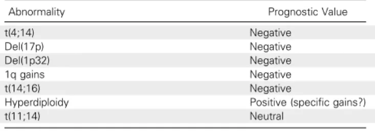
Table 1. Prognostic Value of the Main Chromosomal Abnormalities of Multiple Myeloma
Abnormality Prognostic Value
t(4;14) Negative
Del(17p) Negative Del(1p32) Negative 1q gains Negative t(14;16) Negative
Hyperdiploidy Positive (specific gains?) t(11;14) Neutral
2 © 2017 by American Society of Clinical Oncology JOURNAL OFCLINICALONCOLOGY
enzymes, and is mostly found in cases with MAF/MAFB translocations. Finally, activation-induced deaminase–driven mutations are observed in several genes involved in immuno-globulin translocations, such as MYC or CCND1, as previously described.33
The role of these mutations in patient outcome is still questionable. The analysis of large cohorts of homogeneously treated patients failed to relate recurrent mutations such as KRAS, NRAS, or BRAF to specific survival rates.36
The only recurrent mutations that significantly affect survival are those observed in TP53, which is not novel. Larger cohorts might be needed, but specific mutations unlikely will affect survival given the very low frequency of these mutations. Future studies in whole-genome sequencing may provide new insights into the broad mutational landscape. Preliminary data have revealed a large set of mutations (in the 5,000 to 10,000 range) that mainly affect the nontranscribed genome; that may target microRNA, small nucleolar RNA, or long noncoding RNA; and that could modify prognosis. The large majority of these mu-tations probably are passenger mumu-tations, with a few of them being drivers. With the objective of successful targeted therapy in the future, the determination of which mutations are really drivers versus passengers will be important. These studies also will detect all the translocations; some of them might be re-current, as preliminary RNA sequencing studies of fusion genes have shown (unpublished data).
SUBCLONALITY
One of the most striking results of the sequencing studies is the description of oligoclonality in MM. Even though the disease is characterized by the secretion of a unique monoclonal protein in the majority of patients, a degree of heterogeneity is ob-served at the molecular level, which suggests a Darwinian evolution of MM.29-32,39-41 In exome sequencing studies, all the mutations were not necessarily present in all the tumor PCs. This subclonal status not only is present at the single base level but also is observed (even less frequently) at the copy number level. This heterogeneity is observed as soon as the monoclonal gammopathy of unknown significance stage (a premalignant state), meaning that immortalized PCs diverge very early in their evolution.42A tumor at diagnosis contains an estimatedfive to six subclones. This number could be much larger as a result of more-sensitive sequencing approaches. Of note, this subclonal process is not limited to supposed pas-senger mutations but is observed in supposed driver muta-tions, such KRAS, NRAS, and BRAF. This means that the mutational process is dynamic and that some mutations of supposed driver genes might occur late in the evolution of the disease.
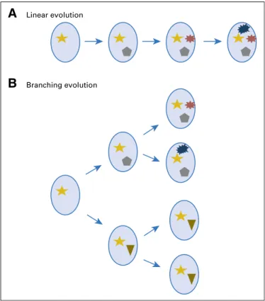
This oligoclonality leads to various types of evolution of the disease over time (Fig 1). The sequencing of tumors from a same patient at different time points (eg, diagnosis, relapse) showed two types of evolution.30 The first one is a linear evolution, which means that the major clone observed on the first time point is still present on the second one and eventually acquires novel mutations. The second one is a branching evolution,
which means that the major clone observed at the second time point is different from the first one, even though this major clone was already present early but as a minor subclone. The linear evolution suggests that all the subclones observed at diagnosis, for instance, responded to therapy but repopulated the tumor at the same speed and led to relapse. In contrast, the branching evolution suggests a different kinetics of repopula-tion or a different sensitivity to the chosen therapy. Whether these two modes of evolution are related to the therapeutic pressure or to the natural history of each patient is still an unresolved issue.
Finally, this oligoclonality raises the question of the devel-opment of targeted therapies in MM because it is currently used in many solid tumors. Some of the mutations recurrently observed in MM are drugable. For instance, the V600E BRAF mutation can be targeted with BRAF inhibitors. In the same way, RAS mutations might be targeted by MEK inhibitors. This ap-proach could be highly successful as shown in a case of a harbored V600E BRAF mutation.43 However, this spectacular result sup-poses that the drugable mutation is fully clonal. It is easy to speculate that if this mutation is subclonal (eg, in 50% of the PCs), the response will be limited to the PCs that present the mutation but will be totally inefficient in the 50% of the PCs that lack the mutation. Currently, therapeutic approaches in MM are mainly based on broadly active drugs (proteasome in-hibitors, immunomodulatory drugs, antibodies, HDAC inhibi-tors), but if physicians want to take advantage of the sequencing efforts for targeted therapeutic approaches, the detection of
Linear evolution
A
Branching evolution
B
Fig 1. Myeloma is characterized by two types of subclonal evolution: (A) a linear one with acquisition of novel mutations over time in the clone and (B) a branching evolution where subclones diverge with subclonal acquisition of novel mutations.
mutations and the clonal or subclonal status of these mutations are important.
CONCLUSION
In the past 2 years, major studies have addressed the issue of the molecular landscape of MM by using NGS. If these studies have markedly improved our knowledge of the biology of the disease, they have failed (so far) to translate this knowledge into practical clinical application. We have no doubt that in the next 3 to 5 years, with more patients, and with more powerful technologies (whole-genome se-quencing, RNA sese-quencing, targeted sequencing panels), sequencing will be a new tool in the routine evaluation of patients that could lead to significant improvements in patient management.
AUTHORS’ DISCLOSURES OF POTENTIAL CONFLICTS OF INTEREST
Disclosures provided by the authors are available with this article at
jco.org.
AUTHOR CONTRIBUTIONS
Conception and design: Nikhil Munshi, Jill Corre, Herv´e Avet-Loiseau Data analysis and interpretation: Sebastien Robiou du Pont, Alice Cleynen, Charlotte Fontan, Michel Attal, Nikhil Munshi, Jill Corre Manuscript writing: All authors
Final approval of manuscript: All authors
Accountable for all aspects of the work: All authors
REFERENCES
1. La¨ı JL, Zandecki M, Mary JY, et al: Improved cytogenetics in multiple myeloma: A study of 151 patients including 117 patients at diagnosis. Blood 85:2490-2497, 1995
2. Smadja NV, Bastard C, Brigaudeau C, et al: Hypodiploidy is a major prognostic factor in multiple myeloma. Blood 98:2229-2238, 2001
3. Calasanz MJ, Cigudosa JC, Odero MD, et al: Cytogenetic analysis of 280 patients with multiple myeloma and related disorders: Primary breakpoints and clinical correlations. Genes Chromosomes Cancer 18:84-93, 1997
4. Nishida K, Tamura A, Nakazawa N, et al: The Ig heavy chain gene is frequently involved in chromo-somal translocations in multiple myeloma and plasma cell leukemia as detected by in situ hybrid-ization. Blood 90:526-534, 1997
5. Avet-Loiseau H, Li JY, Facon T, et al: High incidence of translocations t(11;14)(q13;q32) and t(4; 14)(p16;q32) in patients with plasma cell malignan-cies. Cancer Res 58:5640-5645, 1998
6. Fonseca R, Debes-Marun CS, Picken EB, et al: The recurrent IgH translocations are highly associ-ated with nonhyperdiploid variant multiple myeloma. Blood 102:2562-2567, 2003
7. Drach J, Ackermann J, Fritz E, et al: Presence of a p53 gene deletion in patients with multiple my-eloma predicts for short survival after conventional-dose chemotherapy. Blood 92:802-809, 1998
8. Avet-Loiseau H, Attal M, Moreau P, et al: Genetic abnormalities and survival in multiple mye-loma: The experience of the Intergroupe Franco-phone du My ´elome. Blood 109:3489-3495, 2007
9. Bergsagel PL, Kuehl WM, Zhan F, et al: Cyclin D dysregulation: An early and unifying pathogenic event in multiple myeloma. Blood 106:296-303, 2005
10. Zhan F, Hardin J, Kordsmeier B, et al: Global gene expression profiling of multiple myeloma, monoclonal gammopathy of undetermined signifi-cance, and normal bone marrow plasma cells. Blood 99:1745-1757, 2002
11. Magrangeas F, Nasser V, Avet-Loiseau H, et al: Gene expression profiling of multiple myeloma reveals molecular portraits in relation to the patho-genesis of the disease. Blood 101:4998-5006, 2003 12. Zhan F, Huang Y, Colla S, et al: The molecular classification of multiple myeloma. Blood 108: 2020-2028, 2006
13. Broyl A, Hose D, Lokhorst H, et al: Gene ex-pression profiling for molecular classification of multiple myeloma in newly diagnosed patients. Blood 116:2543-2553, 2010
14. Decaux O, Lod ´e L, Magrangeas F, et al: Pre-diction of survival in multiple myeloma based on gene expression profiles reveals cell cycle and chromo-somal instability signatures in high-risk patients and hyperdiploid signatures in low-risk patients: A study of the Intergroupe Francophone du My ´elome. J Clin Oncol 26:4798-4805, 2008
15. Carrasco DR, Tonon G, Huang Y, et al: High-resolution genomic profiles define distinct clinico-pathogenetic subgroups of multiple myeloma pa-tients. Cancer Cell 9:313-325, 2006
16. Avet-Loiseau H, Li C, Magrangeas F, et al: Prognostic significance of copy-number alterations in multiple myeloma. J Clin Oncol 27:4585-4590, 2009 17. Chng WJ, Kumar S, Vanwier S, et al: Molecular dissection of hyperdiploid multiple myeloma by gene expression profiling. Cancer Res 67:2982-2989, 2007 18. Chesi M, Nardini E, Brents LA, et al: Frequent translocation t(4;14)(p16.3;q32.3) in multiple mye-loma is associated with increased expression and activating mutations offibroblast growth factor re-ceptor 3. Nat Genet 16:260-264, 1997
19. Chesi M, Nardini E, Lim RS, et al: The t(4;14) translocation in myeloma dysregulates both FGFR3 and a novel gene, MMSET, resulting in IgH/ MMSET hybrid transcripts. Blood 92:3025-3034, 1998
20. Chesi M, Bergsagel PL, Shonukan OO, et al: Frequent dysregulation of the c-maf proto-oncogene at 16q23 by translocation to an Ig locus in multiple myeloma. Blood 91:4457-4463, 1998
21. Hurt EM, Wiestner A, Rosenwald A, et al: Overexpression of c-maf is a frequent oncogenic event in multiple myeloma that promotes pro-liferation and pathological interactions with bone marrow stroma. Cancer Cell 5:191-199, 2004
22. Hanamura I, Iida S, Akano Y, et al: Ectopic expression of MAFB gene in human myeloma cells carrying (14;20)(q32;q11) chromosomal translocations. Jpn J Cancer Res 92:638-644, 2001
23. Shaughnessy J Jr, Gabrea A, Qi Y, et al: Cyclin D3 at 6p21 is dysregulated by recurrent chromo-somal translocations to immunoglobulin loci in mul-tiple myeloma. Blood 98:217-223, 2001
24. Chretien ML, Corre J, Lauwers-Cances V, et al: Understanding the role of hyperdiploidy in myeloma prognosis: Which trisomies really matter? Blood 126:2713-2719, 2015
25. Hebraud B, Leleu X, Lauwers-Cances V, et al: Deletion of the 1p32 region is a major independent prognostic factor in young patients with myeloma: The IFM experience on 1195 patients. Blood 28: 675-679, 2014
26. Hanamura I, Stewart JP, Huang Y, et al: Frequent gain of chromosome band 1q21 in plasma-cell dyscrasias detected byfluorescence in situ hybridization: Incidence increases from MGUS to relapsed myeloma and is related to prognosis and disease progression following tandem stem-cell transplantation. Blood 108: 1724-1732, 2006
27. Lod ´e L, Eveillard M, Trichet V, et al: Mutations in TP53 are exclusively associated with del(17p) in multiple myeloma. Haematologica 95:1973-1976, 2010
28. Liu Y, Chen C, Xu Z, et al: Deletions linked to TP53 loss drive cancer through p53-independent mechanisms. Nature 531:471-475, 2016
29. Palumbo A, Avet-Loiseau H, Oliva S, et al: Revised International Staging System for multiple myeloma: A report from International Myeloma Working Group. J Clin Oncol 33:2863-2869, 2015
30. Chapman MA, Lawrence MS, Keats JJ, et al: Initial genome sequencing and analysis of multiple myeloma. Nature 471:467-472, 2011
31. Bolli N, Avet-Loiseau H, Wedge DC, et al: Heterogeneity of genomic evolution and mutational profiles in multiple myeloma. Nat Commun 5:2997, 2014
32. Lohr JG, Stojanov P, Carter SL, et al: Wide-spread genetic heterogeneity in multiple myeloma: Implications for targeted therapy. Cancer Cell 25: 91-101, 2014
33. Walker BA, Wardell CP, Murison A, et al: APOBEC family mutational signatures are associated with poor prognosis translocations in multiple mye-loma. Nat Commun 6:6997, 2015
34. Tiacci E, Trifonov V, Schiavoni G, et al: BRAF mutations in hairy-cell leukemia. N Engl J Med 364: 2305-2315, 2011
35. Treon SP, Xu L, Yang G, et al: MYD88 L265P somatic mutation in Waldenstr ¨om’s macroglobuli-nemia. N Engl J Med 367:826-833, 2012
36. Alexandrov LB, Nik-Zainal S, Wedge DC, et al: Signatures of mutational processes in human cancer. Nature 500:415-421, 2013 [Erratum: Nature 502:258, 2013]
37. Walker BA, Boyle EM, Wardell CP, et al: Mutational spectrum, copy number changes, and outcome: Results of a sequencing study of patients
4 © 2017 by American Society of Clinical Oncology JOURNAL OFCLINICALONCOLOGY
with newly diagnosed myeloma. J Clin Oncol 33: 3911-3920, 2015
38. Rashid NU, Sperling AS, Bolli N, et al: Differ-ential and limited expression of mutant alleles in multiple myeloma. Blood 124:3110-3117, 2014
39. Magrangeas F, Avet-Loiseau H, Gouraud W, et al: Minor clone provides a reservoir for relapse in multiple myeloma. Leukemia 27:473-481, 2013
40. Walker BA, Wardell CP, Melchor L, et al: Intraclonal heterogeneity and distinct molecular mechanisms characterize the development of t(4; 14) and t(11;14) myeloma. Blood 120:1077-1086, 2012
41. Morgan GJ, Walker BA, Davies FE: The ge-netic architecture of multiple myeloma. Nat Rev Cancer 12:335-348, 2012
42. Walker BA, Wardell CP, Melchor L, et al: Intraclonal heterogeneity is a critical early event in the development of myeloma and precedes the devel-opment of clinical symptoms. Leukemia 28:384-390, 2014
43. Andrulis M, Lehners N, Capper D, et al: Tar-geting the BRAF V600E mutation in multiple mye-loma. Cancer Discov 3:862-869, 2013
Affiliations
Sebastien Robiou du Pont, Charlotte Fontan, Michel Attal, Jill Corre, and Herv´e Avet-Loiseau, L’Institut Universitaire du Cancer Oncopole, Toulouse; Alice Cleynen, Centre National de la Recherche Scientifique, and Montpellier University, Montpellier, France; and Nikhil Munshi, Dana-Farber Cancer Institute, Boston, MA.
Support
Supported by a grant from the National Cancer Institute (CA100707-12) and the CAPTOR (Cancer Pharmacology of Toulouse-Oncopole and Region) program. Cancer Research Center of Toulouse Team 13 is labeled by Fondation ARC pour la Recherche sur le Cancer.
AUTHORS’ DISCLOSURES OF POTENTIAL CONFLICTS OF INTEREST
Genomics of Multiple Myeloma
The following represents disclosure information provided by authors of this manuscript. All relationships are considered compensated. Relationships are self-held unless noted. I5 Immediate Family Member, Inst 5 My Institution. Relationships may not relate to the subject matter of this manuscript. For more information about ASCO’s conflict of interest policy, please refer towww.asco.org/rwcorascopubs.org/jco/site/ifc.
Sebastien Robiou du Pont No relationship to disclose Alice Cleynen No relationship to disclose Charlotte Fontan No relationship to disclose Michel Attal No relationship to disclose Nikhil Munshi Leadership: OncoPep
Stock or Other Ownership: OncoPep
Consulting or Advisory Role: Celgene, Takeda Pharmaceuticals, Janssen Pharmaceuticals, Merck, Pfizer, OncoPep
Patents, Royalties, Other Intellectual Property: OncoPep Jill Corre
No relationship to disclose Herv´e Avet-Loiseau No relationship to disclose
© 2017 by American Society of Clinical Oncology JOURNAL OFCLINICALONCOLOGY