Publisher’s version / Version de l'éditeur:
Acta Crystallographica, B35, 6, pp. 1360-1363, 1979
READ THESE TERMS AND CONDITIONS CAREFULLY BEFORE USING THIS WEBSITE. https://nrc-publications.canada.ca/eng/copyright
Vous avez des questions? Nous pouvons vous aider. Pour communiquer directement avec un auteur, consultez la
première page de la revue dans laquelle son article a été publié afin de trouver ses coordonnées. Si vous n’arrivez pas à les repérer, communiquez avec nous à [email protected].
Questions? Contact the NRC Publications Archive team at
[email protected]. If you wish to email the authors directly, please see the first page of the publication for their contact information.
NRC Publications Archive
Archives des publications du CNRC
This publication could be one of several versions: author’s original, accepted manuscript or the publisher’s version. / La version de cette publication peut être l’une des suivantes : la version prépublication de l’auteur, la version acceptée du manuscrit ou la version de l’éditeur.
Access and use of this website and the material on it are subject to the Terms and Conditions set forth at
The structure of chloramphenicol
Acharya, K. Ravindra; Gowda, D. S. Sake; Post, Michael
https://publications-cnrc.canada.ca/fra/droits
L’accès à ce site Web et l’utilisation de son contenu sont assujettis aux conditions présentées dans le site LISEZ CES CONDITIONS ATTENTIVEMENT AVANT D’UTILISER CE SITE WEB.
NRC Publications Record / Notice d'Archives des publications de CNRC:
https://nrc-publications.canada.ca/eng/view/object/?id=f8615cdc-ac51-4d4c-bb0f-74c5f3fc915e
https://publications-cnrc.canada.ca/fra/voir/objet/?id=f8615cdc-ac51-4d4c-bb0f-74c5f3fc915e
1360 THE STRUCTURE OF 3-CARBOXYMETHYLTHIO-1,5-DIPHENYLFORMAZAN
OGILVIE, J. W. & CORWIN, A. H. (1961). J. Am. Chem. Soc.
83, 5023-5027.
PIERCE, L. & HAYASHI, M. (1961). J. Chem. Phys. 35, 479- 485.
PREUSS, J. & GIEREN, A. (1975). Acta Cryst. B31, 1276- 1282.
ROBERTS, P. & SHELDRICK, G. M. (1975). Private communi- cation.
SHELDRlCK, G. M. (1976). The SHELX Program System.
Univ. Chemical Laboratory, Cambridge.
SIM, G. A., ROBERTSON, J. M. & GOODWlN, T. H. (1955).
Acta Cryst. 8, 157-164.
Acta Cryst. (1979). B35, 1360-1363
The Structure of Chloramphenieol
BY K. RAVINDRA ACHARYA AND D. S. SAKE GOWDA
Department of Physics, Central College, Bangalore University, Bangalore-1, India A N D M I C H A E L P O S T
National Research Council, Division of Chemistry, Ottawa K1A 0R9, Canada
(Received 14 November 1978; accepted 20 February 1979)
Abstract Experimental
The structure of chloramphenicol, D-(--)-threo-2,2-
dichloro-N- [ fl-hy drox y-ct- (hy droxy methyl)-p- nitrophen- ethyl]acetamide, C l~Hx2C12N2Os, an important broad- spectrum antibiotic, has been solved by direct methods with X-ray diffraction data collected using Mo
Ka radiation. The crystals are orthorhombic, a = 7.335 (3), b = 17.552 (8), c = 22.159 (6) A, with space group C222~, and the structure has been refined by Fourier and least-squares techniques to an R of 0.069 for 940 observed reflections. The side chain exists in the 'alicyclic' form, stabilized by hydrogen bonding between the hydroxyl groups. The dichloro- acetamido moiety is folded back over the phenyl ring.
Introduction
Chloramphenicol is a widely used antibiotic produced by Streptomyces Venezuelae (Ehrlich, Bartz, Smith, Joslyn & Burkholder, 1947) and cultures of Strepto- myces lavendulave (Carter, Gottlieb & Anderson, 1948). It has also been obtained synthetically by several routes (Controulis, Rebstock & Crooks, 1949; Long & Troutman, 1949). The crystal structure of chloramphenicol has been shown to be isomorphous with bromamphenicol, for which two-dimensional X- ray work has been reported (Dunitz, 1952). The present work describes the three-dimensional structure of chloramphenicol.
0567-7408/79/061360-04501.00
Chloramphenicol in powder form was obtained from Parke-Davis (India) Ltd, Bombay. Transparent crystalline needles were grown from ethanol. Precession and Weissenberg photographs showed the crystal system to be orthorhombic, with systematic absences
hkl, h + k = 2n + 1; 00l, l = 2n + 1 indicating the space group C2221. Accurate cell parameters were obtained by least-squares treatment of the 20 values of high-angle reflections centred on a diffractometer. The crystal density was measured by a flotation technique using bromoform and m-xylene. The crystal data are given in Table 1.
The crystal used for data collection, 0-32 x 0.29 x 0.36 mm, was mounted on a Picker card-automated diffractometer. Data were collected employing Nb- filtered Mo Ka radiation within the range 20 < 49 ° using the 0-20 scanning mode operating at 2 ° min -1 in 20 with a scan width of 1.2 ° in 20. Individual back- ground counts were recorded at the higher 20 limit and three check reflections were monitored periodically for
Table I. Crystal data of chloramphenicol
Chemical f o r m u l a : C11HI2CI2N20~ M r = 323.1 Crystal system and space g r o u p : o r t h o r h o m b i c , C222~ a = 7.335 (3) A Z = 8
b = 17.552 (8) F ( 0 0 0 ) = 1328 c = 2 2 . 1 5 9 (6) 2 ( M o K~t) = 0 . 7 1 0 6 9 A d m = 1.49 Mg m -3 /.t(Mo Kt0 = 0 . 4 7 4 1 m m - t d c = 1.50 M.p. = 423 K
K. R A V I N D R A A C H A R Y A , D. S. S A K E G O W D A A N D M I C H A E L P O S T 1361
scaling purposes. O f the 1367 reflections accessed, 427 which had an intensity /(net) < /(threshold) [where /(threshold) was set as the greater quantity of 0.1 x total background or 108 counts], were treated as unobserved. Lorentz and polarization corrections were applied to the raw data of derived F ' s and E's.
The structure was solved with M U L T A N (Declercq, Germain, Main & Woolfson, 1973), using 150 E ' s ___
1.40. All the non-H atoms were located in an E m a p computed for these data with the phases generated by the multisolution set with highest figure of merit. Suc- cessive block-diagonal least-squares refinement cycles using initially isotropic and then anisotropic thermal parameters reduced R to 0.072. The H atoms were located in difference Fourier syntheses and their posi- tions corrected by the Booth method (Stout & Jensen, 1968); in subsequent cycles each H atom was allotted an isotropic thermal parameter equal to that of the atom to which it was attached, but neither thermal nor posi- tional parameters were refined. In the final stages, the unobserved reflections with Fo/F c < 2 were included in the least-squares calculations. Throughout, the function minimized was ~ w(IFol - IFcl) 2 with weights, w =
1/cr2(Fo), and convergence was attained at an R of 0.069 for 940 observed data* with no shift being > 0 . 2 5 in the final cycle. Scattering factors were taken from
International Tables f o r X-ray Crystallography (19 74) and computations were carried out with M U L TAN, the N R C crystallographic program system (Ahmed, Hall,
* Lists of structure factors and anisotropic thermal parameters have been deposited with the British Library Lending Division as Supplementary Publication No. SUP 34213 (11 pp.). Copies may be obtained through The Executive Secretary, International Union of Crystallography, 5 Abbey Square, Chester CH 1 2HU, England.
Table 2. Positional parameters (x 104) with standard deviations f o r non-hydrogen atoms
x y z Cl(1) -3068 (5) 3890 (2) -179 (1) C1(2) -1945 (7) 2629 (2) 584 (2) O(1) 1682 (20) 1586 (6) 2880 (4) O(2) 2978 (24) 1112 (6) 2079 (6) 0(3) 4398 (10) 4720 (4) 942 (3) 0(4) -2107 (10) 4191 (4) 1210 (3) 0(5) 2276 (22) 5811 (6) 418 (6) N(I) 2440 (11) 1638 (4) 2384 (4) N(2) 624 (15) 4408 (6) 773 (4) C(1) 3092 (17) 3872 (6) 1701 (5) C(2) 3482 (18) 3254 (7) 1326 (6) C(3) 3223 (20) 2505 (7) 1544 (5) C(4) 2705 (18) 2433 (6) 2149 (6) C(5) 2410 (15) 3035 (6) 2526 (4) C(6) 2614 (14) 3770 (5) 2306 (4) C(7) 3221 (13) 4672 (6) 1450 (4) C(8) 1320 (12) 4937 (5) 1232 (4) C(9) -933 (15) 4082 (6) 814 (5) C(10) -1285 (18) 3510 (6) 282 (5) C(II) 1315 (11) 5754 (4) 984 (3)
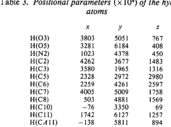
Table 3. Positional parameters (x 104) o f the hydrogen atoms x y z H(O3) 3803 5051 767 H(O5) 3281 6184 408 H(N2) 1023 4378 450 H(C2) 4262 3677 1483 H(C3) 3580 1965 1316 H(C5) 2328 2972 2980 H(C6) 2259 4261 2597 H(C7) 4005 5009 1758 H(C8) 503 4881 1569 H(C 10) -76 3350 69 H(CI 1) 1742 6127 1257 H(CAll) -138 5811 894
Pippy & Huber, 1973) and the IISc program set (Shiono, 1968). Final atomic parameters are presented in Tables 2 and 3.
D i s c u s s i o n
It has been suggested (Gale, Cundliffe, Reynolds, R i c h m o n d & Waring, 1972)that chloramphenicol (Fig. la) exists in an alicyclic form (Fig. lb). The diagram of the structure of the compound, Fig. 2, shows that this is indeed the case in the solid state; this form is stabilized by an intramolecular hydrogen bond between 0 ( 3 ) and 0 ( 5 ) [ 0 ( 3 ) . . . 0 ( 5 ) = 2.727 ( 1 0 ) A ] . The orientation of the side chain, with a trans arrangement of the C ( 7 ) - 0 (3) and C ( 8 ) - N (2) bonds, causes the dichloro- acetamido group to fold back over the aromatic ring.
The bonding parameters for the structure are presented in Tables 4 and 5, with details of inter- molecular approaches and mean planes being given in Tables 6 and 7. The structural geometry shows no remarkable deviation from that expected. The lattice
OH--oH H NH-CO-CHCI2 _ ~ ~ 7 ~ O N- ~ 2 ~ ')"'-C -- C -- CH2OH [ [ O2N ~ / I I HN---~ HO H [ /CI OC--C-~cI (a) (b) H
Fig. 1. (a) The chloramphenicol molecule. (b) Alicyclic ring form of chloramphenicol.
.cl0)
C1(2) ? . 0(4) 10~9 [
1362 T H E S T R U C T U R E O F C H L O R A M P H E N I C O L
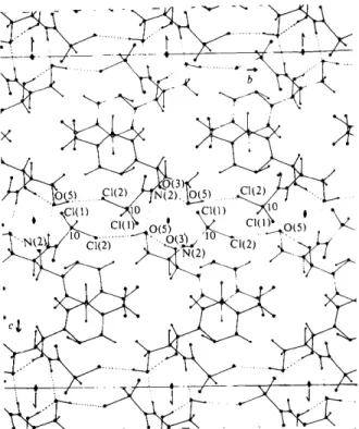
packing (Fig. 3) is achieved through hydrogen bonding between 0 ( 5 ) and the C1(2) atom of a molecule related by a carbon centre [ 0 ( 5 ) . . . C 1 ( 2 ) = 3.262 (7) A], and between 0 ( 5 ) and the N(2) atom of a twofold (a axis) symmetry-related molecule I N ( 2 ) . . . 0 ( 5 ) = 2.929 (10) A], with all other contacts being of the van der Waals type (Table 6).
Inhibition of protein synthesis is described as the general mode of action of chloramphenicol (Gottlieb & Shaw, 1967). It has been reported that the antibiotic binds preferentially with the 50S subunit of the ribosomal particle (Gale, Cundliffe, Reynolds, Rich-
Table 4.
Bond lengths (A)
c(1)-c(2) 1.396 ( 1 5 ) N(I)-O(I) 1.235 (17) C(2)-C(3) 1.414 ( 1 6 ) N(I)-O(2) 1.210 (17) C(3)-C(4) 1.398 ( 1 7 ) C(8)-C(11) 1.536 (14) C(4)-C(5) 1.364 (17) C(I 1)-O(5) 1.441 (13) C(5)-C(6) 1.388 ( 1 5 ) C(8)-N(2) 1.469 (12) C(6)-C(1) 1.397 ( 1 3 ) N(2)--C(9) 1.280 (12) C(1)-C(7) 1.513 ( 1 4 ) C(9)-O(4) 1.244 (11) C(7)-C(8) 1.547 ( 1 4 ) C(9)-C(10) 1.571 (13) C(7)-O(3) 1.422 ( 1 2 ) C(10)-CI(1) 1.789 (11) C(4)--N(1) 1.503 ( 1 6 ) C(10)-C1(2) 1.753 (11) O(3)-H(O3) 0.825 C(6)-H(C6) 1.017 O(5)-H(O5) 0.990 C(7)-H(C7) 1.070 N(2)-H(N2) 0.775 C(8)-H(C8) 0.963 C(2)-H(C2) 0.999 C(10)-H(C 10) 1.042 C(3)-H(C3) 1.105 C(11)-H(CI 1) 0.946 C(5)-H4C5) 1.015 C(I I)--H(CA 11) 1.089
Table 5.
Bond angles (o) and their standard deviations
O(I)-N(1)-O(2) 126.0 ( 1 4 ) C(1)-C(7)-C(8) 109.7 (8) O(I)-N(1)-C(4) 115.8 ( 1 2 ) O(3)-C(7)-C(8) 106.4 (8) O(2)-N(1)-C(4) 118.1 ( 1 3 ) C(7)-C(8)-C(11) 113.2 (8) N(1)-C(4)--C(5) 119.1 (1 I) C(7)-C(8)-N(2) 109.9 (8) N41)-C(4)--C(3) 116.8 ( 1 1 ) N(2)-C(8)-C(1 I) 110.0 (8) C(3)-C(4)-C(5) 124.0 ( 1 2 ) C(8)-C(11)-O(5) 112.0 (8) C(4)-C(5)-C(6) 119.2 ( 1 1 ) C(8)-N(2)-C49) 122.8 (8) C(5)-C(6)-C(1) 118.8 ( 1 0 ) N(2)-C(9)-O(4) 126.8 (9) C(2)-C(3)-C(4) 116.6 ( 1 1 ) N(2)-C(9)-C(10) 112.2 (8) C(1)-C(2)-C(3) 119.5 ( 1 0 ) O(4)-C(9)-C(10) 120.9 (8) C(6)-C(1)-C(2) 121.5 ( 1 0 ) C(9)-C(10)-C1(2) 108.8 (7) C46)-C41)-C47) 119.2 49) C1(1)-C410)-C1(2) 110.2 46) C(2)-C(1)-C(7) 119.3 (9) C(9)-C(10)-CI(I) 108.1 (7) C(1)-C(7)-O(3) 112.6 (8)
Table 6.
Intermolecular distances
(A)less than 3.6 A
Symmetry code: (i)
x,y,z;
(ii) x,)),~; (iii) ~, )3, 1 + z; (iv) ~, y, ~ - z; (v) ½ + x, ½ + y , z ; (vi) ½ + x, ½ - y , ~ ; (vii) ½ - x , ½ - y , ½ + z; (viii) ½ - x, ½ + y, ½ - z. Cl(l)...0(3) 3.502 (8) l~ O(1)...0(2) 3.519 (23) ~v El(l)...0(5) 3.496 ( 9 ) " C(6)...C(6) 3.60 (15) Iv 0(3)-..0(5) 3.518 ( 1 0 ) " O(1)...O(1) 2.987 (19) ~v 0(5)...0(5) 3.397 (9) ~ O(1)...N(I) 3.081 (22) Iv 0(2)...0(2) 3.505 (24) ~ O(1)...C(4) 3.546 (20) ~v 045)...C(10) 3.263 4 1 3 ) " C45)...C(5) 3.537 418) ~v 0(4)-..C(5) 3.467 (14) ~ 0(2)...C(11) 3.503 (19) ~ 0(4)...C(6) 3.391 (11) ~ O(1)...C(l 1) 3.26 (15) ~"~ _ - . i t - ~ - - - - - - O(3) O ~ ( ~ ) . . • . ~I ' ~ Cl(l~ .O(5~ [ ~ C 1 ( l ~ O ~ 5 ) , . . .Fig. 3. Crystal packing of chloramphenicol viewed down the a axis.
Table 7.
Least-squaresplanes
The equations are ofthe form p x + q y + r z = d, where x , y . z and d are in A.
Plane Atoms in plane Equation of plane
1 C(1). C/2l, C(3). C(4), C(5), 0.8772x - 0.0069)' + 0.4802z = 3-8566
C(6), C/7)
2 Oil), Oi2), Nil) 0.9546x * 0.01093'+ 0.2977z = 3,3093
3 O(3). C(7). C(8), CIll), O(5) 0-6951x + 0.0055y + 0.7189z = 3.1741
Deviations from the least-squares planes
Plane Atom Deviation IA) Plane Atom Deviation (A)
1 C(I) -0.102 2 Oil) -0.199 CI2) -0.245 Oi2) 0.176 C(3) -0.169 N(I) 0.001 C(4) 0. 142 3 0(3) 0-614 C(5) 0.344 C(7) 0-822 C(6) 0.234 C(8) -0.491 C(7) -0.298 C ( l l ) -0.882 Ot5) -1-292
mond & Waring, 1972). As it combines with the subunit, the hydrogen bond between the two aliphatic hydroxyl groups might break and bind with groups having a greater affinity for H, thus inhibiting protein synthesis.
We thank Professor M. A. Viswamitra, Indian Institute of Science, Bangalore, for his interest in the work and his group for their kind help with the computer programming. We thank Professor K. N. Kuchela, Head of the Department of Physics, Ban- galore University, for his encouragement. Michael Post thanks the National Research Council of C a n a d a for
K. R A V I N D R A A C H A R Y A , D. S. SAKE G O W D A A N D M I C H A E L POST 1363 the award of a Research Associateship, and D. S. Sake
Gowda wishes to acknowledge the award of the research grant by the Indian Council of Medical Research, New Delhi.
References
AHMED, F. R., HALL, S. R., PlPpY, M. E. & HUBER, C. P. (1973). NRC Crystallographic Programs for the IBM/360 System. National Research Council, Ottawa, Canada.
CARTER, H. E., GOTTLIEB, D. ~ ANDERSON, H. W. (1948).
Science, 107, 113.
CONTROULIS, J., REBSTOCK, M. C. & CROOKS, H. M. (1949). J. Am. Chem. Soc. 71, 2463-2468.
DECLERCQ, J. P., GERMAIN, G., MAIN, P. & WOOLESON,
M. M. (1973). Acta Cryst. A29, 231-234.
DUNITZ, J. D. (1952). J. Am. Chem. Sue. 74, 995-999.
EHRLICH, J., BARTZ, Q. R., SMITH, R. M., JOSLYN, D. A. &
BURKHOLDER, P. R. (1947). Science, 106, 417.
GALE, E. F., CUNDLIFFE, E., REYNOLDS, P. E., RICHMOND, M. H. & WARING, M. J. (1972). Molecular Basis of
Antibiotic Action. London:John Wiley.
GOTTLIEB, D. & SHAW, P. D. (1967). Antibiotics. I.
Mechanism of Action. Berlin, Heidelberg, New York:
Springer.
International Tables for X-ray Crystallography (1974). Vol.
IV. Birmingham: Kynoch Press.
LONG, L. M. & TROUTMAN, H. D. (1949). J. Am. Chem. Soc.
71, 2469-2472; 2473-2475.
SHIONO, R. (1968). IISc Crystallographic Programs, revised by S. REDDY.
STOUT, G. H. & JENSEN, L. H. (1968). X-ray Structure
Determination. New York: Macmillan.
Acta Cryst. (1979). B35, 1363-1372
Crystal and Molecular Structure ofp-Methoxybenzyl 2~t-Methyl-2fl-
[ ( R)-acetoxy(methoxy)methyl ]-6fl-phenoxyacetamidopenam- 3~t-carboxylate
BY PAOLO DOMIANO AND MARIO NARDELLI
Centro di Studio per la Strutturistica Diffrattometrica del CNR, Istituti di Chimica Generale e Strutturistica Chimiea dell'Universitd di Parma, Italy
AND ALDO BALSAMO, BRUNO MACCHIA AND FRANCO MACCHIA
Istituti di Chimiea Farmaeeutiea e Chimiea Organiea dell'Universitd di Pisa, Italy
(Received 12 June 1978; accepted 25 January 1979)
Abstract
The title compound, C27H30N209S, crystallizes in the orthorhombic space group P2~212~ with a = 20.526 (5), b = 12.756 (2), c = 10.298 (3) A, Z = 4. The structure has been solved by direct methods and refined by a least-squares procedure to a conventional R value of 0.033 (absolute configuration) for 2562 observed independent reflections. A comparison of several known structures of penicillin derivatives is made. Only small differences are observed for the characteristic moiety of this class of compounds. In the title compound the thiazolidine ring exhibits slight puckering, the C(3) atom is 0.39 A out of the plane through the remaining four atoms. The N atom of the fl-lactam ring is 0.37 A out of the plane of its ligand C atoms. The phenoxymethyl moiety is compared with that of other penicillin and cephalosporin derivatives. Packing results from normal van der Waals contacts.
0567-7408/79/061363-10501.00
Introduction
In recent years the study of interconversion reactions of penicillins and cephalosporins has received consider- able attention (Cooper & Spry, 1972; Cooper, Hatfield & Spry, 1973; Kukolja, Lammert, Gleissner & Ellis, 1975; Tanida, Tsuji, Tsushima, Ishitobi, Irie, Yano, Matsumura & Tori, 1975). This interconversion is usually supposed to occur through a mechanism implying the formation and the rearrangement of thiiranium ion intermediates (Cooper & Spry, 1972; Cooper, Hatfield & Spry, 1973; Kukolja et al., 1975; Barton, Comer, Greig, Lucente, Sammes & Un- derwood, 1970).
As a continuation of our studies of the chemistry of the dihydrothiazine ring moiety of cephalosporins (Balsamo, Crotti, Domiano, Macchia, Macchia, Nan- nini & Rosai, 1978; Domiano, Nardelli, Balsamo, Macchia, Macchia & Meinardi, 1978), we started an © 1979 International Union of Crystallography