doi: 10.3389/fnins.2020.00792
Edited by: Domenico De Berardis, Azienda Usl Teramo, Italy Reviewed by: Carla Masala, University of Cagliari, Italy Federica Vellante, University of Studies G. d’Annunzio Chieti and Pescara, Italy *Correspondence: Lavinia Alberi [email protected]
Specialty section: This article was submitted to Neurodegeneration, a section of the journal Frontiers in Neuroscience Received: 07 May 2020 Accepted: 06 July 2020 Published: 25 August 2020 Citation: Brai E, Hummel T and Alberi L (2020) Smell, an Underrated Early Biomarker for Brain Aging. Front. Neurosci. 14:792. doi: 10.3389/fnins.2020.00792
Smell, an Underrated Early
Biomarker for Brain Aging
Emanuele Brai
1, Thomas Hummel
2and Lavinia Alberi
3,4*
1Center for Brain and Disease Research, Flanders Institute for Biotechnology (VIB) – Catholic University (KU) Leuven, Leuven, Belgium,2Smell and Taste Clinic, Department of Otorhinolaryngology, University Hospital Dresden, Dresden, Germany,3Swiss Integrative Centre for Human Health (SICHH), Fribourg, Switzerland,4Department of Oncology, Microbiology and Immunology, Faculty of Science and Medicine, University of Fribourg, Fribourg, Switzerland
Olfaction is in addition to touch the most ancient of our senses, developing already in the
womb it decays progressively from 65 years of age with a more pronounced impairment
associated with dementia. Despite its clinical relevance and testing accessibility, smell
remains an overlooked biomarker, which is rarely used by neurologists in the early
screening phase. In this perspective article, we outline the reasons underlying the lack
of awareness for this sense. In an attempt to put olfaction forward as an early biomarker
for pathological brain aging, we draw a comparison with vision and hearing, regarded
as more relevant for general health. This perspective article wants to encourage
further studies aimed at understanding the mechanisms responsible for the early smell
dysfunction in individuals a decade or more before the onset of cognitive symptoms.
Keywords: smell, Alzheimer’s disease, vision, hearing, biomarkers brain alterations
SMELL – AN INVISIBLE BUT VITAL SENSE
Olfaction, from an evolutionary aspect, is the oldest of our senses. Across different species, it
modulates the interactions between an organism and the surrounding environment even before
birth (
Brai and Alberi, 2018
). From early postnatal life into adulthood, the sense of smell regulates
many of our behaviors, from nutrition to social interaction (
Schaal et al., 2020
). For example,
human neonates are attracted by a pad impregnated with the mother’s breast odor (promoting
locomotor activity) compared to an empty control pad, supporting an odor memory developed
already during the fetal stage (
Han and Hummel, 2019
). Nevertheless, the majority of the studies on
chemo-sensation have been developed in rodents, with a less rich literature in humans (
Zhou et al.,
2019
). The incomplete understanding of human olfaction may relate to the complexity of studying
the multiple olfactory centers distributed in several brain regions comprising the cortical and the
subcortical pathways, e.g., olfactory bulb, piriform and entorhinal cortex, amygdala, orbitofrontal
cortex and hypothalamus (
Shepherd, 2004
;
Atanasova et al., 2008
;
Fagundo et al., 2015
;
Hummel
et al., 2017
). This anatomical heterogeneity implies an extensive connection among the olfactory
sensory areas which constitute a complex network essential to associate the olfactory stimulus with
other cerebral regions, such as those involved in the processing of memories and emotions and
multisensory integration with other senses (
Shepherd, 2004
;
McGann, 2017
).
Another challenge facing smell research in humans relates to its minor clinical implication as
compared to impairment of vision and hearing: the occurrence of blindness or deafness produces
a massive personal and social deficit which severely disrupts someone’s life. In line with these
observations, the different attention paid to these three senses has been also described by
Murphy
et al. (2002)
who reported that older adults in the US received assistance for vision and hearing
deficits, whereas no testing for olfactory dysfunction was
performed. While vision and hearing have been treated as
primary senses for general health, olfaction is gaining increasing
importance in clinical settings since its impairment represents
an overarching non-invasive biomarker in predicting dementia
during aging (
MacDonald et al., 2018
). With the frequent
decline in smell acuity, mostly attributed to the reduced
turnover of the olfactory neuroepithelium with aging (
Doty
et al., 2011
), the early and pronounced olfactory deficit
described in different neurodegenerative diseases, ranging from
Alzheimer’s to Parkinson’s and Huntington’s diseases remains yet
poorly understood.
In daily life, smell is an “invisible” sense influencing much
of our daily existence from social interactions to our most
ancestral memories and healthy aging. Without the sense
of smell, there is no flavor perception during eating and
drinking, so that everything tastes bland (
Croy et al., 2014
).
Without a functioning sense of smell, many people develop
mood and anxiety disorders. This body of evidence can
be partially explained from an anatomical perspective since
several brain regions, such as the limbic system and the
orbitofrontal cortex, are involved in olfactory processing and
also in the pathophysiology of these psychiatric disturbances
(
Atanasova et al., 2008
;
Stevenson, 2010
;
Croy et al., 2014
;
Takahashi et al., 2015
;
Kohli et al., 2017
;
Kamath et al., 2018
;
Rochet et al., 2018
).
For instance, Croy and colleagues have shown that depressed
patients display a decreased olfactory acuity. In particular,
a decrement in odor discrimination and activation of the
olfactory areas was observed in these individuals compared to
healthy subjects. However, upon anti-depressive intervention, the
olfactory parameters were comparable in the two groups (
Croy
et al., 2014
). Additional data supporting the interconnection
between olfaction and depression report that a dysfunction in
odor identification can also be experienced by patients affected
by this disorder (
Kamath et al., 2018
).
Among the studies suggesting a potential link between anxiety
and olfactory impairment, Takahashi and coworkers explored
whether anxiety traits could influence olfactory tasks in healthy
subjects. Specifically, the authors observed that individuals
suffering from states of anxiety presented remarkable odor
detection and identification deficits (
Takahashi et al., 2015
).
Anxiety and mood disorders may impinge the cognitive
function in older adults as indicated by neuroimaging
data,
showing
that
this
condition
may
be
possibly
considered as an early marker or an independent risk
component for Alzheimer’s disease (AD) (
Becker et al.,
2018
). Furthermore, subjects affected by mild cognitive
impairment or dementia show a higher probability to develop
anxiety (
Lyketsos et al., 2002
). Therefore, accounting for
olfactory impairment and co-symptomatic depression may
represent a multifactorial risk factor for the development of
dementia with aging.
Altogether these observations suggest that understanding
more deeply how olfactory processing modulates odor perception
across the lifespan will add considerable value for our well-being
and health status along the age continuum.
OLFACTION, VISION, AND HEARING AS
NON-INVASIVE BIOMARKERS IN
DEMENTIA
In an effort to halt the progressive spread of Alzheimer’s disease
worldwide, more studies in the past decade have focused on
the senses, vision, hearing, and smell, as sources of stable
non-invasive biomarkers (
MacDonald et al., 2018
;
Murphy, 2019
;
Lin, 2020
), which may enable a preventive treatment during its
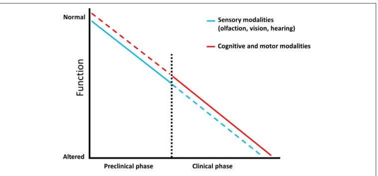
preclinical phase (Figure 1). In this section we summarize some
of the advances in sensory biomarkers offering a predictive value
for dementia conversion/progression.
Olfactory Biomarkers
Olfactory performance reaches its peak at the age of 40 years
(
Buschhüter et al., 2008
) and then progressively declines with
aging (
Eibenstein et al., 2005
;
Doty and Kamath, 2014
;
Palmquist
et al., 2020
;
Parvand and Rankin, 2020
;
Van Regemorter et al.,
2020
). In addition, in different neurological pathologies, like
Parkinson’s and Alzheimer’s disease, dysosmia is exacerbated
compared to the physiological decrease and occurs prior to
motor and cognitive disabilities (
Doty, 2012
;
Masala et al., 2018
;
Bathini et al., 2019
;
Cecchini et al., 2019
;
Dintica et al., 2019
;
Murphy, 2019
). Many studies sustain that the alteration of the
olfactory system may be used as an early predictor for detecting
AD because several brain olfactory regions are impaired in the
asymptomatic phase of this disorder due to the deposition of
pathological hallmarks (
Braak and Braak, 1991
). Consequently,
monitoring the olfactory sensitivity through different tests offers
a supplementary approach which, together with other medical
evaluations, can help to forecast the risk of developing memory
decline, favoring the implementation of prevention strategies and
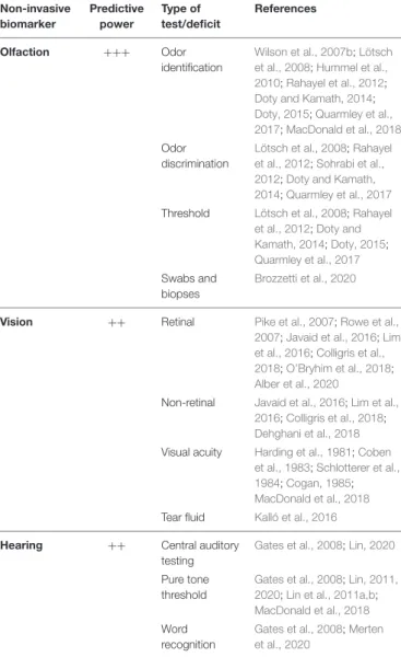
promotion of clinical trials development (Table 1).
There is a battery of tests which can be adopted to gauge
olfactory loss (
Doty and Kamath, 2014
), but the most used
in determining early-stage AD include odor identification,
discrimination and threshold (
Lötsch et al., 2008
;
Hummel et al.,
2010
;
Doty, 2015
).
In particular, the first two olfactory tasks provide a more
accurate evaluation in diagnosing prodromal dementia (
Rahayel
et al., 2012
;
Sohrabi et al., 2012
;
Quarmley et al., 2017
). This can be
explained by the fact that odor discrimination and identification
rely, to a greater extent than odor threshold, on higher brain
centers, such as the piriform, entorhinal, orbitofrontal cortices
and the amygdala (
Rahayel et al., 2012
;
Sohrabi et al., 2012
;
Quarmley et al., 2017
), therefore indicating a more complex
processing for their execution. On the contrary, odor threshold
performance is mainly associated with peripheral olfactory
stimuli as perceived by the olfactory receptors placed in the nasal
neuroepithelium (
Doty et al., 1994
;
Atanasova et al., 2008
) and
is susceptible to changes in the anatomy of the nasal cavity and
clogging of the cribriform plate which occurs naturally with aging
(
Damm et al., 2002
;
Doty and Kamath, 2014
;
Masala et al., 2019
).
Olfactory function can also be influenced by other factors,
e.g., smoking or chronic rhinosinusitis may also trigger olfactory
loss and should be taken into consideration (
Devanand, 2016
).
FIGURE 1 | Predictive factors characterizing dementia. Schematic representation showing that sensory modalities are affected from the prodromal phase of Alzheimer’s disease and represent an early prognostic tool (blue line) which is less efficient in the late stage of the pathology, as sensory testing can be influenced by additional impairments (blue dotted line). Complex cognitive and motor abilities display a low predictive power in the preclinical phase since they are mainly intact (red dotted line), however, they have an increased diagnostic value in the clinical stage where they progressively worsen (red line).
Psychophysical tests of olfactory function are non-invasive, fast,
inexpensive, and may be self-administered. Moreover, they can
spare some patients from undergoing costly examinations, such
as positron emission tomography (PET) scans, especially when
administered together with other analysis (
Devanand, 2016
).
In order to obtain reliable results, an important aspect which
is taken into account when performing odor identification
tasks is that people from different regions are familiar with
distinct odors related to their cultural and traditional background
(
Konstantinidis et al., 2008
;
Neumann et al., 2012
;
Tekeli et al.,
2013
;
Cavazzana et al., 2017
). In addition to olfactory tests,
nasal biopsies and swabs represent a supplementary procedure
which can be adopted to evaluate the integrity of the olfactory
system and its possible impairment in neurodegenerative diseases
(
Brozzetti et al., 2020
). This multivariate approach on studying
the olfactory system may provide complementary results useful
for the diagnosis of neurological pathologies during their
preclinical phase, therefore further elucidating whether a person
is “silently” developing a pathology.
In conclusion, fully characterizing olfaction in its diverse
aspects, as done with the other senses, will provide an added value
in understanding the development of cognitive deficits and their
prevention, offering new horizons for therapeutic applications
during the preclinical phase of AD and possibly tackling its
progression before it becomes irreversible.
Visual Biomarkers
Additionally to olfactory decline, AD patients can experience
visual disturbances before displaying memory impairment
(
Harding et al., 1981
;
Coben et al., 1983
;
Schlotterer et al.,
1984
). Poor vision, spatial disorientation, and visual agnosia
were characterized in patients with presumable AD (
Cogan,
1985
). Therefore, detecting these deficits in the asymptomatic
stage of the pathology might play an important role in its early
diagnosis (
Javaid et al., 2016
;
Lim et al., 2016
;
Colligris et al.,
2018
;
Alber et al., 2020
). There is a number of retinal and
non-retinal biomarkers which may be used to predict the risk
of dementia in its prodromal phase (
Javaid et al., 2016
;
Lim
et al., 2016
;
Colligris et al., 2018
;
Dehghani et al., 2018
;
Alber
et al., 2020
; Table 1). For instance, some reports show that eye
tests conducted using PET and optical coherence tomographic
angiography could detect retinal microvascular abnormalities
in cognitively normal subjects with preclinical AD (
O’Bryhim
et al., 2018
). Furthermore, PET analysis revealed that intraretinal
aggregation of
β-amyloid and Tau occurs before the onset of AD
clinical profile (
Pike et al., 2007
;
Rowe et al., 2007
). In particular,
the structural variation observed in the eye components along
with the accumulation of amyloid deposits cause visual deficits
in the early stages of AD, and such variations could also alter the
composition and the production of tears. Notably, it has been
shown that the protein level in this ocular fluid is increased in
AD subjects compared to that in healthy controls (
Kalló et al.,
2016
). This data is in line with a previous study describing the
use of tears to detect other neurodegenerative pathologies, like
glaucoma (
Tezel, 2013
). While the detection of structural and
functional changes through retinal scans may be informative of
early neurodegenerative processes, it requires medical assistance
and will be less accessible and compliant than olfactory testing
to aging adults. On the other hand, point-of-care tear biomarker
diagnostics may have a wider acceptance and usability across the
population, but studies in this area remain few to date and there
is so far no approved or marketed product.
TABLE 1 | Sensory biomarkers and their predictive power to diagnose Alzheimer-related dementia at early stages.
Non-invasive biomarker Predictive power Type of test/deficit References Olfaction +++ Odor identification Wilson et al., 2007b;Lötsch et al., 2008;Hummel et al., 2010;Rahayel et al., 2012;
Doty and Kamath, 2014;
Doty, 2015;Quarmley et al., 2017;MacDonald et al., 2018
Odor discrimination
Lötsch et al., 2008;Rahayel et al., 2012;Sohrabi et al., 2012;Doty and Kamath, 2014;Quarmley et al., 2017
Threshold Lötsch et al., 2008;Rahayel et al., 2012;Doty and Kamath, 2014;Doty, 2015;
Quarmley et al., 2017
Swabs and biopses
Brozzetti et al., 2020
Vision ++ Retinal Pike et al., 2007;Rowe et al.,
2007;Javaid et al., 2016;Lim et al., 2016;Colligris et al., 2018;O’Bryhim et al., 2018;
Alber et al., 2020
Non-retinal Javaid et al., 2016;Lim et al., 2016;Colligris et al., 2018;
Dehghani et al., 2018
Visual acuity Harding et al., 1981;Coben et al., 1983;Schlotterer et al., 1984;Cogan, 1985;
MacDonald et al., 2018
Tear fluid Kalló et al., 2016
Hearing ++ Central auditory
testing
Gates et al., 2008;Lin, 2020
Pure tone threshold
Gates et al., 2008;Lin, 2011, 2020;Lin et al., 2011a,b;
MacDonald et al., 2018
Word recognition
Gates et al., 2008;Merten et al., 2020
Auditory Biomarkers
During aging the prevalence of hearing reduction becomes
predominant and it is estimated that about 9–14% of people
aged
>65 years show auditory processing disorders (
Rosenhall,
2015
). With age-dependent hearing damage communication is
affected, contributing to social isolation and loneliness which
may alter the integrity of cognitive processes (
Bennett et al.,
2006
;
Wilson et al., 2007a
). In addition, increasing studies
suggest the association between hearing loss and cognitive deficit
and dementia-related disorders (
Gates et al., 2008
;
Lin, 2011
;
Lin et al., 2011a, 2014
). Furthermore, such deficits appear to
be stronger in concurrent multi-sensory impairments, like in
auditory and vision domains (
Yamada et al., 2016
). Auditory
performance can be tested through several measures, such as pure
tone threshold, word recognition and central auditory testing
(
Gates et al., 2008
;
Lin, 2011, 2020
;
Lin et al., 2011a,b
;
Merten
et al., 2020
) (Table 1). Overall, hearing loss alters the input
and therefore the information from external stimuli, leading to
reduction in general awareness of the person with respect to
time and space. Taken altogether, these observations sustain a
potential involvement of hearing loss in dementia and to the late
AD pathobiology. However, further studies need to target the key
mechanisms underlying the link between auditory dysfunction
and dementia/AD condition and therefore promote hearing
monitoring as a reliable early predictor of cognitive decline.
CONCLUSION
In the challenge for finding relevant diagnostic markers to
target dementia in its early stage, sensory modalities are offering
themselves as promising non-invasive predictors. Different
studies provide increasing evidence that monitoring olfaction
acuity and, to some extent, vision and hearing could contribute to
the early detection of neuronal changes linked to mental decline,
therefore promoting a more immediate intervention long before
the onset of the cognitive symptoms (Table 1).
As shown by several studies, a reduced olfactory activity
is a valuable predictor for cognitive decline in non-demented
older adults (
Wilson et al., 2007b
;
Sohrabi et al., 2012
).
The sequential association between olfactory dysfunction and
cognitive deficit is not completely resolved. Nevertheless, the
spreading rostral to caudal tauopathy in the limbic brain
areas may influence olfactory transmission first and cognitive
processing later (
Doty and Kamath, 2014
;
Dintica et al., 2019
).
These observations further support the association between
the sense of smell and cognition and promote olfactory
testing to identify those subjects with a higher risk of
developing dementia.
Since visual symptoms are experienced by many AD
patients, increasing studies are investigating whether this
sensory system may represent another potential source of
early biomarkers to diagnose cognitive decline and dementia.
Among the features corroborating the pathophysiological
link
between
visual/ocular
impairments
and
cognitive
dysfunction, it can be acknowledged that the eye and
the central nervous system have some commonalities,
such as the same embryologic origin and the presence
of physiological barriers (
Alber et al., 2020
). In addition,
advanced imaging techniques revealed the accumulation of
AD hallmarks in visual structures during the prodromal
stage of dementia (
Lim et al., 2016
;
Colligris et al., 2018
;
Alber et al., 2020
).
Many research projects foster the use of hearing assessment
as a supplemental diagnostic measure to target cognitive decline
and dementia during their preclinical stage. For instance,
Lin
et al. (2011a,b)
explored whether hearing impairment could
be mirrored by decreased cognitive tests scoring. Interestingly,
they observed that hearing decrement was related with lower
cognitive scores. Further evidence proposing this interplay have
been correlated with cerebral atrophy in regions implicated in
speech processing, common degenerative mechanisms, social
isolation and altered communication (
Lin et al., 2011a, 2014
;
Lin and Albert, 2014
).
Overall, if the use of sensory functions would be considered
as a reliable tool to evaluate the risk of developing dementia, this
would enrich the array of multi-step screening protocols that can
be adopted in clinical practice. As with other chronic pathologies,
such as diabetes and hypertension, where a medical evaluation
is regularly recommended in many countries, the monitoring
of olfactory performance during aging through simple smell
testing could inform about a possible neuropathological risk.
A strong informative campaign about the potential benefits
which could be obtained by administering olfactory tests might
lead to a broader consensus in providing a first screening
phase in classifying individuals with a higher risk of developing
cognitive impairment and then fostering the development of
novel clinical trials. Obviously, supplementary examinations like
cognitive tests and brain imaging techniques would be required
to confirm this evidence.
In conclusion, we see olfactory testing as a potentially
first-line non-invasive diagnostic measure with great compliance
and flexibility that may inform the patient and the family
and aid in planning healthcare interventions. This primary
monitoring would be of substantial value for clinical studies
where the selection and the recruitment of subjects in
prodromal stages remain challenging. Furthermore, a first
diagnostic screening based on olfactory testing will allow
the swift implementation of preventive approaches aimed at
improving cognition and brain health. In the future, we see
the integration of analog tests with IT interfaces expanding the
capabilities of olfactory monitoring as part of the personalized
health program aimed at improving the quality of life
for a long term.
DATA AVAILABILITY STATEMENT
All datasets generated for this study are included in the
article/supplementary material, further inquiries can be directed
to the corresponding author.
AUTHOR CONTRIBUTIONS
EB wrote the manuscript. TH reviewed and gave the feedback. LA
advised and wrote part of the text. All authors contributed to the
article and approved the submitted version.
FUNDING
This study was supported by the Schweizerischer Nationalfonds
zur Förderung der Wissenschaftlichen Forschung (163470).
REFERENCES
Alber, J., Goldfarb, D., Thompson, L. I., Arthur, E., Hernandez, K., Cheng, D., et al. (2020). Developing retinal biomarkers for the earliest stages of Alzheimer’s
disease: what we know, what we don’t, and how to move forward.Alzheimers
Dement. J. Alzheimers Assoc. 16, 229–243. doi: 10.1002/alz.12006
Atanasova, B., Graux, J., El Hage, W., Hommet, C., Camus, V., and Belzung, C. (2008). Olfaction: a potential cognitive marker of psychiatric disorders. Neurosci. Biobehav. Rev. 32, 1315–1325. doi: 10.1016/j.neubiorev.2008.05.003 Bathini, P., Brai, E., and Auber, L. A. (2019). Olfactory dysfunction in the
pathophysiological continuum of dementia.Ageing Res. Rev. 55:100956. doi:
10.1016/j.arr.2019.100956
Becker, E., Orellana Rios, C. L., Lahmann, C., Rücker, G., Bauer, J., and Boeker, M. (2018). Anxiety as a risk factor of Alzheimer’s disease and vascular dementia. Br. J. Psychiatry J. Ment. Sci. 213, 654–660.
Bennett, D. A., Schneider, J. A., Tang, Y., Arnold, S. E., and Wilson, R. S. (2006). The effect of social networks on the relation between Alzheimer’s disease pathology and level of cognitive function in old people: a longitudinal cohort study.Lancet Neurol. 5, 406–412. doi: 10.1016/s1474-4422(06)70417-3
Braak, H., and Braak, E. (1991). Neuropathological stageing of Alzheimer-related
changes.Acta Neuropathol. 82, 239–259. doi: 10.1007/bf00308809
Brai, E., and Alberi, L. (2018).Olfaction, Among the First Senses to Develop and Decline Sensory Nervous System. Thomas Heinbockel: IntechOpen.
Brozzetti, L., Sacchetto, L., Cecchini, M. P., Avesani, A., Perra, D., Bongianni, M., et al. (2020). Neurodegeneration-associated proteins in human olfactory neurons collected by nasal brushing.Front. Neurosci. 14:145. doi: 10.3389/fnins. 2020.00145
Buschhüter, D., Smitka, M., Puschmann, S., Gerber, J. C., Witt, M., Abolmaali, N. D., et al. (2008). Correlation between olfactory bulb volume and olfactory
function.NeuroImage 42, 498–502. doi: 10.1016/j.neuroimage.2008.05.004
Cavazzana, A., Wesarg, C., Schriever, V. A., Hummel, T., Lundström, J. N., and Parma, V. (2017). A cross-cultural adaptation of the sniffin’ sticks olfactory
identification test for US children.Chem. Senses 42, 133–140. doi: 10.1093/
chemse/bjw113
Cecchini, M. P., Federico, A., Zanini, A., Mantovani, E., Masala, C., Tinazzi, M., et al. (2019). Olfaction and taste in Parkinson’s disease: the association with mild
cognitive impairment and the single cognitive domain dysfunction.J. Neural
Transm. 1996, 585–595. doi: 10.1007/s00702-019-01996-z
Coben, L. A., Danziger, W. L., and Hughes, C. P. (1983). Visual evoked potentials in mild senile dementia of Alzheimer type.Electroencephalogr. Clin. Neurophysiol. 55, 121–130. doi: 10.1016/0013-4694(83)90178-5
Cogan, D. G. (1985). Visual disturbances with focal progressive dementing
disease. Am. J. Ophthalmol. 100, 68–72. doi: 10.1016/s0002-9394(14)74
985-2
Colligris, P., Perez de Lara, M. J., Colligris, B., and Pintor, J. (2018). Ocular manifestations of Alzheimer’s and other neurodegenerative diseases: the prospect of the eye as a tool for the early diagnosis of Alzheimer’s disease. J. Ophthalmol. 2018, 1–12. doi: 10.1155/2018/8538573
Croy, I., Nordin, S., and Hummel, T. (2014). Olfactory disorders and quality of
life–an updated review.Chem. Senses 39, 185–194. doi: 10.1093/chemse/bjt072
Damm, M., Vent, J., Schmidt, M., Theissen, P., Eckel, H. E., Lötsch, J., et al.
(2002). Intranasal volume and olfactory function.Chem. Senses 27, 831–839.
doi: 10.1093/chemse/27.9.831
Dehghani, C., Frost, S., Jayasena, R., Masters, C. L., and Kanagasingam, Y. (2018). Ocular biomarkers of Alzheimer’s disease: the role of anterior eye and potential future directions.Invest. Ophthalmol. Vis. Sci. 59, 3554–3563.
Devanand, D. P. (2016). Olfactory identification deficits, cognitive decline, and dementia in older adults.Am. J. Geriatr. Psychiatry 24, 1151–1157. doi: 10.1016/ j.jagp.2016.08.010
Dintica, C. S., Marseglia, A., Rizzuto, D., Wang, R., Seubert, J., Arfanakis, K., et al. (2019). Impaired olfaction is associated with cognitive decline and
neurodegeneration in the brain.Neurology 92, e700–e709. doi: 10.1212/wnl.
0000000000006919
Doty, R. L. (2012). Olfaction in Parkinson’s disease and related disorders. Neurobiol. Dis. 46, 527–552. doi: 10.1016/j.nbd.2011.10.026
Doty, R. L. (2015). Olfactory dysfunction and its measurement in the clinic.World J. Otorhinolaryngol. Head Neck Surg. 1, 28–33. doi: 10.1016/j.wjorl.2015.09.007 Doty, R. L., and Kamath, V. (2014). The influences of age on olfaction: a review.
Front. Psychol. 5:20. doi: 10.3389/fpsyg.2014.00020
Doty, R. L., Petersen, I., Mensah, N., and Christensen, K. (2011). Genetic and environmental influences on odor identification ability in the very old.Psychol. Aging 26, 864–871. doi: 10.1037/a0023263
Doty, R. L., Smith, R., McKeown, D. A., and Raj, J. (1994). Tests of human olfactory function: principal components analysis suggests that most measure a common source of variance.Percept. Psychophys. 56, 701–707. doi: 10.3758/bf03208363 Eibenstein, A., Fioretti, A., Lena, C., Rosati, N., Ottaviano, I., and Fusetti,
M. (2005). Olfactory screening test: experience in 102 Italian subjects.Acta Otorhinolaryngol. Ital. 25, 18–22. doi: 10.1001/archderm.98.1.18
Fagundo, A. B., Jiménez-Murcia, S., Giner-Bartolomé, C., Islam, M. A., de la Torre, R., Pastor, A., et al. (2015). Modulation of higher-order olfaction components on executive functions in humans.PLoS One 10:e0130319. doi: 10.1371/journal. pone.0130319
Gates, G. A., Anderson, M. L., Feeney, M. P., McCurry, S. M., and Larson, E. B. (2008). Central auditory dysfunction in older persons with memory
impairment or Alzheimer dementia.Arch. Otolaryngol. Head Neck Surg. 134,
771–777.
Han, P., and Hummel, T. (2019). “Orthonasal and retronasal olfaction,” inFood
Aroma Evolution, 1st Edn, eds M. Bordiga and L. M. L. Nollet (Boca Raton: CRC Press), 99–120. doi: 10.1201/9780429441837-5
Harding, G. F. A., Doggett, C. E., Orwin, A., and Smith, E. J. (1981). “Visual evoked potentials in pre-senile dementia,” inVisual Pathways: Electrophysiology and Pathology, eds H. Spekreijse and P. A. Apkarian (Dordrecht: Springer), 193–202. doi: 10.1007/978-94-009-8656-5_20
Hummel, T., Pfetzing, U., and Lötsch, J. (2010). A short olfactory test based on the identification of three odors.J. Neurol. 257, 1316–1321. doi: 10.1007/s00415-010-5516-5
Hummel, T., Whitcroft, K. L., Andrews, P., Altundag, A., Cinghi, C., Costanzo, R. M., et al. (2017). Position paper on olfactory dysfunction.Rhinol. Suppl. 54, 1–30.
Javaid, F. Z., Brenton, J., Guo, L., and Cordeiro, M. F. (2016). Visual and ocular manifestations of Alzheimer’s disease and their use as biomarkers for diagnosis and progression.Front. Neurol. 7:55. doi: 10.3389/fneur.2016.00055
Kalló, G., Emri, M., Varga, Z., Ujhelyi, B., T˝ozsér, J., Csutak, A., et al. (2016). Changes in the chemical barrier composition of tears in Alzheimer’s Disease
reveal potential tear diagnostic biomarkers.PLoS One 11:e0158000. doi: 10.
1371/journal.pone.0158000
Kamath, V., Paksarian, D., Cui, L., Moberg, P. J., Turetsky, B. I., and Merikangas, K. R. (2018). Olfactory processing in bipolar disorder, major depression, and anxiety.Bipolar Disord. 20, 547–555. doi: 10.1111/bdi.12625
Kohli, P., Naik, A. N., Harruff, E. E., Nguyen, S. A., Schlosser, R. J., and Soler, Z. M. (2017). The prevalence of olfactory dysfunction in chronic rhinosinusitis. Laryngoscope 127, 309–320. doi: 10.1002/lary.26316
Konstantinidis, I., Printza, A., Genetzaki, S., Mamali, K., Kekes, G., and Constantinidis, J. (2008). Cultural adaptation of an olfactory identification test: the Greek version of Sniffin’.Sticks Rhinol. 46, 292–296.
Lim, J. K. H., Li, Q.-X., He, Z., Vingrys, A. J., Wong, V. H. Y., Currier, N., et al. (2016). The eye as a biomarker for Alzheimer’s disease.Front. Neurosci. 10:536. doi: 10.3389/fnins.2016.00536
Lin, F. R. (2011). Hearing loss and cognition among older adults in the United States.J. Gerontol. A Biol. Sci. Med. Sci. 66, 1131–1136. doi: 10.1093/ gerona/glr115
Lin, F. R. (2020). Making sense of the senses in aging research.J. Gerontol. Ser. A 75, 529–530. doi: 10.1093/gerona/glaa028
Lin, F. R., and Albert, M. (2014). Hearing loss and dementia – who’s listening? Aging Ment. Health 18, 671–673. doi: 10.1080/13607863.2014.915924 Lin, F. R., Ferrucci, L., An, Y., Goh, J. O., Doshi, J., Metter, E. J., et al. (2014).
Association of hearing impairment with brain volume changes in older adults. NeuroImage 90, 84–92. doi: 10.1016/j.neuroimage.2013.12.059
Lin, F. R., Ferrucci, L., Metter, E. J., An, Y., Zonderman, A. B., and Resnick, S. M. (2011a). Hearing loss and cognition in the baltimore longitudinal study of
aging.Neuropsychology 25, 763–770. doi: 10.1037/a0024238
Lin, F. R., Metter, E. J., O’Brien, R. J., Resnick, S. M., Zonderman, A. B., and
Ferrucci, L. (2011b). Hearing loss and incident dementia.Arch. Neurol. 68,
214–220.
Lötsch, J., Reichmann, H., and Hummel, T. (2008). Different odor tests contribute differently to the evaluation of olfactory loss.Chem. Senses 33, 17–21. doi: 10.1093/chemse/bjm058
Lyketsos, C. G., Lopez, O., Jones, B., Fitzpatrick, A. L., Breitner, J., and DeKosky, S. (2002). Prevalence of neuropsychiatric symptoms in dementia and mild cognitive impairment: results from the cardiovascular health study.JAMA 288, 1475–1483.
MacDonald, S. W. S., Keller, C. J. C., Brewster, P. W. H., and Dixon, R. A. (2018). Contrasting olfaction, vision, and audition as predictors of cognitive change
and impairment in non-demented older adults.Neuropsychology 32, 450–460.
doi: 10.1037/neu0000439
Masala, C., Käehling, C., Fall, F., and Hummel, T. (2019). Correlation between olfactory function, trigeminal sensitivity, and nasal anatomy in healthy subjects. Eur. Arch. Oto-Rhino-Laryngol. 276, 1649–1654. doi: 10.1007/s00405-019-05367-y
Masala, C., Solla, P., Liscia, A., Defazio, G., Saba, L., Cannas, A., et al. (2018). Correlation among olfactory function, motors’ symptoms, cognitive impairment, apathy, and fatigue in patients with Parkinson’s disease.J. Neurol. 265, 1764–1771. doi: 10.1007/s00415-018-8913-9
McGann, J. P. (2017). Poor human olfaction is a 19th-century myth. Science
356:eaam7263. doi: 10.1126/science.aam7263
Merten, N., Fischer, M. E., Tweed, T. S., Breteler, M. M. B., and Cruickshanks, K. J. (2020). Associations of hearing sensitivity, higher-order auditory processing, and cognition over time in middle-aged adults.J. Gerontol. A Biol. Sci. Med. Sci. 75, 545–551. doi: 10.1093/gerona/glz189
Murphy, C. (2019). Olfactory and other sensory impairments in Alzheimer disease. Nat. Rev. Neurol. 15, 11–24. doi: 10.1038/s41582-018-0097-5
Murphy, C., Schubert, C. R., Cruickshanks, K. J., Klein, B. E. K., Klein, R., and Nondahl, D. M. (2002). Prevalence of olfactory impairment in older adults. JAMA 288, 2307–2312.
Neumann, C., Tsioulos, K., Merkonidis, C., Salam, M., Clark, A., and Philpott, C. (2012). Validation study of the “Sniffin’ Sticks” olfactory test in a British
population: a preliminary communication.Clin. Otolaryngol. 37, 23–27. doi:
10.1111/j.1749-4486.2012.02431.x
O’Bryhim, B. E., Apte, R. S., Kung, N., Coble, D., and Van Stavern, G. P. (2018). Association of preclinical Alzheimer disease with optical coherence
tomographic angiography findings.JAMA Ophthalmol. 136, 1242–1248.
Palmquist, E., Larsson, M., Olofsson, J. K., Seubert, J., Bäckman, L., and Laukka, E. J. (2020). A prospective study on risk factors for olfactory dysfunction in aging.J. Gerontol. Ser. A 75, 603–610. doi: 10.1093/gerona/glz265
Parvand, M., and Rankin, C. H. (2020). Is there a shared etiology of olfactory impairments in normal aging and neurodegenerative disease?J. Alzheimers Dis. 73, 1–21. doi: 10.3233/jad-190636
Pike, K. E., Savage, G., Villemagne, V. L., Ng, S., Moss, S. A., Maruff, P., et al. (2007). Beta-amyloid imaging and memory in non-demented individuals: evidence for preclinical Alzheimer’s disease.Brain J. Neurol. 130, 2837–2844. doi: 10.1093/ brain/awm238
Quarmley, M., Moberg, P. J., Mechanic-Hamilton, D., Kabadi, S., Arnold, S. E., Wolk, D. A., et al. (2017). Odor identification screening improves diagnostic classification in incipient Alzheimer’s disease.J. Alzheimers Dis. 55, 1497–1507. doi: 10.3233/jad-160842
Rahayel, S., Frasnelli, J., and Joubert, S. (2012). The effect of Alzheimer’s disease and Parkinson’s disease on olfaction: a meta-analysis.Behav. Brain Res. 231, 60–74. doi: 10.1016/j.bbr.2012.02.047
Rochet, M., El-Hage, W., Richa, S., Kazour, F., and Atanasova, B. (2018). Depression, olfaction, and quality of life: a mutual relationship.Brain Sci. 8:80. doi: 10.3390/brainsci8050080
Rosenhall, U. (2015). Epidemiology of age related hearing loss.Hear. Bal. Commun. 13, 46–50. doi: 10.3109/21695717.2015.1013775
Rowe, C. C., Ng, S., Ackermann, U., Gong, S. J., Pike, K., Savage, G., et al.
(2007). Imaging beta-amyloid burden in aging and dementia.Neurology 68,
1718–1725.
Schaal, B., Saxton, T. K., Loos, H., Soussignan, R., and Durand, K. (2020). Olfaction scaffolds the developing human from neonate to adolescent and beyond. Philos. Trans. R. Soc. Lond. B Biol. Sci. 375:20190261. doi: 10.1098/rstb.2019. 0261
Schlotterer, G., Moscovitch, M., and Crapper-McLachlan, D. (1984). Visual processing deficits as assessed by spatial frequency contrast sensitivity and
backward masking in normal ageing and Alzheimer’s disease.Brain J. Neurol.
107(Pt 1), 309–325.
Shepherd, G. M. (2004). The human sense of smell: are we better than we think? PLoS Biol. 2:e146. doi: 10.1371/journal.pbio.0020146
Sohrabi, H. R., Bates, K. A., Weinborn, M. G., Johnston, A. N. B., Bahramian, A., Taddei, K., et al. (2012). Olfactory discrimination predicts cognitive decline
among community-dwelling older adults.Transl. Psychiatry 2:e118. doi: 10.
Stevenson, R. J. (2010). An initial evaluation of the functions of human olfaction. Chem. Senses 35, 3–20. doi: 10.1093/chemse/bjp083
Takahashi, T., Itoh, H., Nishikawa, Y., Higuchi, Y., Nakamura, M., Sasabayashi, D., et al. (2015). Possible relation between olfaction and anxiety in healthy subjects. Psychiatry Clin. Neurosci. 69, 431–438. doi: 10.1111/pcn.12277
Tekeli, H., Altundað, A., Salihoðlu, M., Çayönü, M., and Kendirli, M. T. (2013). The applicability of the “Sniffin’ Sticks” olfactory test in a Turkish population. Med. Sci. Monit. Int. Med. J. Exp. Clin. Res. 19, 1221–1226. doi: 10.12659/msm. 889838
Tezel, G. (2013). A proteomics view of the molecular mechanisms and biomarkers
of glaucomatous neurodegeneration.Prog. Retin. Eye Res. 35, 18–43. doi: 10.
1016/j.preteyeres.2013.01.004
Van Regemorter, V., Hummel, T., Rosenzweig, F., Mouraux, A., Rombaux, P., and Huart, C. (2020). Mechanisms linking olfactory impairment and risk of mortality.Front. Neurosci. 14:140. doi: 10.3389/fnins.2020.00140
Wilson, R. S., Krueger, K. R., Arnold, S. E., Schneider, J. A., Kelly, J. F., Barnes, L. L., et al. (2007a). Loneliness and risk of Alzheimer disease.Arch. Gen. Psychiatry 64, 234–240.
Wilson, R. S., Schneider, J. A., Arnold, S. E., Tang, Y., Boyle, P. A., and Bennett, D. A. (2007b). Olfactory identification and incidence
of mild cognitive impairment in older age. Arch. Gen. Psychiatry 64,
802–808.
Yamada, Y., Denkinger, M. D., Onder, G., Henrard, J.-C., van der Roest, H. G., Finne-Soveri, H., et al. (2016). Dual sensory impairment and cognitive decline: the results from the shelter study.J. Gerontol. A Biol. Sci. Med. Sci. 71, 117–123. doi: 10.1093/gerona/glv036
Zhou, G., Lane, G., Cooper, S. L., Kahnt, T., and Zelano, C. (2019). Characterizing functional pathways of the human olfactory system.eLife 8:e47177.
Conflict of Interest:The authors declare that the research was conducted in the absence of any commercial or financial relationships that could be construed as a potential conflict of interest.
Copyright © 2020 Brai, Hummel and Alberi. This is an open-access article distributed under the terms of the Creative Commons Attribution License (CC BY). The use, distribution or reproduction in other forums is permitted, provided the original author(s) and the copyright owner(s) are credited and that the original publication in this journal is cited, in accordance with accepted academic practice. No use, distribution or reproduction is permitted which does not comply with these terms.