HAL Id: hal-01211578
https://hal.archives-ouvertes.fr/hal-01211578
Submitted on 26 Apr 2016
HAL is a multi-disciplinary open access archive for the deposit and dissemination of sci-entific research documents, whether they are pub-lished or not. The documents may come from teaching and research institutions in France or abroad, or from public or private research centers.
L’archive ouverte pluridisciplinaire HAL, est destinée au dépôt et à la diffusion de documents scientifiques de niveau recherche, publiés ou non, émanant des établissements d’enseignement et de recherche français ou étrangers, des laboratoires publics ou privés.
Echocardiographic characteristics and outcome of
straddling mitral valve
Alain Fraisse, Pedro del Nido, Jean Gaudart, Tal Geva
To cite this version:
Alain Fraisse, Pedro del Nido, Jean Gaudart, Tal Geva. Echocardiographic characteristics and outcome of straddling mitral valve. Journal of the American College of Cardiology, Elsevier, 2001, 38 (3), pp.819-826. �10.1016/S0735-1097(01)01441-3�. �hal-01211578�
Valve Disease
Echocardiographic Characteristics
and Outcome of Straddling Mitral Valve
Alain Fraisse, MD,* Pedro J. del Nido, MD, PHD,† Jean Gaudart, MD,‡ Tal Geva, MD, FACC*
Boston, Massachusetts and Marseille, France
OBJECTIVES This study sought to characterize the echocardiographic features of straddling mitral valve (SMV) and to determine its surgical implications and midterm outcome in a large clinical cohort.
BACKGROUND Despite a relatively large body of literature on the postmortem anatomy of SMV, there is a paucity of information regarding its echocardiographic features, surgical implications and preoperative predictors of outcome.
METHODS A retrospective review identified 46 patients with SMV between 1982 and 1999 who underwent echocardiography and surgery and had follow-up data. A detailed review of the echocardiograms, surgical reports and all pertinent records was undertaken.
RESULTS Review of the echocardiograms revealed a widely varying anatomy among the study patients. However, four distinct groups with relatively uniform morphologic features could be distinguished on the basis of segmental analysis. Cardiac malposition associated with right ventricular hypoplasia, superior-inferior ventricles and criss-cross atrioventricular relations were common among patients with {S,D,L} (S ⫽ visceroatrial situs solitus, D ⫽ D-ventricular loop, L⫽ L-malposition of the great arteries) (n ⫽ 6) and {S,L,D} (n ⫽ 5) segmental combinations but were rare among patients with {S,D,D} (n⫽ 26) and {S,L,L} (n ⫽ 9) combinations. Surgical management consisted of a functional single-ventricle palliation in 38 patients (83%) and biventricular repair in 8 patients (17%). Overall mortality was 22%, but none of the seven patients who were operated on during the cohort’s last five years (1994 to 1999) has died. By multivariate analysis, noncommitted ventricular septal defect was the strongest independent predictor of death (relative risk⫽ 10.2), followed by multiple ventricular septal defects (relative risk⫽ 4.7).
CONCLUSIONS This study demonstrates that echocardiography provides detailed noninvasive imaging of the complex anatomic features of SMV and its associated anomalies. Anatomic classification based on segmental analysis allows the distinction of four groups with more uniform morphologic features. Although a biventricular approach is feasible in selected patients, a functional univentricular palliation is indicated in those with major straddling and markedly hypoplastic ventricles. (J Am Coll Cardiol 2001;38:819 –26) © 2001 by the American College of Cardiology
Straddling mitral valve (SMV) is a rare congenital cardiac malformation that is invariably associated with a ventricular septal defect (VSD) and almost always with conotruncal anomalies (1–3). The etiology and morphogenesis of SMV remain incompletely understood, but its anatomic features have been well described on the basis of autopsy material (1– 8). These reports indicate that the mitral valve (MV) straddles the anterior aspect of the septum through a malalignment conoventricular-type VSD, that the most common associated conotruncal anomaly is double-outlet right ventricle (DORV) followed by transposition of the great arteries (TGA) and that left and right ventricular size varies considerably (2– 6). However, because of its rare occurrence, most published reports on SMV are based on a
relatively small number of cases, and no consistent anatomic classification has been proposed. The morphologic and functional features of SMV in living patients have not been described in detail, and few reports have addressed the echocardiographic features of SMV (9 –12). Moreover, in-formation on midterm outcome and preoperative echocar-diographic features predictive of mortality is scarce. Only a few surgical reports have included patients with SMV (13–16); the largest published clinical experience included only 11 patients (14). Thus, the spectrum of echocardio-graphic findings and the optimal surgical management of patients with SMV remain unclear. The goals of this study, therefore, are to propose a classification of SMV based on echocardiographic findings in 46 patients, to report their midterm clinical outcome and to identify preoperative variables predictive of mortality.
MATERIALS AND METHODS
Patients. The computerized database of the Department of
Cardiology at Children’s Hospital (Boston, Massachusetts)
From the Departments of *Cardiology and †Cardiac Surgery, Children’s Hospital, and the Departments of Pediatrics and Surgery, Harvard Medical School, Boston, Massachusetts; and the ‡Laboratoire de Biomathe´matiques, Universite´ de la Me´dit-erane´e, Marseille, France. Dr. Fraisse was supported by the Federation Francaise de Cardiologie.
Manuscript received January 22, 2001; revised manuscript received May 10, 2001, accepted May 23, 2001.
was searched for all patients with the coded diagnosis of SMV from 1982 through February 1999. Patients were included in this study if they had: 1) a preoperative echocardiogram performed in our laboratory with documen-tation of SMV; 2) surgical management at Children’s Hospital; and 3) follow-up data regarding outcome. The Committee on Clinical Investigations at Children’s Hospi-tal approved the study protocol.
Clinical and echocardiographic data. The clinical records,
echocardiograms, surgical reports and, when applicable, autopsy reports were reviewed. The following data were recorded for each patient, based on clinical records: demo-graphic information, age at diagnosis, age at first surgery, age at last surgery and type of surgical procedures. The following anatomical details were documented based on review of the echocardiograms: segmental anatomy (17), atrioventricular (AV) relationship, ventricular size and func-tion, mitral valve morphology and attachments (including number and location of the papillary muscles), tricuspid valve (TV) morphology and function, type of conus, semi-lunar valve morphology and function, outflow tract obstruc-tion (defined as peak Doppler gradientⱖ20 mm Hg) and the morphology of the great arteries. All associated malfor-mations were recorded. Ventricular size was graded as normal or hypoplastic (defined as ventricular sinus apex reaching ⱕ2/3 of the cardiac apex). Ventricular function was qualitatively graded as normal or mildly, moderately or severely depressed. The type of VSD and its relationship to the semilunar valves were classified as subaortic, subpulmo-nary, doubly committed or uncommitted according to pub-lished criteria (18). Straddling mitral valve was defined as attachments of MV chordae tendineae on both sides of the ventricular septum (1– 4). Patients with chordal attachments on the left ventricular septal surface were not considered to have SMV and were not included in this study. Major degree of SMV was defined as more than half of the MV related to the infundibulum on the basis of qualitative assessment from a short-axis view (either subxiphoid or parasternal) and from the apical four-chamber view during diastole. The anatomical definitions of right ventricle (RV) sinus, infundibulum and the nomenclature of segmental anatomy were previously published (17–20).
Statistical analysis. The frequency of anatomic findings
was compared between groups using Fisher exact test. The
demographic and anatomic findings of patients who were alive at the time of the most recent follow-up evaluation were compared with those of nonsurvivors. The Kaplan-Meier method was used to evaluate the probability of survival over time. Univariate analysis of predictors of death was performed using the Breslow test for qualitative vari-ables and the Cox proportional hazards model for continu-ous variables. To identify independent predictors of mor-tality, variables that reached a p value ⬍0.2 by univariate analysis were entered into a multivariate Cox proportional hazards model with a backward elimination algorithm. A p value⬍0.05 was considered statistically significant.
RESULTS
Patients. Forty-six patients fulfilled inclusion criteria for
this study. Two of the patients included in this series were previously reported (6). Of the 46 patients included, 27 were males and 19 were females (male/female ratio⫽ 1.4). Their median age at echocardiography was 1.1 years (range 0 to 13). Their mean (⫾ standard deviation) follow-up was 3.9⫾ 4.8 years (range 3 days to 17 years).
Anatomic findings. All patients had visceroatrial situs
solitus. Thirty-two patients (70%) had D-ventricular loop and the remaining 30% had L-ventricular loop. Based on review of the echocardiograms, patients with visceroatrial situs solitus (S), D-ventricular loop (D) and L-malposition of the great arteries (L), or {S,D,L} segmental anatomy, differed from those with {S,D,D} pattern in that the prevalence of dextrocardia, superior-inferior ventricles, criss-cross AV relations and RV sinus hypoplasia was significantly higher in the former group. Similarly, levocar-dia, superior-inferior ventricles, criss-cross AV relations and RV sinus hypoplasia were more common in patients with {S,L,D} than in those with {S,L,L} anatomy. Consequently, patients were grouped according to the following four segmental anatomic patterns: {S,D,D} in 26 cases, {S,D,L} in 6 cases, {S,L,D} in 5 cases and {S,L,L} in 9 cases. Table 1 summarizes the anatomic characteristics of the four anatomic groups and Figures 1 to 4 show their echocardio-graphic features.
Several anatomic features were common to all patients. The MV was good-sized and straddled into the RV infun-dibulum over the anterior aspect of the ventricular septum through a conoventricular-type VSD. Malalignment be-tween the conal septum superiorly and the muscular inter-ventricular septum inferiorly was also present in all patients. In 12 patients (26%) the VSD extended posteriorly into the AV canal septum (inlet septum), in 2 patients (4%) the VSD was restricted by the straddling MV leaflets and in 4 patients (9%) there were additional muscular VSDs. The most common location of the VSD was subpulmonary (61%), followed by subaortic in 22%, noncommitted in 11% and doubly committed VSD in 6%. All patients had an abnormal conotruncus, with DORV being the most com-mon anomaly (83%) followed by TGA (17%). However, the
Abbreviations and Acronyms
AV ⫽ atrioventricular
DORV⫽ double-outlet right ventricle LV ⫽ left ventricle
MV ⫽ mitral valve RV ⫽ right ventricle SMV ⫽ straddling mitral valve
TGA ⫽ transposition of the great arteries TV ⫽ tricuspid valve
VSD ⫽ ventricular septal defect
820 Fraisseet al. JACC Vol. 38, No. 3, 2001 Straddling Mitral Valve September 2001:819 –26
prevalence of TGA in patients with L-ventricular loop was slightly higher (29%) than in those with D-ventricular loop (12%). The most common type of conus was subaortic (67%), followed by bilateral (30%). One patient with {S,D,D} DORV segmental anatomy had a subpulmonary conus.
The morphology of the MV leaflets, the extent of straddling and the anatomy of the chordae tendineae and papillary muscles varied considerably among patients. In 87% of cases, the anterior leaflet of the MV was large and a portion of it straddled through an anteriorly positioned conoventricular VSD into the RV infundibulum. Less frequently (13%), both anterior and posterior MV leaflets straddled into the infundibulum (Fig. 3). Based on the defined criteria, the degree of SMV was judged to be major in 11 patients (24%) and minor in the other 35 patients (76%). In 29 patients (63%), the SMV attached to a single papillary muscle or to a group of small papillary muscles within the infundibulum. In the remaining 17 patients, most of whom had minor straddling, the SMV had direct chordal attachments within the infundibulum. The site of infundibular attachment(s) included septal band (35 pa-tients), conal septum (12 papa-tients), and the infundibular free wall (23 patients). In the LV, the MV attached to two distinct papillary muscles in 17 patients (37%), to two fused papillary muscles in 12 patients (26%) and to a single
posteromedial papillary muscle in 17 patients (37%). The anterior leaflet of the MV was frequently divided by an additional commissure, resulting in a trileaflet MV. The chordae tendineae from both sides of the commissure converged into one or more closely spaced papillary muscles within the infundibulum, thus creating a functional com-missure. By Doppler, there was mild mitral regurgitation in nine patients (20%) and mild tricuspid regurgitation in eight patients (17%). None of the patients had more than mild AV valve regurgitation.
Both ventricles were well developed in 29 patients (63%), the RV was hypoplastic in 10 patients (22%) and the left ventricle (LV) was hypoplastic in 7 patients (15%). The MV was well developed in all patients whereas the TV was hypoplastic in seven patients (15%). In the three patients with hypoplasia of the RV sinus and a well-developed TV, the TV straddled through the posterior aspect of the VSD (AV canal-type VSD)—i.e., bilateral straddling.
Obstruction of the pulmonary outflow tract was common in this series (33 patients, 72%). Subvalvar and valvar pulmonary stenosis was present in 24 patients and pulmo-nary atresia in 9 patients. The mechanism of obstruction was judged to be secondary to conal septal malalignment in all patients. In two of these patients, the SMV was felt to further contribute to the pulmonary outflow obstruction. Aortic outflow was obstructed in seven patients (15%), with
Table 1. Anatomic Characteristics of 46 Patients with Straddling Mitral Valve
Segmental Anatomy Group I {S,D,D} (nⴝ 26) Group II {S,D,L} (nⴝ 6) Group III {S,L,D} (nⴝ 5) Group IV {S,L,L} (nⴝ 9) All Patients (nⴝ 46) Dextrocardia 0 2* 0 4 6 SI ventricles 2 5 5 0 12 Crisscross AV relations 0 2 2 0 4 VA alignment DORV 22 6 4 6 38 TGA 4 0 1 3 8 Ventricular size No hypoplasia 20 1 1 7 29 Hypoplastic RV sinus 1 4 3 2 10 Hypoplastic LV 5 1 1 0 7 Type of conus Subaortic 15 5 4 7 31 Bilateral 10 1 1 2 14 Subpulmonary 1 0 0 0 1 VSD Subpulmonary 19 4 4 1 28 Subaortic 5 1 0 4 10 Noncommitted 1 0 1 3 5 Doubly committed 1 1 0 1 3
Extension of VSD into AV canal 8 0 1 3 12
Restrictive VSD 1 1 0 0 2 Additional VSDs 4 0 0 0 4 Outflow obstruction Pulmonary 16 6 3 8 33 Aorta 6 0 1 0 7 Neither 4 0 1 1 6 TV straddling 2 0 0 1 3 Major straddling 6 2 1 2 11
*In one patient the heart was predominantly in the left hemithorax on chest x-ray but with a rightward-pointing apex.
anterior malalignment of the conal septum in all patients and MV tissue interposition in one patient. In six patients there was no outflow obstruction. Associated anomalies included coarctation (5 patients), type A interrupted aortic arch (1 patient), secundum atrial septal defect (16 patients), left superior vena cava to coronary sinus (2 patients), additional muscular VSDs (4 patients), coronary artery anomaly (3 patients), patent ductus arteriosus (6 patients) and severe dilation of the main pulmonary artery (1 patient).
Clinical profile and outcome. The clinical presentation
was predominantly influenced by the relative distribution of pulmonary and systemic blood flow. Cyanosis asso-ciated with decreased pulmonary blood flow led to an early placement of a systemic-to-pulmonary artery shunt in 25 patients (54%). Increased pulmonary blood flow domi-nated the clinical picture in 11 patients (24%). Pulmonary artery banding was performed in seven patients, coarctation repair in one patient and stage I Norwood procedure in an additional three patients who had severe LV and aortic
hypoplasia. Four patients (9%) presented with well-balanced pulmonary-to-systemic flow ratio and underwent a modified Fontan operation as their first operation. Five patients (11%) underwent a biventricular repair as their first operation: LV-to-aortic valve intracardiac baffle and a con-duit from the RV to the pulmonary arteries (Rastelli procedure) in two patients, arterial switch operation with VSD closure in two patients and an atrial switch with VSD closure in one patient. Three additional patients underwent a biventricular repair after an initial palliative procedure at another institution. The most recent operation was a biven-tricular procedure in eight patients (17%), whereas 38 patients (83%) underwent a functional single-ventricle palliation. Among the 38 patients who had a functional single-ventricle palliation, 11 (29%) had major MV straddling, seven had LV
Figure 1. Straddling mitral valve in a patient with double outlet right ventricle and {S,D,D} segmental anatomy. (A) Subxiphoid long-axis view showing levocardia, situs solitus of the atria with a right-sided right atrium (RA), left-sided left atrium (LA), and D-ventricular loop. The mitral valve is seen entering the left ventricle (LV). (B) Cranial angulation of the transducer demonstrates straddling of the MV into the infundibulum (Inf). PA ⫽ pulmonary artery; S/A ⫽ superior/anterior; S/P ⫽ superior/ posterior; R⫽ right; TV ⫽ tricuspid valve.
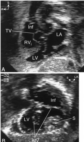
Figure 2. Echocardiogram in a patient with straddling mitral valve (MV) and {S,D,L} segmental anatomy. (A) Subxiphoid long-axis view showing dextrocardia, left-sided left atrium (LA) with an MV that enters an inferior left ventricle (LV). The right ventricle inflow (RVi) is superior to the LV and the ventricular septum is horizontal (superior-inferior ventricles). Notice that the tricuspid valve (TV) is seen in a “short-axis” orientation whereas the MV is seen in a “long-axis” orientation. This indicates that the axes of the MV and TV are angled relative to each other, typical for criss-cross atrioventricular relations. (B) Clockwise rotation of the trans-ducer from the long-axis plane demonstrates major straddling of the MV into a large left-sided infundibulum (Inf). L/A⫽ left/anterior; R ⫽ right; S⫽ superior.
822 Fraisseet al. JACC Vol. 38, No. 3, 2001 Straddling Mitral Valve September 2001:819 –26
hypoplasia and nine had RV sinus hypoplasia. All patients who underwent a biventricular repair had minor SMV and two well-developed ventricles, except for one patient with a mildly hypoplastic RV who successfully underwent an arte-rial switch operation and VSD closure. None of the patients with {S,L,D} segmental anatomy and only two patients with {S,D,L} segmental combination underwent biventricular repair. Overall, the patients underwent an average of 2.2 operations each (range 1 to 5). The median age at last surgery was 2.3 years (range 0 to 13) and the mean duration of follow-up was 3.9⫾ 4.8 years.
Ten patients died during the study period, yielding a mortality rate of 22%. The clinical characteristics of the patients who died are summarized in Table 2. The mortality rate did not change significantly from 1982 to 1988 (4/17, 24%) to 1989 to 1993 (6/22, 27%). However, all seven patients who were operated on from 1994 through February 1999 survived. There were 7 deaths (18%) among the 38 patients who underwent single-ventricle palliation and 3 deaths (38%) in the 8 patients who underwent a biventricu-lar repair (p⫽ 0.25). Kaplan-Meier analysis of the
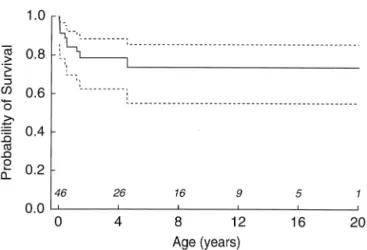
proba-bility of survival for the entire cohort demonstrated that the likelihood of survival was 87% at 1 year, 79% at 8 years and 74% at 20 years (Fig. 5). When the same analysis was
Figure 4. Straddling mitral valve (MV) associated with double-outlet right ventricle and {S,L,D} segmental anatomy. (A) Cranial angulation of the transducer in the subxiphoid long-axis plane shows straddling of the MV into a right-sided infundibulum (Inf). Notice the rightward position of the aorta (Ao). (B) Parasternal long-axis view showing the posterior MV leaflet entering the left ventricle (LV) (open arrow) and the anterior MV leaflet inserting into the infundibulum (Inf) (white arrow). A⫽ anterior; R ⫽ right.
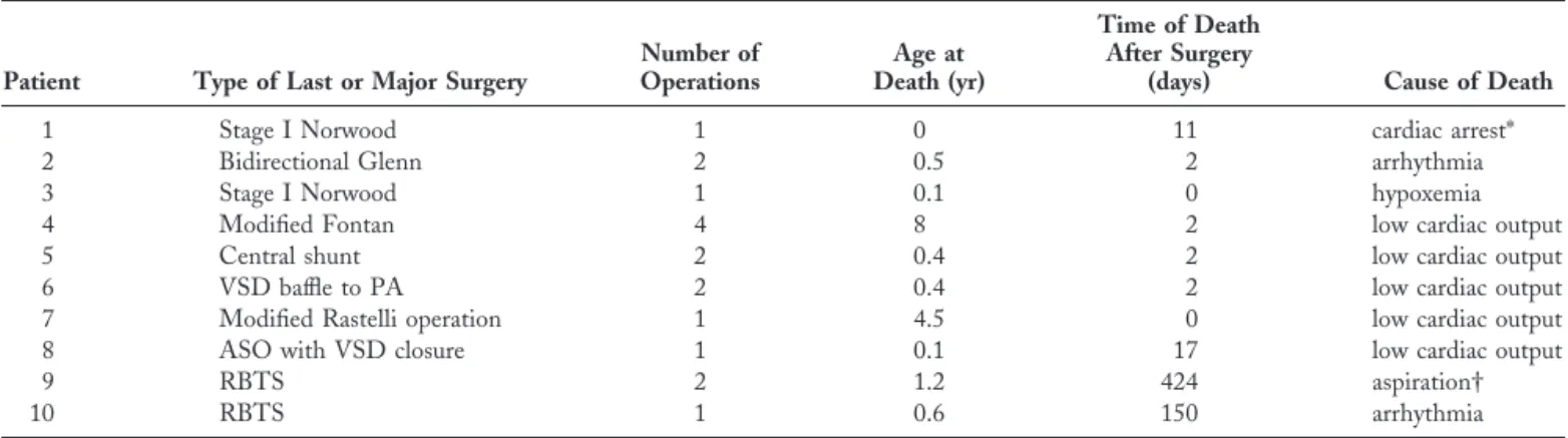
Table 2. Clinical Characteristics of the Patients Who Died
Patient Type of Last or Major Surgery
Number of Operations Age at Death (yr) Time of Death After Surgery
(days) Cause of Death
1 Stage I Norwood 1 0 11 cardiac arrest*
2 Bidirectional Glenn 2 0.5 2 arrhythmia
3 Stage I Norwood 1 0.1 0 hypoxemia
4 Modified Fontan 4 8 2 low cardiac output
5 Central shunt 2 0.4 2 low cardiac output
6 VSD baffle to PA 2 0.4 2 low cardiac output 7 Modified Rastelli operation 1 4.5 0 low cardiac output 8 ASO with VSD closure 1 0.1 17 low cardiac output
9 RBTS 2 1.2 424 aspiration†
10 RBTS 1 0.6 150 arrhythmia
*During tracheal suctioning on the 11thpostoperative day. †During sedation with oral chloral hydrate.
ASO⫽ arterial switch operation; PA ⫽ pulmonary artery; RBTS ⫽ right Blalock-Taussig shunt; VSD ⫽ ventricular septal defect.
Figure 3. Straddling mitral valve in a patient with double-outlet right ventricle {S,L,L} segmental anatomy and straddling of both anterior (white
arrow) and posterior (black arrow) mitral leaflets. Ao⫽ aorta; Inf ⫽ infundibulum; LV⫽ left ventricle; RA ⫽ right atrium.
applied separately for patients who underwent a single versus biventricular repair, the probability of survival for the functional single-ventricle palliation group was 77% at 16 and 20 years compared with 50% at 16 years for the biventricular group. However, the difference between groups was not statistically significant (p⫽ 0.34).
Table 3 summarizes the results of univariate and multi-variate analyses of potential demographic, anatomic and functional predictors of mortality. Patients with a noncom-mitted VSD had the highest risk of death (relative risk 10.2, p⫽ 0.005), followed by multiple VSDs (relative risk 4.7, p ⫽ 0.06). All five patients with noncommitted VSD underwent a functional single-ventricle palliation, with three deaths. Of the four patients with multiple VSDs, three underwent a functional single-ventricle palliation, with two deaths. In the remaining patient who underwent a biven-tricular repair and subsequently died, the additional VSDs were not detected preoperatively. Overall, 8 patients with multiple or noncommitted VSDs had a functional single-ventricle palliation, with 5 deaths (62.5%), compared with only 2 deaths among the 30 patients who had other types of a single VSD and had a functional single-ventricle palliation (6.7%) (p⫽ 0.02).
DISCUSSION
This largest clinical cohort of patients with SMV studied to date demonstrates five key findings: 1) despite the wide morphological spectrum that characterizes this lesion, it is possible to identify four distinct groups with more uniform anatomy; 2) biventricular repair is feasible in selected patients with SMV; 3) patients with {S,D,L} and {S,L,D} segmental combinations had a higher incidence of cardiac malposition and hypoplastic TV and RV and were less likely to undergo a biventricular repair; 4) the presence of a noncommitted VSD was the strongest independent risk factor for death in the multivariate model followed by multiple VSDs; and 5) mortality rate decreased from 24% to 27% before 1994 to 0% during the cohort’s most recent five years (1994 to 1999).
Morphologic considerations. This echocardiographic
study confirms several anatomic observations that were previously made in postmortem series (1– 8). The MV straddled over the anterior aspect of the interventricular septum through a malalignment-type conoventricular septal defect into the RV infundibulum in all patients. Another uniform finding was an abnormal conotruncus with either DORV or TGA. However, the relatively small number of cases included in most previous studies did not permit classification of anatomic subgroups. We have previously recognized a distinct anatomic pattern in patients with SMV who had hypoplasia of the RV sinus and TV, superior-inferior ventricles and criss-cross AV relations (6). These patients differed from the more common group with two well-developed ventricles in that the degree of ventric-ular malposition (measured as the angle between the atrial and ventricular septa) was much larger (6). In the present study, patients were easily categorized into one of the following four anatomic groups based on segmental situs: {S,D,D}, {S,D,L}, {S,L,D} or {S,L,L}. On further analysis, patients with {S,D,L} and {S,L,D} segmental anatomy had a significantly higher incidence of cardiac malposition (dex-trocardia in patients with D-ventricular loop and levocardia in patients with L-ventricular loop), RV sinus and TV hypoplasia, superior-inferior ventricles and criss-cross AV relations. In general, the anatomy in these patients was less likely to be amenable to a biventricular repair, as opposed to the patients with {S,D,D} or {S,L,L} segmental anatomy.
Echocardiographic considerations. This study
demon-strates that echocardiography is ideally suited for noninva-sive evaluation of SMV. This conclusion is in agreement with previous reports (9 –12) and clinical practice (21). Review of the echocardiograms from the 46 study patients indicates that the MV and its support apparatus must be systematically imaged from multiple views to fully charac-terize the anomaly. In young patients, the subxiphoid window offers excellent views of the MV, chordae tendineae and papillary muscles, as well as simultaneous cross-sectional images of the LV, RV sinus and infundibulum. Off-axis images are often necessary to obtain simultaneous views of all the anatomical elements involved in SMV. For example, counterclockwise rotation of the transducer from the standard subxiphoid short-axis (parasagittal) plane pro-duces an oblique coronal image that displays both ventricles and infundibulum. Any straddling MV leaflets and chordae can be seen from this view, as well as the potential for construction of a surgical pathway from the LV to the semilunar valves. The apical and parasternal windows are also informative. The apical four-chamber view is especially useful to determine the extent of SMV (minor vs. major straddling). The parasternal short-axis view is particularly helpful in visualizing the MV attachments within the infundibulum, including the presence and location of any accessory papillary muscles. The experience from this study suggests that the importance of systematic echocardio-graphic examination from multiple acoustic windows and
Figure 5. Kaplan-Meier curve (with 90% confidence intervals) showing the probability of survival of 46 patients with straddling mitral valve.
824 Fraisseet al. JACC Vol. 38, No. 3, 2001 Straddling Mitral Valve September 2001:819 –26
views cannot be overemphasized. Flow mapping with color Doppler is useful for assessment of MV function but does not aid in the diagnosis of SMV or with surgical planning. The potential benefit of three-dimensional echocardiogra-phy in the preoperative evaluation of SMV deserves further study.
Surgical considerations. The nearly universal association
of SMV with a malalignment conoventricular septal defect and with conotruncal anomalies (DORV or TGA), and the recognition of four distinct segmental anatomic patterns, has important surgical implications. Another important observation is that a significant proportion of these children (29 of 46) had two ventricles of normal or nearly normal size. Therefore, discussion as to whether a biventricular repair is feasible in these patients will revolve around the ability of the surgeon to baffle the LV outflow through the VSD to the closest semilunar valve—pulmonary or aortic— without distorting the chordal apparatus of the mitral valve and without obstructing LV outflow (22).
Techniques have been described for repair of these defects in children when the degree of straddling is relatively minor (14,23). Frequently, enlargement of the VSD is necessary to avoid subaortic obstruction from the chordal apparatus crossing the VSD. In patients with DORV, the VSD patch is usually placed around the chordal insertion site (achieved in seven patients in this study). In TGA with SMV, the chordae can be retracted flush with the edge of the VSD and they are then fixed in that position by suturing of the VSD patch onto the crest of the septum (performed in one patient in this series). Despite this apparent
distor-tion, mitral regurgitation is an uncommon complication of biventricular repair.
Recognition of the segmental anatomy is also important for determination of the likely position of the conduction tissue before VSD closure or baffling. The conduction axis in patients with SMV, unlike straddling TV, follows the usual pattern for the segmental situs. Specifically, in patients with solitus atria and D-ventricular loop the conduction axis is expected to run along the posterior-inferior rim of the VSD. In those with L-ventricular loop the conduction axis is expected to run along the superior-anterior rim of the VSD. Therefore, injury to conduction tissue, even when VSD enlargement is necessary, should be avoidable (22).
For the subgroup of children with SMV and hypoplasia of one ventricle, the usual management for functional single ventricle with total cavopulmonary connections—avoiding complex intracardiac procedures—is a good alternative with excellent early and midterm results (24). The only exception is the small subgroup (2 of 46) with DORV where the MV leaflet attaches to the crest of the septum, creating a restrictive VSD. In this group, VSD enlargement is likely to be required to prevent LV outflow obstruction.
Study limitations. The retrospective nature of the analysis
imposes inherent limitations. The surgical management of the patients was decided on a case-by-case basis, and selection of single versus biventricular approach was not based on predefined criteria. Hence, it is not possible to know whether some of the patients who underwent a functional single-ventricle palliation could have survived a biventricular repair. Similarly, it is not possible to determine
Table 3. Predictors of Death in 46 Patients With SMV
Variables
Relative Risk
(95% confidence limits) p Value
Univariate analysis
Male gender 1.8 (0.5–7) 0.25
Age at initial surgery⬍0.1 year old 0.8 (0.5–1.2) 0.28 Weight at last surgery⬍2 SD 1 (0.5–1.8) 0.98 Biventricular repair 2.3 (0.6–8.9) 0.25 Dextrocardia 0.04 (1.3⫻ 10⫺5–125.6) 0.24 Superior-inferior ventricles 1.1 (0.3–4.2) 0.85 Crisscross AV valves 1.3 (0.2–10) 0.78 Ventriculo-arterial alignment (DORV) 1.9 (0.2–14.6) 0.39 Hypoplastic RV 0.3 (0.04–2.7) 0.33 Hypoplastic LV 0.6 (0.1–4.8) 0.81 Bilateral conus 0.5 (0.1–2.5) 0.31 Noncommitted VSD 4.9 (1.2–18.9) 0.006 Extension of VSD into AV canal 1.2 (0.3–4.7) 0.65 Multiple VSDs 5.6 (1.4–22) 0.002 Major straddling 0.8 (1.2–3.8) 0.95 Mitral regurgitation 0.5 (0.1–4.1) 0.52 Nonrestrictive pulmonary blood flow 0.3 (0.1–1.1) 0.04 Oxygen saturation before last surgery 0.9 (0.2–4.8) 0.98 Multivariate analysis
Noncommitted VSD 10.2 (2–51.8) 0.005 Multiple VSDs 4.7 (0.9–23.8) 0.06 Nonrestrictive pulmonary blood flow 0.3 (0.05–1.2) 0.08
AV⫽ atrioventricular valves; DORV ⫽ double-outlet right ventricle; LV ⫽ left ventricle; RV ⫽ right ventricle; SD ⫽ standard deviation; SMV⫽ straddling mitral valve; VSD ⫽ ventricular septal defect.
whether patients who did not survive a biventricular ap-proach would have survived a functional single-ventricle palliation.
Acknowledgments
We thank Emily Flynn McIntosh for the artwork and Bill McIntosh for the photography.
Reprint requests and correspondence: Dr. Tal Geva,
De-partment of Cardiology, Children’s Hospital, 300 Longwood Avenue, Boston, Massachusetts 02115. E-mail: geva_t@ a1.tch.harvard.edu.
REFERENCES
1. Kitamura N, Takao A, Ando M, Imai Y, Konno S. Taussig-Bing heart with mitral valve straddling: case reports and postmortem study. Circulation 1974;49:761–7.
2. Freedom RM, Bini R, Dische R, Rowe RD. The straddling mitral valve: morphological observations and clinical implications. Eur J Car-diol 1978;8:27–50.
3. Bharati S, McAllister HA, Lev M. Straddling and displaced atrioven-tricular orifices and valves. Circulation 1979;3:673– 84.
4. Milo SM, Ho SY, Macartney FJ, et al. Straddling and overriding atrioventricular valves: morphology and classification. Am J Cardiol 1979;44:1122–34.
5. Wenink ACG, Gittenberger-de Groot AC. Straddling mitral and tricuspid valves: morphologic differences and developmental back-ground. Am J Cardiol 1982;49:1959 –71.
6. Geva T, Van Praagh S, Sanders SP, Mayer JE, Van Praagh R. Straddling mitral valve with hypoplastic right ventricle, crisscross atrioventricular relations, double outlet right ventricle and dextrocar-dia: morphologic, diagnostic and surgical considerations. J Am Coll Cardiol 1991;17:1603–12.
7. Muster AJ, Bharati S, Aziz KU, et al. Taussig-Bing anomaly with straddling mitral valve. J Thorac Cardiovasc Surg 1979;77:832– 42. 8. Wenink ACG, Gittenberger-de Groot AC, Brom AG.
Developmen-tal considerations of mitral valve anomalies. Int J Cardiol 1986;11:85– 98.
9. Rice MJ, Seward JB, Edwards WD, et al. Straddling atrioventricular valve: two-dimensional echocardiographic diagnosis, classification and surgical implications. Am J Cardiol 1985;55:505–13.
10. Marino B, Sanders SP, Pasquini L, Giannico S, Parness IA, Colan SD. Two-dimensional echocardiographic anatomy in criss-cross heart. Am J Cardiol 1986;58:325–33.
11. Smallhorn JF, Tommasini G, Macartney FJ. Detection and assessment of straddling and overriding atrioventricular valves by two-dimensional echocardiography. Br Heart J 1981;46:254 – 62.
12. Vargas Barron J, Sahn DJ, Valdes-Cruz LM. Two-dimensional echocardiographic evaluation of overriding and straddling atrioventric-ular valves associated with complex congenital heart disease. Am Heart J 1984;107:1006 –14.
13. Danielson GK, Tabry IF, Riter DG, Fulton RE. Surgical repair of criss-cross heart with straddling atrioventricular valve. J Thorac Car-diovasc Surg 1979;77:847–51.
14. Serraf A, Nakamura T, Lacour-Gayet F, et al. Surgical approaches for double-outlet right ventricle or transposition of the great arteries associated with straddling atrioventricular valves. J Thorac Cardiovasc Surg 1996;111:527–35.
15. Belli E, Serraf A, Lacour-Gayet F, et al. Biventricular repair for double outlet right ventricle. Results and long-term follow-up. Circulation 1998;98:II360 –7.
16. Aoki M, Forbess JM, Jonas RA, Mayer JE, Castaneda AR. Result of biventricular repair for double-outlet right ventricle. J Thorac Cardio-vasc Surg 1994;107:338 –50.
17. Van Praagh R. Terminology of congenital heart disease. Glossary and commentary. Circulation 1977;56:139 – 43.
18. Van Praagh S, Davidoff A, Chin A, Shiel FS, Reynolds J, Van Praagh R. Double-outlet right ventricle: anatomic type and developmental implications based on a study of 101 autopsied cases. Arch Mal Coeur 1982;13:389 – 440.
19. Kumar K, Lock JE, Geva T. Apical muscular ventricular septal defects between the left ventricle and the right ventricular infundibulum: diagnostic and interventional considerations. Circulation 1997;95: 1207–13.
20. Van Praagh R, Geva T, Kreutzer J. Ventricular septal defects: how shall we describe, name and classify them? J Am Coll Cardiol 1989;14:1298 –9.
21. Geva T. Echocardiography and Doppler ultrasound. In: Garson A, Jr., Bricker JT, Fisher DJ, Neish SR, editors. The Science and Practice of Pediatric Cardiology. Baltimore, MD: Williams & Wilkins, 1997: 789 – 843.
22. Anderson RH, Ho SY. Which hearts are unsuitable for biventricular repair? Ann Thorac Surg 1998;66:621– 6.
23. Pacifico AD, Soto B, Bargeron LMJ. Surgical treatment of straddling tricuspid valves. Circulation 1979;60:655– 64.
24. Stamm C, Friehs I, Mayer JE, Jr., et al. Long-term results of the lateral tunnel Fontan operation. J Thorac Cardiovasc Surg 2001;121:28 – 41.
826 Fraisseet al. JACC Vol. 38, No. 3, 2001 Straddling Mitral Valve September 2001:819 –26